1
UNIVERSITA’ DI PISA
Facoltà di Medicina e Chirurgia
Scuola di dottorato in Neuroscienze e Scienze endocrinometaboliche
[SD06/EM] Esplorazione molecolare, metabolica e funzionale del sistema nervoso e degli organi di senso
Thesis submitted for the degree of Doctor
IN VITRO CYTOTOXIC AND GENOTOXIC EFFECTS OF
DIFFERENT METAL NANOPARTICLES
AUTHOR: SUPERVISORS:
Maria Rita Fabbrizi
Prof. Lucia Migliore
Prof. Gabriele Siciliano
2 “Do not worry about your difficulties in Mathematics.
I can assure you mine are still greater.” Albert Einstein
“Έτσι, δεν γνωρίζω” Σωκράτης
3
Summary
ABSTRACT ... 5
INTRODUCTION ... 6
NANOPARTICLES: DEFINITION AND APPLICATIONS ... 6
ROUTES OF EXPOSURE ... 9
NANOPARTICLE-CELL INTERACTIONS ... 14
EFFECT OF NPs PHYSICOCHEMICAL PROPERTIES ON TOXICITY ... 22
BIOLOGICAL ENDPOINTS OF IN VITRO ASSAYS ... 25
MTT assay ... 25
Trypan Blue dye exclusion assay ... 27
Cytokinesis-block micronucleus assay ... 28
Comet assay ... 31
FISH analysis ... 34
AIM OF THE STUDY ... 36
MATERIALS AND METHODS ... 37
Nanoparticles set ... 37
Cell Cultures and Exposure to NPs ... 38
MTT Assay ... 38
Trypan blue dye exclusion assay ... 39
Cytokinesis-block micronucleus cytome assay ... 39
Fluorescence in situ hybridization ... 40
Comet Assay and oxidative DNA damage ... 41
Statistical Analysis ... 42
RESULTS ... 43
Copper oxide nanoparticles ... 43
MTT assay ... 43
Trypan blue dye exclusion ... 45
CBMN cytome assay... 47
Primary and oxidative DNA damage ... 52
Fluorescence in situ hybridization ... 57
Gold nanoparticles ... 58
MTT assay ... 58
CBMN cytome assay... 60
4
Fluorescence in situ hybridization ... 70
Silver nanoparticles ... 72
MTT assay ... 72
Trypan blue dye exclusion ... 74
CBMN cytome assay... 76
Primary and oxidative DNA damage ... 81
Silica nanoparticles ... 85
MTT assay ... 85
Trypan blue dye exclusion ... 86
CBMN cytome assay... 87
Primary and oxidative DNA damage ... 91
DISCUSSION ... 94 CuO nanoparticles ... 94 Gold nanoparticles ... 98 Silver nanoparticles ... 102 Silica nanoparticles ... 105 CONCLUSIONS ... 109 REFERENCES ... 112
5
ABSTRACT
Several commercial nano-agents are already available for biomedical applications and many nanomedicine products are near obtaining final approval for clinical use. Besides biomedical applications, nanoparticles (NPs) are used commercially in products such as electronic components, scratch-free paint, sports equipment, cosmetics, food colour additives, and surface coatings. Hence, our exposure to nanomaterials is significant and increasing, yet there is little understanding of the unique toxicological properties of NPs and their long-term impact on human health.
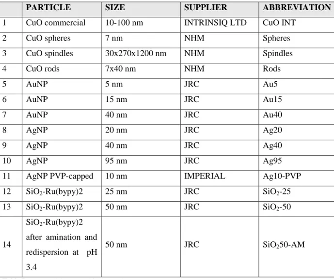
For this reason, in this study we address the issue of cytotoxic and genotoxic effects of different metallic nanoparticles two different cell systems. Moreover, this study aim is to understand if, modifying the physicochemical characteristics of NPs, it is possible to mitigate the toxic effects they are able to induce. Thus a set of 14 different NPs was screened: 4 CuO NPs, 3 Au NPs, 4 Ag NPs and 3 SiO2 NPs. Inside every group, the NPs differ for size, shape and\or capping. The tests were performed in Raw 264.7 macrophage cells and peripheral blood lymphocytes (PBL). Macrophages are one of the principal immune effector cells that play essential roles as secretory, phagocytic and antigen-presenting cells in the immune system while PBL are present in the circulatory system and are representative of the major pathway of NPs distribution in the whole organism.
The results showed how NPs caused cytotoxicity and genotoxicity with different degrees of damage, often in a dose-dependent manner. The NPs set tested induced micronuclei formation and DNA damage at different levels. Oxidative stress seemed to be the most probable cause of damage, followed by the activation of apoptotic and necrotic pathways. Moreover, CuO and Au NPs induced aneuploidogenic events in cells after exposure compared to negative control. Nevertheless, it was not possible to ascribe the damage to one single physicochemical parameter of the nanoparticles, as all the characteristics seemed to act altogether in cell damaging.
Further investigations are required to better understand which mechanism(s) is(are) involved in the NPs toxicity and if modification in one or more physicochemical parameters could be sufficient to make NPs harmless or at least less toxic.
6
INTRODUCTION
NANOPARTICLES: DEFINITION AND APPLICATIONS
In the last two decades, new terms with the prefix `nano' have rushed into the scientific vocabulary - nanoparticle, nanostructure, nanotechnology, nanomaterial, nanocluster, nanochemistry, nanocolloids, nanoreactor and so on. A series of new journals are devoted particularly to this subject, monographs with the corresponding names have appeared, `nano'-specialised institutes, chairs and laboratories have been founded; and numerous conferences are held. In most cases, new names were applied to long known objects or phenomena; however, new objects inaccessible for researchers some 20 years ago have also appeared. These include fullerenes, quantum dots, nanotubes, nanofilms and nanowires, i.e., the objects with at least one nanometer dimension (Gubin et al., 2005). However the definition of nanoparticle (NP) is a complex issue. The European community has discussed the topic and issued a document: “Scientific Committee on Emerging and Newly Identified Health Risks (SCENIHR)” that offers a more complex approach. This document subdivides nanoparticles into three categories as explained below.
Category 1: size > 500 nm
If the size (e.g. mean, median, etc.) of the material is above 500 nm it is assumed that the size distribution at the lower end will most likely be above the designated lower threshold of 100 nm. This should be confirmed by determination of the size distribution. So, the need for further evaluation regarding possible nano-specific properties may be of lower priority and thus for the moment classical risk assessment should be performed taking into consideration the particulate nature of the material.
Category 2: 500 nm> size >100 nm
When the size is <500 nm it is more likely that part of the size distribution will be lower than 100 nm and that a material may be considered a nanomaterial and that therefore a more detailed characterization and specific risk assessment will be necessary. A nano-specific risk assessment should be undertaken if the characterization demonstrates that >0.15% (or any specified percentage) of the number size distribution is <100 nm. If these characteristics are not met, the need for further evaluation regarding possible nano-specific
7
properties may be of lower priority and thus for the moment classical risk assessment should be performed taking into consideration the particulate nature of the material.
Category 3: 100 nm> size >1 nm
The material is considered a nanomaterial and nano-specific risk assessment has to be performed. A volume specific surface area (VSSA) above the threshold (e.g. >60 m2/cm3) may be used as an additional qualifier to indicate a size below 100 nm (SCHENIR).
Several commercial nano-agents are already available for biomedical applications and many nanomedicine products are near obtaining final approval for clinical use (Xu and Sun, 2007; Lewin et al., 2000). Besides biomedical applications, NPs are used commercially in products such as electronic components, scratch-free paint, sports equipment, cosmetics, food colour additives, and surface coatings. Hence, our exposure to nanomaterials is significant and increasing, yet there is little understanding of the potential toxicological properties of NPs and their long-term impact on human health (Mahmoudi et al., 2011).
Outcomes from studies on nano-bio interactions in the past decade have greatly influenced nanoparticle design. The evolution of nanoparticles destined for biomedical applications has occurred in parallel with studies investigating the biological responses to the nanomaterials themselves. Material design evolved whenever the effect of size, shape, or surface charge was further elucidated. To date, three generations of nanoparticles have been engineered for biomedical applications (Figure 1). The first generation consisted of novel nanomaterials functionalized with basic surface chemistries to assess biocompatibility and toxicity. The second generation produced nanomaterials with optimized surface chemistries that improved stability and targeting in biological systems. The third generation shifted the paradigm of design from stable nanomaterials to “intelligent” environment-responsive systems that should improve targeted compound delivery. The first generation of nanomaterials was synthesized to demonstrate the potential applications of novel materials in biomedical research. Before this generation, liposome studies in the supra-nanoscale range (100–1,000 nm) established a basic research template for evaluation of more modern, smaller materials (1–100 nm). Seminal papers from this first generation include those describing the surface modification of organic-soluble quantum dots and magnetic nanoparticles to render them water soluble and stable enough for biological applications (Nie and Chan, 1998; Gupta and Gupta, 2005). First-generation nanoparticles were modified with non-stealth surface chemistries and used in experiments to assess cell uptake and toxicity (Chithrani et al., 2006; Kirchner et al., 2005).
8
The main focus of these materials was to determine the effects of surface charge (Cho et al., 2009a; Hauck et al., 2008). However, most of these studies included experiments in serum-free media or did not account for serum-protein interactions with their nanomaterials, which makes the findings difficult to interpret when considering physiological conditions within the body. First generation nanomaterials also did not use PEG; thus, most in vivo data show the rapid clearance of nanomaterials (De Jong et al., 2008; Cherukuri et al., 2006). These early studies were important in highlighting the biocompatibility of these novel materials and marked a major step in the transition from chemical synthesis to biological uses. However, the poor stability in cell-culture media and rapid clearance from the body shifted research focus to increase stability and prolong blood half-life. With nanoparticle synthesis established, research shifted to surface chemistry optimization for diagnosing and treating cancer. Although nanoparticles were a promising avenue for targeted delivery in many organs and tissues, most studies using second-generation nanoparticles focused on tumour delivery as a proof of concept. Consequently, second-generation nanomaterials are characterized by two important design concepts: stealth and active targeting. The goal of stealth nanoparticles is to maximize blood circulation half-life to ensure continuous delivery of nanoparticles into the tumour via leaky vasculature. The longer a nanoparticle remains in circulation, the higher likelihood it has of entering the tumour. Several studies showed the increased half-life of nanomaterials by simply adding PEG to their surface (Owens and Peppas, 2006). The chemistry used to attach PEG, overall PEG length, and surface density all affect nanoparticle stability (Perrault et al., 2009; Zhang et al., 2009b; Gref et al., 2000). Alternative molecules such as lipids and silica were investigated, but PEG remains the most widely used approach (Van Schooneveld et al., 2008).
Researchers recently developed a new generation of nanomaterials that do not rely on passive retention of nanoparticles or on endogenous tumour ligands. The third generation of nanomaterials has “environment-responsive” properties. These dynamic nanoparticles use biological, physical, or chemical cues in their target environment to trigger a change in their properties to maximize tumour delivery. Two approaches have been used so far: the first uses hallmark cues inside the tumour environment such as low pH, low O2, or matrix-metalloproteinase enzymatic activity; the second provides an artificial cue such as near-infrared light inside the target tissue.
9
Figure 1. Evolution of nanoparticle design, highlighting the interplay between evolution of nanomaterial design and fundamental nano-bio studies. Abbreviations: Ab, antibody; EPR, enhanced permeation and retention; MPS, mononuclear phagocyte system; PEG, poly(ethylene) glycol. From Albanese et al., 2012
ROUTES OF EXPOSURE
Due to the increased use of nanomaterials it is inevitable that the human being are exposed to NPs, which are able to penetrate into the body. The main routes of entry are through the skin, lungs or intestinal tract causing adverse biological effects (Davoren et al., 2007; Li et al., 2007; Oberdörster et al., 2005; Hoet et al., 2004; Warheit et al., 2004). Other potential routes of NPs in the case of biomedical applications include parental administration such as intravenous, intradermal and peritoneal exposures into experimental systems (Stern and McNeil, 2008; De Jong et al., 2008). Factors that may influence NPs entry include size, charge, surface area and shape. According to Auffan and co-authors nanosized particles have an elevated surface/volume ratio of approximately 35- 40% of atoms localized at the surface of a 10nm NPs compared with less than 20% for particles larger than 30nm (Auffan et al., 2008). The toxicity of these materials depends on their persistence or clearance from the different organs due to the immune response of the host (Jeffrey et al., 2008). Much research has been done on NPs toxicity inside the respiratory tract. These nanomaterials can be inhaled naturally in the form of aerosol, powders or artificially by instillation into the respiratory tract for toxicity studies (Oberdörster et al., 2005; Hoet et al., 2004; Warheit et al., 2004). The respiratory system is the part of the organs that deal with the process of respiration and it is responsible for taking in and sending out air from living animals. The lungs are part of the respiratory tract responsible for exchange of gases. Inhalation is the most common route of exposure to NPs in the workplaces. Once inhaled, these materials are carried by electrostatic
10
force of the air from the upper to the lower respiratory tract (Cross et al., 2007; Oberdörster et
al., 2005). The particles are usually inhaled in the form of airborne NPs, systemic
administration of drugs, chemicals and other compounds to the lungs through direct cardiac output to the pulmonary arteries (Jeffrey et al., 2008). Immediately the NPs are in the pulmonary sites, translocation to blood circulation through the lymphatic pathways can occur depending on the nanomaterial size. Also when the NPs are deposited in the alveolar, they are usually attacked by the process of phagocytosis. This also led to chemotactic activities which trigger the complement system cascade and the inflammatory cell response to the site of NPs. According to Oberdörster and colleagues the effect of the inflammatory and the complement cascade may take up to 10 days in rat and roughly two years in humans to be cleared. Ingestion is another route whereby NPs may enter the body (Oberdörster et al., 2005). Most of the toxicity studies pertaining to NPs are focused mainly on respiratory tract (RT) exposures with few studies describing the gastrointestinal tract exposures. The gastrointestinal tract (GT) exposures usually occur either unintentional from hand to mouth transfer or from traditional materials. Furthermore, it could occur during handling of the materials that contain the NPs. Other possible gastrointestinal tract exposures may come from particles cleared from the respiratory system through the mucociliary escalator (Li et al., 2007; Chen et al., 2006; Oberdörster et al., 2005). The GT can be exposed to NPs also by water, food, cosmetics, drugs, drug delivery devices. Some studies have investigated the potential intestinal absorption and the translocation of NPs and generally found uptake within the GT. More critical findings concerning the fate of ingested NPs can be viewed from radioactive metal studies, where NPs have been shown to translocate from the gastrointestinal tract to other organs (Borm and Kreyling, 2004). Furthermore, NPs administered orally are usually absorbed, through the epithelial cells of the Peyer’s patches in the gut-associated lymphoid tissue (GALT) and also through the gut enterocytes (Chen et al., 2006; Florence, 2005). Skin barrier alterations (such as the wounds, scrapes, or dermatitis) could act as exposures routes to NPs into the body and should not be overlooked. Debilitated skin represents a good channel for entry of finer and even larger particles (0.5-7μm) as reported by Blundell and collaborators (1989). Most of the penetration and distribution of nanomaterials in skin and toxicity are minimal and limited to the uppermost stratum corneum layers and areas near hair follicles. This usually led to irritation of the inflammation area in experimental animals. This is because the stratum corneum is the primary barrier for skin and any type of perturbations of the skin (such as an open wound, cut, or alteration to this skin barrier) could expose viable skin cells to NPs (Zhang et al., 2008; Cross et al., 2007). Therefore, more toxicological
11
assessment such as abrasion should be conducted to determine if penetration to this barrier would allow an enhancement of NPs absorption. This raises the question whether nanomaterials could penetrate the dermis, be eventually absorbed systemically, and be responsible for an acute/chronic and local/systemic potential health risk. It is already known that the skin is nanoporous at the nanoscale, having orifices of hair follicles and glands open on skin surface providing alternative entrance routes. There are few literatures reports which indicate the eye as route of NPs exposure into animals. Drug delivery is achieved through topical application (Aniruddha et al., 2008). However, topical treatment of posterior eye infection is not effective due to the rapid precorneal elimination due to solution drainage, long diffusional path length, induced lacrimation, and corneal epithelial impermeability (Jarvinen
et al., 1995). However, NPs have generated considerable interest for drug delivery into the
eye (Aniruddha et al., 2008). According to Herrero-Vanrell and Refojo (Herrero- Vanrell and Refojo, 2001), intravitreally administrations of NPs showed to sustain drug delivery to the eye. Recent reports by Farjo and co-authors explained how DNA added on NPs can be implored to transfer genes into the mouse retina (Farjo et al., 2006). Jani and colleagues reported that albumin encapsulated NPs, when injected into the corneas of uninjured mice, were detected in the corneal keratocyte cytoplasm (Jani et al., 2007). The albumin NPs can be used to express intraceptors for extended periods that are effective in suppressing injury-induced corneal neovascularization. The highly efficient transfer of the reporter gene into photoreceptor cells could lead to effective treatments for conditions such as retinitis pigmentosa. Therefore, by modifying the properties of NPs, they could be made to target specific organs. Vertebrates have a pair of ears, which are symmetrically placed on opposite sides of the head. Their arrangement and the ability to localize sound sources (waves) can passively facilitate the entry of NPs into the inner ear and to the other vital parts of the body via blood. However, very few researches had published about the auditory pathway as a channel for NPs transport into the ear. This is due the complex natures of the anatomies of the ears which contains hollow channels filled with fluid, and have sensory cells that are surrounded by hair cells. Some preliminary reports by Dormen and co-authors showed that superparamagnetic NPs can be used as drug delivery into the inner ear of guinea pigs and into the prilymphatic fluid (Dormen et al., 2005). Another pilot report by Xianxi and colleagues (Xianxi et al., 2007) also showed that polylactic/glycolic acid (PLGA) polymer coated with iron oxide NPs, applied to the round window membrane of chinchillas, induced by magnetic field can enter the inner ear and will be found in multiple locations within the cochlea tissue. In biological assessment, intravenous administration of NPs is very important route used in
12
determining toxicological assessment in experimental animals. In the study of De Jong and collaborators to determine particle size-dependent organ distribution of gold NPs they intravenously injected experimental rat in the tail vein with gold NPs with diameters of 10, 50, 100 and 250 nm, respectively. Their results gave an oxidative stress in the rat’s liver cells. The 10nm gold NPs showed the most widespread presence in the various organ systems including brain, heart, kidneys, lungs, testis and thymus (De Jong et al., 2008). Intravenous administration of NPs is followed by inflammatory responses, characterized by an increased synthesis and secretion of cytokines. Experimental animals absorb NPs from the site of injection into the lymphatic system (Thanos et al., 1999). The subcutaneous route involves a complex sequence of nanoparticle movement, mostly involving lymph and blood. The relevance of intravenous administration of NPs into experimental animals studies to humans have been questioned not only in drug delivery but also in vaccination, a modality which requires systematic absorption of the encapsulated active drug to achieve a biological response (Rocio et al., 1997). The nano-mucus membrane pathway is the lining of most endodermal cells that cover the epithelium and are involved in absorption and secretion. They line various body cells and cavities that are exposed to the external environment and internal organs. It is continuous with the skin, nostrils, lips, ears, the genital and the anus. NPs deposited on the various mucus tissues pathway, encounter mucus or epithelial lining fluid. This may trigger phagocytosis or contact fibroblasts B cells or endothelial cells resulting into the NPs removal (Oberdörster et al., 2005; Brayden, 2001). The mucus membrane is the first barrier that confronts NPs that are deposited in the conducting epithelium. Other reports by Moghimi and collegues have shown that NPs can be translocated through the mucosal lining and epithelial cells of the intestine and associate with the GALT and the blood circulatory system (Moghimi et al., 2001).
It is quite likely that nanoparticles, when reaching the circulation will influence endothelial cell membrane function, either due to their direct toxic effects or through some indirect mechanisms, e.g., by inducing a cascade of events leading to disruption of tight junctions or increasing the membrane permeability of the blood-brain barrier (BBB) (Sharma et al., 2009; Lanone and Boczkowski, 2006). The nanoparticles may also stimulate vesicular transport to gain access in to the central nervous system (CNS) microenvironment to induce neurotoxicity. However, translocation of nanoparticles from blood to brain across the BBB requires additional investigation. Another possibility of nanoparticles-induced BBB disruption could be due to formation of free radicals and increased oxidative stress. Apart from nanoparticle induced disruption of the BBB leading to neurotoxicity, recent advances in nanotechnology
13
resulted in production of polymer nanoparticles that can be used as carriers to transport the entrapped or adsorbed drugs across the BBB for better therapeutic potential (Alyautdin et al., 1997). However, the toxicity of nanocarriers across the BBB is still not well investigated. Nanoparticles without surfactant coating are engulfed by mononuclear phagocytes and thus unable to reach brain tissue or in cerebrospinal fluid (Calvo et al., 2001). Thus, PEGylated polybutylcyanoacrylate nanoparticles are needed to crosses the BBB in a significant amount loaded with drugs (Tiwari and Amiji, 2006). It appears that endocytosis within the brain endothelial cells play major roles for transfer of coated nanoparticles with drugs into the brain. Surface-coated nanoparticles cross the BBB through receptor mediated mechanisms, as they could mimic low-density lipoprotein (LDL) (Sharma et al., 2009; Lanone and Boczkowski, 2006; Liu et al., 2006). Nanoparticles combined with the apolipoprotein acts as LDL leading to receptor-mediated transcytosis (Kreuter, 2004). For this purpose, nanoparticles with similar size to LDL (in the range of 20-25 nm) are most effective in drug delivery across the BBB (Zheng et al., 2005; Hagenbuch and Meier, 2003). This is largely due to their small size that makes them degradable, and their translocation into endothelial cells results in a rapid release of the drug particles into the brain (Lanone and Boczkowski, 2006). However, polysorbate 80 has some toxic and haemolytic effects that restrict its usage in drug delivery to the brain (Sharma et al., 2009; Lanone and Boczkowski, 2006). Apart from the physical properties of the nanoparticles, electrostatic charges play important roles in transport of nanomaterials across the BBB (Fenart et al., 1999). The cationized nanoparticles in vitro have higher brain translocation compared to anionic or neutral nanoparticles (Nagy et
al., 1983). The cationic charged molecules may occupy anionic areas at the BBB endothelium
resulting in an increased permeability of cell membranes across the tight junctions (Lockman
et al., 2004). However, in vivo data regarding brain permeability of cationized nanoparticles
are still lacking. On the other hand, nanoparticles with either neutral or low concentrations of anionic surface coating charges do not disrupt the BBB integrity (Saija et al., 1997). Whereas high anionic and/or cationic charged nanoparticles at low concentration (25 µg/kg) in rats induce BBB breakdown in less than 60 seconds in a dose dependent manner. This BBB disruption is reversible in nature (Koziara et al., 2006). Administration of negatively charged nanoparticles in a dose range of 100 μg-200 μg per animal resulted in their accumulation within the cellular matrix without apparent neurotoxicity. However, only the free fraction of anionic nanoparticles is related to BBB opening (Koziara et al., 2003; Sahagun et al., 1990). Thus, both the size and the charge of colloidal drug carriers are important factors in nanoparticles-induced drug delivery across the BBB or in brain parenchyma (Kim et al.,
14
2006). Since the tight junctions maintaining BBB integrity have a gap of 4-6 nm (Ambruosi et
al., 2005), these nanoparticles may pass through the endothelial cell membrane rather than via
inter-endothelial junctions. As nanoparticles are lipid insoluble and endothelial cells lack pinocytosis, their penetration into the brain through circumventricular organs lacking a tight barrier is also quite likely (Teeguarden et al., 2006). Using this route, nanoparticles may gain access to the brain without affecting BBB permeability. However, further studies are needed to clarify these issues.
NANOPARTICLE-CELL INTERACTIONS
A prototypical nanoparticle is produced by chemical synthesis; then coated with polymers, drugs, fluorophores, peptides, proteins, or oligonucleotides, and eventually administered into cell cultures or animal models. Nanoparticles were initially considered as benign carriers, but multiple studies have demonstrated that their interaction with serum proteins and cell membrane receptors is determined by the nanoparticle design, in effect influencing cell uptake, gene expression, and toxicity. Nanoparticles can interact with the cell surface membrane in multiple scenarios, as shown in Figure 2.
Interactions between nanoparticle-bound ligands and cellular receptors depend on the engineered geometry and the ligand density of a nanomaterial. The nanoparticle acts as a scaffold whose design dictates the number of ligands that interact with the receptor target. A multivalent effect occurs when multiple ligands on the nanoparticle interact with multiple receptors on the cell. The binding strength of complexed ligands is more than the sum of individual affinities and is measured as the avidity for the entire complex. A ligand’s binding affinity increases proportionally to the size of a nanoparticle owing to a higher protein density on the nanoparticle surface. However, when viewed in terms of the downstream signalling via the ErbB2 receptor, 40–50-nm gold nanoparticles induced the strongest effect, suggesting that other factors beyond binding affinity must be considered. Nevertheless, several studies have shown that nanoparticle design can cause differential cell signalling when compared with free ligand in solution. For example, the before mentioned 40–50-nm Herceptin-coated gold nanoparticles altered cellular apoptosis by influencing the activation of caspase enzymes (Jiang et al., 2008b). Similarly, receptor-specific peptides improved their ability to induce angiogenesis when conjugated to a nanoparticle surface (Kanaras et al., 2011). Presentation of the peptide on a structured scaffold increased angiogenesis, which is dependent on receptor-mediated signalling. These findings highlight the advantages of having a ligand bound to a nanoparticle as opposed to its being free in solution. The nanoparticle surface creates a region of highly concentrated ligand, which increases avidity and, potentially, alters cell signalling.
15
However, this benefit comes at a cost: nanomaterials can also cause unexpected changes in cell signalling. For example, a study showed that 14-nm carbon nanoparticles interact with epidermal growth receptors and β1-integrins on rat alveolar II epithelial cells and induce the activation of the Akt signalling pathway, causing cell proliferation (Unfried et al., 2008). An additional concern with nanoparticle-ligand complexes is the potential denaturation of proteins when bound to the engineered surface. The denaturation of a protein can affect binding to its receptor, increase non-specific interactions or provoke inflammation. Lysozyme, for example, when bound onto gold nanoparticles will denature and interact with other lysozyme molecules and produce protein-nanoparticle aggregates (Zhang et al., 2009a). Fibrinogen also unfolds when bound to the surface of polyacrylic acid–coated gold nanoparticles. The denatured fibrinogen can then bind to the integrin receptor Mac-1 and lead to inflammation (Deng et al., 2010). In many cases, nanoparticles enter the cell after binding to the receptor target. Once bound, several factors can dictate the behaviour of nanomaterials at the nano-bio interface. For instance, a nanoparticle shape directly influences uptake into cells: rods show the highest uptake, followed by spheres, cylinders, and cubes. Gratton and co-authors determined this ordering using synthesized nanoparticles larger than 100 nm, but using sub-100-nm nanoparticles, spheres show an appreciable advantage over rods (Qiu et al., 2010; Chithrani et al., 2006) probably due to the increase in the aspect ratio of nanorods (the aspect ratio of a shape is defined as the length of the major axis divided by the width of the minor axis) (Gratton et al., 2008). Although few studies have focused on non-spherical nanoparticles thus far, research indicates their interactions with cells may be much more complex. For the engineering process, asymmetrical nanoparticles may provide another level of control in presenting ligands to the target receptors. Within a given geometric shape, a nanomaterial’s dimensions are a strong determinant of total cell uptake. For spherical gold nanoparticles, silica nanoparticles, single-walled carbon nanotubes, and quantum dots, a 50-nm diameter is optimal to maximize the rate of uptake and intracellular concentration in certain mammalian cells (Lu et al., 2009; Jin et al., 2009; Chithrani and Chan, 2007). In addition to size and shape, the composition of the nanomaterials also affects uptake because single-walled carbon nanotubes and gold nanoparticles, each 50 nm in diameter, possess endocytosis rates of 10−3 min−1 and 10−6 min−1, respectively. Which ligand is used to coat the nanomaterial will also affect downstream biological responses. For example, the uptake and cytotoxicity of nanoparticles were significantly altered when the nanoparticles were coated with two different proteins targeting the same receptor (Wang et al., 2010). Once bound to their receptor, nanoparticles will typically enter the cell via receptor-mediated endocytosis
16
(Jiang et al., 2008a; Chithrani et al., 2006; Gao et al., 2005). The binding of the nanoparticle-ligand conjugate to the receptor produces a localized decrease in the Gibbs free energy, which induces the membrane to wrap around the nanoparticle to form a closed-vesicle structure (Gao
et al., 2005). The vesicle eventually buds off the membrane and fuses with other vesicles to
form endosomes, which fuse with lysosomes where degradation occurs. The size-dependent uptake of nanoparticles is likely related to the membrane-wrapping process. Small nanoparticles have less ligand-to-receptor interaction than larger nanoparticles do. A 5-nm nanoparticle coated with 50-kDa proteins may interact with only one or two cell receptors. By contrast, a 100-nm nanoparticle has many more ligand-receptor interactions per particle. Several small ligand-coated nanoparticles must bind to receptors in close proximity to one another to produce enough free energy to drive membrane wrapping. Larger nanoparticles can act as a cross-linking agent to cluster receptors and induce uptake. Thermodynamically, a 40– 50-nm nanoparticle is capable of recruiting and binding enough receptors to successfully produce membrane wrapping. Above 50 nm, nanoparticles bind such a large number of receptors that uptake is limited by the redistribution of receptors on the cell membrane via diffusion to compensate for local depletion. Nanoparticles larger than 50 nm bind with high affinity to a great number of receptors and may limit the binding of additional nanoparticles. Mathematical model of this phenomenon has demonstrated that optimal endocytosis occurs when there is no ligand shortage on the nanoparticle and no localized receptor shortage on the cell surface (Yuan et al., 2010). This situation occurs in nanoparticles from 30 to 50 nm in diameter where ligand density is optimal. Because each cell type possesses a unique phenotype, optimal nanoparticle uptake size may depend on the cell being assayed. Each cell type can express varying levels of target receptor and can utilize different internalization pathways. The behaviour of nanoparticles in endolysosomal vesicles remains a mystery. Some evidence suggests that the nanoparticle ligands are cleaved by the protease Cathepsin-L inside endolysosomal vesicles (See et al., 2009). In macrophages, quantum dots seem to remain in intracellular vesicles for an extended period of time as degradative enzymes slowly decompose the core structure (Fischer et al., 2010). The localization of nanoparticles in the intracellular space may be directed using peptides such as the mitochondrial localization sequence. If a nanoparticle is engineered to escape the endolysosomal system, it can enter the cytosol, where it is free to interact with a wide number of organelles and can affect cell behaviour. Once in the cytosol, nanoparticles can elicit biological responses by disrupting mitochondrial function, eliciting production of reactive oxygen species and activation of the oxidative stress-mediated signalling cascade (AshaRani et al., 2009). The production of
17
reactive oxygen species can have detrimental effects on the mitochondrial genome, induce oxidative DNA damage, and promote micronuclei formation (Berneburg et al., 2006). Furthermore, certain types of nanoparticles can induce nuclear DNA damage, leading to gene mutations, cell-cycle arrest, cell death, or carcinogenesis (Onuma et al., 2009). Once in the cytoplasm, nanoparticles will persist unless they are sorted back into the endolysosomal system where they can be exocytosed. Nanoparticles that persist in the cytosol during mitosis will be distributed within the daughter cells (Rees et al., 2011). However, the effect of the nanoparticles on subsequent cell generations remains unclear. To date, there is no consensus on the toxicity and properties of nanoparticles inside the cytoplasm. When compared with other sizes, 30-nm amorphous TiO2 and 15-nm silver nanoparticles induce the highest generation of reactive oxygen species (Carlson et al., 2008; Jiang et al., 2008a).However, in other studies, the effect of nanoparticle size does not appear to be significant. For instance, the uptake of silver nanoparticles and quantum dots into macrophages induces the expression of inflammatory mediators such as TNF-α, MIP-2, and IL-1β independent of size (Fischer et al., 2010; Carlson et al., 2008). In addition to a nanomaterial’s size, shape, and ligand density, surface charge is also important in dictating cellular fate. Compared with nanoparticles with neutral or negative charge, positively charged nanoparticles are taken up at a faster rate (Thorek and Tsourkas, 2008; Slowing et al., 2006). It has been suggested that the cell membrane possesses a slight negative charge and cell uptake is driven by electrostatic attractions (Wang et al., 2010; Jin et al., 2009). A recent study demonstrated that this electrostatic attraction between membrane and positively charged nanoparticles favours adhesion onto the cell’s surface, leading to uptake. For small nanoparticles (2 nm), a positive charge can perturb the cell membrane potential, causing Ca2+ influx into cells and the inhibition of cell proliferation (Mukherjee et al., 2010). For larger nanoparticles (4– 20 nm), surface charge induces the reconstruction of lipid bilayers (Wang et al., 2008). Binding of negatively charged nanoparticles to a lipid bilayer causes local gelation, whereas binding of positively charged nanoparticles induces fluidity. Several studies have confirmed the pivotal role surface charge plays in downstream biological responses to nanoparticles. It is important to remember that, in the presence of serum or other biological environments, the nanomaterial surface charge is quickly covered by a corona made up of multiple proteins. Because the surface charge affects corona composition, studies comparing positively and negatively charged nanoparticles may be describing the effects of corona composition. This phenomenon was observed when negatively charged citrate-capped gold nanoparticles were incubated with cells in culture (Chithrani and Chan, 2007; Chithrani et al., 2006). Another example using
18
negatively charged DNA-coated nanoparticles shows their interaction with serum proteins is responsible for cell uptake (Giljohann et al., 2007).
Figure 2. Nanoparticle-cell interactions. (a) List of factors that can influence nanoparticle-cell interactions at the nano-bio interface. (b) Ligand-coated nanoparticles interacting with cells. 1)The ligand-coated nanoparticles bind to receptors on the membrane and induce a signalling cascade without entering the cell. 2) The ligand-coated nanoparticles can also be internalized and exocytosed by the cell, without ever leaving the vesicle. They bind to the membrane receptor, enter the cells, and then exit from the cell.3) Internalized nanoparticles can escape the vesicle and interact with various organelles. They bind on membrane receptors, enter the cell, and target subcellular structures. 4) Nanoparticles can interact non-specifically with the cell surface membrane. They are subsequently internalized. (Albanese et al., 2012)
As already described, when nanoparticles come into contact with a biological fluid their surface will be covered with a “corona” of biological macromolecules. The composition of the corona depends on the nanoparticle size and surface characteristics,(Lundqvist et al., 2008; Lynch and Dawson, 2008) which determine protein binding specificities and affinities. Thus, some particles will have a stable hard core of biological macromolecules (named hard corona) that interact strongly with the surface as well as a more loosely bound outer layer of biological macromolecules that associate less strongly both to the particle surface and to the strongly associated biological macromolecules. Some particles will only have a “weak” corona, meaning that most of the biological macromolecules will have a weak association to the surface, for example, pegylated particles. Assuming a sufficiently long residence time, the biological macromolecules that surround a nanoparticle will determine its biological fate, because cells “see” and interact with this corona of biomolecules. The majority of the
19
identified biological macromolecules surrounding nanoparticles are proteins, though recently the presence of some lipids has been reported (Hellstrand et al., 2009). Cederval and co-authors have reported detailed pictures for the “hard” corona formed around nanoparticles of different materials, including copolymer and polystyrene nanoparticles, of different sizes and with different surface properties (Lundqvist et al., 2008; Cedervall et al., 2007a; Cedervall et
al., 2007b). The importance of the protein corona for determining any possible toxicity from
different nanoparticles has been reported (Clift et al., 2010; Maiorano et al., 2010) as well as the interactions between the proteins that build up the corona and the copolymer particles (Dell’Orco et al., 2010; Cedervall et al., 2007a). These data were used to generate a theoretical model for the formation of the corona around the copolymer particles over time. The model shows that immediately after being introduced into the blood the particle will be surrounded by serum albumin, but with time the serum albumin will be replaced with less abundant proteins that have a higher association rate constant and lower dissociation rate constant (Dell’Orco et al., 2010). Casals and collaborators have also shown the transition from a loosely attached protein corona from media containing 10% of foetal bovine serum around gold nanoparticles that over time, evolves toward an irreversible attached protein corona (Casals et al., 2010). All these data suggested that the nanoparticle biomolecule corona is not static, but rather evolves as the particles are trafficked with cells (Lynch et al., 2006). As previously described, NPs can exert toxicity once they enter into the cell environmental. Several studies were focussed particularly on genotoxicity: one of the most important effects the NPs determine is the DNA damage, since the cancer development is strictly linked to the increase of genomic instability. Although nuclear pores have a diameter less than 8 nm, some studies demonstrated the ability of some NPs to penetrate inside the nucleus. (Chen et al., 2006, AshaRani et al., 2009). Probably, the contact with the DNA occur during the mitosis, when a direct interaction with the DNA is possible due to the nuclear membrane breakage: NPs can interfere with mitotic spindle originating aneuploidy (Gonzalez et al., 2008). Another possible mechanism is represented by the NPs capacity of increasing the permeability of the lysosomal membrane, which cause the release of lysosomal enzymes: these enzymes can enter inside the nucleus, causing DNA breakage (Banasik et al., 2005). A key mechanism which can explain NPs ability in inducing DNA damage is the increase of oxidative stress (Nel et
al.,2006; Schins, 2002). The oxidative pressure determined by the increase of reactive oxygen
species (ROS), such as H2O2 and OH∙, is caused by the NPs capacity of generating oxidants and causing DNA damage, simulating cellular targets to product oxidative and genotoxic
20
compounds, such as those which are involved in the electron chain in the mitochondria or those which induce P450 enzyme, and causing inflammation.
There is a direct relationship between the surface area, ROS-generating capability, and proinflammatory effects of nanoparticles in the lung. From a mechanistic perspective, ROS generation and oxidative stress is the best developed paradigm to explain the toxic effects of inhaled nanoparticles (Nel, 2005; Shvedova et al., 2005; Oberdoster et al., 2005; Donaldson
et al., 2004; Bell, 2003; Donaldson and Tran, 2002; Donalsdon et al., 2001). Under normal
coupling conditions in the mitochondrion, ROS are generated at low frequency and are easily neutralized by antioxidant defences such as glutathione (GSH) and antioxidant enzymes. However, under conditions of excess ROS production, such as may occur in the lung and possibly the circulatory system during ambient or occupational nanoparticle exposures (Nel, 2005), the natural antioxidant defences may be overwhelmed. Oxidative stress refers to a state in which GSH is depleted while oxidized glutathione (GSSG) accumulates (Halliwell and Gutteridge, 1999). Cells respond to this drop in the GSH/GSSG ratio by mounting protective or injurious responses (Bell, 2003; Xiao et al., 2003). The oxidative stress resulting from real-life ambient and occupational particle exposures, as well as experimental challenge with ambient particulate matter (PM), quartz, carbon black or TiO2 nanoparticles, results in airway inflammation and interstitial fibrosis (Nel, 2005; Shvedova et al., 2005; Oberdoster et al., 2005; Donaldson et al., 2004; Bell, 2003; Donaldson and Tran, 2002; Donalsdon et al., 2001). Mechanistic studies that use discovery tools such as proteomics and genomics have proven useful for substantiating mechanistic hypotheses explaining the biology of oxidative stress, and developing biomarkers. According to the hierarchical oxidative stress hypothesis, the lowest level of oxidative stress is associated with the induction of antioxidant and detoxification enzymes (Xiao et al., 2003). The genes that encode the phase II enzymes are under the control of the transcription factor Nrf-2. Nrf-2 activates the promoters of phase II genes via an antioxidant response element (ARE) (Xiao et al., 2003). Defects or aberrancy of this protective response pathway may determine disease susceptibility during ambient particle exposure. At higher levels of oxidative stress, this protective response is overtaken by inflammation and cytotoxicity (Fig. 3). Inflammation is initiated through the activation of pro-inflammatory signalling cascades [e.g., mitogen-activated protein kinase (MAPK) and nuclear factor kB (NF-kB) cascades], whereas programmed cell death could result from mitochondrial perturbation and the release of pro-apoptotic factors (Fig. 3). It is noteworthy that several different types of nanoparticles, including ambient ultrafines, target mitochondria directly (Oberdoster et al., 2005; Xiao et al., 2003). A number of responses at each level of oxidative
21
stress have now been successfully incorporated as screening assays for toxicological effects of ambient PM in vivo, for example, increased expression of antioxidant enzymes and cytokines in the lungs of exposed animals. Moreover, knockout or genetic polymorphisms of genes that encode for phase II enzymes establish a susceptibility mechanism that may determine why only some individuals develop PM-induced injury (Gilliland et al., 2004). Characterization of particle size and physical characteristics, together with in vitro assays for ROS and oxidative stress (phase II responses, inflammation, and mitochondrion mediated apoptosis) plus in vivo markers of oxidative stress (e.g., lipid peroxidation and signature cytokines), is an example of a predictive paradigm for toxicity screening. Similar paradigms can be developed for engineered NP. These assays can be supplemented with nanosensor systems that are designed to interrogate the abilities of NP to generate ROS. In addition to the paradigm of oxidative stress and inflammation, it is important to consider that some of the NP interactions may also results in other forms of injury, such as protein denaturation, membrane damage, DNA damage, immune reactivity, and the formation of foreign body granulomas. It is also possible that new NP properties may emerge that can lead to novel mechanisms of toxicity.
Figure 3. The hierarchical oxidative stress model. At a lower amount of oxidative stress (tier 1), phase II antioxidant enzymes are induced via transcriptional activation of the antioxidant response element by Nrf-2 to restore cellular redox homeostasis. At an intermediate amount of oxidative stress (tier 2), activation of the MAPK and NF-kB cascades induces pro-inflammatory responses. At a high amount of oxidative stress (tier 3), perturbation of the mitochondrial PT pore and disruption of electron transfer results in cellular apoptosis or necrosis. N/A means not applicable. From Nel et al., 2006
NPs can also induce peroxidation of lipids. Lipid peroxidation products are among the most common types of oxygenated molecules implicated in oxidative stress responses. Overall, lipid peroxidation reactions are sub-categorized into two types: enzymatic and non-enzymatic.
22
The latter proceed as typical “free radical” reactions whereby the oxidation rates are defined mainly by the number of double bonds in their polyunsaturated fatty acid residues. Consequently, this random process should affect all different (phospho)lipid classes with six, five and four double bonds.
EFFECT OF NPs PHYSICOCHEMICAL PROPERTIES ON TOXICITY
Nanomaterial structures are more likely to be toxic than the same materials of conventional sized samples. The very small size distribution and large surface area of nanoparticles available to undergo reactions may play a significant role in nanotoxicity, but other physic-chemical parameters must be take into account in the evaluation of NPs toxicity. Dissolution, chemical composition, size, shape, agglomeration state, crystal structure, specific surface area, surface energy, surface charge, surface morphology and surface coating influence the biological interaction of NPs, and hence it is important to evaluate these properties in determining toxic potential of nanomaterials. Particle size and surface area are crucial material characteristics from a toxicological point of view, as interactions between nanomaterials and biological organisms typically take place at the surface of the NPs. As the particles size decreases, the surface area exponentially increases and a greater proportion of the particles atoms or molecules will be displayed on the surface rather than within the bulk of the material. Thus, the nanomaterial surface becomes more reactive toward itself or surrounding biological components with decreasing size, and the potential catalytic surface for chemical reactions increases (Sharifi et al., 2012). Since it is known that endocytic mode, cellular uptake and efficiency of particle processing in the endocytic pathway are dependent on size of material (Aillon et al., 2009; Nel et al., 2006; Lanone and Boczkowski, 2006; Rejman et al., 2004); size plays a key role in physiological response, distribution, and elimination of materials. A series of studies in rodents using a variety of different NPs showed that surface area is a critical factor in provoking lung and other epithelial-induced inflammatory responses. When equal mass doses of fine or ultrafine particles of the same composition were inhaled by rats, the latter caused greater pulmonary inflammation. However, there was not any difference between them when the particle dose was normalized to the equivalent total particle surface area (Holgate, 2010). The lung is an effective barrier against the uptake and distribution of NPs. In order to show toxic effects, NPs first need to traverse the epithelial barrier. NPs usually enter cells through energy-dependent endocytosis, non-phagocytic mechanisms or through receptor mediated endocytosis. Although it seems that size can be useful for assessing the toxic potential of some NPs, there is a consensus that
23
NP surface area or size is not the only physicochemical property that determines toxicity. Usually there is no precision in size determination as particle aggregation and agglomeration and the physicochemical properties of dispersion medium can also influence the ultimate particle size and related toxicity (Wiebert et al., 2006). The harmfulness of NPs may arise from their size-related ability to readily enter biological systems and modify the structure of proteins through formation of new NP–protein complexes or enhanced protein degradation (Lovric et al., 2005). Several studies indicated that small size, and consequently a large surface area, enhance the generation of ROS. The electron donor or acceptor sites on the NPs react with molecular oxygen, resulting in formation of superoxide anions or hydrogen peroxide, which subsequently oxidizes other molecules (Aggarwal et al., 2009; Mailander and Landfester, 2009). This phenomenon plays a role in the ability of NPs to induce another tissue injury. Recently Jiang and co-authors showed that binding and activation of membrane receptors and subsequent protein expression strongly depend on NP size (Jiang et al., 2008b). Apart from size dependent toxicity of NPs toward respiratory organs, oral toxicity of NPs has been shown to have significant correlation with size in spite of the fact that the gastrointestinal tract offers physical, chemical, and cell-based barriers against the uptake of NPs. Nanoparticle size has an important effect on the rate and route of clearance from the body, especially in parenteral dosage forms. For example, although the inert nature of bulk gold suggests it is a safe substrate for nanomaterials, NPs smaller than 50 nm administrated by intravenous injection are potentially toxic and disperse quickly to nearly all tissues, accumulating in blood, heart, lungs, liver, spleen, kidney, thymus, brain and reproductive organs. Larger particles (100–200 nm) were found in the RES tissues but not as widely dispersed into other tissues as were the smaller particles (De Jong and Borm, 2008). This implies that although NP surface area is important in toxicity, other factors such as chemistry play a role. Particle shapes and aspect ratios are two additional key factors that determine the toxicity of NPs. Nanomaterials can have very different shapes including fibres, spheres, tubes, rings, and planes. It had been suggested that endocytosis of spherical NPs is easier and faster compared to rod-shaped or fibre-like NPs (Verma and Stellacci, 2010). Rod-shaped or needle-like NPs can have a larger contact area with the cell membrane receptors than spherical NPs when the longitudinal axis of the rods interacts with the receptors. Hence, the ends with high curvature at the half-cup stage of endocytosis are very likely to cause a higher membrane surface energy, resulting in a large distorting force that exceeds the maximum force provided by the actin polymerization. This effect stalls the growing ends of the phagocytic cup and results in impaired phagocytosis and the macrophage spreading onto the material rather than
24
internalizing it (Champion and Mitragotri, 2006). Because of this, disc-like, cylindrical and hemispherical particles substantially outperform spherical particles in terms of evading uptake by phagocytic cells; consequently these non-spherical particles are more disposed to flow through capillaries and adhere to blood vessel walls, thus causing other biological consequences (Lu et al., 2010; Doshi and Mitragotri, 2010). Long fibres such as long carbon nanofibers block stomata pores and meanwhile damage mesothelial and endothelial cell. Accumulation of pleural macrophages attempting to phagocytose these retained fibres results in frustrated phagocytosis. The macrophages release cytokines and oxidants that cause further inflammation, fibrosis and genotoxicity to the bystander mesothelial cells in these areas of congestion around the stomatal entrances (Donaldson et al., 2010). A shape dependent toxicity has been observed with silica and TiO2 allotropes as well (Murphy et al., 2011). Surface charge also plays a role in toxicity, as it influences the adsorption of ions and biomolecules that may change organism or cellular responses toward particles. In addition, surface charge is a major determinant of colloidal behaviour, which influences the organism response by changing the shape and size of NPs through aggregate or agglomerate formation. In general, it is believed that cationic surfaces are more toxic than anionic surfaces, whereas neutral surfaces are the most biocompatible (Hoshino et al., 2004). This may be due to the affinity of cationic particles to the negative phospholipid head groups or protein domains on cell membranes. In addition surface charge influences plasma protein binding, which in turn affects the in vivo organ distribution and clearance of NPs from the circulation (Goodman et
al., 2004). Nanoparticle surface charge has been observed to alter blood–brain barrier
integrity and permeability. Particle surface charge can also impact transdermal permeation of charged NPs. It was found that after dermal administration, negatively charged NPs of about 50 and 500 nm permeated the skin, while positively charged and neutral particles of all sizes did not. NPs of 50 nm permeate the skin due to the small size and large specific surface area, whereas 500 nm particles permeate the skin because the high number and density of charged groups leads to a high charge concentration that overcomes the skin barrier (Lockman et al., 2004).
Although it has been suggested that size or surface area may be more important than chemical composition in conferring NPs toxicity, particle chemistry is more relevant in relation to cell molecular chemistry and oxidative stress. It appears that the toxicity of NPs is not a generic response to nanoscopic dimensions; rather, it seems that multiple particular characteristics affect toxicity, including but not limited to chemical composition, surface charge, size, and shape. The adverse effects of NPs maybe mitigated or eliminated by incorporation of surface
25
coatings. Proper surface coatings can stabilize particles and avoid agglomeration. Coating is also an effective means of preventing the dissolution and release of toxic ions (Kirchner et al., 2005). Furthermore the steric hindrance of coatings can retard the cellular uptake and accumulation of NPs, or coatings can facilitate NP endocytosis. Surface coatings can modify the surface charge or surface composition, which can impact intracellular distribution and the production of ROSs that cause further toxicity. Many coatings are environmentally labile or degradable and may shed or degrade after exposure to biological media, thus rendering an initially nontoxic material a hazardous one. Severe inflammatory and immunological responses can occur dependent on the density and type of surface coating. In some NPs such as QDs, a coating is unavoidable as the metallic core is hydrophobic, and the core itself is toxic as it is composed of heavy metals such as cadmium. Thus, a secondary coating is needed to increase the QD core’s durability, prevent ion leaching, and increase water dispersibility (Zhao et al., 2010; Su et al., 2009; Guo et al., 2007; Derfus et al., 2004). Coatings may not be stable under oxidative or photolytic conditions thus exposing the metalloid core, which may be toxic or pave the way for unforeseen reactions of the QD inside the body (Su et al., 2009). The charge of surface coatings may affect the toxicity of QD NPs (Hardman, 2006; Mancini
et al., 2008). Polyethylene glycol (PEG) is a FDA (Food and Drug Administration) approved
biocompatible polymer that generally does not induce any toxicity, so PEG has been used extensively for coating QDs. Although PEGylation may reduce the potential of harmful biological interactions, Cho and co-authors found that 13 nm sized Au NPs coated with PEG 5000 induce acute inflammation and apoptosis in the mouse liver. A relatively high concentration of PEG on the NPs surface alone does not lead to a lower NP uptake, but rather the spatial configurational freedom of PEG chains on the particle surface plays a determinant role (Cho et al., 2009b). Altogether, most studies have indicated that surface coatings can alter the pharmacokinetics, distribution, accumulation, and toxicity of NPs (Hu et al., 2007).
BIOLOGICAL ENDPOINTS OF IN VITRO ASSAYS
MTT assay
The MTT (3-[4,5-dimethylthiazol-2-yl]-2,5 diphenyl tetrazolium bromide) assay is based on the conversion of MTT into formazan crystals by living cells, which determines mitochondrial activity. The general purpose of the MTT (3-[4,5-dimethylthiazol-2-yl]-2,5 diphenyl tetrazolium bromide) assay is to measure viable cells in relatively high throughput (96-well plates) without the need for elaborate cell counting. Therefore the most common use is to determine cytotoxicity of several drugs at different concentrations. The principle of the
26
MTT assay is that for most viable cells mitochondrial activity is constant and thereby an increase or decrease in the number of viable cells is linearly related to mitochondrial activity. The mitochondrial activity of the cells is reflected by the conversion of the tetrazolium salt MTT into formazan crystals, which can be solubilised for homogenous measurement. Thus, any increase or decrease in viable cell number can be detected by measuring formazan concentration reflected in optical density (OD) using a plate reader at 540 and 720 nm. For drug sensitivity measurements the OD values of wells with cells incubated with drugs are compared to the OD of wells with cells not exposed to drugs. The MTT assay is suitable for the measurement of drug sensitivity in established cell lines as well as primary cells. For dividing cells (usually cell lines) the decrease in cell number reflects cell growth inhibition and the drug sensitivity is then usually specified as the concentration of the drug that is required to achieve 50% growth inhibition as compared to the growth of the untreated control (50% inhibitory concentration, IC50). For primary cells, drug sensitivity is measured as enhanced cell kill of treated cells as compared to the loss of cells already commonly seen in untreated cells (50% lethal concentration, LC50) (Van Meerloo et al., 2011).
Many biological experimental readout parameters are based on transmission, i.e., a detector measures the quantity of transmitted light at a given wavelength, and this measure is translated into an optical density. These systems have been designed for testing transparent matter, such as diluted cell lysates and cell culture media, and cannot be applied to optically dense materials such as NPs without accurate validation. Normally, NPs do not emit light but they can absorb it, thus resulting in optically dense samples. Density depends on their chemistry (gold absorbs more light than iron oxide), size (larger NPs absorb more than small NPs) and concentration. The optical density due to formazan (which is proportional to the number of living cells) can be significantly increased by the presence of NPs, giving the false impression of an improved viability and increased proliferation caused by the NPs (Monteiro-Riviere et al., 2009; Lewinski et al., 2008). In the case of toxic NPs, the decreased formation of formazan (due to reduced cell metabolism) could be masked by the NPs optical density, providing a false impression of toxicity lack. Small NPs (4-15 nm) of Au, Ag, AgO, Fe3O4, CeO2, and CoO, all absorb at the wavelengths used in most biological assay readouts: 340, 380, 405, 440, 540/ 550 nm. In addition, some NPs can inhibit colour formation, thereby mimicking a cytotoxic effect. Indeed, it has been observed that carbon nanotubes can absorb formazan and protect it from metabolisation by the cells. The decreased colour formation, due to the direct effect of nanotubes on the dye rather than to decreased number of living cells, may thus lead to the false interpretation of a cytotoxic effect (Patri et al., 2007;
Wörle-27
Knirsch et al., 2006). The best option, when possible, is to remove NPs before performing the assay. The appropriate controls, including cell free systems, have to be included to ensure the analysis of true NP-induced effects and, whenever it is possible, alternative assays that are not based on optical readouts should be used (e.g., flow cytometry, visual counting of dead cells). Not only nanoparticles can interfere with the measurement during the readout in the MTT assay. Funk and colleagues described that different preparations of serum albumin from bovine or human origin can lead to a concentration-dependent increase in the signals of the MTT assay; therefore leading to an overestimation of cell numbers and to an underestimation of potential cytotoxic effects of compounds to be tested (Funk et al., 2007). Additionally, antioxidant such as vitamin E, ascorbic acid and N-acetylcysteine interfere with the MTT tetrazolium salt, as suggested by Bruggiser and collaborators, which showed that MTT tetrazolium assay may lead to false positive results when compounds with intrinsic reductive potential interact with the tetrazolium salt (Bruggiser et al., 2001).
Trypan Blue dye exclusion assay
The Trypan Blue dye exclusion assay is used to determine the number of viable cells present in a cell suspension. It is based on the principle that live cells possess intact cell membranes that exclude certain dyes, such as trypan blue, eosin, or propidium, whereas dead cells do not. In this test, a cell suspension is simply mixed with dye and then visually examined to determine whether cells take up or exclude dye: a viable cell will have a clear cytoplasm whereas a nonviable cell will have a blue cytoplasm. Dye exclusion is a simple and rapid technique measuring cell viability but it is subject to the problem that viability is being determined indirectly from cell membrane integrity. Thus, it is possible that cell viability may have been compromised (as measured by capacity to grow or function) even though its membrane integrity is (at least transiently) maintained. Conversely, cell membrane integrity may be abnormal yet the cell may be able to repair itself and become fully viable. Another potential problem is that because dye uptake is assessed subjectively, small amounts of dye uptake indicative of cell injury may go unnoticed. In this regard, dye exclusion performed with a fluorescent dye using a fluorescence microscope routinely results in the scoring of more non-viable cells with dye uptake than tests performed with trypan blue using a transmission microscope. A more sophisticated method of measuring cell viability is to determine the cell lightscatter characteristics or propidium uptake. However, this technique is far more time consuming and is necessary only when precise measurements on the number of dead cells in a cell mixture must be obtained. The trypan blue exclusion is used more as a