CLINICAL REVIEW
Mark K. Wax, MD, Section Editor
INFRAHYOID FASCIO-MYOCUTANEOUS FLAP AS AN
ALTERNATIVE TO FREE RADIAL FOREARM FLAP
IN HEAD AND NECK RECONSTRUCTION
Alberto Deganello, MD,1Valentina Manciocco, MD,1Gilles Dolivet, MD, PhD,2 C. Rene´ Leemans, MD, PhD,3Giuseppe Spriano, MD1
1
Department of Otolaryngology/Head and Neck Surgery, National Cancer Institute ‘‘Regina Elena,’’ Rome, Italy. E-mail: [email protected]
2Head and Neck Surgery Unit, Centre ‘‘Alexis Vautrin,’’ Nancy, France
3
Department of Otolaryngology/Head and Neck Surgery, VU Medical Center, Amsterdam, The Netherlands Accepted 1 June 2006
Published online 17 January 2007 in Wiley InterScience (www.interscience.wiley.com). DOI: 10.1002/hed.20512
Abstract: Background. The use of microvascular free flaps is currently the favored method for the reconstruction of defects af-ter resection of head and neck cancer. The flap most commonly used for head and neck reconstruction is the free radial forearm flap, but the less popular infrahyoid flap represents a good alter-native in selected cases. This flap has proven to be helpful in the reconstruction of a wide range of moderate-sized head and neck defects.
Methods. We reviewed a series of 13 patients with defects resulting from cancer of the head and neck, who underwent infra-hyoid flap reconstruction as an alternative to free radial forearm flap. The series includes 12 squamous cell carcinomas arising from the oral cavity and oropharynx, and 1 Merkel cell carcinoma of the submental skin. In the harvesting of the flap, the technical modifica-tions recently suggested by Dolivet et al were used in all cases. Furthermore, another technical change has been introduced so creating a new infrahyoid facio-myocutaneous flap (IHFMCF).
The surgical technique is described in detail.
Results. No total or partial flap necrosis was experienced. All reconstructions healed quickly without wound complications and with good functional results. The healing process in the do-nor site was excellent in every case with good aesthetic results.
Conclusions. The IHFMCF is a versatile, reliable, and con-venient flap suitable for repairing small and medium-sized
defects of the oral cavity and oropharynx and obviates the need for a microvascular reconstruction. VVC2007 Wiley Periodicals, Inc. Head Neck 29: 285–291, 2007
Keywords: head and neck reconstruction; infrahyoid flap; free radial forearm flap
T
he main goals of modern head and neck recon-structive surgery are adequate wound healing, restoration of function, and appearance. In the preoperative planning, it is crucial to determine which reconstructive procedure will be most suit-able for the patient to optimize functional outcome after cancer ablation. As a general rule, an optimal reconstruction should enhance the re-sidual function allowing good motility of the pre-served structures around the resected area, guar-antee a quick and safe healing process, and pro-vide a restoration of form with acceptable aesthetic results. The application of microvascu-lar free flaps is the most widespread method cur-rently employed for the reconstruction of exten-sive defects after resection of head and neck can-cer because of their versatility and reliability. The Correspondence to: A. DeganelloV
flap most commonly used for head and neck recon-struction is the free radial forearm flap (FRFF).1
The realization that not all patients are suita-ble for a free flap reconstruction and that not every defect strictly requires a free flap transfer to achieve a good functional result raises the necessity to find alternatives. The pectoralis major flap and tempo-ralis flap are the most used pedicled flaps in head and neck reconstruction, but the less popular infrahyoid myocutaneous flap (IHMCF) repre-sents an interesting alternative in selected cases.
In 1980, Wang and Shen2 first described the IHMCF for head and neck reconstruction. In spite of its limited rotation arch, this flap has proven to be helpful in the reconstruction of a wide range of moderate-sized head and neck defects (intraoral, pharyngeal, and parotid region).3–8 The major blood supply of the IHMCF is derived from the superior thyroid artery, which is the first branch of the external carotid artery. The higher the bifurcation of the common carotid artery, the more convenient it is to transfer the IHMCF upward. All the branches of the superior thyroid artery, except its posterior branch to the thyroid gland, have tiny tributaries entering the infrahyoid muscles and the overlying cervical skin. To in-crease success rate, Wang in 1991 recommended including the sternal edge of the sternocleidomas-toid (SCM) muscle to protect the platysma and the SCM branches of the superior thyroid artery.9
The complication rate reported in the litera-ture is extremely variable, ranging from 3% to 47%3–7; the main problems are related to the reli-ability of the skin paddle for insufficient venous drainage. Recently, Dolivet et al10 described a modification of the original surgical technique to improve drainage2–5: the detachment of the infrahyoid muscles from the hyoid bone is carried out in a subperiosteal plane to preserve microve-nous drainage toward digastric triangle network. They also changed the cervical incision from an inverted T to an inverted Z, with better aesthetic results. In this article, a series of 13 infrahyoid flap reconstructions for selected tumors of the oral cavity, pharynx, and cervical skin is pre-sented as an alternative to FRFF reconstruction. All patients underwent reconstruction using the improvements of Dolivet et al10with the addition of a personal surgical modification.
MATERIALS AND METHODS
From October 2003 to April 2005, 13 patients, 11 men and 2 women, underwent infrahyoid flap
reconstruction after cancer ablation and neck dis-section, in a single-stage procedure. The ages of the patients ranged from 29 to 81 years, with median age of 60 years. The series included 12 squamous cell carcinomas (SCC) arising from the mucosa of the oral cavity and oropharynx, and 1 Merkel cell carcinoma (MCC) of the submental skin. Disease was staged according to the 6th edi-tion of the TNM classificaedi-tion established by the UICC/AJCC.11All the reconstructions were per-formed by the first author.
In this series, infrahyoid flap reconstruction was chosen as an alternative to FRFF reconstruc-tion. Selection criteria were a defect estimated as small or medium size or the presence of relative general contraindications to a microvascular free flap reconstruction. In the harvest of the flap, the technical modifications suggested by Dolivet et al10were used in all cases with an extra techni-cal change. In this series, to increase venous drainage toward the median cervical fascia, a por-tion of the superficial cervical fascia is included in the flap creating actually a new infrahyoid fascio-myocutaneous flap (IHFMCF).
The dimensions of the skin paddle ranged from a minimum of 5 cm in length and 3 cm in width for a pharyngolaryngeal reconstruction to a maxi-mum of 9 cm in length and 5 cm in width used for oropharyngeal reconstruction; the average size was 7.15 cm long and 3.73 cm wide. An overview of the clinical series is described in Table 1.
Seven patients had an oral cavity carcinoma. Three were T2 lesions of the floor of the mouth, 1 was a T4a of the left mobile tongue, 1 was a T1 ret-romolar trigone carcinoma, 1 was a second pri-mary T1 buccal mucosa carcinoma (in the field treated 7 years before with brachytherapy), and 1 was an alveolar process T4a carcinoma with bony invasion that required a segmental mandib-ular resection. In this patient, an intraoral soft tissue reconstruction using the IHFMCF was combined with osseous fibula free flap reconstruc-tion. Five patients had oropharyngeal carcinoma: 1 was a T2 unilateral soft palate carcinoma, 1 was a T4a base of tongue (this patient required the widest skin paddle of the series, 93 5 cm, so that a deltopectoral flap was used to close the donor site), while the remaining 3 patients presented with an unilateral T4a vallecula carcinoma (the tumor extended inferiorly to the epiglottis and homolateral aryepiglottic fold and superiorly to the base of tongue). A partial pharyngolaryngec-tomy with a controlateral pharyngopharyngolaryngec-tomy approach was combined with IHFMCF reconstruction of
the resected pharyngolaryngeal unit. One patient had a Merkel cell carcinoma of the submental skin region and was treated with wide local exci-sion and bilateral selective neck dissection.8
All the flaps were harvested from the same neck side as the primary tumor during homolat-eral neck dissection; 11 patients had bilathomolat-eral neck dissection. In 1 case, the internal jugular vein and external carotid artery were ligated above the braching of the superior thyroid pedicle to allow for a safe removal of a lymph node metas-tasis at level IIa without compromising the vital-ity of the flap. The reliabilvital-ity of the IHFMCF reconstruction was evaluated in terms of possibil-ity to reach the recipient site, shape matching between the defect and the skin paddle, vitality after transposition, and definitive integration. Postoperative vitality of the flap was checked by clinical observation only. In the series, 9 patients underwent postoperative therapy; 2 patients received concomitant chemoradiation, and the re-maining 7 patients received radiation therapy alone. Twelve patients, excluding the patient with Merkel cell carcinoma, required a temporary tra-cheotomy that was closed within 2 weeks. The nasogastric feeding tube was removed within a week in 10 patients and within 20 days in the 3 patients that underwent partial pharyngolaryn-geal resection.
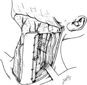
Surgical Technique. The cervical incision is
out-lined as shown in Figure 1, the skin paddle always being located at the same neck side of the tumor resection. The medial limit of the IHFMCF lies at the midline, the upper limit at the level of the hyoid bone, and the lower limit at the supraster-nal notch, the lateral limit lies 3 to 5 cm from the midline. The shape of the flap is rectangular in a vertical position.
While performing the incision, we suggest immediately incising the skin and platysma all
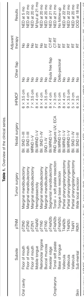
Ta b le 1 . Ov ervie w o f the cli nical serie s. Su bsite pTNM Tum or surgery Nod al surg ery IHFM CF Other flap Adjuva nt th erapy Re sults Oral cavity Floor of mou th pT 2N0 Marg inal mandi bulecto my Bil. SND I– III 6 3 3. 5 c m N o N o NED at 5 m o Floor of mou th pT 2N0 Marg inal mandi bulecto my Bil. SND I– III 9 3 4 c m N o N o NED at 22 mo Floor of mou th pT 2N1 Marg inal mandi bulecto my MRND I– V 7 3 3. 5 c m N o N o NED at 20 mo Mob ile tong ue pT 4aN0 Hemigl ossecto ly Bil.MRND I– V 8 3 4 c m N o R T M1pul at 21 mo Retro mola r trigo ne pT 1N0 Marg inal mandi bulecto my Bil. SND I– IV 8 3 4 c m N o N o NED at 20 mo Bucc al mucos a p T 1N0 Marg inal mandi bulecto my SND I– IV 6 3 3. 5 c m N o R T NED at 20 mo Alve olar ridge pT 4aN2b Se gmental m andibu lectomy Bil.MRND I– V 8 3 4 c m Fibula fre e flap CT-R T NED at 21 mo Oroph arynx Tons il soft palate pT 4aN2b Tons il þ soft palate rese ction RND ligat ure IJV , ECA 7 3 3 c m N o CT-R T NED at 21 mo Base of to ngue pT 4aN2c Tong ue ba se rese ction Bil.MRND I– V 9 3 5 c m Delto-pe ctoral RT No follow-up Va llecul a T 4 aN2b Pa rtial pharyn golaryn gect omy Bil.MRND I– V 8 3 4 c m N o R T NED at 22 mo Va llecul a T 4 aN2c Pa rtial pharyn golaryn gect omy Bil.MRND I– V 6 3 3. 5 c m N o R T NED at 22 mo Va llecul a T 4 aN1 Pa rtial pharyn golaryn gect omy Bil. SND I– IV 5 3 3 c m N o R T NED at 20 mo Skin Su b-me ntal R0N 1 Wide local excision Bil. SND I-III 6 3 3. 5 N o R T DOD at 18 m o Abbreviations: IHFMCF, infrahyoidfasciomyoc utaneous flap; B il., bilateral; SND, selective neck dissection; NED, no evidence of disease; MRND, mo dified radical n eck dissection; RT, radiotherapy; CT, chemotherapy; RND, radical neck dissection; IJV, internal jugular vein; ECA, external carotid artery; DOD, dead of disease.
FIGURE 1. (A) Cervical incision if unilateral neck dissection is needed. (B) Cervical incision if bilateral neck dissection is needed.
around the skin paddle to allow prompt choke per-forator vessels opening.
The cervical skin flap are elevated as during a standard neck dissection, the superficial cervical fascia along the anterior border of the sternoclei-domastoid muscle, from the sternal insertion up to the level of the hyoid bone, is incised, and the dis-section of the fascia proceeds until the intermedi-ate tendon of the omohyoid muscle is identified at its intersection with the internal jugular vein. The tendon is divided and subfascial dissection is car-ried on toward the flap, suturing this portion of the fascia and the stump of the omohyoid muscle to the lateral edge of the skin paddle (Figure 2).
After modified radical or selective neck dissec-tion is completed (the preservadissec-tion of the superior thyroid and internal jugular veins is mandatory), the dissection of the flap starts by dividing the an-terior jugular vein and sectioning the sternohyoid and sternothyroid muscles distally at the level of the suprasternal notch (Figure 3).
The skin paddle is stitched to the underlying muscles and then the IHFMCF is raised over the avascular plane of the proper capsule of the thy-roid gland; when the dissection reaches the upper pole, the cricothyroid artery and vein, all the dis-tal branches of the superior thyroid artery and vein that supply the thyroid gland and the poste-rior branch of the supeposte-rior thyroid artery and vein at their entrance in the upper pole of the gland are legated, divided, and kept with the flap (Figure 4).
The sternothyroid muscle is detached from the thyroid cartilage.
Special care must be taken in preserving the external branch of the superior laryngeal nerve; therefore, the thyrohyoid muscle is usually spared and left in place. Finally, the hyoid insertion of the
FIGURE 3. The dissection of the flap starts by dividing the an-terior jugular vein and sectioning the sternohyoid and sternothy-roid muscles distally at the level of the suprasternal notch.
FIGURE 4. When the dissection reaches the upper pole of the thyroid gland, all the distal branches of the superior thyroid ar-tery and vein that supply the thyroid gland are individually legated, divided, and kept with the flap.
FIGURE 2. The tendon of the omohyoid muscle is divided and subfascial dissection is carried on toward the flap.
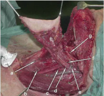
sternohyoid and omohyoid muscles are sectioned inside out in a subperiostial plane. The pedicle of the flap is formed by the neurovascular pedicle (superior thyroid artery and vein and ansa cervi-calis), by fascial connections between the superfi-cial and median cervical fascia, and by periosteal connections to the digastric muscle (Figure 5).
Those facial connections are important to di-rectly provide microvascular venous return to-ward the median cervical fascia and to protect the superior thyroid vein from twisting or kneeing, so creating the new infrahyoid fascio-myocutaneous flap.
The flap is ready to be transferred to recon-struct the defect.
The donor site can be usually primarily closed, if the width of the skin paddle is greater then 5 cm a deltopectoral flap could be needed.
RESULTS
In this series, all the flaps reached the recipient area without extensive vascular pedicle stretch-ing, even in case of soft palate reconstruction. The rectangular shape of the skin paddle matched per-fectly with the shape of the resections that re-sulted mostly oval or rectangular (Figure 6). No total or partial flap necrosis was experienced. All
reconstructions healed timely and without wound complications. Also, the healing process at the do-nor site was excellent in every case, with good aes-thetic results, including the patient who needed a deltopectoral flap to achieve donor site closure.
In this series, good functional results were achieved; all patients were decannulated and the nasogastric feeding tube was removed with resto-ration of oral intake in all cases. In every case, the flap withstood adjuvant treatments without any local complication, and the long-term results as to reconstruction appearance were excellent. We experienced no flap fibrosis, and only 1 patient experienced hair growth in the skin paddle.
DISCUSSION
In this series, 7 patients of 13 presented with a rel-ative contraindication for a FRFF reconstruction: 2 patients had systemic vascular insufficiency, 1 patient was HIV positive with poor general condi-tion, 1 patient was a professional piano player, and 3 patients were elderly and in poor general condition.
The advantages of the IHFMCF include its easy and relatively quick preparation, and a flap that is harvested during neck dissection so there is no need for a second surgical team. The skin paddle is hairless in most cases, and in almost ev-ery case the donor area can be primarily closed avoiding skin grafting or scars beyond the head and neck area, with absence of significant cos-metic and functional squeals. On the other hand, FRFF reconstruction mostly requires 2 surgical teams, an expert microsurgeon, and vigilant
mon-FIGURE 5. a: cricothyroid artery; b: posterior branch of the superior thyroid artery at the entrance in the upper pole of the thyroid gland; c: fascial connections between superficial and median cervical fascia; d: superior thyroid artery; e: superior thyroid vein; f: ansa cervicalis; g: periosteal connections to the digastric muscle. [Color figure can be viewed in the online issue, which is available at www.interscience.wiley.com.]
FIGURE 6. Postoperative result after 6 months. The flap covers the marginal mandibulectomy and reconstructs the floor of mouth allowing good tongue mobility. The rectangular shape of the skin paddle matched perfectly with the shape of the resec-tion. [Color figure can be viewed in the online issue, which is available at www.interscience.wiley.com.]
itoring of the free flap during the first postopera-tive days.
The majority of myocutaneous flaps for head and neck reconstruction (eg, pectoralis major, tra-pezius, latissimus dorsi) are quite bulky, and for this reason, we found that the IHFMCF repre-sents an excellent alternative to FRFF reconstruc-tion for medium-sized defects of the oral cavity and oropharynx, which can also easily reach sites such as retromolar trigone and soft palate.
The IHFMCF is thin and pliable, and even if it is not as thin and pliable as the FRFF, it appears to be extremely suitable in case of floor of mouth recon-struction, especially in case of marginal mandibulec-tomy and en bloc resections, because it is able to pro-vide thigh closure preventing salivary fistulas in the neck and it allows good motility of the tongue.
In 1 case, where a segmental mandibulectomy was performed, we combined the IHFMCF with a free fibula reconstruction. If the mucosal loss is not very large, the IHFMCF suits the defect perfectly and the osseous microvascular transfer is well cov-ered by vascularized infrahyoid muscles.
In case of tongue reconstruction, it is useful to preserve the motor innervation of the infrahyoid muscles provided by the ansa cervicalis. The main advantage of this voluntary innervated flap is that the innervation prevents scarring and atrophy of the reconstructed tongue.6 For intermediate oro-pharyngeal defects, we found that the IHFMCF suits the defect perfectly if the defect does not extend into the oral cavity. On the other hand, if a pharyngeal defect does extend to the oral cavity, a complex reconstruction in terms of dimensions and shape is needed, and the FRFF appears to be pref-erable. For soft palate reconstruction, if the resec-tion is strictly uniolateral and does not include the uvula, the IHFMCF can be used with good func-tional results preventing open rhinolalia and pro-viding soft palate competence without nasal regur-gitation. If a larger soft palate defect exists, a dou-ble-folded FRFF is functionally superior. In case of partial pharyngolaryngectomy for vallecula carci-noma, the IHFMCF provided an excellent restora-tion of form and funcrestora-tion, being small and pliable.
All flaps in this series were harvested with the inclusion of the fascia cervicalis superficialis, which assists microvascular tissue drainage to-ward the median cervical fascia. For this reason, it is important to keep a certain amount of fascia around the superior thyroid pedicle without thin-ning it too much during the dissection. This ma-neuver does not interfere with the oncologic radi-cality of the neck dissection.
CONCLUSIONS
A critical recodification of the role of the infra-hyoid flap in modern days could be of great help for the microvascular surgeon looking for alterna-tives, because in management of head and neck tumors the tool box of a wide range of reconstr-uctive options is of a great advantage. In our series, the IHFMCF has shown to be a reliable flap even in elderly patients and in patients in general poor condition or with peripheral vessel insuffi-ciency who are not optimal candidates for free flap reconstruction. The use of the described technical modifications together with the inclusion of part of the superficial cervical fascia in the harvest has led to a complete success rate without venous problems in this series.
This flap is thin and pliable, so that it is par-ticularly useful in oral cavity reconstructions. In this preliminary experience, our impression is that for small and medium-sized defects the func-tional results are comparable to those with the FRFF reconstruction. FRFF appears to be prefer-able for the reconstruction of extensive oropha-ryngeal defects where a large amount of skin is needed, but for the closure of small and medium-sized defects and after partial pharyngolaryn-gectomy, IHFMCF has proved to be an excellent alternative.
Contraindications such as previous neck dissec-tion, previous thyroid surgery, and presence of N3 neck disease must be respected. A relative contra-indication is previous radiotherapy. If it is possible to preserve the superior thyroid pedicle dividing the internal jugular vein and external carotid ar-tery just above its branching, the infrahyoid flap can be harvested also if metastatic lymph nodes are present at level II. In our series, 8 patients pre-sented a pN+ neck at the side of the flap, and it was always possible to use the planned IHFMCF despite the proximity to a metastatic node.
Acknowledgment. The authors thank Fran-cesca Brunone for the surgical diagrams.
REFERENCES
1. Evans GR, Schusterman MA, Kroll SS, Miller MJ, et al. The radial forearm flap in head and neck reconstruction: a review. Am J Surg 1994;168:446–50.
2. Wang HS, Shen JW. Preliminary report on a new approach to the reconstruction of the tongue. Acta Acad Med Prim Hanghai 1980;7:256–259.
3. Magrin J, Kowalski LP, DiPaula RA, et al. Infrahyoid myocutaneous flap in head and neck reconstruction. Head Neck 1993;15:522–525.
4. Wang H, Junwen S, Tian A, et al. The infrahyoid myocu-taneous flap for reconstruction after resection of head and neck cancer. Cancer 1986;57:663–668.
5. Rojanin S, Suphaphongs N, Ballantyne AJ. The infra-hyoid musculocutaneous flap in head and neck recon-struction. Am J Surg 1991;162:400–403.
6. Remmert SM, Sommer KD, Majocco AM, Weerda HG. The neurovascular infrahyoid flap: a new method for tongue reconstruction. Plast Reconstr Surg 1997;99:613– 618.
7. Zhao YF, Zhang WF, Zhao JH. Reconstruction of intra-oral defects after cancer surgery using cervical pedicle flaps. J Oral Maxillofac Surg 2001;59:1142–1146.
8. Deganello A, De Bree R, Dolivet G, Leemans CR. Infra-hyoid myocutaneous flap reconstruction after wide local excision of a Merkel cell carcinoma. Acta Otorhinolar-yngol Ital 2005;25:50–54.
9. Wang H. 10 years’ experience on infrahyoid myocutane-ous flap. Zhonghua Er Bi Yan Hou Ke Za Zhi 1991;26: 332–334, 382.
10. Dolivet G, Gangloff P, Sarini J, Ton Van J, Garron X, Guillemin F, Lefebvre JL. Modification of the infra hyoid musculo-cutaneous flap. Eur J Surg Oncol 2005;31:294–298. 11. Sobin LH, Wittekind Ch, editors. TNM classification of malignant tumors, 6th ed. International Union Against Cancer; New York: Wiley-Liss; 2002.