Research Article
Corticosteroids for Acute and Recurrent Idiopathic Pericarditis:
Unexpected Evidences
Antonio Perrone, Anna Castrovilli , Giuseppina Piazzolla, Sabina Savino,
Alessia D’Introno , and Carlo Sabb`a
Interdisciplinary Department of Medicine, Internal Medicine Unit “Cesare Frugoni”, School of Medicine, University of Bari, Piazza G. Cesare 11, 70100 Bari, Italy
Correspondence should be addressed to Anna Castrovilli; [email protected] Received 21 May 2019; Accepted 25 November 2019; Published 16 December 2019 Academic Editor: Lihua Duan
Copyright © 2019 Antonio Perrone et al. This is an open access article distributed under the Creative Commons Attribution License, which permits unrestricted use, distribution, and reproduction in any medium, provided the original work is properly cited.
Pericarditis is a common disease, often postviral or “idiopathic,” diagnosed in about 5% of emergency room visits for non-ischemic chest pain. Although pericarditis often occurs as a benign and self-limiting disease, it may present recurrences. The first-line therapy includes aspirin/nonsteroidal anti-inflammatory drugs (ASA/NSAIDs) plus colchicine. Steroids especially at high-dose have been associated with a higher recurrence rate. In this retrospective study, we evaluated efficacy and safety of ASA/ NSAIDs and prednisone in the treatment of acute or recurrent idiopathic pericarditis (colchicine was off-label in the period of the study). The cohort included 276 patients diagnosed with acute idiopathic pericarditis. Mean age was 45.4 ± 12.7 years, and males were significantly higher in number and younger than females. Sixty-one patients (22.1%) were treated with prednisone and 215 with ASA/NSAIDs (77.9%). 171 patients experienced at least one recurrence (62%). No difference in recurrence rate was observed (p � 0.257) between the groups treated with prednisone (55.7%) vs. ASA/NSAIDs (63.7%). The recurrences were treated with steroids at low doses and very gradual tapering, and the dose reduction was slower as the number of relapses was higher. Steroids alone were administered to about 80% of patients, while in the remaining 20% of cases, they were associated with ASA/NSDAIDs or colchicine. Approximately 90% of patients had a very favorable course, that is no more than 2 relapses and no patients presented serious side effects. Steroids at low dose, did not act, surprisingly, as an independent risk factor for recurrences and therefore may be considered a successful and safe treatment for acute and recurrent idiopathic pericarditis.
1. Introduction
Acute pericarditis (AP), with or without effusion, is the most common pericardial disease, it accounts approximately for 0.2% of all cardiovascular admission [1], and 5% of patients visited in an Emergency Department for nonischemic car-diac chest pain [2].
It occurs more often in men aged 20 to 50 years; however, the prevalence data in elderly subjects (>65 years) are poor due to the limited studies [3].
There are multiple causes of AP which can be broadly divided into infectious and noninfectious [4–6], but most of AP (80–90%) are of unknown etiology, labeled as idiopathic [7–9].
The AP prognosis is generally good, and recurrences are reported after 15–30% of all acute idiopathic pericarditis [3, 10]. It is supposed an occult autoimmune etiology for these recurrent subtypes [10, 11]. In a few cases, severe complications, such as cardiac tamponade and constrictive pericarditis [12, 13], may occur and they are usually related to the etiology of pericarditis.
The treatment of AP is largely empirical except those cases with a specific etiology. Medical therapy includes nonsteroidal anti-inflammatory drugs (NSAIDs), aspirin (ASA), colchicine, and steroids. ASA/NSAIDs plus colchi-cine represents the first line of idiopathic pericarditis treatment [10]. Steroids are recommended in those patients with contraindications or failure of ASA/NSAIDs plus
Volume 2019, Article ID 1348364, 7 pages https://doi.org/10.1155/2019/1348364
colchicine [10] as they are considered an independent risk factor for recurrent pericarditis [10, 14].
However, some issues remain unclear about the therapy of acute idiopathic pericarditis (AIP), i.e., what is the best duration of therapy, what are the best doses, how best ta-pering steroids, and why the association steroid-colchicine does not reduce and may even increase the AP recurrence rate [15].
The aim of the present study was to evaluate the efficacy and safety of ASA/NSAIDs and steroids for the treatment of acute or recurrent idiopathic pericarditis.
2. Materials and Methods
This study is a retrospective analysis of a cohort of patients admitted to the Internal Medicine Unit “Cesare Frugoni” of Bari University Hospital with a diagnosis of AP during the time period of January 1993 to December 2016.
The diagnosis of AP was based on at least two of the following criteria: (a) typical pericardial chest pain, (b) pericardial friction rubs upon auscultation, (c) characteristic electrocardiogram (ECG) changes, and (d) new pericardial effusion mainly detected by echocardiography [10].
We descriptively analysed demographic parameters, clinical presentation, physical examination findings, labo-ratory (creatinine, electrolytes, troponin I, liver enzymes, CRP, haemoglobin, leucocytes, thyroid hormones, antinu-clear antibodies, anti-DNA antibodies, rheumatoid-factor, serum electrophoresis, urine analysis, viral and neoplastic markers, and Mantoux or QuantiFERON-TB Gold test) and instrumental (ECG, chest X-ray, echocardiogram, chest CT/ MRI scan) investigations, underlying etiology, comorbid-ities, and therapies. In no case, HIV test and pericardial biopsy were performed.
These clinical investigations allowed classifying AP into idiopathic and secondary to a known etiology.
Pericardial effusion was evaluated by echocardiography and classified as a mild (<10 mm, estimated volume <200 ml), moderate (10–20 mm, estimated volume 200– 500 ml), and large effusion (>20 mm, estimated volume >500 ml) [16, 17]. Pericardiocentesis and the subsequent analysis of pericardial fluid were performed only in patients with cardiac tamponade and hemodynamic impairment. Follow-up was performed only in AIP patients, re-evaluating clinical and laboratory features approximately every 3 months. The median follow-up time was 23.5 months (range 11–36 months).
Pericarditis relapse was attained according to the same clinical criteria utilized for diagnosis of acute pericarditis. A diagnosis of recurrent pericarditis was made if the relapse occurred after 4–6 or more weeks symptom-free interval, during therapy discontinuation or pharmacological tapering [10].
2.1. Statistical Analysis. Continuous data were reported as
mean ± SD; categorical variables were reported as frequency and percentage. Patient groups were compared by use of Student’s t test for continuous variables and χ2analysis or
Fisher’s exact test, as appropriate, for categorical variables. A value of p < 0.05 was considered statistically significant.
3. Results and Discussion
3.1. Clinical Presentation and Baseline Features. During the
study period, 313 cases of AP (1.2% of the annual hospi-talized patients) were recorded. The patients were more likely to be male (214; 68.4%) than female (99; 31.6%) with mean age of 45 ± 12.6 years (range 17–76 years).
Diagnosis of AIP was made in 276 of 313 patients (88.2%).
Among 37 patients with pericarditis of known aetiology (11.8%), metastatic neoplasms were found in 40.5% of cases, 8 cases developed AP after cardiac surgery (21.6%), a di-agnosis of autoimmune disease was made in 7 patients (18.9%), 3 subjects had post-traumatic pericarditis (8.1%), tuberculosis pericarditis was diagnosed in 2 cases (5.4%), and uremic pericarditis was shown in 2 patients with end-stage renal disease. Detailed features of patients with a pericarditis secondary to a known aetiology are reported in Table 1.
Table 2 describes demographic, clinical and laboratory characteristics of AIP patients. Males were significantly more numerous than females (67.8%, M/F: 2.1, p < 0.001), mean age 45.4 ± 12.7 years. However, when the subjects were stratified by age, the sex-based differences in prevalence were statistical significant only in the group aged ≤65 years (M/F: 2.3, p � 0.002), while no differences were detected in the group aged >65 years (M/F ratio � 0.6, p � 0.78). Male patients were significantly younger than female (43.5 ± 11.8 yrs vs. 49.7 ± 13.3 yrs, p < 0.001). The most prevalent presenting symptom was chest pain (98.6%), followed by cough (43%) and dyspnea (18%). On physical examination, pericardial rub was detected in 104 patients (38%), and fever was present on admission in 102 cases (37%). Typical ECG changes were found in 204 subjects (74%). Pericardial effusion on echocardiography was shown in 182 subjects (66%) and was estimated as severe in 44 patients (24%), moderate in 51 patients (28%), and mild in 87 patients (48%). Pleural effusion was observed in 96 subjects (35%). Cardiac tamponade was detected in 11 patients, and in 7 patients with hemodynamic compromise, an emergency pericardiocentesis was performed, showing in all cases a serohematic fluid. Only in those cases, pericardial fluid analysis was carried out and did not yield a specific diagnosis in any case. Pericardial biopsy was not performed. White blood cells, C-reactive protein (CRP), and erythrocyte sedimentation rate (ESR) elevations were observed in almost all patients as reported in Table 2.
3.2. Patient Management. As shown in Table 3, ASA/
NSAIDs were administered to 215/276 patients with AIP (78%), in particular ASA (1.5–3 g daily) to 194/276 patients (70.2%) and NSAIDs, namely, ibuprofen (1.2–1.8 g daily), to 21/276 patients (7.6%). Corticosteroids (prednisone) were prescribed in 61/276 patients (22.1% of cases) after the exclusion of tubercular or purulent pericarditis, at variable
doses (0.25–0.5 mg/kg/daily, until the resolution of symp-toms and normalization of the inflammatory markers, generally for two-three weeks, followed by gradual tapering of 5 mg each two-three weeks). Patients treated with cor-ticosteroids were significant older (57 ± 7.1 vs. 42 ± 12
p< 0.001) and presented more relevant pericardial effusion
(44 vs. 0, p < 0.001), dyspnea (36.1% vs 12.6), and TnI (41% vs 24%) than ASA/NSAIDs-treated patients. Moreover steroids were used more frequently in patients affected by diseases that requested their use as haemolytic autoimmune anemia, De Quervain thyroiditis (5 vs. 0, p < 0.001).
3.3. Follow-Up Data. After a median follow-up of 23.5
months (range, 11–36 months), recurrences of pericarditis were diagnosed in 171 AIP patients (62%) and they were more common in ASA/NSAIDs-treated patients than in the ones treated with corticosteroids (64% vs 56%); however, the difference was not statistically significant (p � 0.257) (Table 4).
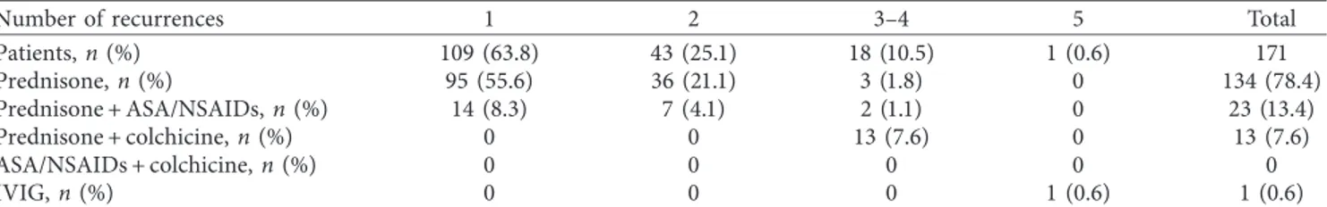
As shown in Table 5, among patients with recurrent pericarditis, 109 patients (63.8%) had one episode, 43 pa-tients (25.1%) had 2 episodes, 18 papa-tients (10.5%) had three to four episodes, and only one patient had five episodes.
Despite the recommendations of ESC 2015 guidelines [10], in Italy, until April 2017, colchicine was off-label for the AIP treatment; therefore, it was prescribed in a small number of patients of this study; in particular, it was added (off-label) to corticosteroids in 13 patients who experienced ≥3 episodes, whereas the other patients were treated with prednisone alone or in combination with ASA or NSAIDs, except for the patient who had 5 relapses and was suc-cessfully treated with IVIG.
No cases of deaths, severe side effects (heart failure, osteoporosis, myopathies, neurological disorders, and bleedings, severe progression of renal disease), or steroid dependent recurrent pericarditis were recorded.
4. Discussion
Currently available diagnostic tests (hematochemical, se-rological, cultural, and histological examination on biopsies) in most cases do not allow to establish the exact etiology of AP that therefore is named acute idiopathic pericarditis (AIP).
The high prevalence of AIP patients observed in our study (Table 1) is consistent to that reported from other studies [4–6].
As shown in Table 1, the neoplastic pericarditis prevailed among the secondary forms and in particular those asso-ciated with melanoma that are rarely highlighted in the literature [18]. Neoplastic pericarditis are frequently asso-ciated with cardiac tamponade, as shown in this study.
Table 2 shows age and sex significant differences that were found in AIP subjects. The observed sex differences are unknown, although a role of male sex hormones can be supposed, as suggested by some experimental studies on myocarditis in mice [19, 20]. It seems that testosterone may play an important role by the inhibition of anti-in-flammatory cells [21], the increase of viral binding to the myocytes [22], and the stimulation of immune responses mediated by Th1-lymphocytes [20].
In our study, the mean age at onset of AIP was lower in male subjects compared to that in women. However, when the patients were stratified by age, the difference of mean age
Table 1: Etiology of acute pericarditis in the overall study population. Etiology Incidence Idiopathic, n (%) 276 (88.2) Secondary 37 (11.8) Infective, n (%) 2 (0.6) Autoimmune, n (%) 7 (2.2) SLE, n (%) 3 (1) Undifferentiated connectivitis, n (%) 2 (0.6) Still disease, n (%) 1 (0.3) Horton disease, n (%) 1 (0.3) Neoplastic, n (%) 15 (4.8) Lung, n (%) 5 (1.6) Lymphoma, n (%) 3 (1) Melanoma, n (%) 3 (1) Breast, n (%) 2 (0.6) Myeloma, n (%) 1 (0.3) Gastric, n (%) 1 (0.3) ESRD, n (%) 2 (0.6) Post-surgery, n (%) 8 (2.6) Post-trauma, n (%) 3 (1)
Values are n (%). SLE indicates systemic erythematous lupus; ESRD, end-stage renal disease.
Table 2: Demographic and clinical data of the 276 patients with acute idiopathic pericarditis.
Feature Value Age, y (mean ± SD) 45.4 ± 12.7 Male 43.5 ± 11.8∗ Female 49.7 ± 13.3∗ Male, n (%) 187 (67.8)∗ Thoracic pain, n (%) 272 (98.6) Fever, n (%) 102 (36.9) Cough, n (%) 118 (42.8) Dyspnea, n (%) 49 (17.8)
Upper respiratory tract infections in the previous 2
weeks, n (%) 149 (53.9) Diarrhea, n (%) 35 (12.7) Pericardial rub, n (%) 104 (37.7) Pleural effusion, n (%) 96 (34.8) Pericardial effusion, n (%) 182 (65.9) Severe 44 (24.2) Moderate 51 (28) Mild 87 (47.8) Cardiac tamponade, n (%) 11 (3.9) Emergency pericardiocentesis n, (%) 7 (2.5)
ECG typical changes, n (%) 204 (73.9)
WBC > 103/mm3, n (%) 240 (87.7)
CRP > 2.9 mg/L, n (%) 273 (98.9)
cTnI > 0.045 ng/ml, n (%) 77 (27.8)
ESR > 20 mm/h, n (%) 266 (96.4)
Values are mean ± SD or n (%). ECG indicates electrocardiogram; WBC, white blood cells; CRP, C-reactive protein; cTnI, cardiac troponin I; ESR,
at clinical onset between male and female subjects was observed only in the younger age group (<65 years), while the gender distribution was nearly balanced in the patients aged >65 years. These results agreed with those reported in another study, where the male predominance was much more pronounced in patients younger than 45 [23].
Compared with the literature, clinical, laboratory, and instrumental features were equally frequent.
As shown in Table 3, in first line, about 80% of patients were treated with aspirin or NSAIDs while for the remaining 20% steroids, at variable doses and variable tapering, were used.
Steroids were generally administered to elderly, to those patients with more severe clinical features, comorbidities that needed steroids (autoimmune haemolytic anemia, thyroiditis, etc.), contraindications to or intolerance of ASA/ NSAIDs.
No other differences in clinical variables were found between patients treated with ASA/NSAIDs vs. prednisone. As for steroids, high doses (until 2 mg/kg/day) were used in the most severe forms (associated with myocarditis or severe pericardial effusion). In the literature, although corticoste-roids produce a fast and satisfactory clinical response, their use has been identified as an independent risk factor for
Table 4: Follow-up data of the study population.
Feature Total (276) ASA/NSAIDs (215) Prednisone (61) pvalue
Recurrence, n (%) 171 (62) 137 (63.7) 34 (55.7) 0.257
Complete remission, n (%) 105 (38) 78 (36.3) 27 (44.3) 0.257
Values are n (%). The recurrences rate was not statistically different between the two groups.
Table 5: Follow-up data of the population with recurrent pericarditis.
Number of recurrences 1 2 3–4 5 Total
Patients, n (%) 109 (63.8) 43 (25.1) 18 (10.5) 1 (0.6) 171 Prednisone, n (%) 95 (55.6) 36 (21.1) 3 (1.8) 0 134 (78.4) Prednisone + ASA/NSAIDs, n (%) 14 (8.3) 7 (4.1) 2 (1.1) 0 23 (13.4) Prednisone + colchicine, n (%) 0 0 13 (7.6) 0 13 (7.6) ASA/NSAIDs + colchicine, n (%) 0 0 0 0 0 IVIG, n (%) 0 0 0 1 (0.6) 1 (0.6)
Values are n (%). In the lines are listed the treatments of the recurrences and in the columns the n, % of patients who experienced the relative recurrence. IVIG indicates intravenous immunoglobulins.
Table 3: Baseline clinical characteristics of AIP patients treated with prednisone vs ASA/FANS.
Feature Prednisone (61) ASA/FANS (215) pvalue∗
Age, y 57 ± 7.1 42 ± 12 <0.001 Female, n (%) 22 (36.1) 67 (31.2) Thoracic pain, n (%) 59 (96.7) 213 (99.1) Fever, n (%) 21 (34.4) 81 (37.7) Cough, n (%) 27 (44.3) 91 (42.3) Dyspnea, n (%) 22 (36.1) 27 (12.6) <0.001
Upper respiratory tract infections in the previous 2
weeks, n (%) 33 (54.1) 116 (54) Diarrhea, n (%) 8 (13.1) 27 (12.6) Pericardial rub, n (%) 12 (19.7) 92 (42.8) �0.001 Pleural effusion, n (%) 25 (41) 71 (33) Pericardial effusion, n (%) Mild/moderate 17 (28) 121 (56) <0.001 Severe 44 (72.1) 0 (0) <0.001 Cardiac tamponade, n (%) 11 (18) 0 (0) <0.001 Emergency pericardiocentesis n, (%) 7 (11.4) 0 (0) <0.001
ECG typical changes, n (%) 45 (73.8) 159 (74)
WBC > 103/mm3, n (%) 53 (86.9) 187 (87)
CRP > 2.9 mg/L, n (%) 61 (100) 212 (98.6)
cTnI > 0.045 ng/ml, n (%) 25 (41) 52 (24.2) �0.012
ESR > 20 mm/h, n (%) 61 (100) 205 (95.3)
Comorbidities†, n (%) 5 (8.1) 0 (0) <0.001
Values are mean ± SD or n (%). ECG indicates electrocardiogram; WBC, white blood cells; CRP, C-reactive protein; cTnI, cardiac troponin I; ESR, erythrocyte
sedimentation rate.∗pvalue is reported in the table only when it was <0.05.†
Comorbidities were Hashimoto’s disease, De Quervain’s thyroiditis, and haemolytic autoimmune anemia.
recurrence based on observational studies [12, 24–30]. Be-cause most cases of AIP are presumed to be postviral, it is believed that corticosteroids causing immunosuppression may impair antiviral immune response. Patients treated with high-dose steroids showed higher rate of side effects and a higher rate of relapses and hospitalizations [25]. So it could be believed that low-dose steroids are better, but the data are still limited and controversial. Currently, the recommended dose of prednisone is 0.25 to 0.5 mg/kg/day for 2–4 weeks, followed by a slow tapering [10, 14, 25].
Table 4 shows that the rate of first recurrences was more common in ASA/NSAIDs-treated patients (63.7%) than in patients treated with corticosteroids (55.7%), but the dif-ference was not statistically significant (p value � 0.257). This unexpected result shows the following: (i) same efficacy of low-dose steroids when used as first-line therapy, compared to ASA/NSAIDs, since they determined not statistically different recurrence rates; (ii) steroids, in this study, were not an independent risk factor for recurrences, unlike literature statements [10, 14].
In the literature, the frequency of first recurrences varies from different studies from 20% and 30% [24, 31], while the recurrence rate after the first relapse varies between 20% and 50% especially in those patients not treated with colchicine or after corticosteroids therapy [32, 33]. In this study, the patients treated with ASA/NSAIDs or steroids had both a first recurrence rate higher than the one reported in the literature (Table 4) [6, 34, 35]. This high rate of relapse is probably related to (a) the absence of colchicine as first-line therapy (in Italy, colchicine was off-label until April 2017) and (b) a “selection bias,” as our clinic, in Southern Italy, was considered a reference centre for the most severe or re-sistant-to-therapy AIPs. Many patients arrived directly to our department with very severe clinical manifestation, while others were treated, at first line, in different hospitals and transferred to our department for the severity of clinical manifestations or resistance to the treatment. Unfortunately, many of them presented a history of inappropriate first-line therapy (low doses of ASA/NSAIDs or too short duration of the therapy).
A limitation of this study is that the number of patients treated in the first line with steroids alone is considerably lower than the number of patients treated with ASA/ NSAIDs; therefore, it is not possible to express a definitive judgment on the rate of first recurrences.
As shown in Table 5, all recurrences were treated with steroids. Steroids alone were administered in about 80% of patients, while in the remaining 20% of cases, they were associated with ASA/NSDAIDs or colchicine. Recurrences were treated with low-dose steroids and very gradual ta-pering and the dose reduction was much slower as the number of relapses was higher. As a result of this recurrence treatment scheme, approximately 90% of patients had a very favorable course, that is no more than 2 relapses. In addition to this result, no patients presented serious side effects (i.e., myopathies, neurological/psychiatric disorders, osteoporo-sis, and dispeptic gastric disorders), and in no cases, pre-ventive treatments were administrated. Patients who, instead, had a less favorable prognosis (≥3 recurrences)
constituted only 10% of the total. By evaluating first-line therapies, these subjects had a too rapid steroid tapering. In these subjects, colchicine was added to the steroids. All of these patients had no more recurrences except only one (5 recurrences). This was a young patient diagnosed with re-current pericarditis associated with multiple sclerosis. The use of intravenous immunoglobulins (IVIG) for the fifth recurrence treatment definitively interrupted the pericarditis recurrences and resolved the symptomatology of the mul-tiple sclerosis.
Based on the results of this retrospective study and our clinical experience, we suggest that prednisone 0.25–0.5 mg/ kg/day given until the resolution of symptoms and nor-malization of the inflammatory markers, generally for three weeks, followed by gradual tapering of 5 mg each two-three weeks (based on the clinical and laboratory response), and further 2.5 mg each two-three weeks, until complete suspension, is a successful and safe treatment for AIP.
In resistant forms, multidrug therapy should be con-sidered, adding ASA or NSAIDs or colchicine to steroids, even if the optimal treatment has not yet been established. The treatment with immunosuppressive agents (azathio-prine, cyclosporine, cyclophosphamide, and methotrexate) [36, 37], IVIG, anakinra [38] (IL-1 receptor antagonist), and canakinumab [39] (IL-1β blocking monoclonal antibody), may be considered only in cases of recurrent pericarditis which is refractory or intolerant to conventional treatments, but data for their use are limited, and should be tailored to the specific individual patients. Finally, pericardiectomy may be performed for frequent and highly symptomatic re-currences of pericarditis, which is resistant to medical treatment.
5. Conclusions
In our study, prednisone showed an equal efficacy compared to ASA/NSAIDs, having determined the same first re-currence rate, as it was not really an independent risk factor for recurrences, unlike the literature evidences [10, 14].
The recurrence rate after the first line treatment was, in the entire study population, higher than the expected on the basis of literature data, probably due to the absence of colchicine in first line and a selection bias of the patients.
The treatment of recurrent pericarditis showed the ef-ficacy of steroids at low doses and very gradual tapering, resulting in a notable reduction of recurrence rate and a low percentage of prolonged courses, unlike the literature evi-dences [10, 14].
Steroids, at low doses and gradual tapering, were, also particularly safe as no serious side effects occurred without any preventive therapy (i.e., vitamin D), while ASA/NSAIDs at high doses, for prolonged time, may get worse gastric dyspeptic disease, renal failure, arterial hypertension, and heart failure, especially in elderly.
The AIP therapy must be tailored on the phenotype of the patient, considering important variables such as severity of the clinical manifestations, age, comorbidities, and side effects of therapies and interactions between the pharma-cological treatments.
Therefore, based on the results presented, although in the literature there are discordant evidences and opinions not favorable to the use of steroids for the first line treatment of idiopathic pericarditis [10, 14], we believe that they may be considered safe and effective when administered at low doses and very gradual tapering, especially in the current “aging era,” in which many more patients will not be able to tolerate high-dose NSAIDs.
Data Availability
Our excel data are protected and we couldn’t give it to anyone else on the basis of what our ethic committee established and of what the patients accepted in the in-formed consense.
Disclosure
This abstract was presented as conference abstract in “Programma Scientifico 14 Congresso Nazionale FADOI” according to the following link. This research article was not published elsewhere. https://planning.it/files/EventDocuments/ Simposio_Tavola%20Rotonda_2019_81465.pdf. The research did not receive specific funding and was performed as of the employment of the authors. The employer is the Italian “Ministero dell’Universit`a e della Ricerca.”
Conflicts of Interest
The authors declare that there are no conflicts of interest regarding the publication of this paper.
References
[1] V. Kyt¨o, J. Sipil¨a, and P. Rautava, “Clinical profile and in-fluences on outcomes in patients hospitalized for acute pericarditis,” Circulation, vol. 130, no. 18, pp. 1601–1606, 2014. [2] M. M. LeWinter, “Acute pericarditis,” New England Journal of
Medicine, vol. 371, no. 25, pp. 2410–2416, 2014.
[3] P. Mody, B. Bikdeli, Y. Wang, M. Imazio, and
H. M. Krumholz, “Trends in acute pericarditis hospitaliza-tions and outcomes among the elderly in the USA, 1999– 2012,” European Heart Journal—Quality of Care and Clinical
Outcomes, vol. 4, no. 2, pp. 98–105, 2018.
[4] B. Maisch, P. M. Seferovi´c, A. D. Risti´c et al., “Guidelines on the diagnosis and management of pericardial diseases exec-utive summary: the task force on the diagnosis and man-agement of pericardial diseases of the European Society of Cardiology,” European Heart Journal, vol. 25, no. 7, pp. 587–610, 2004.
[5] S. Azam and B. D. Hoit, “Treatment of pericardial disease,”
Cardiovascular Therapeutics, vol. 29, no. 5, pp. 308–314, 2011.
[6] M. Imazio, A. Brucato, F. G. DeRosa et al., “Aetiological diagnosis in acute and recurrent pericarditis: when and how,”
Journal of Cardiovascular Medicine, vol. 10, no. 3, pp. 217–
230, 2009.
[7] R. Zayas, M. Anguita, F. Torres et al., “Incidence of specific etiology and role of methods for specific etiologic diagnosis of primary acute pericarditis,” The American Journal of
Cardi-ology, vol. 75, no. 5, pp. 378–382, 1995.
[8] G. Permanyer-Miralda, “Acute pericardial disease: approach to the aetiologic diagnosis,” Heart, vol. 90, no. 3, pp. 252–254, 2004.
[9] B. Maisch, H. Rupp, A. Ristic, and S. Pankuweit, “Peri-cardioscopy and epi- and pericardial biopsy-a new window to the heart improving etiological diagnoses and permitting targeted intrapericardial therapy,” Heart Failure Reviews, vol. 18, no. 3, pp. 317–328, 2013.
[10] Y. Adler, P. Charron, M. Imazio et al., “2015 ESC guidelines for the diagnosis and management of pericardial diseases: the task force for the diagnosis and management of pericardial diseases of the European Society of Cardiology (ESC),”
Eu-ropean Heart Journal, vol. 36, no. 42, pp. 2921–2964, 2015.
[11] G. Markel, M. Imazio, A. Brucato, and Y. Adler, “Colchicine for the prevention of recurrent pericarditis,” Israel Medical
Association Journal, vol. 10, pp. 69–72, 2008.
[12] M. Imazio, D. H. Spodick, A. Brucato, R. Trinchero, and Y. Adler, “Controversial issues in the management of peri-cardial diseases,” Circulation, vol. 121, no. 7, pp. 916–928, 2010.
[13] M. Imazio, A. Brucato, S. Maestroni et al., “Risk of constrictive pericarditis after acute pericarditis,” Circulation, vol. 124, no. 11, pp. 1270–1275, 2011.
[14] M. Imazio, A. Brucato, R. Trinchero, R. Shabetai, D. Spodick, and Y. Adler, “Corticosteroid therapy for pericarditis: a double-edged sword,” Nature Clinical Practice Cardiovascular
Medicine, vol. 5, no. 3, pp. 118-119, 2008.
[15] A. Mager, Y. Talmor, C. Chezar Azzerad et al., “Does col-chicine decrease the rate of recurrence of acute idiopathic pericarditis treated with glucocorticoids?,” Journal of
Cardi-ology, vol. 71, no. 4, pp. 409–413, 2018.
[16] M. S. Horowitz, C. S. Schultz, E. B. Stinson, D. C. Harrison, and R. L. Popp, “Sensitivity and specificity of echocardio-graphic diagnosis of pericardial effusion,” Circulation, vol. 50, no. 2, pp. 239–247, 1974.
[17] L. B. Weitzman, W. P. Tinker, I. Kronzon, M. L. Cohen, E. Glassman, and F. C. Spencer, “The incidence and natural history of pericardial effusion after cardiac surgery-an echocardiographic study,” Circulation, vol. 69, no. 3, pp. 506–511, 1984.
[18] P. T. Vaitkus, H. C. Herrmann, and M. M. LeWinter, “Treatment of malignant pericardial effusion,” JAMA: The
Journal of the American Medical Association, vol. 272, no. 1,
pp. 59–64, 1994.
[19] S. A. Huber, L. P. Job, and K. R. Auld, “Influence of sex hormones on Coxsackie B-3 virus infection in Balb/c mice,”
Cellular Immunology, vol. 67, no. 1, pp. 173–189, 1982.
[20] S. A. Huber and B. Pfaeffle, “Differential Th1 and Th2 cell responses in male and female BALB/c mice infected with coxsackievirus group B type 3,” Journal of Virology, vol. 68, pp. 5126–5132, 1994.
[21] S. Frisancho-Kiss, M. J. Coronado, J. A. Frisancho et al., “Gonadectomy of male BALB/c mice increases Tim-3+ al-ternatively activated M2 macrophages, Tim-3+ T cells, Th2 cells and Treg in the heart during acute coxsackievirus-in-duced myocarditis,” Brain, Behavior, and Immunity, vol. 23, no. 5, pp. 649–657, 2009.
[22] D. C. Lyden, J. Olszewski, M. Feran, L. P. Job, and S. A. Huber, “Coxsackievirus B-3-induced myocarditis. Effect of sex ste-roids on viremia and infectivity of cardiocytes,” The American
Journal of Pathology, vol. 126, pp. 432–438, 1987.
[23] E. Schwegler, M. Bachmann, N. Krasniqi, and U. Eriksson, “Pericarditis in a Swiss regional hospital,” Cardiovascular
[24] M. Imazio, M. Bobbio, E. Cecchi et al., “Colchicine in addition to conventional therapy for acute pericarditis: results of the COlchicine for acute PEricarditis (COPE) trial,” Circulation, vol. 112, no. 13, pp. 2012–2016, 2005.
[25] M. Imazio, A. Brucato, D. Cumetti et al., “Corticosteroids for recurrent pericarditis,” Circulation, vol. 118, no. 6, pp. 667–671, 2008.
[26] U. Lange, C. Schumann, and K. L. Schmidt, “Current aspects of colchicine therapy—classical indications and new thera-peutic uses,” European Journal of Medical Research, vol. 6, no. 4, pp. 150–160, 2001.
[27] A. Brucato, Y. Adler, and D. H. Spodick, “Letter regarding article by Imazio et al. “Colchicine in addition to conventional therapy for acute pericarditis”,” Circulation, vol. 113, no. 14, p. e693, 2006.
[28] T. M. Melchior, V. Ringsdal, P. Hildebrandt, and C. Torp-Pedersen, “Recurrent acute idiopathic pericarditis treated with intravenous methylprednisolone given as pulse therapy,”
American Heart Journal, vol. 123, no. 4, pp. 1086–1088, 1992.
[29] R. A. Lange and L. D. Hillis, “Clinical practice. Acute peri-carditis,” New England Journal of Medicine, vol. 351, no. 21, pp. 2195–2202, 2004.
[30] M. Imazio, B. Demichelis, I. Parrini et al., “Recurrent pain without objective evidence of disease in patients with previous idiopathic or viral acute pericarditis,” The American Journal of
Cardiology, vol. 94, no. 7, pp. 973–975, 2004.
[31] M. Imazio, A. Brucato, R. Cemin et al., “A randomized trial of colchicine for acute pericarditis,” New England Journal of
Medicine, vol. 369, no. 16, pp. 1522–1528, 2013.
[32] M. Imazio, M. Bobbio, E. Cecchi et al., “Colchicine as first-choice therapy for recurrent pericarditis: results of the CORE (COlchicine for REcurrent pericarditis) trial,” Archives of
Internal Medicine, vol. 165, no. 17, pp. 1987–1991, 2005.
[33] M. Imazio, R. Belli, A. Brucato et al., “Efficacy and safety of colchicine for treatment of multiple recurrences of pericar-ditis (CORP-2): a multicentre, double-blind, placebo-con-trolled, randomized trial,” The Lancet, vol. 383, no. 9936, pp. 2232–2237, 2014.
[34] M. Imazio, A. Brucato, R. Cemin et al., “Colchicine for re-current pericarditis (CORP): a randomized trial,” Annals of
Internal Medicine, vol. 155, no. 7, pp. 409–414, 2011.
[35] M. Imazio, B. Demichelis, I. Parrini et al., “Day-hospital treatment of acute pericarditis: a management program for outpatient therapy,” Journal of the American College of
Cardiology, vol. 43, no. 6, pp. 1042–1046, 2004.
[36] R. Marcolongo, R. Russo, F. Laveder, F. Noventa, and C. Agostini, “Immunosuppressive therapy prevents recurrent pericarditis,” Journal of the American College of Cardiology, vol. 26, no. 5, pp. 1276–1279, 1995.
[37] F. Vianello, F. Cinetto, M. Cavraro et al., “Azathioprine in isolated recurrent pericarditis: a single centre experience,”
International Journal of Cardiology, vol. 147, no. 3,
pp. 477-478, 2011.
[38] G. Lazaros, M. Imazio, A. Brucato et al., “Anakinra: an emerging option for refractory idiopathic recurrent pericar-ditis a systematic review of published evidence,” Journal of
Cardiovascular Medicine, vol. 17, no. 4, pp. 256–262, 2015.
[39] N. Kougkas, A. Fanouriakis, I. Papalopoulos et al., “Cana-kinumab for recurrent rheumatic disease
associated-peri-carditis: a case series with long-term follow-up,”
Stem Cells
International
Hindawi www.hindawi.com Volume 2018 Hindawi www.hindawi.com Volume 2018 INFLAMMATIONEndocrinology
International Journal ofHindawi www.hindawi.com Volume 2018 Hindawi www.hindawi.com Volume 2018
Disease Markers
Hindawi www.hindawi.com Volume 2018 BioMed Research InternationalOncology
Journal of Hindawi www.hindawi.com Volume 2013 Hindawi www.hindawi.com Volume 2018Oxidative Medicine and Cellular Longevity
Hindawi
www.hindawi.com Volume 2018
PPAR Research
Hindawi Publishing Corporation
http://www.hindawi.com Volume 2013 Hindawi www.hindawi.com
The Scientific
World Journal
Volume 2018 Immunology Research Hindawi www.hindawi.com Volume 2018 Journal ofObesity
Journal of Hindawi www.hindawi.com Volume 2018 Hindawi www.hindawi.com Volume 2018 Computational and Mathematical Methods in Medicine Hindawi www.hindawi.com Volume 2018Behavioural
Neurology
Ophthalmology
Journal of Hindawi www.hindawi.com Volume 2018Diabetes Research
Journal ofHindawi
www.hindawi.com Volume 2018
Hindawi
www.hindawi.com Volume 2018
Research and Treatment
AIDS
Hindawi
www.hindawi.com Volume 2018
Gastroenterology Research and Practice
Hindawi www.hindawi.com Volume 2018