The Rockefeller University Press $30.00
The Wiskott-Aldrich syndrome (WAS OMIM
301000) is an X-linked disorder characterized
by thrombocytopenia, eczema, and
immuno-deficiency (Thrasher and Burns, 2010). T
lym-phocytes from WAS patients fail to proliferate
and secrete IL-2 in response to cross-linking of
the TCR–CD3 complex. Moreover, they exhibit
actin cytoskeletal defects illustrated by defective
F-actin polymerization after TCR–CD3
cross-linking, reduced ability to spread upon contact
with anti-CD3–coated surfaces, impaired
chemo-taxis in vitro, and poor homing in vivo to
lym-phoid organs (Thrasher and Burns, 2010). NK cell
function and immune synapse formation with
target cells is also impaired in WAS (Orange et al.,
2002; Gismondi et al., 2004; Stabile et al., 2010).
In T lymphocytes, WASP is almost totally
complexed with the WASP-interacting
pro-tein (WIP; de la Fuente et al., 2007). A major
function of WIP is to stabilize WASP and
pre-vent its degradation. WASP protein levels, but
not mRNA levels, are severely reduced in T cells
from WIP-deficient mice. Introduction of
full-length WIP, but not of WIP that lacks the
WASP binding domain, restores WASP levels
in these cells (de la Fuente et al., 2007). We
de-scribe for the first time a patient who presented
in early infancy with a phenotype of WAS who
was found to have a homozygous mutation in
the WIPF1 gene, which encodes WIP.
RESULTS AND DISCUSSION
Clinical characteristics
The index patient was the second female child
of consanguineous Moroccan parents. She was
referred at 11 d of age with poor weight gain,
CORRESPONDENCE Silvia Giliani: [email protected] Abbreviations used: CFSE, carboxyfluorescein succinimidyl ester; hWIP, human WIP; IP10, C-X-C motif chemokine immune protein-10; rIl-2, recombinant IL-2; WAS, Wiskott-Aldrich syndrome; WASP, WAS protein; WIP, WASP interacting protein.
G. Lanzi and D. Moratto contributed equally to this paper. R.S. Geha and S. Giliani contributed equally to this paper.
A novel primary human immunodeficiency
due to deficiency in the WASP-interacting
protein WIP
Gaetana Lanzi,
1Daniele Moratto,
1Donatella Vairo,
1Stefania Masneri,
1Ottavia Delmonte,
2Tiziana Paganini,
1Silvia Parolini,
3Giovanna Tabellini,
3Cinzia Mazza,
1Gianfranco Savoldi,
1Davide Montin,
2Silvana Martino,
2Pierangelo Tovo,
2Itai M. Pessach,
4Michel J. Massaad,
4Narayanaswamy Ramesh,
4Fulvio Porta,
6Alessandro Plebani,
1Luigi D. Notarangelo,
4,5Raif S. Geha,
4and Silvia Giliani
11A. Nocivelli Institute for Molecular Medicine, Pediatric Clinic, University of Brescia, and Laboratory of Genetic Disease of Childhood, Spedali Civili, 25123 Brescia, Italy
2Department of Pediatrics, University of Turin, 10126 Turin, Italy
3Department of Biomedical Sciences and Biotechnologies, University of Brescia, 25123 Brescia, Italy 4Division of Immunology and 5The Manton Center for Orphan Disease Research, Children’s Hospital,
Harvard Medical School, Boston, MA 02115
6Division of Hematology and Oncology, Spedali Civili, 25123 Brescia, Italy
A female offspring of consanguineous parents, showed features of Wiskott-Aldrich
syndrome (WAS), including recurrent infections, eczema, thrombocytopenia, defective T cell
proliferation and chemotaxis, and impaired natural killer cell function. Cells from this
patient had undetectable WAS protein (WASP), but normal WAS sequence and messenger
RNA levels. WASP interacting protein (WIP), which stabilizes WASP, was also undetectable.
A homozygous c.1301C>G stop codon mutation was found in the WIPF1 gene, which
encodes WIP. Introduction of WIP into the patient’s T cells restored WASP expression.
These findings indicate that WIP deficiency should be suspected in patients with features of
WAS in whom WAS sequence and mRNA levels are normal.
© 2012 Lanzi et al. This article is distributed under the terms of an Attribution– Noncommercial–Share Alike–No Mirror Sites license for the first six months after the publication date (see http://www.rupress.org/terms). After six months it is available under a Creative Commons License (Attribution–Noncommercial–Share Alike 3.0 Unported license, as described at http://creativecommons.org/licenses/ by-nc-sa/3.0/).
The Journal of Experimental Medicine
on October 26, 2012
jem.rupress.org
Downloaded from
rotavirus enteritis at 2 mo of age, and acute hepatitis of
un-known etiology at 3 mo of age. No clinical manifestations of
autoimmunity or bleeding tendency were noted. Because of
persistent deterioration, failure to gain weight, and poor T cell
function, at the age of 4.5 mo she underwent unrelated cord
blood transplantation. 16 mo after the procedure, she is alive and
well with >98% of T cells, >98% of B cells, 94% of NK cells,
50% of monocytes, and 41% of granulocytes of donor origin.
The oral ulcerations resolved after bone marrow transplant,
sug-gesting that they were secondary to deficient immune function.
Immunological analysis
Analysis of peripheral blood revealed low percentages and
numbers of CD3
+cells (809 cells/µl), with CD8
+cells more
affected than CD4
+cells (Fig. 1 A and Table S1). 90% of
an eczematous rash, papulovesicular lesions on the scalp, and
ulcerative lesions on the hard palate and tongue. A previous
female sibling suffered from ulcerative and vesicular skin
lesions and died of sepsis at 4 mo of age.
Laboratory findings included thrombocytopenia (59 ×
10
3/µl platelets) with normal platelet volume and elevated
levels of C reactive protein (10.3 mg/dl;
Table S1
). Blood
and urine cultures were negative. Stool cultures revealed no
pathogenic organisms. S. epidermidis and K. pneumoniae grew
from the skin vesicular lesions. The patient developed
respi-ratory distress and required oxygen supplementation. A
tra-cheal aspirate was positive for respiratory syncytial virus by
PCR. She was placed on wide-spectrum antibiotic therapy,
fungal and viral prophylaxis, immunoglobulin replacement, and
platelet and red cell transfusions as needed. She developed
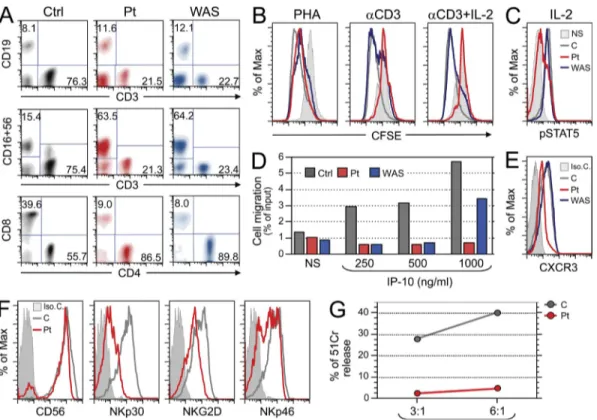
Figure 1. Functional characterization of WIP deficiency. (A) Evaluation of the proportion of T, B, and NK cells on gated lymphocytes in whole blood (top
and middle row), and of CD4- and CD8-expressing cells on the CD3+ gated cells (bottom row) of a control (Ctrl), the patient (Pt), and a WAS-null patient. Data are representative of three independent analyses. (B) T cell proliferation to PHA and anti-hCD3 with or without rIL-2, as assessed by CFSE dilution. PBMCs were used and FACS analysis was performed on CD3+ gated cells. The solid gray histogram represents exemplificative profile of unstimulated cells. An overlapping profile was obtained from unstimulated cells of control and WAS patients (not depicted). The red, gray, and blue profiles represent CFSE content in stimulated cells from patient, control, and a WAS-null patient, respectively. Data are representative of two independent experiments. (C) PBMCs were stimulated with IL-2, and gated CD3+ cells were analyzed by FACS for intracellular pSTAT5 using anti-pY694STAT5. The solid line represents the signal in unstimulated patient’s cells, which was comparable to that of unstimulated control and WAS-null patient cells (not depicted). The pSTAT5 signal in the rIL-2 stimulated cells from patient, control, and WAS patient are indicated in red, gray, and blue, respectively. Data are representative of three independent experiments. (D) Migration of PHA blasts from a healthy control, the patient and from a WAS-null patient toward the chemokine IP-10. The gray, red, and blue histograms represent migration of cells from con-trol, patient, and WAS-null patient, respectively. Data are representative of two independent experiments. (E) Expression of the IP-10 receptor CXCR3 on PHA blasts from patient, control, and WAS-null patient. Isotype control staining of patient PHA blasts is shown by the filled histogram and was comparable to that of control T cells and WAS patient (not depicted). The CXCR3 signals in cells from patient, control, and WAS patient are indicated in red, gray, and blue, respectively. Data are representative of two independent experiments. (F) Expression of CD56, NKp30, NKGD2, and NKp46 on NK cells from patient (red) and control (gray). The filled histogram represents isotype control staining of patient NK cells, and was comparable to that of control NK cells (not depicted). Data are representative of two analyses. (G) Cytolytic activity by patient and control NK cell lines against the LCL 721.221 target cells, measured as the percentage of net 51Cr release at effector (E) to target (T) ratios of 3:1 and 6:1. Data are representative of two independent experiments on the same expanded NK cells.
on October 26, 2012
jem.rupress.org
Expression of the IP-10 receptor CXCR3 was lower in the
patient’s T cells (Fig. 1 E), and may have contributed to their
failure to migrate toward IP-10. Analysis of cytolytic activity
of freshly isolated NK cells from the patient was precluded
by the amount of blood that could be drawn. To circumvent
this limitation, NK cells were expanded by stimulating T cell–
depleted PBMCs from patient and a control with PHA and
rIL-2 in the presence of feeder cells (Castriconi et al., 2007).
The resulting cell lines contained >90% CD56
+NK cells,
which expressed comparable levels of CD56 on their surface.
However, expression of NKp30, NKp46, and NKG2D, all of
which are implicated in NK cytotoxicity, were reduced on the
patient’s cells (Fig. 1 F). Analysis of cytolytic activity against
LCL 721.221 target cells demonstrated a drastic reduction in
the functional activity of the patient’s NK cell line compared
with that from control (Fig. 1 G). Previous studies have shown
impaired cytotoxicity of freshly isolated NK cells from WAS
patients (Gismondi et al., 2004). We have been unable to
de-rive NK cell lines from the WAS patient for direct comparison
with the NK cell line from our patient.
WIPF1 is mutated in the patient
Although the WAS gene is located on the X-chromosome,
several cases of WAS in females caused by extreme
lyoni-zation or biallelic WAS mutations have been described
CD3
+cells were TCR
+, and 7% were TCR
+. B cell
number was low (319 cells/µL), whereas the percentage and
number of CD16
+CD56
+NK cells were increased (2,485
cells/µl; Fig. 1 A and Table S1), as reported in patients with
WAS (Gismondi et al., 2004). Serum immunoglobulins were
normal, except for elevated IgE (32 IU/ml).
Because of the clinical similarities between our patient and
WAS patients we studied in parallel the function of her T cells
and those from a WAS patient who expressed no detectable
WASP protein. Analysis of T cell proliferation by CFSE
dilu-tion revealed normal proliferadilu-tion to PHA, but completely
de-fective proliferation to immobilized anti-CD3 in the patient
(Fig. 1 B). In contrast, the proliferation of T cells from the
WAS patient to anti-CD3 was only partially decreased.
Ad-dition of IL-2 failed to correct the inability of the patient’s
T cells to proliferate to anti-CD3 (Fig. 1 B), and induced
only modest STAT5 phosphorylation compared with control
T cells (Fig. 1 C). In contrast, IL-2 completely corrected the
proliferation of WASP-deficient T cells to anti-CD3 (Fig. 1 B)
and caused normal STAT5 phosphorylation in these cells
(Fig. 1 C). Migration toward the C-X-C motif chemokine
immune protein-10 (IP-10) was abolished in PHA blasts from
the patient at all three concentrations tested (Fig. 1 D).
WASP-deficient PHA blasts responded to the highest
concentra-tion of IP-10 tested, but not to the lower two concentraconcentra-tions.
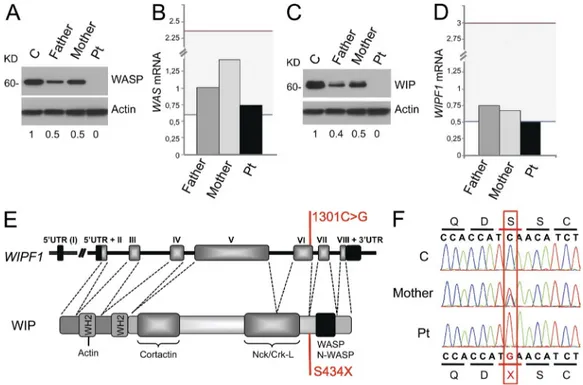
Figure 2. WIP is mutated in the patient. (A and C) Western blot analysis of WASP (A) and WIP (C) in lysates of PHA T cell blasts. Numbers at the
bottom indicate densitometric quantification corrected for the actin signal. Data are representative of two experiments. (B and D) qPCR analysis of mRNA expression of WAS (B) and WIPF1 (D) in PHA T blasts, relative to the housekeeping gene GAPDH, normalized to 1 for the mean of 25 controls. The gray area indicates the range for the 25 controls. Data are representative of three experiments conducted on triplicates. (E) Genomic organization of WIPF1 (top) and protein structure (bottom) of WIP showing the localization of the mutation and the resulting amino acidic change (in red) from Serine to a pre-mature termination codon (S434X). The genomic organization shows coding exons, indicated with Roman numbers, and 5 and 3 untranslated regions (UTR). Protein structure includes known WIP-interacting protein domains and WASP homology 2 (WH2). (F) Electropherogram depicting the homozygous c.1301C>G mutation in exon 6 in the patient and the presence of the same mutation in the heterozygous state in the mother.
on October 26, 2012
jem.rupress.org
had a homozygous point mutation c.1301C>G in exon 6 of
the WIPF1 gene located on chromosome 2 (Fig. 2, E and F).
This mutation results in a change from serine to premature
termination in codon 434 (S434X), situated immediately
up-stream of the region encoding the WASP-binding domain of
WIP (451–485 aa; Ramesh et al., 1997). Both parents were
heterozygous for the c.1301C>G substitution, confirming
the autosomal recessive inheritance of the mutation.
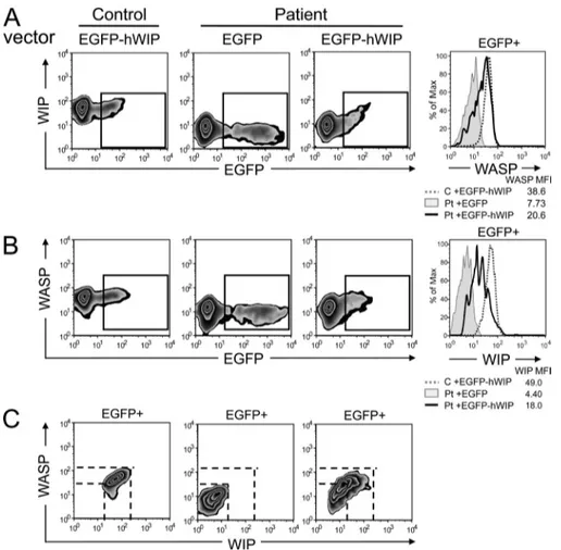
Expression of WIP in the patient’s cells corrects the defect
in WASP expression
We examined whether expression of human WIP (hWIP) in
the patient’s cells corrects their defective WASP expression.
PHA T blasts from the patient and a control were transfected
with vectors expressing EGFP-hWIP fusion protein or EGFP
alone, and their content of WIP and WASP was determined
by FACS analysis of intracellular staining. As expected,
intro-duction of EGFP-hWIP, but not EGFP, in the patient’s T cells
resulted in WIP expression, as indicated by the detection
of WIP
+cells in the EGFP
+gate (Fig. 3 A). More
impor-tantly, introduction of EGFP-hWIP, but not EGFP, in the
patient’s T cells resulted in increased WASP expression by
EGFP
+cells (Fig. 3 B). WASP expression in the
EGFP-hWIP–transfected cells from the patient correlated with WIP
expression (Fig. 3 C). These findings indicate that the
absence of WIP in the patient’s T cells resulted in WASP
instability and degradation. Because
the patient was transplanted at 4.5 mo
of age and because of the limited
amounts of blood we could obtain
pretransplant, we were not able to
examine whether transduction of WIP
in the patients T cells would restore
their function or examine the
corti-cal actin meshwork in the patient’s
T cells, which is severely attenuated
in WIP
/but not WASP
/mice
(Antón et al., 2002).
Several of the clinical
characteris-tics and laboratory findings in the
patient resemble those found in WAS.
They include recurrent infections,
eczematous skin rash,
thrombocyto-penia, T cell lymphopenia affecting
(Lutskiy et al., 2002; Proust et al., 2005). Western blot
analysis revealed no detectable WASP in lysates of the
pa-tient’s PHA T blasts (Fig. 2 A). WASP expression in the
PHA T blasts from both parents was reduced to 50% of
nor-mal as assessed by Western blot compared with the mean
of four controls (Fig. 2 A and not depicted). Similar results
were obtained by flow cytometric analysis of PBMCs from
the patient and her parents (unpublished data). The levels
of WAS mRNA in PHA T blasts from the patient and
her parents were within the normal range of 25 controls
(Fig. 2 B). Sequencing of the WAS gene in the patient
re-vealed no mutations.
The absence of WASP expression in the patient’s cells,
despite normal WAS sequence and mRNA expression level,
suggested that WASP was being degraded. Because WIP
stabilizes WASP, we examined its expression. Western blot
analysis of PHA T blasts from the patient revealed no
detect-able WIP (Fig. 2 C). WIP expression in the PHA T blasts
from both parents was reduced to 50% of normal (n = 4).
These results were confirmed by flow cytometric analysis of
PBMCs from the patient and her parents (unpublished data).
Quantitative PCR analysis revealed that the WIPF1
mRNA level in the patient’s PHA T blasts was at the lowest
limit of the normal range of 25 controls (Fig. 2 D). WIPF1
mRNA levels in the parents were in the lower range of
nor-mal. Sequencing of genomic DNA revealed that the patient
Figure 3. Introduction of WIP restores WASP levels in the patient T cells.
(A and B) Two-color FACS analysis of expres-sion of WIP (A) and WASP (B) versus EGFP in PHA T cell blasts transfected with vectors encoding EGFP-hWIP for patient and control and with EGFP alone for the patient. Histo-grams depicting WIP and WASP expression in gated EGFP+ cells are shown in the right pan-els. (C) Two-color FACS analysis of WASP ver-sus WIP expression in gated EGFP+ cells. Data are representative of two different analyses.
on October 26, 2012
jem.rupress.org
(Beckman Coulter). CD3 contamination was <1%. Purified NK cells were cultured on irradiated feeder cells in the presence of 100 U/ml of rIL-2 and 5 µg/ml of PHA (Invitrogen) to obtain activated polyclonal NK cell popu-lations. Activated polyclonal NK cells were maintained in complete RPMI medium containing 1,200 U/ml of rIL-2.
Molecular genetic analyses. Genomic DNA was isolated using the
auto-matic DNA extractor Maxwell 16 (Promega). Sequencing of genomic DNA corresponding to the coding regions of WAS (ENSG00000015285) and WIPF1 (ENSG00000115935) genes was performed by direct sequencing after PCR amplification of exons with flanking intronic regions. Primers and conditions are reported in Table S2.
Total RNA was isolated from PBMCs using the RNeasy Mini kit (QIAGEN) and transcribed into complementary DNA (cDNA). Quanti-tative PCR experiments were performed by reverse transcription of 200 ng of DNase-treated total RNA to synthesize the first strand of cDNA by the GeneAmp RNA PCR kit (Applied Biosystems). Analysis of WASP, WIPF1, and GAPDH gene expression was assessed by RealTime PCR using Assays-on-Demand products and TaqMan Master Mix from Ap-plied Biosystems. The level of expression was normalized using GAPDH as a reference.
Immunoblotting. Cell lysates from PHA T blasts were prepared and 10 µg
protein were loaded on a 10% SDS-PAGE and transferred onto a polyvinyli-dene fluoride membrane (GE Healthcare). Specific proteins were detected using mouse IgG1 anti-WIP mAb (3D10; Koduru et al., 2007), mouse IgG2a anti-WASP mAb 5A5 (BD), and mouse anti-actin mAb (US Biological).
Flow cytometry. Flow cytometry for surface and intracellular proteins was
performed on 100 µl of whole blood or isolated cells (1.5 × 10⁶) resuspended in 100 µl of the appropriate medium. To assess WASP and WIP expression, cells were treated with the Fix and Perm kit (Invitrogen) for FACS analysis. Cells were stained with mouse IgG1 anti WIP (3D10; Koduru et al., 2007) and mouse IgG2a anti-WASP (5A5; BD), followed by anti IgG1PE and bio-tinylated anti–IgG2a-streptavidinPECy5, respectively. Acquisition was per-formed using a FACSCalibur (BD), and data were analyzed with FlowJo Software v7.5 (Tree Star).
Standard flow cytometric methods were also used for CFSE assay. CFSE labeling (100 nM) was performed in PBS for 6 min at 37°C. Cells were then washed twice in RPMI 10% FCS. Cells were cultured for 96 h in the ab-sence of stimulation or stimulated with anti-CD3 (clone OKT3, 100 ng/ml) and anti–human CD28 (1 µg/ml) or PHA (5 µg/ml) and then analyzed for CFSE dilution.
For the characterization of NK cells, the following mAbs, which were produced in our laboratories, were used in this study: BAB281 (IgG1anti-NKp46), AZ20 (IgG1, anti-NKp30), c218 (IgG1, anti-CD56), BAT221 (IgG1, anti-NKG2D), and 289 (IgG2a, anti-CD3). For cytofluorimetric analysis, cells were stained with mAb PE-conjugated isotype-specific goat anti–mouse secondary antibody (SouthernBiotech). Cell acquisition was performed on a FACScan flow cytometer (BD), and data were analyzed using the CellQuest software (BD).
Lymphocyte functional analysis. Analysis of STAT5 phosphorylation in
T cells in response to IL-2 was assessed by flow cytometry: 100 µl of whole blood was incubated with or without 100 ng/ml rIL-2 (Proleukin-Chiron) for 10 min at 37°C. Activation was stopped using Lyse/Fix buffer (BD). After washing, the samples were permeabilized by PBIII Buffer (BD) for 30 min on ice and incubated with anti–phospho-STAT5 (Y694)-PE and CD3-FITC, or with isotype-matched mAb PE (BD) for 30 min at room tempera-ture. Samples were then washed and resuspended in 200 µl of BD CellFix and analyzed with FACSCalibur instrument (BD). To analyze NK cytolytic activity, PBMCs were depleted of T cells and expanded in vitro with rIL-2, resulting in highly enriched (>95%) CD56+ NK lymphocytes, which were tested for cytolytic activity against the NK-susceptible tumor target cell line LCL 721.221 in a 4-h 51Cr-release assay. Target cells were used at 5 × 10³ cells/well with final E/T ratios of 6:1 and 3:1.
CD8
+lymphocytes more severely, impaired T cell
prolifer-ation to immobilized anti-CD3, defective T cell chemotaxis,
and increased NK cell number but decreased NK cell
func-tion. In addition, the patient displayed immune abnormalities
that are observed in WIP-deficient mice, but not in WAS
patients or WASP-deficient mice. They include complete
failure to proliferate to TCR ligation with anti-CD3,
im-paired response of T cells to IL-2, and complete abrogation
of T cell chemotaxis (Fig. 1; Haddad et al., 2001; Gallego
et al., 2006; Le Bras et al., 2009). In contrast to WAS patients,
platelet volume was normal in the patient, like in WIP-deficient
mice (Curcio et al., 2007). However, we hesitate to draw a
firm conclusion on platelet size in human WIP deficiency
based on a single patient.
Despite undetectable WASP in the patient, no mutations
were detected in the coding region of the WAS gene and
WAS mRNA levels were normal, findings strongly
indica-tive of WASP instability. WIP, which is critical for the
sta-bility of WASP, was undetectable in the patient’s cells, and
a homozygous mutation that introduces a premature stop
codon in the WIPF1 coding sequence was identified in the
patient. The same mutation was found in the heterozygous
state in both parents, indicating that WIP deficiency was
inherited as an autosomal recessive trait. This is consistent
with an older sibling having died from a similar condition.
The fact that both parents showed reduced levels of WIP,
to approximately half of normal, suggests a gene dose effect.
The correspondingly reduced WASP level in the parents
suggests that WIP tightly regulates WASP levels. We have
observed a similar reduction of WASP levels in mice
het-erozygous for a WIP-null allele (unpublished data).
Impor-tantly, we demonstrated that reconstitution of the patient’s
T cells with WIP restores WASP levels, indicating that loss
of WIP expression was the cause of WASP instability in the
patient’s cells.
Collectively, the data indicate that WIP deficiency is
re-sponsible for the immune dysfunction in the patient. Based
on our findings, WAS cannot be diagnosed solely on the basis
of lack of WASP expression, but requires sequence analysis of
WAS. WIP deficiency should be suspected in patients with
features of WAS in whom WAS sequence and mRNA levels
are normal, and the diagnosis should be confirmed by
se-quencing WIPF1.
MATERIALS AND METHODS
Cell purification and culture. Patient’s samples were obtained upon
in-formed consent by the local Institution Review Board and in respect to the Helsinki declaration. Human studies were approved by the Ethical Commit-tee of Azienda Ospedaliera Spedali Civili, Brescia. Isolation of peripheral blood mononuclear cells (PBMCs) and of specific cell populations and estab-lishment of cell lines were performed as follow: to obtain PHA T blasts, PBMCs were stimulated with 5 µg/ml PHA (Sigma-Aldrich) and 600 U/ml rIL-2 (Cairon-Novartis) in RPMI medium containing 10% FCS, 1% l-glutamine, and 1X antibiotics (EuroClone). To generate NK cell lines, NK cells were purified by NK cell separation cocktail (Rosette Sep; StemCell Technologies). The purity of NK cells was >96% as assessed by flow cyto-metric analysis of cells stained with a mixture of CD56-PC5 and CD3-FITC
on October 26, 2012
jem.rupress.org
that resembles Wiskott-Aldrich syndrome. J. Pathol. 211:67–75. http:// dx.doi.org/10.1002/path.2088
de la Fuente, M.A., Y. Sasahara, M. Calamito, I.M. Antón, A. Elkhal, M.D. Gallego, K. Suresh, K. Siminovitch, H.D. Ochs, K.C. Anderson, et al. 2007. WIP is a chaperone for Wiskott-Aldrich syndrome protein (WASP). Proc. Natl. Acad. Sci. USA. 104:926–931. http://dx.doi.org/10 .1073/pnas.0610275104
Gallego, M.D., M.A. de la Fuente, I.M. Anton, S. Snapper, R. Fuhlbrigge, and R.S. Geha. 2006. WIP and WASP play complementary roles in T cell homing and chemotaxis to SDF-1alpha. Int. Immunol. 18:221– 232. http://dx.doi.org/10.1093/intimm/dxh310
Gismondi, A., L. Cifaldi, C. Mazza, S. Giliani, S. Parolini, S. Morrone, J. Jacobelli, E. Bandiera, L. Notarangelo, and A. Santoni. 2004. Impaired natural and CD16-mediated NK cell cytotoxicity in patients with WAS and XLT: ability of IL-2 to correct NK cell functional defect. Blood. 104:436–443. http://dx.doi.org/10.1182/blood-2003-07-2621 Haddad, E., J.L. Zugaza, F. Louache, N. Debili, C. Crouin, K. Schwarz, A.
Fischer, W. Vainchenker, and J. Bertoglio. 2001. The interaction between Cdc42 and WASP is required for SDF-1-induced T-lymphocyte chemo-taxis. Blood. 97:33–38. http://dx.doi.org/10.1182/blood.V97.1.33 Koduru, S., M. Massaad, C. Wilbur, L. Kumar, R. Geha, and N. Ramesh.
2007. A novel anti-WIP monoclonal antibody detects an isoform of WIP that lacks the WASP binding domain. Biochem. Biophys. Res. Commun. 353:875–881. http://dx.doi.org/10.1016/j.bbrc.2006.12.079 Le Bras, S., M. Massaad, S. Koduru, L. Kumar, M.K. Oyoshi, J. Hartwig,
and R.S. Geha. 2009. WIP is critical for T cell responsiveness to IL-2. Proc. Natl. Acad. Sci. USA. 106:7519–7524. http://dx.doi.org/10.1073/ pnas.0806410106
Lutskiy, M.I., Y. Sasahara, D.M. Kenney, F.S. Rosen, and E. Remold-O’Donnell. 2002. Wiskott-Aldrich syndrome in a female. Blood. 100:2763– 2768. http://dx.doi.org/10.1182/blood-2002-02-0388
Orange, J.S., N. Ramesh, E. Remold-O’Donnell, Y. Sasahara, L. Koopman, M. Byrne, F.A. Bonilla, F.S. Rosen, R.S. Geha, and J.L. Strominger. 2002. Wiskott-Aldrich syndrome protein is required for NK cell cyto-toxicity and colocalizes with actin to NK cell-activating immunologic synapses. Proc. Natl. Acad. Sci. USA. 99:11351–11356. http://dx.doi.org/ 10.1073/pnas.162376099
Proust, A., B. Guillet, I. Pellier, P. Rachieru, C. Hoarau, S. Claeyssens, C. Léonard, S. Charrier, W. Vainchenker, G. Tchernia, and J. Delaunay. 2005. Recurrent V75M mutation within the Wiskott-Aldrich syndrome protein: description of a homozygous female patient. Eur. J. Haematol. 75:54–59. http://dx.doi.org/10.1111/j.1600-0609.2005.00415.x Ramesh, N., I.M. Antón, J.H. Hartwig, and R.S. Geha. 1997. WIP, a
pro-tein associated with wiskott-aldrich syndrome propro-tein, induces actin polymerization and redistribution in lymphoid cells. Proc. Natl. Acad. Sci. USA. 94:14671–14676. http://dx.doi.org/10.1073/pnas.94.26 .14671
Stabile, H., C. Carlino, C. Mazza, S. Giliani, S. Morrone, L.D. Notarangelo, L.D. Notarangelo, A. Santoni, and A. Gismondi. 2010. Impaired NK-cell migration in WAS/XLT patients: role of Cdc42/WASp pathway in the control of chemokine-induced beta2 integrin high-affinity state. Blood. 115:2818–2826. http://dx.doi.org/10.1182/blood-2009-07- 235804
Thrasher, A.J., and S.O. Burns. 2010. WASP: a key immunological mul-titasker. Nat. Rev. Immunol. 10:182–192. http://dx.doi.org/10.1038/ nri2724
Chemotaxis assay. In vitro chemotaxis of PHA T blasts toward IP-10
was examined using a standard Transwell chamber assay: a total of 3 × 10⁵ PHA T blasts were added to the upper chamber of a plate 6.5 mm in diameter with a pore size of 5 µm (Costar). 600 µl of RPMI 1640 with 0.1% FCS was added to the bottom chamber with or without IP10 (250, 500, and 1,000 ng/ml; PeproTech). After 2 h at 37°C, cells that migrated to the lower chamber were collected and counted. The experiment was performed in duplicates. Expression of the IP-10 receptor CXCR3 was examined by FACS using a mouse anti–CXCR3-FITC mAb (R&D Systems).
WIP reconstitution experiment. hWIP cDNA was generated from
normal PBMCs by RT-PCR and cloned into the BglII–PstI cloning sites of the pAC-EGFP vector. Confirmation of the WT WIP sequence and its in-frame insertion in the vector was obtained by direct sequencing. PHA T blasts were transfected using the Amaxa nucleofection technology (Lonza) according to the manufacturer’s protocol, which is optimized for stimulated human T cells. In brief, 2–3 × 10⁶ cell suspension was mixed with 1 µg/106 cell of both mock and hWIP-containing plasmid DNA, transfected using the T-020 program, then cultured for 48 h in the growth medium. Mock- and hWIP-transfected cells were treated with the Fix/Perm kit (Invitrogen) for FACS analysis. Cells were stained with mouse IgG1 anti WIP (3D10) and mouse IgG2a anti WASP (5A5), followed by anti–IgG1-PE and biotinylated anti–IgG2a-streptavidin-PECy5, respectively. Acquisition was performed using a FACSCalibur (BD), and analysis was performed by FlowJo software (Tree Star). Transfected PHA-T cells were evaluated for WASP and WIP expression level gating on EGFP+ cells.
Online supplemental material. Table S1 provides the Laboratory data of
the patient at 3 wk of age. Table S2 provides the list of the primers used for the amplification of the WIPF1 gene. Online supplemental material is avail-able at http://www.jem.org/cgi/content/full/jem.20110896/DC1. This work was supported by Fondazione ‘A. Nocivelli’ to S. Giliani; United States Public Health Services grant 5PO1HL059561 to S. Giliani, L.D. Notarangelo, and R.S. Geha; Fondazione C. Golgi to A. Plebani; the Jeffrey Modell Foundation; and the Perkin fund. D. Vairo is recipient of a Rotary Brescia fellowship.
The authors have no conflicting financial interests.
Submitted: 4 May 2011 Accepted: 7 December 2011
REFERENCES
Antón, I.M., M.A. de la Fuente, T.N. Sims, S. Freeman, N. Ramesh, J.H. Hartwig, M.L. Dustin, and R.S. Geha. 2002. WIP deficiency reveals a differential role for WIP and the actin cytoskeleton in T and B cell activa-tion. Immunity. 16:193–204. http://dx.doi.org/10.1016/S1074-7613(02) 00268-6
Castriconi, R., A. Dondero, M. Cilli, E. Ognio, A. Pezzolo, B. De Giovanni, C. Gambini, V. Pistoia, L. Moretta, A. Moretta, and M.V. Corrias. 2007. Human NK cell infusions prolong survival of metastatic human neuroblastoma-bearing NOD/scid mice. Cancer Immunol. Immunother. 56:1733–1742. http://dx.doi.org/10.1007/s00262-007-0317-0 Curcio, C., T. Pannellini, S. Lanzardo, G. Forni, P. Musiani, and I.M.
Antón. 2007. WIP null mice display a progressive immunological disorder