1
UNIVERSITA’ POLITECNICA DELLE MARCHE
FACOLTA’ DI MEDICINA E CHIRURGIA
Dottorato di Ricerca XVI CICLO
in
SCIENZE BIOMEDICHE
PANCREATIC DUCTAL ADENOCARCINOMA: BIOMARKER IDENTIFICATION, EXOSOME
CHARACTERIZATION, APOPTOSIS AND NECROPTOSIS INDUCTION BY SULFORAPHANERelatore: Dottoranda:
Dott. Francesco Piva Dr.ssa Giulia Occhipinti
2
Table of content
1. Introduction ... 5
1.1 Epidemiology of pancreatic cancers ... 5
1.2 Risk factors ... 5
1.3 Pancreatic tumours and PDAC pathogenesis ... 6
1.4 Genetic mutations ... 8
1.5 Microenviroment ... 9
1.6 Biomarkers ... 10
1.7 Treatment... 12
2. Aim of the thesis ... 15
3. Exosome characterization in pancreatic cancer patients ... 16
3.1 Introduction ... 16
3.1.1 Exosomes ... 16
3.1.2 Biogenesis, secretion and uptake ... 17
3.1.3 Exosomes composition ... 17
3.1.4 Roles of exosomes in cancer ... 20
3.1.5 Exosome isolation and quantification techniques ... 21
3.2 Materials and Methods ... 24
3.2.1 Patients ... 24
3.2.2 Blood samples ... 25
3.2.3 Reagents ... 25
3.2.4 ELISA assay ... 25
3.3 Results ... 27
3.3.1 Evaluation of the best washing and blocking buffers ... 27
3.3.2 Evaluation of the appropriate antibody dilution buffer ... 28
3.3.3 Characterization of exosome tumour markers from plasma of PDAC patients... 29
3.4. Discussion ... 36
3.4.1 Elisa assay development and optimization ... 36
3.4.2 Exploring correlations between exosomal markers and clinical variables ... 37
4 Identification of gene and miRNA biomarkers for pancreatic ductal adenocarcinoma by weighted gene co-expression network analysis ... 41
4.1 Introduction ... 41
4.2 Materials and Methods ... 43
3
4.2.2 Dataset comparability analyses ... 44
4.2.3 Construction of weighted gene co-expression networks and their modules ... 44
4.2.4 Modules preservation analyses ... 45
4.2.5 Detection of hub genes and their functional annotation ... 45
4.2.6 Detection of hub miRNAs and their functional annotation ... 46
4.2.7 Survival analyses ... 46
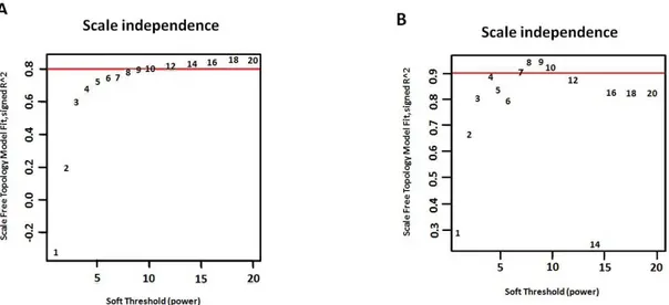
4.3 Results ... 47
4.3.1 Pre-processing of the Normal and PDAC dataset ... 47
4.3.2 Weighted gene and miRNA co-expression networks and their modules ... 48
4.3.3 Identification of hub genes and their functional annotations ... 51
4.3.4 Identification of the hub miRNAs and functional enrichment analysis of their targets ... 53
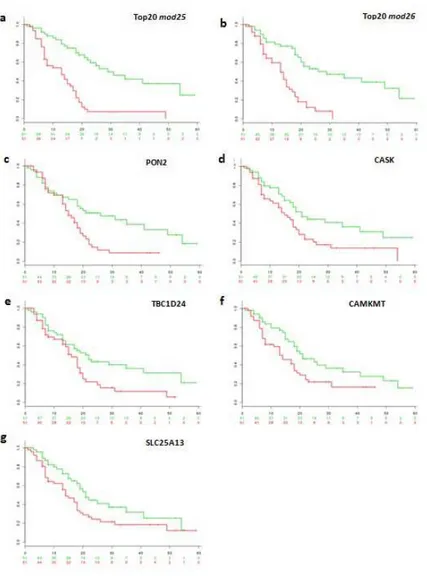
4.3.5 Stratification of PDAC patients into high- and low- risk groups based on novel candidate biomarkers ... 55
4.4 Discussion ... 59
5. Evaluation of cell death pathways induced by sulforaphane in pancreatic cancer cell lines ... 63
5.1. Introduction ... 63
5.1.1 Chemoprevention by phytochemicals in pancreatic cancer ... 63
5.1.2 Anti-cancer effects of sulforaphane ... 63
5.1.3 Apoptosis and Necroptosis ... 64
5.2. Materials and Methods ... 67
5.2.1 Cell culture ... 67
5.2.2 Reagents ... 67
5.2.3. Cell treatments ... 67
5.2.4 Cell viability detection by MTT assay ... 67
5.2.5 Detection of apoptosis by Annexin V/PI staining and FACS analysis ... 68
5.2.6 Detection of active caspase-3 by immunocytochemistry ... 68
5.2.7 Cell lysis and determination of protein concentration ... 68
5.2.8 Detection of RIP1 and MLKL by Western blot analysis ... 69
5.2.9 Statistical analysis ... 70
5.3. Results ... 71
5.3.1 Sulforaphane-reduced viability involves caspases, whereas necroptosis plays a minor role ... 71
4
5.3.3 Sulforaphane-induced caspase-3 cleavage ... 74
5.3.4 Sulforaphane reduces RIP1 and MLKL protein expression ... 74
5.4. Discussion ... 77
5
1. Introduction
1.1 Epidemiology of pancreatic cancers
Cancer is one of the major global public health problems, nevertheless the huge scientific research efforts have achieved, in the last decades, important results in terms of prevention, therapy and reduction of mortality rates. The deadliest cancers are considered those with 5-years relative survival rates below 50% and, according to Recalcitrant Cancer Research Act, pancreatic cancer, with 6% of survival, has the lowest percentage followed by lung (16,6%), liver (18%), esophagus (19%), stomach (29%), brain (35%), ovary (44%), and multiple myeloma (45%) [1]. While stomach, lung and breast cancer have shown falls in mortality since the late 1980s, pancreatic cancer mortality has the opposite trends in EU, with a steady rise in women of 3,9% and a stable mortality rate in men in 2016 [2]. Every year worldwide pancreatic cancer deaths are 200000 and it is predicted to be the second cause of cancer death in USA by
2030 [1]. Notably, African-Americans have a 30-50% higher incidence than other ethnic groups in the United States. Indigenous populations seem to be 30% more affected than other populations living in Oceania whereas the lowest rates are recorded in India, Africa and Southeast Asia. In this geographical variation, the quality of clinical diagnoses and the differential access to health care have to be taken into account because these evaluations could be altered by under diagnosis [3].
1.2 Risk factors
Pancreatic cancer incidence rate could be attenuated modifying some aspects of the life style. Among preventable factors, tobacco smoking is the most common agent, and it increases threefold the risk of developing pancreatic cancer [4]. It has been estimated that 25% of pancreatic cancers are attributable to cigarette smoking. Interestingly, the sequencing of pancreatic cancer genome has revealed that smoker patients have more somatic mutations than never-smoker patients. Other life habits connected with the frequency of this disease are low physical activity and some dietary factors like high consumption of saturated fats, red and processed meat and low intake of vegetables and fruits. Moreover, it has been observed that heavy alcohol consumption is correlated with the tumour incidence, while moderate alcohol intake does not induce an increased risk [4]. Additionally, some pathological states can
6
contribute to the neoplasm onset. Obesity is positively associated with pancreatic cancer, specifically, high body-mass index and centralized fat distribution may increase the risk [5]. Diabetes mellitus is not only a consequence of early-stage pancreatic cancer but it is also a risk factor. Diabetic patients have a 30% possibility of developing a tumour for more than 20 years after diagnosis [6].
Some hereditary conditions can also increase the risk of developing the disease. Several studies have demonstrated that 10% of patients have a family history of ductal adenocarcinoma of the pancreas, but the majority of the genetic basis remains unknown. Some germline genetic syndromes have been associated with an increased risk of developing this neoplasia. Inherited mutations in onco-suppressor genes BRCA2 and BRCA1 are associated with elevated risk of breast and ovarian cancer. While BRCA2 mutations account for familial pancreatic cancer, the role of mutated BRCA1 remains uncertain. Germline mutations in the p16/CDKN2A causes familial atypical multiple mole melanoma syndrome with a high lifetime risk of melanoma, as well as an increased risk of pancreatic cancer. Patients with Peutz-Jeghers syndrome, caused by germline mutations in STK11, have been shown to have an 11-32% lifetime risk of pancreatic cancer in addition to hamartomatous polyps in the gastrointestinal tract. Hereditary pancreatitis is a rare form of pancreatitis caused by mutations in PRSS1 and SPINK1. Patients with these alterations have a 30-40% possibility of developing cancer. Individuals with Lynch Syndrome, a disease caused by germline mutations in genes encoding DNA mismatch repair proteins, are characterized by early onset colon cancer and have an elevated risk of vary cancer types, among which is pancreatic cancer [4, 7].
1.3 Pancreatic tumours and PDAC pathogenesis
Pancreatic ductal adenocarcinoma (PDAC) is the most common and aggressive malignancy arising from the pancreas. Less frequent pancreatic neoplasms are neuroendocrine tumours, solid-pseudopapillary neoplasms, pancreatoblastomas, acinar carcinomas and colloid carcinomas.
Pancreatic neuroendocrine tumours (PanNETs) are the second most common neoplasm, representing 1-5% of all pancreatic tumours, with a mortality rate of 60%. They could arise as part of hereditary syndromes: multiple endocrine neoplasia type 1
7
(MEN-1), von Hippel-Lindau syndrome (VHL) and tuberous sclerosis complex (TSC). PanNETs are classified into functioning tumours which give early symptoms caused by excessive hormone production and non-functioning tumours that manifest only when they are large [8]. Solid-pseudopapillary neoplasms represent 1-2% of primary pancreatic tumours. This type appears as a heterogeneous low-grade malignant tumour with solid components and cysts and it is frequent in young women. Pancreatoblastoma accounts for 0,2% of all pancreatic neoplasms and it mostly occurs in children. At presentation, it is a large mass with aggressive behaviour and metastasis. Pancreatic ductal adenocarcinoma occurs in 90% of cases and has a remarkably poor prognosis. PDAC incidence is 50% higher in men than in women and old age is also a significant factor with most cases occurring in patients between 60 and 80 years. Pancreatic adenocarinomas are located in the head of the pancreas in 60-70% of patients, 10–20% in the body, and 5–10% in the tail. Principal marks, that appear too late in the disease course, are abdominal pain, weight loss and jaundice [9]. These tumours are solid and firm, derived from neoplastic cells which infiltrated into tissues forming glands and spread far from the primary tumour. Invasive cancer colonizes nerves, perineural and lymphatic spaces until it spreads to the liver. One of its important histologic traits is the intense desmoplastic reaction, which impedes biopsy to reach neoplastic glands and constitutes an obstruction for chemotherapeutic agents [3, 4]. Lack of early symptoms, absence of sensitive and specific biomarkers and the challenging identification of early-stage tumours are the principal reasons of a late diagnosis. Moreover, PDAC is characterized by high aggressiveness that leads to exclude surgical resection in most patients due to vascular invasion and early spread of metastasis. Traditional treatments like chemotherapy and radiotherapy are not effective since pancreatic tumour cells have resistance to the majority of anti-cancer agents [3].
Pancreatic ductal adenocarcinoma originated from non-invasive lesions. In 82% of pancreas with cancer, intraductal non-invasive proliferations called pancreatic intraepithelial neoplasia (PanIN) are observed. Timely detection of these precursor lesions may be crucial for an early diagnosis but so far their detection is challenging due to the lack of symptoms, specific biomarkers and mainly because of their microscopic size, which makes them not easily detectable by Magnetic Resonance
8
Imagery (MRI) or by Computed Tomography (CT). PanINs are classified into three grades depending on the cytological atypia. Low grade PanINs are found in 16-80% of normal pancreas without neoplasia while high grade PanINs are related to adenocarcinoma [10]. Less frequently, pancreatic cancer derives from macroscopic cystic precursors: intra-ductal papillary mucinous neoplasm (IPMN) and mucinous cystic neoplasm (MCN). IPMNs involve the larger pancreatic ducts, are larger than 1 cm in size and approximately a third of them are associated to a higher risk of developing invasive adenocarcinoma. MCNs occur particularly in women, arise in the body and the tail of pancreas, and do not involve the ductal system [7].
1.4 Genetic mutations
Cancer is a genetic disease caused not only by inherited mutations but also by somatic mutations involving tumour suppressor genes and oncogenes. The sequencing of infiltrating pancreatic ductal adenocarcinoma revealed that there are four major driver genes, KRAS, p16/CDKN2A, TP53 and SMAD4, somatically mutated in more than 50% of the cases. Precursor lesion analyses have sketched out the timing of the genetic alterations in pancreatic tumourigenesis. KRAS and CDKN2A mutations occur in low-grade PanINs, suggesting that they are the earliest alterations in pancreatic tumourigenesis. KRAS is an oncogene that encodes a small GTPase protein involved in the activation of MAP Kinase and/or the PI3K pathways that increase mitogenic activity. Its mutational activation results in the downstream activation of effector proteins that sustain proliferation, cell migration and metastasis. CDKN2A is a tumour suppressor gene inactivated in 95% of PDAC. Its mutation is associated to unlimited cell growth caused by the loss of function of its protein product, p16, an important cell cycle regulator. TP53 and SMAD4 mutations take place at an advanced stage of the carcinoma, suggesting that they are late events. The tumour suppressor TP53 codes for p53 protein, which responds to several cellular stresses inducing growth arrest and cell death. Loss of p53 function is observed in 75% of pancreatic cancers. The protein product of SMAD4 gene, Smad4, plays an important role in the TGFβ pathway and in transcription of cell cycle inhibitory factors like p21. Thereby, SMAD4 inactivation, in about 50% of cases, is linked to poor prognosis and metastatic disease [4, 7].
9
1.5 Microenviroment
Pancreatic ductal adenocarcinoma is different from other solid cancers because of the desmoplastic reaction, an abundant and dense collagenous stroma, which envelops malignant cells. This stroma contains extracellular matrix (ECM) proteins, such as collagens, fibronectin and laminin, non-collagenous proteins (glycoproteins, proteoglycans and glycosaminoglycans), as well as growth factors, osteopontin, periostin and serine protein acidic and rich in cysteine that may mediate the interaction of cancer cells and ECM [3]. Cellular component of desmoplasia consists of cancer-associated fibroblasts (CAFs), which produce the collagenous matrix and immune cells that could regulate the cancer growth. For these reasons, there has been growing interest in the role of desmoplasia in malignant and aggressive behaviour of pancreatic cancer and its resistance to treatment. Pancreatic stellate cells (PaSCs) are the principal cellular source of CAFs in pancreatic cancer. They are present in the exocrine pancreas in a normal quiescent state but are transformed in an activated state during the pathogenesis. Activated PaSCs have high proliferation rate and start to produce ECM proteins and the other components of desmoplasia. Several studies have demonstrated that this activated state is maintained not only by autocrine but also paracrine mechanisms with inflammatory and cancer cells. This relationship results in an increased tumour growth and metastasis development [11]. Interaction of pancreatic stellate cells with cancer cells and other stromal cancer cells promotes cancer progression. They are probably implicated in the formation of new blood vessels (angiogenesis) through the production of the proangiogenic factors: vascular endothelial growth factor (VEGF), platelet-derived growth factor (PDGF) and hepatocyte growth factor (HGF). These factors are found to be up-regulated in tumour tissue and are associated with poor prognosis [12]. Furthermore PaCSs promote stem-cell like phenotype conferring chemo-resistance upon pancreatic cancer stem-cells [13]. These cells are also able to travel from the primary tumour, surviving in the circulation, and seed metastatic niches to distant organs [14]. The desmoplastic stroma is composed also by different type of inflammatory cells: macrophages, T cells and neutrophilic granulocytes, that contribute mostly to the tumour progression. For example, CD4+ regulatory T cells have the decisive role in keeping away the host immune system, and M2 macrophages synthesize cytokines and chemokines involved
10
in tumour angiogenesis and metastasis. Factors implicated in the antitumour immunity suppression, for example PD-L1, are clinically important because they could be targets for new immunotherapeutic approaches [3, 15].
Metastasis formation needs a supportive environment that is established prior to the arrival of carcinoma cells, the so-called pre-metastatic niche. This environment is the result of numerous signalling factors from primary tumour to distant tissue that lead to the formation of metastasis [16]. Exosomes, nanovesicles secreted from cells by exocytosis, play an important role in this process. It has been demonstrated that pre-mestatic niches in the liver of naive mice are caused by PDAC-derived exosomes formation. Exosome uptake by hepatic cells induced TGFβ secretion and production of fibrotic microenvironment by hepatic stellate cells. In particular, it seems that the exosomal macrophage migration inhibitory factor (MIF) prepares the liver for metastasis since it was found highly expressed in exosomes derived from patients with late-stage cancer [17]. The role of exosomes in cancer is described in detail in the chapter 3.
1.6 Biomarkers
The importance of finding reliable biomarkers arises from the difficulty to detect the early stage of PDAC but also for evaluation of treatments or post-resection follow-up. Liquid biopsy is an alternative to surgical biopsies that allows detection of circulating tumour cells and so cancer at an early stage. It is a promising approach for the evaluation of new cancer markers in a non-invasive and easy way, generally through blood samples, instead of tumour tissue samples that are often difficult and time-consuming to obtain and to analyse. Moreover, liquid biopsy can be repeated several times during the therapy period without pain and risk for the patients, allowing the monitoring of tumour relapses or the occurrence of resistance mutations [18]. Among serum markers, carbohydrate antigen 19-9 (CA19-9) is the most widely studied. However, CA19-9 lacks sensitivity and specificity since the protein levels are often normal in the early stages of the disease or falsely high in individuals with other pathological conditions. These factors make it an inaccurate marker but it is nonetheless used to control disease progression, recurrence after surgery detection and therapy response. Carcinoembryonic antigen (CEA) could also be a diagnostic tool
11
but with low reliability [19]. Instead, it has been reported that CA19-9 and CEA combination or CA19-9, CA125 and laminin γC (LAMC2), increase specificity compared with the markers alone [7]. Another marker that could be detected non-invasively in the circulation is tumour DNA that can be extracted from circulating tumour cells (CTCs), intact and viable cells that are distinguished and isolated from the normal blood cells. Disadvantage of circulating tumour cells is that they are present only in some patients with advanced neoplasia. On the contrary, circulating tumour DNA (ctDNA), not associated with cells, has been found in blood samples in patients with localized disease. Using digital polymerase chain reaction–based technologies (dPCR) it is possible to evaluate somatic mutations of circulating DNA [18]. In a recent study, mutated KRAS has been detected from ctDNA of 43% of the patients at the time of diagnosis, indicating that this approach is a highly specific tool for early diagnosis [20]. Body fluids are also enriched in exosomes, extra-cellular vesicles participating in intercellular communication. They are a rich source of information since they are constituted by specific microRNAs, proteins, lipids and other nucleic acids, and thus are generating a big interest as tumour biomarkers. In particular, they bring on their external surface specific proteins that allow distinguishing exosomes derived from different subpopulations of pancreatic cancer cells. It is well known that tumour-derived exosomes express the same markers of cancer-initiated cells, which are implicated in metastasis formation, drug resistance and cancer recurrence. A recent work has demonstrated that circulating exosomes, from pancreatic cancer patients and genetically engineered mutant mice models, showed the proteoglycan glypican-1 on the membrane. Glypican-1 positive exosomes carry the KRAS mutation, allowing the distinction between healthy individuals and patients with early or late pancreatic cancer [21].
During my PhD, I analysed plasma samples of PDAC patients, using exosome markers,
with the aim of finding a correlation between different kinds of exosomes and patient clinical data that might be informative as prognostic and diagnostic tools (chapter 3). MicroRNAs enclosed in these nano-vesicles are considered potential biomarkers since they are specifically and differently expressed depending on the disease features: tumour growth, drug resistance or metastasis progression. Therefore, their evaluation in biofluids could be informative for the early detection of pancreatic cancer. To
12
measure exosomal miRNAs accurately is tricky and great care should be paid in all the experimental procedures, starting from samples handling, exosome isolation and miRNA extraction, in order to avoid contaminations that can affect the results. In Occhipinti et al. (2016) I have dealt with the problem of the choice of an RNA that can be used as normalizer for the quantification of miRNAs expression levels in exosomes. To make the measures from different samples by reverse transcription quantitative real-time polymerase chain reaction (RT-qPCR) comparable, it is important to normalize selecting an endogenous control permanently expressed among all the samples examined. In that review, I discussed the studies where the exosomal miRNA profiling was assessed in human biofluids highlighting the specific RNA used as normalizer [22].
Microarray analysis of gene expression profiles have revealed that hundreds of genes resulted to be differently expressed in pancreatic tumour tissues compared to normal tissues, and that therefore may serve as biomarkers. This method has allowed the outlining of a list of genes that are inversely related to PDAC patient survival: keratin 7, laminin gamma 2, stratifin, platelet phosphofructokinase, annexin A2, MAP4K4 and OACT2 (MBOAT2) [23]. Gene expression analysis has also permitted to find that high levels of PIK3R1 expression are correlated with improved survival contrary to SRC [24]. In another study, microarray gene-expression data from tumour and adjacent non-tumour tissues of PDAC patients revealed that DPEP1 could be a prognostic relevant gene in PDAC, since its low expression is related to poor prognosis. Further validation on pancreatic cell lines showed that over-expression of DPEP1, suppressed tumour cells invasiveness increasing sensitivity to Gemcitabine [25].
It is possible to identify new candidate biomarkers by processing expression data with bioinformatic tools that take account of correlation among genes. With this aim, the weighted gene co-expression network analysis (WGCNA) algorithm has been applied,
as described in chapter 4. This approach has allowed the detection of key genes and miRNAs involved in tumourigenesis of PDAC [26, 27].
1.7 Treatment
Surgery, chemotherapy, radiotherapy and palliative care are the treatment choices selected according to the stage of pancreatic cancer.
13
Surgical resection is the option that prolongs life in comparison to the other treatments with 5-year survival rates of 20%. However only 10-20% of patients have resectable diseases, and 80% of these cases undergo a relapse despite tumour resection and adjuvant therapy. Pancreatic cancers without metastases can be classified as resectable, borderline resectable and locally advanced, depending on the degree of local extension that could involve mesenteric or portal vein, gastroduodenal and hepatic artery or could form a tumour abutment of the superior mesenteric artery. Traditionally, surgery is performed by an open procedure, but at the present, the use of laparoscopic or robotically assisted resections is increasing. Tumour surgery that includes venous resection is usually a low risk procedure whereas arterial resection increases the mortality rate. However, frequently, recurrence of PDAC is reported after surgical operations, and the median survival could be similar to that of inoperable patients. The main causes are occult primary metastases and microscopically incomplete resections. For these reasons, specialized surgeons and pathologists, as well as an accurate selection of patients for resection, are crucial. Parameters to consider in selecting patients are the absence of comorbidities, such as cardiac disease, and age, which should be not higher than 75 years, otherwise the patient could be further debilitated. It is also important to consider the tendency of metastatic spread and tumour aggressiveness. Even though there are not validated biomarkers, indicators of aggressive tumour are significantly high serum levels of CA19-9 and SMAD4 alteration. Patients with these traits are unlikely to benefit from resection and therapy compared to patients with low CA19-9 levels and wild-type SMAD4. Surgery alone is anyway associated to poor survival so adjuvant therapy, with gemcitabine or 5-fluorouracil (5-FU) and leucovorin, is the standard care started 1-2 months after resection. Several studies have demonstrated that 6 months treatments improved significantly survival patients compare with no adjuvant treatment after
surgery or with chemotherapy alone [3, 4, 7].
For patients with metastatic and unresectable PDAC the survival rate is 5-9 months, so the main purposes of treatments are pain alleviation and improved survival. Gemcitabine is the standard treatment for elderly patients for its favourable toxicity profile. Agent combinations such as FOLFIRINOX (folinic acid, 5-FU, irinotecan and oxaliplatin) or gemcitabine with nab-paclitaxel result in a longer survival and in an
14
improved quality of life but are more toxic than gemcitabine monotherapy. For this reason, their use is limited only to patients in a good health status. About 50% of patients in good performance status receive second-line chemotherapy after disease progression which might be useful for their benefit. Effective second-line treatment could be combinations like 5-FU with oxaliplatin, FOLFIRI (5-FU, irinotecan and leucovirin) or gemcitabine monotherapy [28].
However, current standard therapies have provided scarce survival advantage highlighting the urgent need to identify new treatment strategies and agents. Among new therapeutic strategies, drugs targeting vital pathways for pancreatic cancer stem cells (CSCs) are promising. Such cells play a role in initiation of new tumour foci, disease relapse and chemotherapeutic-resistance because of their ability to self-renew. The pathways targeted for this purpose are WNT, Notch and Hedgehog but until now completed trials have shown negative results and CSCs continue to confer a shorter survival [29]. Recent studies have focused on the anti-cancer properties of plant-derived compounds that can have effects not only on cell cycle regulation and apoptotic pathways but also on non-apoptotic pathways such as autophagy and programmed necrosis (necroptosis) [30, 31]. In the last part of my thesis, as described in chapter 5, I investigated the question whether Sulforaphane eliminates pancreatic cancer cells by inducing apoptosis and/or necroptosis. Sulforaphane is a plant-derived agent, which has shown health-promoting and anti-cancer properties [32].
15
2. Aim of the thesis
My thesis is focused on the study of pancreatic ductal adenocarcinoma using different approaches: i) exosome characterization and quantification in clinical samples in order to evaluate if exosome levels correlate with progression of the disease; ii) identification of potential diagnostic and prognostic biomarkers through the application of the weighted gene co-expression network analysis (WGCNA) tool; iii) investigation on programmed cell-death pathways induced by the broccoli-derived isothiocyanate sulforaphane in pancreatic cancer cell lines.
To reach the first goal, a convenient enzyme linked immunosorbent assay (ELISA) was developed for the detection of exosomes in plasma samples from PDAC patients. This study was funded by an AIRC grant and patient samples were procured by Medical Oncology Unit of Ospedali Riuniti Ancona.
In the second part of my thesis, WGCNA tool was applied, for the first time, to PDAC microarray-based gene and microRNA expression datasets, respectively from normal and PDAC tissues samples and from serum samples of PDAC and healthy individuals. I conducted the last part of my PhD thesis in the Department of Molecular OncoSurgery at the University Hospital of Surgery in Heidelberg (Germany) where I worked as visiting PhD student supervised by Prof. Ingrid Herr.
16
3. Exosome characterization in pancreatic cancer
patients
3.1 Introduction
3.1.1 Exosomes
Intercellular communication is a vital system for multicellular organisms and it is exerted by several different mechanisms. In addition to direct contact between cells or active and passive transfer of secreted molecules, another communication system, which has gained a large interest, is constituted by vesicular transport. Under physiological and pathological conditions, cells can secrete different types of vesicles, depending on their cellular origin, divided mostly in two classes: microvesicles and exosomes that are differentiated for their biogenesis, size and composition. Microvesicles derive from the outward budding and fission processes of the plasma membrane and their size range from 200 nm to more than 1 μm in diameter. Exosomes are smaller than microvesicles, ranging between 30-120 nm. They originate from endocytic invagination of the plasma membrane and then are progressively released into extracellular space [33, 34]. These nanovesicles were identified for the first time in the 1980s when, during studies about reticulocyte maturation was observed a different mode of vesicles excretion. Later, in 1987, the term “exosomes” was proposed, indicating the endosomal origin of these vesicles and it was demonstrated that they contained active enzymes [35]. In 2007, description of miRNA
and mRNA transported by exosomes led to an increasing interest in discovering their functions [36, 37]. Exosomes have been isolated from almost all body fluids: serum, plasma, breast milk, urine, malignant ascites, amniotic fluid and saliva, leading researchers to consider them as novel tools for early diagnosis [38-40]. Exosomal lipid layer protects miRNAs from endogenous RNases even in extreme conditions such as in faeces [41] or in a simulated gastric and pancreatic digestion [42]. These findings suggest that exosomal miRNAs can exert their influence to distant target cells since their integrity is preserved in body fluids and that genetic material may be transferred via exosomes in breast milk to the infants [42, 43].
17 3.1.2 Biogenesis, secretion and uptake
The process of exosome biogenesis begins with the endocytosis of the plasma membrane that takes place after the ubiquitination of membrane receptors. This process is followed by the formation of early endosomes and, successively, of the multivesicular bodies (MVBs). They generate, through internal budding, intraluminal vesicles (ILVs) that are eventually released in the extracellular space as exosomes. MVBs can also be addressed to the lysosomes for degradation or to the trans-Golgi network for recycling. Endosomal Sorting Complex Required for Transport (ESCRT) machinery intervenes in ILVs formation and exosomal content sorting. ESCRT is made of four main protein complexes involved in ubiquitin-dependent cargo assemblage, bud formation and vesicle scission [33]. Exosomal content can be determinated by other mechanisms ESCRT-independent involving lipids, tetraspanins or heat shock proteins that serve as receptor for enclosing specific cytoplasm components such as proteins and microRNAs. Exosomes are secreted from the sender cell through exocytosis. In this mechanism Rab GTPase plays the role of moving the late endosome towards a site of plasma membrane while SNAREs proteins promote the fusion of MVBs membranes with the plasma membrane. Intracellular Ca+ levels and extracellular/intracellular pH gradients also affect exosome release. It has been shown that low pH in the microenvironment leads to an increased exosome secretion and uptake [33, 36, 44]. Exosomes can transfer material to recipient cells in different ways. The receptor-mediated uptake can occur via some phospholipids and proteins on the exosomal membrane that act as receptors to bind the cell membrane. Endocytosis by phagocytosis is another way to incorporate exosomes that are actively transported by the cytoskeleton. Finally, the uptake can occur through membrane fusion facilitated by low pH [33].
3.1.3 Exosomes composition
Lipids. Lipid composition of exosomes depends of cellular origin but generally consists of cholesterol, sphingomyelin, ceramide, phosphatidylcholine (PC), phosphatidylserine (PS), phosphatidylethanolamine (PE), phosphatidylinositol (PI). Some exosomal lipids, such as sphingomyelin, cholesterol, PS, PC and PI, are present in higher quantities compared to parental cells conferring an elevated membrane rigidity [45]. Exosomes are also composed of lipid rafts associated with proteins such as Flotillin-1 and
18
glycosylphosphatidylinositol-anchored proteins [46]. Lipids also have a functional role in exosomes. Lysobisphosphatidic acid (LBPA) may help the formation of the intraluminal vesicles in MVBs and participates in exosomes segregation [47]. Moreover, these vesicles contain bioactive lipids, such as prostaglandins and leukotrienes, which are delivered to target cells and enzymes involved in their metabolism suggesting that exosomes produce autonomously such lipids [48, 49].
Proteins. Exosomes, depending on their endosomal origin, contain numerous membrane transport and fusion proteins: Rab, GTPase, SNAREs, annexins and flotillins. The exosome membrane is enriched in tetraspanins, a protein family constitute of four transmembrane domains. They are implicated in biological process such as signalling and protein trafficking, cell motility, adhesion and membrane fusion. The first identified in B cell-derived exosomes were CD63, CD81, CD82, CD53 and CD37 while CD9 was identified for the first time in exosomes secreted from dendritic cells. Subsequent studies have demonstrated that tetraspanins are present also in exosomes derived from other cells [46, 50]. Tetraspanins are involved also in selection and incorporation of specific material inside exosomes, for example, CD9 loads the metalloproteinase CD10 [51]. Other typical proteins are the heat shock proteins (Hsp70 and Hsp90), Alix and TSG101 which are involved in MVB biogenesis. tetraspanins, Alix, flotillin, TSG101, and Rab5b are the most routinely used as markers for exosome identification in antibody-based techniques such as western blot and ELISA [52]. Exosomes incorporate also proteins implicated in cell signalling pathway. It has been demonstrated that exosomes secreted from Drosophila and human cells bear on their membrane active Wnt proteins that induce the related signalling pathways in target cells. The study suggests also the evolutionary conserved role of exosomes in Wnt transportation [53]. Notch ligand Delta-like 4 (DIl4) is also included in exosomes implicating the inhibition of Notch signalling [54]. Tumour-derived exosomes are
enriched of all cancer stem cells markers. For examples MART1 has been found in melanoma exosome, EpCAM is included in exosomes derived from epithelial cells and glioma exosomes contain EGFRVIII [55].
Nucleic acids. An important discovery, which has made possible to confirm the regulative role of exosomes in intercellular communication, was the demonstration that they enclose nucleic acids. Microarray analysis allowed Valadi and colleagues
19
(2007) to describe for the first time about 1300 genes in exosomes from mouse and human mast cell lines. Then they showed that mRNAs and microRNAs (miRNAs) contained in exosomes can be functional in target cells since in these cells new proteins were found [37]. After this finding, numerous other studies described the presence of small RNAs (mRNA and microRNA) and ribosomal 18S and 28S RNAs in exosomes purified from other human cells [56]. MicroRNAs are small non-coding RNAs, which regulate numerous developmental and physiological processes by targeting mRNAs, causing their degradation and downregulation of protein expression. miRNAs have attracted a greater interest among molecules contained in exosomes due to their regulatory roles in gene expression. It is well known that pre-miRNAs in the nucleus, after Drosha processing, are exported into the cytoplasm by Exportin-5 (Exp5) where they are digested by the cytoplasmic ribonuclease III-like endonucleases Dicer [57]. Likewise, the sorting of mature miRNAs in exosomes is specifically regulated. It was found that the 3’ portion of miRNA sequence presents some sorting signals. For example, GGAG motif is recognized by sumoylated heterogeneous nuclear ribonucleoproteins A2B1 (hnRPNsA2B1) which import specific miRNAs in exosomes. miRNAs with 3’-end uridylated are preferably distributed inside B cells derived-exosome differently from the B cells where adelylated endogenous miRNAs are found. The neural sphingomyelinase 2 (nSMase2) was demonstrated to be involved in exosomal microRNA secretion in cancer cells. The human AGO2 protein mediates the
interaction among mRNA and microRNA, but recently it has been found a potential involvement of this protein in exosomal miRNA sorting. These evidences suggest that miRNAs are not randomly incorporated in exosomes. Indeed, miR-451 is highly expressed in HMC-1 and HEK293T cell lines or in primary T lymphocytes while miR-320 family members are widely included in exosomes from normal and tumour tissue. The expression level of miR-21 is lower in exosomes isolated in serum from healthy donors
than in glioblastoma patients, and let-7 family are abundant in gastric cancer-derived exosomes but not in other cancer cell lines. Interestingly, exogenous miRNAs from viruses can also enter exosomes in order to use them as a vector towards non-infected cells [58].
Several studies showed that short DNA sequences are in exosomes too. Mitochondrial DNA was found in exosomes from myoblasts [59] and single-strand DNA was isolated
20
in microvesicles from glioma cell line [60]. Kahlert et al.(2014) identified for the first time the presence of double-stranded genomic DNA in exosomes from pancreatic cancer cell lines and in serum patients [61]. Gradually, other studies have confirmed the presence of DNA in exosomes from cancer cell lines and in plasma samples from cancer patients, as well as from seminal fluid, blood and urine [62].
3.1.4 Roles of exosomes in cancer
Tumour cells have the capacity to create a microenvironment where normal cells are recruited in order to support tumour cells survival and propagation. Tumour-derived exosomes play an important role in the communication between cancer cells and tumour environment. The extracellular matrix (ECM) is an important component of the primary tumour as evidenced by several studies that suggested that stromal cells are stimulated by tumour-derived exosomes to secrete biological factors such as the oncogenic receptor EGFRvIII20, Matrix Metalloproteinase 9, Vascular Endothelial Growth Factor, Interleukin 8 that promote tumour metastasis. In addition, exosomes could induce ECM degradation with important consequences on tumour cell motility, adhesion and invasiveness [63]. Moreover, they support the metabolic demands of colonizing tumour cells through the promotion of angiogenesis. It has been found that endothelial derived exosomes included and transferred to other endothelial cells Dll4 which inhibited Notch signalling stimulating the formation of capillary-like structures [64]. Exosomes from LAMA84 chronic myeloid leukaemia (CML) cells secreted IL-8 mediating activation of VCAM-1 and so inducing vascular differentiation [65]. Hypoxia is a specific trait of tumour microenvironment and tumour-derived exosomes can mediate intercellular signalling in this condition too. It has been demonstrated that exosomes from glioblastoma multiforme (GBM) cells, which grow under hypoxic condition, significantly stimulated angiogenesis compared with those produced in normoxic envirorment [66]. Exosomes also play an important role in the development of cancer drug resistance, which is caused by several factors, such as: the epigenetic suppression of tumour suppressor proteins activated by miRNAs, the role of desmoplastic reaction as drugs barrier, the presence of stem-like cells that are highly resistant [43].
21 3.1.5 Exosome isolation and quantification techniques
Techniques that allow exosomes isolation from different body fluids or cell culture supernatants are based on vesicle physical properties, such as size and density. The choice among these techniques usually depends on sample volume and downstream applications. Differential ultracentrifugation is considered the standard method for the isolation of these nanovesicles and it has been the most commonly used for several years. This method uses successive centrifugations at increased speeds in order to eliminate dead cells and large cell debris. Pellets consisting of smaller particles are gradually discarded until the final supernatant, which is centrifuged at the high speed of 100000 g for several hours. The resulting pellet consists of exosomes [67]. However, extravesicular protein complexes or lipoproteins can contaminate the exosome pellet because they sediment at the same speed. Density gradient ultracentrifugation separates particles depending on their density by using a sucrose solution at different concentrations. The latter method can be used as a continuation of ultracentrifugation protocol with the purpose of separating exosomes from non-vesicles contamination, obtaining a purer exosome fraction. Disadvantages of both methods are the time necessary for the overnight spins and the need for extensive sample handling. Because of low-volume and large numbers of clinical samples, ultracentrifugation is not appropriate for routine clinical practice [67, 68]. An alternative method consists in commercial kits such as ExoQuick™ (System BioScience) or Exosome Isolation Reagent (Life Technologies) which precipitate exosomes using water to exclude polymers. They are easy to use and do not require ultracentrifuging but only low speed centrifugation to obtain the exosome pellet. Nevertheless, exosome yields are variable and the purity can be affected by protein association [69].
When exosomes are purified, their quantification can be accomplished by different approaches. Nanoparticle tracking analysis (NTA) examines the light scattering produced by particle movement under Brownian motion in liquid suspension. A drawback of this method is that it cannot discriminate between exosomes or small debris of the same size range because it does not identify the composition (proteins, lipids, etc.) of the particles [22]. Flow cytometry is an effective method for the quantitative and qualitative analysis of cells and it could be adopted for extracellular vesicles even though with several difficulties due to their small size and low refractive
22
index. The most used protocol for flow cytometry consists in labelling isolated exosomes with lipophilic dye PKH67 and antibodies, but it is not easy to calibrate the machine and necessitates an experienced operator [70]. Exosomes can also be bound to beads coupled with antibodies that guarantee a large surface and high specificity but the detection is dependent on the antigen availability [70].
Enzyme linked immunoassay (ELISA) is a commonly used method for protein detection and quantification. It is based on highly specific non-covalent antigen-antibody binding in samples containing myriads of different proteins. There are different formats of this assay. The key step is the antigen immobilization that can be carried out by the direct adsorption to the plate or through the capture antibody that has been attached to the plate. Then, the antigen can be detected by labelled primary antibodies (direct ELISA) or by labelled secondary antibodies, which specifically recognize the primary one (indirect ELISA). Another format is called ELISA sandwich because the antigen is bound between the capture antibody and the detection antibody. The antibodies can be labelled with an enzyme which catalyses the conversion of chromogenic substrates into coloured product (colorimetric ELISA), becomes fluorescent (immunofluorescence assay) or chemiluminescent (chemiluminescence analysis). Immunoassays seem to have more advantages for exosomes isolation compared to the other mentioned precipitation based techniques. Indeed, this method results in an elevated efficiency of recovered exosomes from complex matrices like body fluids and it is much more specific due to the antibodies that can identify one of the different proteins expressed on exosome surfaces. ELISA assay has been demonstrated to be a robust method for the detection and quantification of disease-derived exosomes in human biological samples and tumour models [69, 71].
In this work, I have first optimized an ELISA protocol in order to isolate the exosome
fraction from human plasma samples in order to maximise the signal of plasma samples and reduce the background noise. Specifically, I have evaluated the most suitable blocking agent, washing solution and antibody dilution buffer.
The obtained protocol was used for the project funded by an AIRC (Associazione Italiana per la Ricerca sul Cancro) grant and in collaboration with the Medical Oncology Unit of Ospedali Riuniti Ancona. Blood samples were collected from PDAC patients
23
before and after chemotherapy treatment in order to measure, through our customized protocol, levels of different exosome populations by using primary antibodies that bind both ubiquitous (CD9, CD81, Alix) and tumour markers on the exosome surface. Pointedly, we measured those proteins which have been already found in tumour exosomes of pancreatic cancer such as CD44v6, Tspan8, EpCAM [72] or other pancreatic cancer initiating cells (CICs) markers: CD24, CXCR4, Integrins α6 and β4, CD133 [73, 74] that are transferred on exosome membranes. This could allow the identification of a potential correlation between the levels of exosomal biomarkers and treatment efficacy.
24
3.2 Materials and Methods
3.2.1 Patients
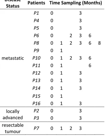
Sixteen patients were selected for the enrolment in the study by the Medical Oncology Unit of Ancona. One patient, P7, was enrolled after having received radical surgery for PDAC with no signs of disease relapse at the time of admission into the study. The remaining 15 were patients with metastatic/locally advanced pancreatic cancer that were treated with palliative chemotherapy. Specifically, 13 patients (P1, P4, P5, P6, P8,
P9, P10, P11, P12, P13, P14, P15, P16) had metastatic disease upon study entry whereas the remaining two (P2 and P3 patients) had locally advanced, and so, unresectable pancreatic cancer. Ten patients received as first-line chemotherapy Gemcitabine in combination with Nab-Paclitaxel whereas three patients received FOLFIRINOX. The remaining two patients received Gemcitabine palliative chemotherapy alone.
Disease
Status Patients Time Sampling (Months)
metastatic P1 0 3 P4 0 3 P5 0 3 P6 0 2 3 6 P8 0 1 2 3 6 8 P9 0 1 P10 0 1 2 3 6 P11 0 1 6 P12 0 1 3 P13 0 1 3 P14 0 1 3 P15 0 1 P16 0 1 3 locally advanced P2 0 3 P3 0 3 resectable tumour P7 0 1 2 3
Table 1 List of enrolled patients with their disease status at the time of admission into the study and
25 3.2.2 Blood samples
Blood samples were taken before chemotherapy treatment (T0) and, when possible,
after one, two and three months (T1, T2, T3). Only in a few cases blood samples were
taken six (T4) and eight (T5) months after the treatment (Tab. 1). Plasma was separated
from blood cells by centrifugation of 1100 g for 20 minutes at room temperature. Supernatant was then centrifuged at 10000 g at 4°C for 7 minutes and pellet with cell debris was then discarded. Finally, plasma samples from each patient were stored at -80°C until use. Moreover, pooled blood samples were used in order to test several different ELISA conditions.
3.2.3 Reagents
The following primary and secondary antibodies were used for exosome characterization in ELISA. Mouse monoclonal antibodies anti-human CD9, CD81 and CD24, Caveolin-1 and Fibronectin were purchased from BD Pharmingen (Milano, Italy). Mouse monoclonal anti-human TSPAN8 was purchased from Sigma-Aldrich (Milano, Italy). Mouse monoclonal antibodies anti-human CD133, PD-L1, CXCR4, EpCAM, Integrin α6, Integrin β4, CD44s and CD44v6 (R&D System, Minneapolis, USA) were reconstituted with sterile PBS to a stock solution. Mouse monoclonal antibodies
anti-human CD151 and Alix were from Santa Cruz Biotechnology (Milano, Italy). Goat Anti-Mouse Biotin conjugated was used as secondary antibody (Thermo Fisher, Monza, Italy). Streptavidin Poly-HRP (Thermo Scientific) is used to amplify the signal.
3.2.4 ELISA assay
Transparent Nunc MaxiSorp™ flat bottom 96 well plates (ThermoFisher), with high protein-binding capacity (600-650 ng IgG/cm2), were used for the ELISA assay according to the following protocol:
1. Blocking procedure. 1 hour and 30 minutes at room temperature (RT) under shaking.
As blocking agents were tested: i) 1% of Bovine Serum Albumine (BSA) protease and fatty acid free (Sigma Aldrich) in PBS, ii) 1% casein (Sigma Aldrich) in PBS, iii) Tween-20 (PanReac Applichem) 0,1% in PBS, iv) 0,2% polyvinylpyrrolidone (PVP) (Sigma Aldrich) in PBS, v) ethanolamine 1M in PBS (Sigma Aldrich).
26
2. Washing of the plate. Three times by using Thermo Scientific™ Wellwash™ Versa Microplate Washer.
Four different washing buffers were tested: i) PBS with 0,05% of Tween-20, ii) PBS with 0,1% of Tween-20, iii) PBS with a double amount of NaCl (274mM), iv) PBS with a double amount of NaCl (274mM) plus 0.1% Tween-20 (PBST-NaCl). 3. Sample incubation. A 100 μl plasma sample from each patient was added to the
wells of the plate and incubated overnight at 4°C. 4. Washing.
5. Primary antibody incubation. 1 μg/ml of primary antibodies was incubated for 3 hours at RT.
6. Secondary antibody incubation. 50 ng/ml of secondary antibody biotin conjugate was incubated for 45 minutes at RT under shaking.
7. Washing.
8. Streptavidin Poly-HRP incubation. 50 ng/ml, 45 minutes at RT under shaking. The antibodies and HRP were tested in five different dilution buffers: i) PBS, ii) PBS with 0,1% Tween-20, iii) PBS with 0,05% Tween-20, iv) PBS with 274mM NaCl and 0,05% Tween-20, v) PBS with 274mM NaCl, 0.5% BSA and 0,05% Tween.
9. Washing.
10. Substrate addition. The substrate solution 1-Step™ Ultra TMB-ELISA (Thermo Fisher) was added for 20 minutes at RT under shaking to detect horseradish peroxidase (HRP) activity, yielding a blue colour.
11. Reactions stop. A 2M sulfuric acid stop solution was added to each well, changing the blue colour of TMB into yellow.
12. Measurement. In order to measure the absorbance (Abs) at 450 nm of each well, Multiskan™ FC Microplate Photometer (Thermo Scientific) was used.
3.2.5 Data elaboration
Absorbance from each well was multiplied by 1000 in order to process data more easily. Absorbance values of the samples (signal) were normalized dividing them by the blank value of the same plate (noise) to obtain the signal/noise ratio.
27
3.3 Results
3.3.1 Evaluation of the best washing and blocking buffers
The term “blank” refers to those wells where, instead of plasma sample, PBS was added with the aim to quantify only the nonspecific signal due to the binding of the antibodies to the plate surface. The term “signal” refers to the absorbance measured in wells with the plasma samples. One of the goals of the protocol optimization phase is to reduce the absorbance values of the blank wells (noise or background) as much as possible, in order to have the highest signal/noise ratio. Washing and blocking procedures are crucial in ELISA assays to reach this goal.
By using the appropriate washing buffer, it is possible to remove all the unbound molecules in the wells without to dissociate those antibodies specifically bound to the proteins. The washing is performed three times between an incubation and the next. The most common washing buffer is usually PBS with variable percentages of Tween-20. I tested PBS with 0,05% or 0,1% Tween-20 and I found that 0,1% concentration was the most effective in reducing noise. Moreover, the addition of NaCl further helped the reduction of unspecific bounds (data not shown). The last washing was performed without Tween-20 because it has the tendency to inhibit the reaction of HRP with the substrate TMB.
The blocking procedure is useful to saturate the unoccupied spaces of the wells that could interact non-specifically with the proteins. There are various agents that could be used as blockers, usually classified into two major categories: proteins and detergents. Among protein-based agents, BSA and casein were tested and, as non-ionic detergent, 0,1% Tween-20 in PBS was chosen. I have also tested
Polyvinylpyrrolidone (PVP), an idrosoluble polymer and the organic chemical compound Ethanolamine. The results of the representative experiment are reported in Table 2, which shows that the most effective blocking buffer is 1% BSA in PBS. Indeed, the absorbance of the blank after BSA treatment was lower (Abs=54) than other blanks (Tween Abs=709; casein Abs=684; PVP Abs=147; ethanolamine Abs=1003). On the contrary, the signals, compared to the blanks, resulted similar or in some cases lower than blanks with all the blocking agents (Tween Abs=739; casein Abs=709; PVP Abs=99; ethanolamine Abs=849) with the exception of BSA (Abs=769).
28 Blocking Sample Absx1000 S/N
PBS+0,1% Tween Plasma 739 1,04 Blanck 709 Casein 1% Plasma 709 1,04 Blanck 684 BSA 1% Plasma 769 14,24 Blanck 54 PVP 0,2% Plasma 99 0,67 Blanck 147 Ethanolamine 1M Plasma 849 0,85 Blanck 1003
Table 2 Comparison of different blocking solutions. Human anti-CD9 has been used as primary antibody.
Absx1000: Absorbance multiplied by 1000.S/N: ratio of signal (plasma) to blank.
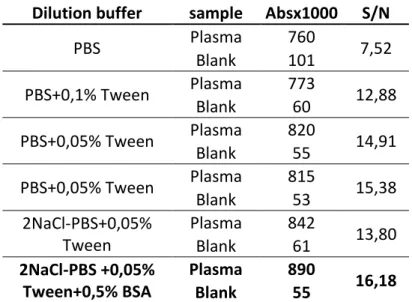
3.3.2 Evaluation of the appropriate antibody dilution buffer
In order to increase sensitivity and decrease nonspecific signals, I needed to test different dilution buffers for the antibodies. First, I compared three different buffers where the primary antibody CD9 was diluted at 1μg/ml: i) PBS, ii) PBS+0,05% Tween and iii) PBS+0,1% Tween. I obtained the highest signal/noise ratio with PBS+0,05% Tween (Plasma Abs=820; Blank Abs=55; S/N=14,91) (Tab. 3). Then, I checked whether there is an improvement by doubling the concentration of NaCl in PBS (referred as 2NaCl-PBS), both with and without 0,5% BSA. According to the results showed in Table 3, I decided to choose the following composition: 2NaCl-PBS + 0,05% Tween + 0,5% BSA (Plasma Abs=890;Blanck Abs=55; S/N=16,18).
29 Dilution buffer sample Absx1000 S/N
PBS Plasma 760 7,52 Blank 101 PBS+0,1% Tween Plasma 773 12,88 Blank 60 PBS+0,05% Tween Plasma 820 14,91 Blank 55 PBS+0,05% Tween Plasma 815 15,38 Blank 53 2NaCl-PBS+0,05% Tween Plasma 842 13,80 Blank 61 2NaCl-PBS +0,05% Tween+0,5% BSA Plasma 890 16,18 Blank 55
Table 3 Comparison of different dilution buffers for antibodies. Human anti-CD9 has been used as
primary antibody. Absx1000: Absorbance multiplied by 1000.S/N: ratio of signal (plasma) to blank
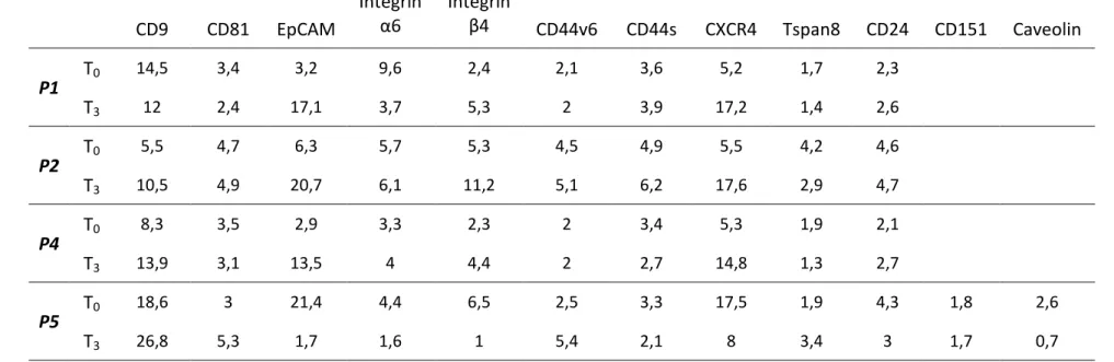
3.3.3 Characterization of exosome tumour markers from plasma of PDAC patients
After the optimization phase, the customized ELISA protocol was used in order to quantify 16 different exosomal protein markers in plasma samples from 16 PDAC patients. From some patients, only plasma samples before the start of chemotherapy treatment (T0) and three months after (T3) were collected, as reported in Table 4.
Principal tumour exosome markers were evaluated and their absorbance values were multiplied by 1000 and successively normalized dividing each sample values with the blank value. Patients P1, P4 and P5 at the time of the admission in the study had a metastatic disease, whereas P2 had locally advanced tumour, probably with micro-metastasis as suggested by the CA19-9 serum level. Three months after chemotherapy, these patients showed increased levels of exosomal EpCAM, Integrin β4, CXCR4 and CD24, with the exception of P5, who showed lower values at T3 than T1 (Tab. 4).
However, all patients in this group had a dismal prognosis. For this reason, I assume that the low exosome marker levels in P5 were due to an error in performing this specific test.
30
Integrin α6
Integrin β4
CD9 CD81 EpCAM CD44v6 CD44s CXCR4 Tspan8 CD24 CD151 Caveolin
P1 T0 14,5 3,4 3,2 9,6 2,4 2,1 3,6 5,2 1,7 2,3 T3 12 2,4 17,1 3,7 5,3 2 3,9 17,2 1,4 2,6 P2 T0 5,5 4,7 6,3 5,7 5,3 4,5 4,9 5,5 4,2 4,6 T3 10,5 4,9 20,7 6,1 11,2 5,1 6,2 17,6 2,9 4,7 P4 T0 8,3 3,5 2,9 3,3 2,3 2 3,4 5,3 1,9 2,1 T3 13,9 3,1 13,5 4 4,4 2 2,7 14,8 1,3 2,7 P5 T0 18,6 3 21,4 4,4 6,5 2,5 3,3 17,5 1,9 4,3 1,8 2,6 T3 26,8 5,3 1,7 1,6 1 5,4 2,1 8 3,4 3 1,7 0,7
Table 4 Principal exosome markers evaluated in plasma samples of PDAC patients (P1, P2, P4, P5). Blood samples taken at the start of chemotherapy (T0)and three months after the treatment (T3). Numbers refer to Abs values x 1000 normalized for the blank values.
31
Successively, in order to better analyse the effects of treatments and the disease progression, blood samples were also taken at the one month (T1), two (T2) and three
(T3) months timepoints after chemotherapy. T3 is when the patients had to undergo to
radiological evaluations. Blood samples were also taken at six (T4) and eight (T5)
months, when possible (Tab. 5a, 5b, 5c).
Patient P3 had locally advanced disease (T0) and, as well as P2, its plasma levels of
EpCAM, Integrin β4, CXCR4 and CD24 increased at T3 timepoint. However, these
markers showed a decrease at T4 that was unexpected since P3 also progressed and
died. Patient P6 had been diagnosed with metastatic pancreatic ductal adenocarcinoma at the admission in the study (T0). Our test showed that only Integrin
α6 and CXCR4 increased, whereas other markers such as EpCAM, Integrin β4, CD151 decreased their levels. At the follow-up, the tumour showed signs of progression but, however, it seems that palliative chemotherapy had shown efficacy since P6 is still alive. Exosome markers remained unvaried for the patient P7, with the exception of CXCR4, which showed a slight increase. P7 is the only patient with resectable tumour and no signs of disease relapse. P8 patient, with metastatic disease, showed increased levels of different exosomal markers: EpCAM, CD44s, CD24 and CD151. At the follow-up (T3), CD44v6 had been decreased, probably due to a beneficial effect of
chemotherapy, but at T5 its level recurred high as at the T0.P8 had a progression even
if its survival was longer than the other patients (Tab. 5a).
Patients from P9 to P16 have been diagnosed with metastatic disease. Levels of CD44v6 and Tspan8 increased at the follow-up in P10, P12, P13, P14 and a growth of EpCAM and CD24 in P10, P12 and P13 was observed (Tab 5b, 5c). Patient P16 have showed reduced levels of EpCAM, CXCR4 and CD24 while Integrin β4, CD44v6 and Tspan8 increased (Tab 5c). In this group, P10 and P16 displayed a progression at three months and P12 at the follow-up, successively P10 and P12 died.
Interestingly, I observed that immediately after chemotherapy (T1) some of the
exosome markers showed a decrease of levels in plasma patients, followed by a growth after two or three months. In particular, exosome markers that followed this trend were: CD24 (P6, P7, P13, P14, P16), CD151 (P7, P10, P12, P14, P16), Integrin β4 (P8, P12, P14, P16), CD44v6 (P6, P10, P13, P16), Caveolin-1 (P7, P10, P14, P16), PD-L1 (P7, P10, P14, P16), Integrin α6 (P6, P14, P16) and CD133 (P13, P14, P16). Their trend
32
could be connected with the worsening of the health conditions and to the poor survival, indeed P6, P8, P10, P16 progressed under chemotherapy treatment and P10 died.
33 CD9 CD81 Epcam Integrin α6 Integrin
β4 CD44v6 CD44s CXCR4 Tspan8 CD24 CD151 Caveolin PD-L1 ALIX CD133 Fibronectin
T0 6,2 2,8 2,5 3,9 1,8 1,6 2,5 5,1 1,4 1,8 1,5 P3 T3 7,3 3,6 16,3 2,5 4,8 1,7 3,4 15,3 1,4 3,2 1,2 T4 10,4 4,7 2,7 1,7 0,3 1,4 2,6 5,2 1,3 1,2 1,3 1,3 P6 T0 26,5 5,1 4,7 1,2 2,5 2,8 3,6 2,6 5,9 1,8 2,6 1,6 0,8 T2 24,8 6,3 2,6 1,0 1,8 1,2 4,0 3,2 7,7 1,6 2,2 1,8 1,1 T3 32,1 4,7 2,1 2,4 1,8 1,1 4,1 3,5 6,8 1,8 1,8 1,1 0,7 T4 4,1 4,6 1,3 7,6 1,8 1,9 3,5 8,8 0,9 1,8 1,8 1,5 2,5 1,4 3,2 5,3 P7 T0 24,4 13,4 6,7 2,4 1,7 2,0 3,6 4,0 6,1 4,6 4,1 2,7 2,2 T1 22,6 12,2 7,0 2,9 3,1 2,1 2,7 4,7 6,3 2,6 3,6 1,8 1,4 T2 23,4 15,4 5,4 2,1 2,4 2,3 2,5 5,1 7,2 3,0 4,1 6,7 3,5 T3 16,3 13,2 5,0 1,5 1,9 1,8 3,2 6,4 6,0 3,3 3,0 2,8 2,1 P8 T0 35,5 14,7 1,0 2,1 3,0 3,6 4,0 8,7 1,7 2,8 1,3 1,1 T1 38,5 8,2 1,2 2,1 1,1 5,6 3,1 8,6 3,9 3,0 1,7 0,9 T2 7,3 6,2 1,3 9,1 1,8 1,6 4,7 9,6 0,8 1,3 1,4 1,8 T3 12,6 5,7 1,3 5,3 1,9 1,9 5,2 8,4 0,9 1,8 1,6 1,9 T4 17,9 13,7 3,9 2,6 1,8 2,6 11,4 2,9 2,1 4,6 2,3 1,9 2,8 3,3 4,5 7,2 T5 20,8 6,5 2,4 1,5 2,6 3,4 4,5 1,5 1,6 3,3 1,9 1,6 2,7 2,3 4,0 7,5 Table 5a
34
Integrin α6
Integrin β4
CD9 CD81 Epcam CD44v6 CD44s CXCR4 Tspan8 CD24 CD151 Caveolin PD-L1 ALIX CD133 Fibronectin
P9 T0 13,9 8,9 8,3 2,8 3 2,7 7,6 3,8 3 8,3 4,1 2,9 5,7 3,3 5,4 7,8 T1 9,1 6,3 6,3 2,4 2,3 2,4 5,6 2,6 2,2 4,2 3 3,4 4,9 2,6 3,4 4,7 P10 T0 11,2 9,8 2,1 7 3,4 3,1 8 9,1 1,7 2,9 3,1 3,7 2,8 2,4 3,8 14,8 T1 12,9 10,9 1,6 7,1 3 3,1 9,1 8,6 1,5 2,7 2,8 2,8 2,4 2,6 3,9 18,5 T2 10,1 11,3 1,8 8,4 3,5 3,1 11,3 9,3 1,6 2,9 3 4,6 2,5 2,6 3,3 13 T3 10,8 11,4 4 3,6 3 4 11,9 3,3 3 4,1 3,8 3,2 4 3,5 4,1 9,6 T4 21,6 9,6 4,7 2,1 3,3 3,8 6,3 2,2 3 4,5 3,5 2,7 4,6 4,4 5,4 9,9 P11 T0 17,9 5,8 2,8 2,3 1,8 2,2 4 2,5 2,2 3,5 2,2 2,1 2,9 2,6 3 4,7 T1 4,7 5,5 2,7 1,5 1,5 1,6 2,6 1,8 1,6 2,2 1,7 1,5 2,2 1,6 2,3 4,3 P12 T0 10,3 4,4 1,2 8,7 1,9 1,5 3,3 8,1 0,9 1,5 1,5 3,9 2,2 1,2 3,1 6,3 T1 11,1 5,3 1,3 7,5 1,9 2,8 3,7 9,8 1 1,7 1,4 2,1 2,5 1,3 2,9 7,8 T4 4,9 2,6 2 0,9 4,3 2,6 2 1,1 2,1 2 1,6 1,4 2,3 1,4 2,8 2,5 Table 5b
35
Integrin α6
Integrin β4
CD9 CD81 Epcam CD44v6 CD44s CXCR4 Tspan8 CD24 CD151 Caveolin PD-L1 ALIX CD133 Fibronectin
P13 T0 15,9 6,6 1,4 1,5 1,7 3,5 2,6 1,5 1,8 6,3 2,1 2 2,2 1,7 2,8 10,7 T1 37,5 6,7 1,6 2 1,7 2,7 3 1,5 2,1 5 2,1 2 2,3 2,3 2,5 12,4 T3 30 8,9 2 1,1 1,7 3,3 4,6 1,8 2,2 7,1 1,8 1,7 3,1 2,1 4,9 6,6 P14 T0 19,7 12,4 2 1,5 1 1,3 1,1 2,2 1 5,3 5,8 1,4 1,6 1,3 2,7 17,4 T1 8 3,9 2,2 1,1 1 1,4 3,1 2,2 0,9 2,3 1,1 1,2 1,5 1,5 1,6 16,1 T3 15,1 4,6 1,4 1,5 1,4 3,3 1,9 1,4 1,5 2,8 1,7 1,6 2,5 1,6 2,7 4,3 P15 T0 7,3 5,8 2,2 1,3 1,1 1,3 7,8 2,6 1,3 3,1 1,2 1,7 2 1,3 2 13,6 T1 6,6 7,7 2,4 1,2 1 1,6 4,8 2,4 1 2,5 1,2 1,3 1,7 1,7 1,9 16,2 P16 T0 26,3 8,4 2,6 2,6 1,5 2 9,2 2,9 1,2 10,2 2,6 3,6 3,1 3,7 6,9 23,9 T1 45,3 6 2,5 1,8 1,1 1,8 6,4 2,6 1,3 5,8 1,8 2,3 2,2 2,2 3,2 14,2 T3 21,4 5,5 1,8 2,3 2 3,1 4 1,9 2,6 7,1 2,4 4 2,5 2,6 3,7 8,5 Table 5c
Principal exosome markers evaluated in plasma samples of PDAC patients (P3, P6-P16). Blood samples taken before chemotherapy (T0), at one month (T1), two months (T2), three months (T3), six months (T4) and eight months (T5) after chemotherapy. Numbers refer to Abs values x 1000 normalized for the blank values.
36
3.4. Discussion
3.4.1 Elisa assay development and optimization
Enzyme-linked immunosorbent assay (ELISA) is a quantitative technique, highly specific and sensitive, which allows the measuring of molecules of interest even using complex matrices such as human plasma or serum. However, it is a time-consuming procedure because it consists of various incubation steps, one overnight, alternated with several washings, and it requires an extremely high precision from the operator in order to have all the wells comparable and reliable. Moreover, due to the high capacity of the plate to bind proteins, it is easy to detect false-positive results. For these reasons, before starting with the actual measurements of samples from the patients enrolled in the study, I performed different experiments in order to detect only exosomal proteins and minimizing unspecific signals.
Washing procedures are crucial in removing all the unbound proteins. It is important that all the wells are filled with the same volume, soaked for the same time and eventually quickly and simultaneously emptied. The microplate washer, used in this work, helped to perform this procedure in a standardized manner, which guarantees that the different signals among wells are not caused by different washing intensity. Commonly, a solution of PBS and Tween-20 is used for the washing method. I found that an increased concentration of NaCl into the buffers provides a better washing. The comparison among different buffer compositions led me to choose a composition of PBS with 274mN NaCl (2NaCl-PBS) with the addition of 0,1% Tween.
The absence of standardized blocking procedures suitable for all the ELISA applications is well known. The two major classes of blocking agents are detergents and proteins.
Regarding the first class, non-ionic detergents such as Tween-20 or Triton-X, are the most used but they are considered weak blockers that do not offer a stable barrier. This consideration is congruent with the results showed in Table 2, where blocking with Tween-20 gave comparable absorbance values between sample and PBS incubation. On the contrary, proteins are permanent blockers and the most widely used for ELISA assays as reported in literature [69, 71, 75-77]. Here I compared two different types, bovine serum albumin and casein. Although both determined the same signal intensity with plasma, somehow primary or secondary antibodies bound unspecifically with casein but not with BSA, as showed in Table 2. An alternative