FIRST RECORD OF A COELACANTH FISH FROM THE MIDDLE TRIASSIC MERIDE LIMESTONE OF MONTE SAN GIORGIO (CANTON TICINO, SWITZERLAND)
SILVIO RENESTO1 & RUDOLF STOCKAR2
1Dipartimento di Scienze Teoriche ed Applicate, Università degli Studi dell’Insubria, Via Dunant 3 I 41100 Varese, Italy.
E-mail: [email protected]
2Repubblica e Cantone Ticino, Dipartimento del territorio, Museo cantonale di storia naturale, Viale Carlo Cattaneo 4, CH-6900 Lugano,
Switzerland.
To cite this article: Renesto S. & Stockar R. (2018) - First record of a coelacanth fish from the Middle Triassic Meride Limestone of Monte San Giorgio (Canton Ticino, Switzerland). Riv. It. Paleontol. Strat., 124(3): 639-653.
Rivista Italiana di Paleontologia e Stratigrafia (Research in Paleontology and Stratigraphy)
vol. 124(3): 639-653. November 2018
Abstract. A new specimen of coelacanth based on a new specimen from the Meride Limestone Formation
of the UNESCO World Heritage area of Monte San Giorgio is described. It represents the first occurrence of an actinistian in this formation. The newly discovered specimen shares many characters with the poorly known Heptanema paradoxum Bellotti, 1857 from the Ladinian Perledo Formation of Northern Italy. A comparison with the holotype and
only existing specimen of H. paradoxum supports the assignment of the new specimen to the genus Heptanema. Some
anatomical differences between the two specimens are most probably due to different ontogenetic stages, while few may support the erection of a new species; since the specimen is a juvenile it is preferred not to erect a new species, but to classify the specimen as Heptanema sp.
New available data from both the holotype of H. paradoxum and form the new specimen allows an attempt to assess
the phylogenetic relationships of Heptanema.
Received: August 27, 2018; accepted: October 29, 2018
Keywords: Actinistia; Middle Triassic (Ladinian); Meride Limestone; New species; Description.
I
ntroductIonCoelacanths (Actinistia) are a clade of sarcop-terygian fishes that are first known from the Early Devonian (Johanson et al. 2006). They peaked in diversity in the Early-Middle Triassic being most-ly known from Europe (see Cavin et al. 2013 and Ferrante et al. 2017 for a reappraisal of European and American taxa) and China (Tong et al. 2006; Wen et al. 2013), their diversity declined in the Cre-taceous and are absent from the Cenozoic fossil record. They were considered extinct until a liv-ing specimen of Latimeria was discovered in 1938 (Smith 1939). Coelacanths are known in the Middle Triassic of Monte San Giorgio, across the Italian Swiss boundary, with nearly complete specimens of Ticinepomis peyeri (Rieppel 1980), and fragments of a larger coelacanth tentatively referred to cf. Holophagus by Rieppel (1985), both from the Ani-sian-Ladinian, Besano Formation. Further findings
have recently been made in the in levels of Prosan-to Formation coeval with the Lower Meride Lime-stone, which yielded large specimens of Ticinepomis cf T. peyeri (Cavin et al. 2013) and the highly derived Foreya maxkuhni (Cavin et al. 2017; a reappraisal of the Middle Triassic coelacanths diversity from Swit-zerland has been recently published by Ferrante et al. 2017).
Here we describe a new coelacanthiform specimen, from the Sceltrich beds at the base of the Upper Meride Limestone (Ladinian). It was discov-ered in 2016 and represents the first record of this clade in over 160 years of excavations in the Meride Limestone Formation
G
eoloGIcalsettInGThe Middle Triassic carbonate succession of Monte San Giorgio (Switzerland-Italy; Figs 1, 2), belonging to the western part of the Southern Alps, has been inscribed in the UNESCO World Heritage List (WHL) because of its unique
paleon-tological value. It is, in particular, world-famous for the exceptionally well-preserved fossil fishes and marine reptiles (e.g. Rieber 1973; Kuhn-Schnyder 1974; Bürgin et al. 1989; Etter 2002). In Middle Triassic times, the South-Alpine domain was situ-ated at a northern intertropical latitude of about 15-18° (Muttoni et al. 2004) and was strongly in-fluenced by monsoonal circulation (Preto et al. 2010). This passive continental margin open to the western Neo-Tethys was progressively submerged by a long-term transgression from the east. The marginal location of the Monte San Giorgio Ba-sin resulted in a peculiar sedimentary succession and in temporarily dysoxic to anoxic bottom water conditions (e.g. Bernasconi 1994; Röhl et al. 2001; Etter 2002; Stockar 2010; Stockar et al. 2013). The Middle Triassic succession (Fig. 2) starts with flu-vio-deltaic deposits (Bellano Formation, Illyrian; Sommaruga et al. 1997), unconformably overlying Lower Triassic transitional clastic deposits (Servi-no, Induan-Olenekian; Frauenfelder 1916; Sciun-nach et al. 2015), in turn onlapping an erosional unconformity at the top of a Lower Permian vol-canic basement. The following upper Anisian sed-iments indicate the progressive transgression of a shallow epicontinental sea and the related expan-sion of carbonate platforms (San Salvatore Dolo-mite; Zorn 1971) north of an emerged land area, which is nowadays covered by the Po Plain (Brusca et al. 1981; Picotti et al. 2007). During the latest Anisian and the Ladinian, although shallow-water sedimentation continued in the north, an intraplat-form basin opened in the area of the Monte San Giorgio, which led to the deposition of the Besa-no Formation, the San Giorgio Dolomite, and the Meride Limestone (Rieber 1973; Bernasconi 1994;
Furrer 1995; Röhl et al. 2001). The Besano Forma-tion (“Grenzbitumenzone”; Frauenfelder 1916) directly overlies the Lower Salvatore Dolomite and is composed of a 16 m thick alternation of black shale and laminated dolostone. Its uppermost part includes the Anisian-Ladinian boundary (Brack & Rieber 1993; Brack et al. 2005). Most of the spec-tacular vertebrate fossils together with important index invertebrate fossils come from this forma-tion, which also yielded the only fossil coelacanths so far described from Monte San Giorgio (Riep-pel 1980; Riep(Riep-pel 1985). The Besano Formation grades upwards into the San Giorgio Dolomite and the Meride Limestone, together constituting a 614-m thick sequence in total (Stockar et al. 2012). Recent studies (Stockar 2012; Stockar et al. 2013) showed that the San Giorgio Dolomite results from early and late diagenetic dolomitization, the latter cutting across stratification and affecting the original limestone in an irregular pattern up to a major volcaniclastic bed (“Val Serrata tuff ”). The Lower Meride Limestone consists of well-bed-ded micritic limestone, laminated limestone and volcaniclastic layers. Three fossiliferous inter-vals, informally known as “Cava inferiore beds”, “Cava superiore beds” and “Cassina beds”, mainly consist of finely laminated limestone and yielded different vertebrate fossil assemblages (e.g. Peyer 1931, 1939a); Sander 1989; Furrer 1995; Stockar 2010; Stockar & Renesto 2011). The top of the Lower Meride Limestone is defined by a very dis-continuous dolostone horizon (“Dolomitband”; Frauenfelder 1916) resulting from late diagenetic dolomitization cutting across the stratification of the Meride Limestone (Stockar 2012; Stockar et al. 2013). The overlying Upper Meride Limestone
Fig. 1 - Map of the Monte San Gior-gio area (Ticino, Southern Switzerland), showing the carbonate Anisian-Ladinian sequence together with the locality.
Coelacanth fish from Meride Limestone of Monte San Giorgio (Canton Ticino, Switzerland) 641
is a sequence of alternating well-bedded micrit-ic limestone and marlstone. The uppermost part comprises the 120 m thick “Kalkschieferzone” (Senn 1924), made up of thinly-bedded, mostly laminated, limestone and marlstone with pecu-liar faunas of fishes, crustaceans and insects (e.g. Wirz 1945; Furrer 1995; Krzeminski & Lombar-do 2001; Bechly & Stockar 2011; LombarLombar-do et al. 2012; Montagna et al. 2018). Finally, the following Carnian regressive phase resulted in the formation of sabkha-type depositional environments and in the related sedimentation of evaporites (Pizzella Marls; Furrer 1995).
The fossiliferous interval yielding the spec-imen described herein belongs to the lowermost part of the Upper Meride Limestone and was in-formally introduced as “Sceltrich beds” in Stockar (2012) and Stockar et al. (2013). Its age is assigned to the transition interval between the Gredleri and Archelaus Ammonoid Zones (sensu Brack &
Rieber 1993) of the Ladinian Stage (Stockar et al. 2012). After a first exploration in 2010 yielding the first fossils from this horizon (Stockar 2012), two small bed by bed excavations on a surface of around 6 and 10 square meters respectively were started in 2012 by the Museo cantonale di storia naturale (MCSN, Lugano) under the direction of the second author. The site is located on the northern bank of a small creek (Valle di Sceltrich; Swiss National Coordinates: 716’910/84’370; WGS8 coordinates 8.4503/45.90084; Fig. 1), northwest of the village of Meride. The fossilif-erous interval consists of a 30 cm thick sequence of prevailing organic-rich laminated limestone (up to 3.1% TOC) intercalated between thick-bedded marly limestone. In the Valle di Sceltrich area, this fossiliferous horizon lies around 2.5 m above the “Dolomitband”. At places, the laminated lime-stone shows storm-generated concentrations of platform-derived skeletal grains and thin-shelled bivalve pavements (Stockar 2012; Stockar et al. 2013). Benthic microbial activity accounts for the microfabrics observed in the laminated limestone, including clotted-peloidal micrite and amorphous organic matter showing EPS (extracellular poly-meric substance) -like structures as well as for the geochemical signature being characterized by high hydrogen indices and prevailing Type-II (Type I) kerogen (Stockar et al. 2013). Preservation of such a labile lipoid-rich organic material requires anoxic/lower dysoxic bottom-water conditions (Stockar et al. 2013 and references therein). Low-er dysoxic conditions wLow-ere able to allow episodic seafloor colonization by thin-shelled opportunis-tic posidonioid bivalves (Stockar 2012; Stockar et al. 2013). On the other hand, both lower dysox-ic and anoxdysox-ic bottom-water conditions ruled out higher macrobenthos, including scavengers, and resulted in complete oxygen depletion within the sediment. Coupled with widespread oxygen deple-tion excluding benthic scavenger organisms, rap-id coating of skeletons by benthic bacterial mats (‘microbial shroud’ effect; Gall, 2001) played the key role in protecting the carcasses from decay and in holding skeletal elements together (see also Furrer 1995, 1999; Stockar 2010), thus account-ing for the microfabrics observed in the laminated limestone and for the exquisite preservation of the vertebrate fossils (Stockar et al. 2013).
So far, the excavation carried out in the
Fig. 2 - Stratigraphic column of the Monte San Giorgio area; coe-lacanth shapes indicate the levels that yielded coecoe-lacanth material.
Sceltrich beds yielded a rich vertebrate fossil fauna (mostly articulated specimens, and rare sauroptery-gian reptile bones and teeth), among fishes, spec-imen of Saurichthys represent the most common finding; in addition, specimens of Archaeosemionotus connectens (López-Arbarello et al. 2016) and of yet unnamed halecomorph fishes have been found. In-vertebrate fossils, mostly bivalves and crustaceans (Stockar & Garassino 2013) and terrestrial plant re-mains have been found.
s
ystematIcP
alaeontoloGy Sarcopterygii Romer, 1955Actinistia Cope, 1891
Heptanema Bellotti, 1857 Type species: H. paradoxum Bellotti, 1857
Diagnosis (emended from Forey 1998): Heptanema is
dia-gnosed by the presence of scales on the flanks of the body orna-mented by one very prominent ridge, ending in a spine that extends beyond the posterior margin of the scale. Additional characters are: body fusiform, head relatively long and shallow with a short postpa-rietal shield equal to approximately half of the parietonasal shield. Anterior and posterior parietals equal in length; lachrymojugal ex-panded anteriorly and curved posterodorsally. Dentary hooked. Operculum large dorsally and narrowing ventrally. Presence of sim-ple anocleitrum. First dorsal fin with seven robust rays ornamented with spines.
Heptanema sp.
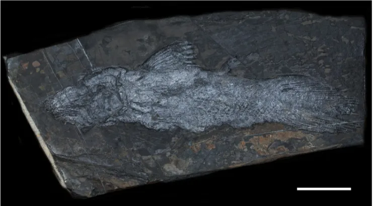
Material: Specimen MCSN 8532 (Fig. 3), housed in the
pa-laeontological collection of the Museo cantonale di storia naturale in Lugano (Switzerland). It is nearly complete and articulated, exposed in left lateral view, which was found in Valle di Sceltrich, Monte San Giorgio, Cantone Ticino, Switzerland from an outcrop of the Upper Meride Limestone, Sceltrich beds, bed 9 (8 cm above the base of the Sceltrich beds); Middle Triassic, early/late Ladinian (transitional interval Gredleri/Archelaus Ammonoid Zones).
Measurements (in mm): Total length 81.7; Standard length
(distance from the tip of the snout to the base of the caudal lobe (Forey 1998) 60.5; Head length 20.5, Head height 19.9, Supplemen-tary lobe length ca 21.2 (11.2 excluding the portion overlapped by caudal lobes).
Description
The specimen is nearly complete and articula-ted (Fig. 3), it lacks only of the pectoral fin, and the tip of the snout is crushed. The outline of the body is revealed by an underlying layer of black organic matter, visible also in areas where the scale covering is missing. Veins of calcite crystals run across the specimen obscuring some details, especially in the skull area.The outer surface of some skull bones is worn out.
The specimen was mechanically prepared with the aid of vibrotools and sharpened steel needles. It has been examined under a stereomicro-scope and photographs have been taken both under
Coelacanth fish from Meride Limestone of Monte San Giorgio (Canton Ticino, Switzerland) 643
visible light and UV light (360 nm) with a SIGMA Sd Quattro H camera. UV photographs were parti-cularly useful in revealing details of the fins and sca-les, while were of little help for the reconstruction of the skull because UV reflexion was very scarce on skull bones in case of poor preservation of the outer surface of the bones.
Skull and pectoral girdle (Fig 4 A-B) - Skull
bones are fractured, and in some case splitted and obliquely embedded in the matrix, thus precise identification of bones is difficult. When preserved, the outer surface of skull bones is smooth, lacking any ornamentation apart for the fragments of pre-maxillae that bear small tubercles. The parietonasal shield is at least two times longer than the postpari-etal shield. The anterior and posterior paripostpari-etals are approximately the same size. A calcite vein obscures part of the contacts between the anterior and pos-terior parietals and between left and right parietals. Nasals are not preserved. The anterior margin of the postparietal is straight, without interdigitations along the intracranial joint. The posterior margin of the postparietal is not well preserved. A small fragment of bone lying posterolaterally to the post-parietal is tentatively identified as a portion of the left supratemporal; like many other skull bones, it does not lie parallel to the slab surface, but it is par-tially sunk in the matrix so that the exact outline cannot be detected. Ventrolaterally to the parietals and postparietals, at least six small bones are
pre-served and are identified as the posterior tectals/ supraorbitals.
The postorbital is high and only moderately expanded, it is also partially sunk in the matrix so that it appears extremely thin. The lachrymojugal is elongate and curved; its dorsal margin forms the ventral margin of the orbit and it extends anterior-ly beneath the preorbital area. Other cheek bones are crushed or embedded obliquely in the matrix so that their exact outlines cannot be determined. Fragments of several bones are visible anterior to the lachrymojugal, but poor preservation does not permit any reliable identification apart for the ante-riormost fragment that may be identified as portion of the premaxilla, which is ornamented by tiny tu-bercles.
The anterior part of the pterygoid is visible in lateral view; it has a usual subtriangular shape as in most coelacanths; a fragment of the narrow anteri-or panteri-ortion of the parasphenoid is also visible.
The squamosal and preoperculum are also preserved, they are flat subrectangular bones but preservation precludes a more detailed description.
The left operculum is also poorly preserved and crossed by elements of the shoulder girdle; it is nevertheless possible to observe that it had a straight anterior margin and a rounded posterior margin, its outline distinctly narrowing ventrally.
Few bones of the lower jaw can be described: the dentary is posteriorly embayed (hook shaped sensu Forey 1998) and edentulous; it appears inclined
Fig. 4 - Heptanema sp., specimen MCSN 8532. A) close up of the skull; B) Sketched outline of recognizable bones abbreviations are: acl)
ano-cleithrum, ar) articular, cl) ano-cleithrum, d) dentary, lj) lachrymojugal, op) operculum, pa) parietals, po) postorbital, pop) preoperculum, psp) parasphenoid, pt) pterygoid, sq) squamosal, stt) supratemporal, sy) symplectic, tec-so) tectals-supraorbitals. The black spots indi-cate the area in which the outer wall of the sensory canal crushed. Scale bar equals 10 mm.
anteroventrally, however it may have been partially rotated from its original position.
The angular is elongate and shallow; its dorsal margin is broken. Some anteroposteriorly elongate irregular openings may be interpreted as areas in which the thin outer walls of the mandibular senso-ry canal have been crushed (Fig. 4).
The gular is large, its lateral margin is slightly convex but the contact with the angular is not clear-ly discernable so that its exact shape is difficult to be reconstructed.
The cleithrum is dorsoventally elongate and sickle shaped as in most coelacanths, other bones of the pectoral girdle are of difficult identification due to the superimposition of left and right halves of the pectoral girdle and the partial rotation of dif-ferent elements. A, slender, and slightly sygmoidal anocleithrum is a visible at the dorsal end of the cleithrum (Fig. 4).
Axial skeleton. No ossified centra are present,
as is typical for coelacanths. The exact number of neural arches remains unknown due to the covering of scales on most of the body. Few neural arches are visible ventral to the first dorsal fin; they show a typical inverted Y-shape with very high but thin neural spines. The neural spines become lower and more robust in the caudal region. Haemal arches are visible just dorsal to the anal fin and at the base of the lower lobe of the caudal fin; their shape is almost identical to that of the corresponding neural arches.
No ossified ribs are visible.
Paired fins. The pectoral fin is not preserved. The pelvic fin (Fig. 5) is located at the level between the first and second dorsal fin lying slightly closer to the anal. The basal plate cannot be seen. At least eight fin rays are present, and they are segmented in their distal half. Fin rays are not expanded.
Median fins. The first dorsal fin (Figs 5-6) is crossed by a calcite vein that obscures some details; the basal plate is not visible. Since the area was cov-ered by matrix before preparation, the bone was not lost during collection, also, careful preparation re-vealed that it is not hidden below scales or by black organic matrix, thus it is feasible that it was not yet ossified. The fin consists of seven robust fin rays segmented in their distal half. The first two rays bear stout and sharp spines.
The second dorsal fin (Fig. 5B) bears at least 10 rays, much thinner than those of the first dorsal fin. Again, the basal plate is not ossified.
Six ray bases are visible but only five-four rays are well preserved, they are segmented in their dis-tal third, and the basal plate does not seem to be ossified.
A
B
C
D
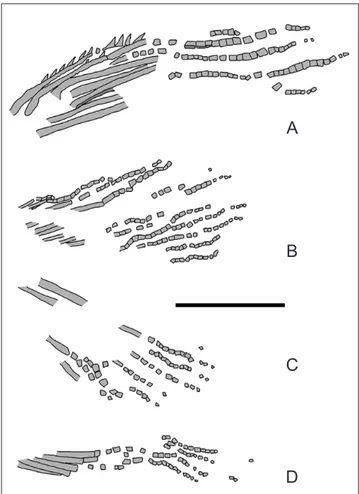
Fig. 5 - Heptanema sp., specimen MCSN 8532. Sketched outline of
the first (A) and second (B) dorsal fin, anal (C) and pelvic (D) fins. Scale bar equals 5 mm.
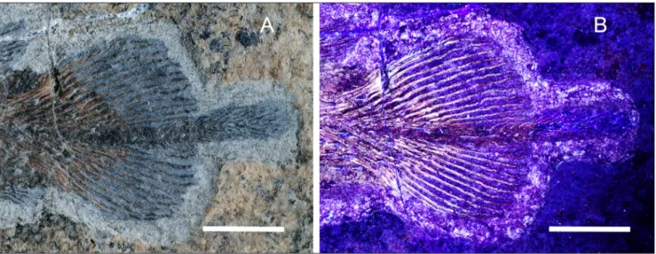
Fig. 6 - Heptanema sp., specimen MCSN 8532, UV photograph of the
Coelacanth fish from Meride Limestone of Monte San Giorgio (Canton Ticino, Switzerland) 645
Caudal fin. The dorsal and ventral lobe are
symmetrical (Fig. 7), both consisting of 13 fin rays; some rays are disjointed in their unsegmented por-tion so that the concave internal part of some of the right hemilepidotrichia can be seen. Fin rays are segmented for about the distal third of their length. The first two rays of the dorsal lobe bear stout den-ticles. The first ray of the ventral lobe is short and unsegmented, while the following are much longer and distally segmented. Few spines are visible on the first segmented ray of the ventral lobe as clearly revealed by UV photographs (Fig. 7B).
Each fin ray is supported by one radial (Figs
7B, 13A). The radials slightly extend along the lead-ing edge of the first five-six distally segmented lep-idotrichia; this feature is clearly visible in the ven-tral lobe, while in the dorsal lobe the area is heavily damaged by a fracture in the stone slab.
The supplementary lobe is elongate; the pro-truding portion is almost as long as the length of the dorsal and ventral lobes and bears up to ten fin rays both on its dorsal and ventral side.
Scales. Scales (Fig. 8) are preserved in patches covering most of the body, apart for the area at the level of the insertion of the dorsal fins. The scales are longer than deep, densely packed together so that the exact outline of each scale is difficult to observe. Each scale bears a stout, prominent medi-an ridge ending posteriorly in a pointed spine that extends beyond the posterior margin of the scale. Lateral line cannot be detected.
Ossified bladder. Fragments of an ossified
structure extends from the pectoral girdle to a point at mid distance between the first and second dorsal fins (Fig. 9). It consists of a series of bony plates slightly superimposed each other with tiny, parallel striations on their internal surface suggesting the possible presence of an ossified bladder (Brito et al. 2010).
d
IscussIonSpecimen MCSN 8532 shows a peculiar mor-phology of the scales that is matched only by the
Fig. 7 - Heptanema sp., specimen MCSN 8532, the tail photographed in visible light (A) ad in UV light (B). The supplementary lobe is more
visible in (A), while the structure of the dorsal and ventral lobe is more visible in (B). Scale bars equal 10 mm.
Fig. 8 - Heptanema sp., specimen MCSN 8532, detail of the scales.
poorly known species Heptanema paradoxum Bellotti, 1857 from the late Ladinian of Perledo (East side of the Como Lake, Lombardy, Northern Italy).
The genus Heptanema was erected by Rüppel (1855-56) including one species, Heptanema paradoxa, but was never described or figured. Bellotti (1857) based on a plaster cast of the original specimen, made a partial diagnosis of the specimen, but in the description he misinterpreted the dorsal region as the ventral one, thus he described the first dorsal fin as a pectoral fin and attributed the species to the Actinopterygia. Later, Deecke (1889) and De Ales-sandri (1910) redescribed the specimen, renamed the species as Heptanema paradoxum and correctly identified it as a coelacanth. The latter description was based on two specimens, but in its work De Alessandri (1910) figured only one. The unfigured specimen was unfortunately destroyed during World War II, when an incendiary bomb hit the Museo di Storia Naturale di Milano where it was housed. For this reason, some characters referred to “the Milano specimen” by De Alessandri (1910) are unverifiable at present. The only existent specimen of
Heptane-ma paradoxum is the holotype, cat. No. SMF P1242 of the vertebrate palaeontological collection of the Senckenberg Forschungsinstitut und Naturmuseum in Frankfurt (Germany). This specimen (Fig. 10-12) has been reexamined for the present study.
Based on the embedding rock, the holotype of Heptanema paraxodum may be tentatively attribut-ed to the uppermost part of the Perlattribut-edo-Varenna Formation (Perledo Member, late Ladinian; Gae-tani et al. 1992) which possibly correlates with the uppermost Meride Limestone (Kalkschieferzone, late Ladinian; Furrer 1995; Tintori 1998). This unit, quarried in the area of Perledo (Lecco, Northern Italy) until the middle of the XIX century for the production of slabs and ornament stones, consists of an up to 100 m thick sequence of dark, finely laminated limestone with thin-bedded shale inter-calations and yielded most of the classical “Perledo Fauna” among which the actinopterygian genera Perleidus and Saurichthys, (De Alessandri 1910) and the sauropterygian reptile Lariosaurus balsami (Peyer 1939b) .
Due to poor preservation, as it is frequent for the Perledo material, most details of the bones of the skull have been obliterated so that their outline and structure cannot be reconstructed. It can only be stated that the skull is elongated, with a well-de-veloped preorbital region, a long parietonasal shield, short postparietals, elongate parietals and an elongate lachrymojugal that extends anterior to the orbit. A coarse granular ornamentation is present on the parietonasal shield and rostral bones, similar to that of Guizhoucoelacanthus Liu et al. 2006 (Geng et al. 2009). Few robust, sharply pointed triangular structures are visible on the ventral margin of the skull: (Fig. 11) they may belong to the lower jaw, but the latter unfortunately is almost completely missing, thus their identification is difficult, despite some similarity with coronoid fangs (sensu Forey 1989). The variable size and the position on differ-ent level suggest that they may instead have been ornamentations, thus in the phylogenetic discussion the character “fang-like coronoid teeth” has been considered absent. Few thinner rods anteroposteri-orly inclined are visible in the posterior third of the abdominal region (Fig. 12A) and are interpreted as fragment of very short ribs. Contrary to previous descriptions, the scale covering of Heptanema para-doxum is differentiated. The scales on the lateral and ventral region (Fig. 12A) show the peculiar
morphol-Fig. 9 - Heptanema sp., specimen MCSN 8532. A) the body region, B)
an enlargement of the area in which fragments of the os-sified bladder are preserved. With the thick white outline it is reconstructed the hypothetical shape of the bladder, with the thin white line is indicated the position of the preserved fragments.
Coelacanth fish from Meride Limestone of Monte San Giorgio (Canton Ticino, Switzerland) 647
ogy described by Deecke (1889) and De Alessandri (1910): they bear a single thick, long and prominent ridge ending in a pointed spine (as it occurs in spec-imen MCSN 8532), sometimes flanked ventrally by a thinner second smaller ridge. In the dorsal region, at the level of the insertion of the dorsal fins, the ornamentation of the scales is different: they lack the prominent central spine, bearing instead 10/12 thin longitudinal ridges, with the central one that is only slightly more robust than the others (Fig. 12B). The first dorsal fin (Fig. 12C) is formed by seven robust rays. The caudal fin (Fig. 12D) tapers posteriorly with the last rays (those adjacent to the supplementary lobe) that start and extend much far-ther than the rays along the outer margin, giving to the tail a slanting outline similar to that of many coelacanths such as Diplurus (Schaeffer 1948, 1952) and, at a lesser extent, to that of Laugia groenlandica Stensiö, 1932, as figured by Wendruff and Wilson (2012, pp. 908-909, figs 4-5). The tail of H. para-doxum seems also asymmetrical, with the dorsal lobe of the caudal fin that starts anteriorly with respect to the ventral lobe and is made by 17 rays, while in the ventral lobe only 12 rays are visible (11 ac-cording to De Alessandri 1910); there would have been a difference of five rays (six, if De Alessandri was possibly correct) between the dorsal and
ven-Fig. 10 - Heptanema paradoxum holotype, specimen SMF P1242. Scale bar equals 50 mm.
Fig. 11 - Heptanema paradoxum SMF P1242, holotype. A) skull, the rectangle indicates the area enlarged in B. B) Tooth-like structures. Scale bar in (A) equals 10 mm.
tral principal lobes, rendering the tail asymmetrical, again as in Laugia (Lambers 1991; Forey 1998). This would have been of taxonomic significance. Close examination of the specimens reveals however that the ventral margin of the tail is incomplete and that there are remains of further radials anterior to the first preserved rays of the ventral lobe; thus it is pos-sible that the asymmetry of the tail is an artifact of preservation, while the difference in the number of fin rays between the dorsal and caudal lobe probably amounts to only to two-three rays.
While Heptanema may superficially look similar to Diplurus, as noted by Forey (1998), it differs from the latter genus by the nuber of rays in the dorsal fin, the ornamentation of the scales and for the absence of long ossified ribs.
A comparison between MCSN 8532 and H. paradoxum holotype shows that MCSN 8532 shares with H. paradoxum all observable skull characters except the ornamentation, the structure of the first dorsal fin (that is in both cases composed of sev-en rays) and the peculiar morphology of the lateral
scales. Since the dorsal part of the squamation is not preserved in MCSN 8532, it is not possible to assess if the scales are also differentiated in this specimen.
MCSN 8532 differs from H. paradoxum holo-type for the size, the body proportions, the presence of a smooth surface on all skull bones except for the premaxilla, and for the symmetrical caudal fin that is rounded posteriorly and has only 13 fin rays in each lobe (Fig. 13).
Some of the differences between MCSN 8532 and H. paradoxum can be related to the early ontoge-netic stage of MCSN 8532. The small size and the proportionally large head (head length reaches near-ly one third of the standard length) suggest a juve-nile condition. Also, in MCSN 8532 endoskeletal basal plates of the unpaired and paired fins are not yet ossified, as it has been reported for juveniles and embryos of some coelacanth taxa such as Rhabdo-derma exiguum and Undina pennicillata (Schultze 1980; Watson 1927) in which the ossification of the basal plates is delayed with respect to other elements of the skeleton.
Fig. 12 - Heptanema paradoxum SMF P1242, holotype. A) lateral scales with prominent ridge and the small ribs on the posterior abdominal region (indicated by the white arrow); B) Scales of the dorsal area just below the dorsal fins; C) first dorsal fin; D) caudal fin. Scale bars equal 5 mm (A-B), 10 mm (C), 25 mm (D).
Coelacanth fish from Meride Limestone of Monte San Giorgio (Canton Ticino, Switzerland) 649
Another character present in MCSN 8532 that is considered as indicative of early ontogene-tic stage for coelacanths (Schultze 1972, 1980; Bri-to & Martill 1999; Anthony & Robineau 1976) is the elongate supplementary lobe of the caudal fin. The supplementary lobe of MCSN 8532 extends well beyond the dorsal and ventral lobes, and its length including the portion overlapped by caudal lobes reaches approximately 1/4th of the total body length (1/7th, when considering only the portion not overlapped by caudal lobes). De Alessandri (1910), possibly on the basis of the lost specimen, stated that the length of the supplementary lobe of H. paradoxum was 32 mm, thus it should have rea-ched 1/7 of the total length. This assumption ho-wever, cannot be verified because the supplementa-ry lobe of the holotype is incomplete and it is not even clear if the measurement given by De Alessan-dri included the portion overlapped by main caudal lobes or not. The possible presence of an ossified bladder is an unusual feature in fossils of juvenile coelacanths, in which the bladder, if present, is pre-served as a mass of phosphatized tissue (Brito &
Martill 1999). However, the presence of an ossified bladder in a juvenile specimen has been already re-ported for Axelrodichthys (Yabumoto & Brito 2013). The same area is poorly preserved and covered by scales in H. paradoxum, thus the presence or absence of an ossified bladder cannot be ascertained in the holotype of H. paradoxum.
In conclusion, MCSN 8532 shares sever-al characters with H. paradoxum holotype, as listed above, and most existing differences between the two specimens can be explained as related to the earlier ontogenetic stage of the former specimen; thus it is proposed here that MCSN 8532 can be-long to the genus Heptanema. The different poste-rior outline of the tail lobes may either be related to different growth stage, or instead have taxonomic significance. The difference in number of caudal fin rays may also have taxonomic significance (in fact, while a difference of one-two rays between con-specific individuals occur frequently, a difference of four-five is rare). The latter two charcters may thus indicate that MCSN 8532 could belong to a differ-ent species of the genus Heptanema. However, taking into account that the specimen is a juvenile, and that the only significant difference with the holotype of H. paradoxum is the number of lepidotrichia on the dorsal lobe of the caudal fin, it is preferred here not to erect a new species but consider specimen MCSN 8532 as Heptanema sp.
P
hyloGenetIcrelatIonshIPsThe only phylogenetic tree that included Hep-tanema was published by Schaeffer (1948, p. 13 fig. 10) and reproduced by Forey (1998, p. 226 fig 9.2) as a cladogram. According to Schaeffer (1941), Hep-tanema was closely related to Scleracanthus, and the two were closely related to Whiteia.
All subsequent phylogenetic analyses of coe-lacanths excluded Heptanema from their species list because the taxon was too poorly known to be in-cluded in a dataset.
Forey (1998) however included Heptanema within the Latimeroidei, but without discussion, and stated (Forey 1998, pp. 350-351) that the overall shape and the ornamentation of the scales may be similar to that of Diplurus.
Some characters, such as the operculum rounded dorsally and posteriorly but pointed
ven-Fig. 13 - Comparison between the caudal fins of Heptanema sp. (A)
trally, the first dorsal fin made of only seven rays, fin rays not expanded distally, and the presence of pointed denticles on the anterior fin rays of the first dorsal and caudal fins, are shared by the Whiteiidae sensu Schultze (1993) but are present also in other taxa. The proportionally slender body and relative-ly large head, a snout (preorbital length) long and more than one-third of the length of the skull roof, a lachrymojugal with a curved posterior region and a long anterior region with a straight dorsal margin, are not only known in the Whiteiidae but are also present in a variety of different taxa (e.g.: Diplurus and Ticinepomis) which fall within the Latimeroidei
(Smith 1939; Lund & Lund 1984, 1985; Forey 1998; Moy-Thomas 1935; Schaeffer 1948; Clément 2005; Arratia & Schultze 2016; Cavin & Gradinaru 2012; Cavin et al. 2013, 2017). In addition, recent analyses (Cavin et al. 2017) do not support the monophyly of Whiteiidae.
We tried to establish the phylogenetic re-lationships of Heptanema, by adding scoring for Heptanema to a slightly modified version of the the matrix by Cavin et al. (2017). With respect to the original matrix the following changes have been made: Rebellatrix has been deleted since not inform-ative and adding instability; Charachter 107, ossified
Fig. 14 - 50% majority rule consen-sus tree of four most parsi-monious trees, obtained by adding Heptanema to a
mod-ified version of Cavin et al. (2017) matrix and dataset. Consistency Index 0.381, Retention Index 0.678.
Coelacanth fish from Meride Limestone of Monte San Giorgio (Canton Ticino, Switzerland) 651
bladder absent (0) or present (1) has been coded [01] for Whiteia, due to the claimed presence of an ossified bladder in Witheia oishii (Yabumoto & Bri-to 2016). The scoring for Heptanema is reported in Tab. 1. Character 92, ossified ribs absent (0), present (1), has been coded (0) for Heptanema paradoxum ac-cording to Forey (1998) who coded (1) the presence of several well developed ribs covering most the ab-dominal region and (0) the presence of few short ribs in the posterior abdominal region.
A parsimony analysis was conducted with TNT (Tree analysis using New Technology, Golo-boff et al. 2008), which allows a faster, exhaustive search of the tree space. In particular we used the parsimony Ratchet (Nixon 1999) and the conven-tional Wagner parsimony models. The Ratchet and Wagner models, performed equally, have yielded two equally parsimonious trees after 3.636.801 rearrange-ments with the best score of 318 with (Consistency Index 0.381, Retention Index 0.678).
The consensus tree (Fig. 14) gives full support to the results by Cavin et al. (2017) and Heptanema is nested after (Wimania + Axelia) in an unresolved tri-chotomy with Dobrogeria and the node at the base of the (Yunnanocoelacanthus (Luopingocoelacanthus (Mawso-niidae (Latimerioidei))) clade. It must be remarked, however, that even if the discovery of specimen MCSN 8532 and the re-examination of the Heptane-ma paradoxum holotype increased at some extent the knowledge of the genus, the scarcity of skull charac-ter still represent an obstacle for a solid phylogenetic analysis. For this reason, the conclusions about phy-logenetic relationships of Heptanema presented here must be considered as tentative.
Acknowledgements: The hard work and unconditional
commit-ment of A. Delmenico, F. Magnani, S. Rampinelli, N. Römer, L. Zulli-ger in the field are gratefully acknowledged. The excellent preparation of the fossil specimen has been carried out by F. Magnani. Fieldwork and fossil preparation have been granted by the Dipartimento del ter-ritorio del Cantone Ticino and the Swiss Federal Office for the Envi-ronment. We also warmly thank the referees, L. Cavin (Geneve) and G. Clément (Paris), their useful comments and remarks improved the paper and are sincerely appreciated. Our sincere thanks also to Dr. Rainer Brocke, curator of the Vertebrate Palaeontological collection of the Senckenberg Forschungsinstitut und Naturmuseum in Frankfurt (Germany), for the permission to access the holotype of H. paradoxum
and kind assistance during the examination of the specimen. Thanks also to my daughter Sara for assistance with computer misfunctions.
referenceS
Anthony J. & Robineau D. (1976) - Sur quelques characteres juveniles de Latimeria chalumnae Smith (Pisces,
Crossop-terygii, Coelacanthidae). Comp. r. Acad. Sci. Paris, D 283:
1739-1742.
Arratia G. & Schultze P.-H. (2015) - A new fossil actinistian from the Early Jurassic of Chile and its bearing on the phylogeny of Actinistia. J. Vert. Paleontol., 35(5): e983524.
DOI:10.1080/02724634.2015.983524.
Bechly G. & Stockar R. (2011) - The first Mesozoic record of the extinct apterygote insect genus Dasyleptus (Insecta:
Ar-chaeognatha: Monura: Dasyleptidae) from the Triassic of Monte San Giorgio (Switzerland). Palaeodiversity, 4: 23-37.
Bellotti C. (1857) - Descrizione di alcune nuove specie di pesci fossili di Perledo e di altre località Lombarde. In: Stoppani A. (Ed.). Studii Geologici e Paleontologici sulla Lombardia:
419-438. Carlo Turati Tipografo Editore Milano.
Bernasconi S.M. (1994) - Geochemical and microbial controls on dolomite formation in anoxic environments: A case study from the Middle Triassic (Ticino, Switzerland). Con-trib. Sedimentol., 19: 1-109.
Brack P. & Rieber H. (1993) - Towards a better definition of the Anisian/Ladinian boundary: New biostratigraphic data and correlations of boundary sections from the Southern Alps. Eclogae Geol. Helv., 86: 415-527.
Brack P., Rieber H., Nicora A. & Mundil R. (2005) - The Global boundary Stratotype Section and Point (GSSP) of the La-dinian Stage (Middle Triassic) at Bagolino (Southern Alps, Northern Italy) and its implications for the Triassic time scale. Episodes, 28: 233-244.
Brito P.M & Martill D. (1999) - Discovery of a juvenile Coel-acanth in the Lower Cretaceous, Crato Formation, North-eastern Brazil. Cybium: int. J. ichthyol., 23(3): 311-314.
Brito P.M., Meunier F.J., Clement G. & Geffard-Kuriyama D. (2010) - The histological structure of the calcified lung of the fossil coelacanth Axelrodichthys araripensis (Actinistia:
Mawsoniidae). Palaeontology, 53: 1281-1290.
Brusca C., Gaetani M., Jadoul F. & Viel G. (1981) - Paleogeogra-fia e metallogenesi del Triassico Sudalpino. Correlazioni tra paleogeografia e mineralizzazioni. Mem. Soc. Geol. It.,
22: 65-82.
Bürgin T., Rieppel O., Sander P.M. & Tschanz K. (1989) - The fossils of Monte San Giorgio. Sci. Am., 260: 74-81.
Cavin L., Furrer H. & Obrist C. (2013) - New coelacanth mate-rial from the Middle Triassic of eastern Switzerland, and comments of the taxic diversity of actinistians. Swiss J. Geosci., 106: 161-177.
Cavin L., Mennecart B., Obrist C., Costeur L. & Furrer H. (2017) - Heterochronic evolution explains novel body shape in a Triassic coelacanth from Switzerland. Nature Sci. Rep., 7: 13695.
Tab. 1 - Characters scoring of Heptanema according to the dataset by Cavin et Al (2017)
Cavin L. & Gradinăru E. (2014) - Dobrogeria aegyssensis, a new
early Spathian (Early Triassic) coelacanth from North Dobrogea (Romania). Acta Geol. Pol., 64: 139-165.
Clement G. (1999) - The actinistian (Sarcopterygii) Piveteau-ia madagascarensis Lehman from the Lower TrPiveteau-iassic of
North-eastern Madagascar: a redescription on the basis of new material. J. Vert. Paleontol., 19: 234-242.
Clement G. (2005) - A new coelacanth (Actinistia, Sarcoptery-gii) from the Jurassic of France, and the question of the closest relative fossil to Latimeria. J. Vert. Paleontol., 25:
481-491.
Cloutier R. (1991) - Interrelationships of Palaeozoic actinistians: patterns and trends. In: Chang M.-M., Liu Y. L. & Zhang G.-N. (Eds) - Early Vertebrates and Related Problems of Evolutionary Biology: 379-428. Science Press. Beijing. De-Alessandri G. (1910) - Studii sui pesci Triasici della
Lom-bardia. Mem. Soc. Ital. Sci. Nat. Mus. Civ. St. Nat. Milano,
7: 1-145.
Deecke J. (1889) - Lieber Fische aus verschiedenen Horizonten der Trias. Palaeontographica, 1888-S9, XXV: 97-138.
Dutel H., Maisey J.G., Schwimmer D.R., Janvier P., Herbin M., Clement G. (2012) - The giant Cretaceous coelacanth (Ac-tinistia, Sarcopterygii) Megalocoelacanthus dobiei Schwimmer,
Stewart & Williams, 1994, and its bearing on Latimerioid-ei interrelationships. PLoS ONE, 7: e49911.
Etter W. (2002) - Monte San Giorgio: Remarkable Triassic ma-rine vertebrates. In: Bottjer D.J., Etter W., Hagadorn J.W. & Tang C.M. (Eds) - Exceptional fossil preservation. A unique view on the evolution of marine life: 220-242. Co-lumbia University Press, New York.
Ferrante C., Martini R., Furrer H & Cavin L. (2017) - Coe-lacanths from the Middle Triassic of Switzerland and the pace of actinistian evolution. Research & Knowledge, 3(2):
59-62. DOI: 10.14456/randk.2017.28.
Forey P. L. (1998) - History of the Coelacanth Fishes. Chapman and Hall, London, 419 pp.
Frauenfelder A. (1916) - Beiträge zur Geologie der Tessiner Kalkalpen. Eclogae Geol. Helv., 14: 247-367.
Furrer H. (1995) - The Kalkschieferzone (Upper Meride Lime-stone; Ladinian) near Meride (Canton Ticino, Southern Switzerland) and the evolution of a Middle Triassic in-traplatform basin. Eclogae Geol. Helv., 88: 827-852.
Furrer H. (1999) - New excavations in marine Middle Triassic Fossil-Lagerstaetten at Monte San Giorgio (Canton Tici-no, Southern Switzerland) and the Ducan mountains near Davos (Canton Graubuenden, Eastern Switzerland). 3rd International Symposium on Lithographic Limestones. Riv. Mus. civ. Sc. Nat. “E. Caffi” 20(suppl.): 85-88.
Gaetani M., Gnaccolini M., Poliani G., Grignani D., Gorza M. & Martellini M. (1992) - An anoxic intraplatform basin in the Middle Triassic of Lombardy (Southern Alps, Italy): Anatomy of a hydrocarbon source. Riv. It. Paleont. Strat.,
97: 329-354.
Gall J.C. (2001) - Role of Microbial Mats. In: Briggs D.E.G. & Crowther P.R. (Eds) - Palaeobiology II: 280-284. Black-well Scientific Publications, Oxford.
Geng B.-H., Zhu M. & Jin F. (2009) - A revision and phyloge-netic analysis of Guizhoucoelacanthus (Sarcopterygii,
Ac-tinistia) from the Triassic of China. Vertebrata PalAsiatica,
47: 165-177.
Goloboff P.A., Farris J.S. & Nixon K.C. (2008) - TNT a free program for phylogenetic analysis. Cladistics ,24 (5):
774-786.
Johanson Z., Long J., Talent J., Janvier P., & Warren J. (2006) - Oldest coelacanth, from the Early Devonian from Aus-tralia. Biology Letters 2: 443-446.
Krzeminski L. & Lombardo C. (2001) - New fossil Ephemer-optera and ColeEphemer-optera from the Ladinian (Middle Trias-sic) of Canton Ticino (Switzerland). Riv. It. Paleont. Strat.
107(1): 69 -78.
Kuhn-Schnyder E. (1974) - Die Triasfauna der Tessiner Kalkalpen. Neu. Jahrb. Naturforsch. Gesellsch. Zürich 176:
1-119.
Lambers P. (1992) - On the ichthyofauna of the Solnhofen Lithographic Limestone (Upper Jurassic, Germany). PhD thesis, Rijksuniversiteit Groningen, 336 pp.
Liu G.B., Yin G.Z, Luo Y.M., Wang X.H. & Wang S.Y. (2006) - Preliminary examination of fish fossil from Upper Tri-assic Wayao Member of Falang Formation in Guanling of Guizhou. Acta Paleontol. Sinica, 45:1-20.
Lombardo C., Sun Z-Y., Tintori A., Jiang D.-Y. & Hao W.-C. (2011) - New species of the genus Perleidus
(Actinoptery-gii: Perleidiformes) from the middle Triassic of Southern China. Boll. Soc. Paleont. Ital., 50(2):75-83. DOI: 10.4435/
BSPI.2011.08.
López-Arbarello A., Bürgin T., Furrer H. & Stockar R. (2016) - New holostean fishes (Actinopterygii: Neopterygii) from the Middle Triassic of the Monte San Giorgio (Canton Ticino, Switzerland). PeerJ 4, e2234.
Lund R. & Lund W. (1985) - Coelacanths from the Bear Gulch Limestone (Namurian) of Montana and the evolution of the coelacanthiformes. Bull. Carnegie Mus. Nat. Hist.,
25:1-74.
Montagna M., Strada L., Dioli P. & Tintori A. (2018) - The Mid-dle Triassic lagerstätte of Monte San Giorgio reveals the oldest lace bugs (Hemiptera: Tingidae): Archetingis ladinica
gen. n. sp. n. Riv. It. Paleontol. Strat., 124(1): 35-44.
Moy-Thomas J. A. (1935) - The coelacanth fishes from Mada-gascar. Geol. Mag. London, 72: 213-227.
Muttoni G., Nicora A., Brack P. & Kent D.V. (2004) - Integrated Anisian/Ladinian boundary chronology. Palaeogeogr., Palae-oclimatol., Palaeoecol., 208: 85-102.
Nixon K. C. (1999) - The Parsimony Ratchet, a New Method for Rapid Parsimony Analysis Cladistics, 15(4): 363-440.
Peyer B. (1931a) - Die Triasfauna der Tessiner Kalkalpen, III. Placodontia: Abh. Schweiz. Palaeont. Gesell., 51: 3-25.
Peyer B. (1939a) - Paranothosaurus amsleri nov. gen. n. sp. Die
Triasfauna der Tessiner Kalkalpen XIV. Abh. Schweiz. Pal-aeont. Gesell. , 62: 1-87.
Peyer B. (1939b) - Neubeschreibung der Saurier von Perledo. Die Triasfauna der Tessiner Kalkalpen VII. Abh. Schweiz. Palaeont. Gesell., 54: 1-130.
Picotti V., Capozzi R., Bertozzi G., Mosca F., Sitta A. & Tor-naghi M. (2007) - The Miocene Petroleum System of the Northern Apennines in the Central Po Plain (Italy). In: Lacombe O., Roure F., Lavé J. & Vergés J. (Eds) - Thrust
Coelacanth fish from Meride Limestone of Monte San Giorgio (Canton Ticino, Switzerland) 653
Belts and Foreland Basins. Frontiers in Earth Sciences. Springer, Berlin, Heidelberg.
Preto N., Kustatscher E., Wignalld P. B. (2010) - Triassic cli-mates - State of the art and perspectives, Palaeogeogr., Pal-aeoclimatol., Palaeoecol., 290: 1-10.
Rieber H. (1973) - Cephalopoden aus der Grenzbitumenzone (Mittlere Trias) des Monte San Giorgio (Kanton Tessin, Schweiz). Schweiz. Paläontol. Abh., 93: 1-96.
Rieppel O. (1980) - A new coelacanth from the Middle Triassic of Monte San Giorgio, Switzerland. Ecl. Geol. Helv., 73:
921-939.
Rieppel O. (1985) -A second actinistian from the Middle Trias-sic of Monte San Giorgio, Kt. Tessin, Switzerland. Ecl. Geol. Helv., 78: 707-713.
Röhl. H.J., Schmid-Röhl A., Furrer H., Frimmel A., Oschmann W. & Schwark L. (2001) - Microfacies, geochemistry and palaeoecology of the Middle Triassic Grenzbitumenzone from Monte San Giorgio (Canton Ticino, Switzerland).
Geol. Insubrica, 6(1): 1-13.
Romer A.S. (1955) - Herpetichthyes, Amphibioidei, Choanich-thyes or Sarcopterygii? Nature, 176: 126.
Schaeffer B. (1941) - A revision of Coelacanthus newarki and notes
on the evolution of the girdles and basal plates of the me-dian fins in the coelacanthini. Amer. Mus. Nov., 1110: 1-17.
Schaeffer B. (1948) - A study of Diplurus longicaudatus with notes
on the body form and locomotion of the coelacanthini.
Amer. Mus. Nov., 1378: 1-32.
Schaeffer B. (1952) - The Triassic coelacanth Diplurus, with
observations on the evolution of the coelacanthini. Bull. Amer. Mus. Nat. Hist., 99(2): 25-78.
Sander M. (1989) - The pachypleurosaurids (Reptilia: Notho-sauria) from the Middle Triassic of Monte San Giorgio (Switzerland) with the description of a new species. Phil. Trans. Roy. Soc. London B, 325: 561-666.
Schultze H.-P. (1972) - Early growth stages in coelacanth fishes.
Nature New Biology, 236: 90-91.
Schultze H.-P. (1993). Osteichthyes: Sarcopterygii. In: Benton M.J. (Ed.) - The Fossil Record 2: 657-663. Chapman and Hall, London.
Schultze H.-P. (2004) - Mesozoic Sarcopterygians. In: Arratia G. & Tintori A. (Eds) - Mesozoic Fishes 3: Systematics, Pa-leoenvironment and Biodiversity: 463-492. Verlag F. Pfeil Munchen.
Sciunnach D., Gaetani M. & Roghi G. (2015) - La successione terrigena pre-Ladinica tra Lugano e Varese (Canton Tici-no, Svizzera; Lombardia, Italia). Geol. Insubrica, 11: 45-61.
Senn A. (1924) - Beitraäege zur Geologie des Alpensüdrandes zwischen Mendrisio und Varese. Ecl. Geol. Helvetiae, 18:
552-632.
Smith J.L.B. (1939) - A living fish of Mesozoic type. Nature, 143:
455-456.
Sommaruga A., Hochuli P.A. & Mosar J. (1997) - The Middle Triassic (Anisian) conglomerates from Capo San Martino, South of Lugano-Paradiso (Southern Alps, Switzerland).
Geol. Insubrica, 2: 1-14.
Stensiö E.A. (1932) - Triassic fishes from East Greenland. Med-delelser om Groenland, 83: 1-305.
Stockar R. & Garassino A. (2013) - Meridecaris ladinica n. gen. n.
sp. (Crustacea, Decapoda, Clytiopsidae) from the Middle Triassic (Ladinian) of Monte San Giorgio (Canton Ticino, Switzerland). Neu. Jahrb. Geol. Paläontol., 270: 347-356.
Stockar R. & Renesto S. (2011) - Co-occurrence of Neustico-saurus edwardsii and N. peyeri (Reptilia) in the Lower Meride
Limestone (Middle Triassic, Monte San Giorgio). Swiss J. Geosci., 104: 167-178.
https://doi.org/10.1007/s00015-011-0077.
Stockar R. (2010) - Facies, depositional environment, and pal-aeoecology of the Middle Triassic Cassina beds (Meride Limestone, Monte San Giorgio, Switzerland). Swiss J. Ge-osci., 103: 101-119.
Stockar R. (2012) - Evolution of a Ladinian (Middle Triassic) intraplatform basin. Stratigraphy, microfacies and palae-oecology of the Meride Limestone (Monte San Giorgio, Canton Ticino, Southern Switzerland). Ph.D. thesis, Uni-versity of Lausanne, Lausanne, 223 pp.
Stockar R., Adatte T., Baumgartner P.O. & Föllmi K.B. (2013) - Palaeoenvironmental significance of organic facies and stable isotope signatures: the Ladinian San Giorgio Dolo-mite and Meride Limestone of Monte San Giorgio (Swit-zerland, WHL UNESCO). Sedimentology, 60(1): 239-269.
Tintori A. (1998) - Ctenognathichthys bellottii (De Alessandri, 1910):
Nomenclatural problems and stratigraphical importance of this Middle Triassic actinopterygian fish. Riv. Ital. Pale-ont. Strat., 104: 417-422.
Tong J.N., Zhou X.G., Erwin D.H., Zuo J.X. & Zhao L.S. (2006) - Fossil fishes from the Lower Triassic of Majiashan, Chaohu, Anhui Province,China. J. Paleontol., 80: 146-161.
Watson D.M.S. (1927) - The reproduction of the coelacanth fish Undina. Proc. Zool. Soc. London, 1927: 453-457.
Wendruff A.J. & Wilson M.V.H.(2012) - A fork-tailed coe-lacanth, Rebellatrix divaricerca, gen. et sp. nov. (Actinistia,
Rebellatricidae, fam. nov.), from the Lower Triassic of Western Gondwana. J. Vert. Paleontol., 32: 499-511.
Wirz A. (1945) - Die Triasfauna der Tessiner Kalkalpen. XV. Beiträge zur Kenntnis des Ladinikums im Gebiete des Monte San Giorgio. Schweiz. Paläontol. Abh., 65: 1-84.
Wen W., Zhang Q.-Y., Hu S.-X., Benton M. J., Zhou C.-Y., Tao X., Huang J.-Y. & Chen Z.-Q. (2013) - Coelacanths from the Middle Triassic Luoping Biota, Yunnan, South China, with the earliest evidence of ovoviviparity Acta Palaeon-tol. Pol., 58(1): 175-193 doi: http://dx.doi.org/10.4202/
app.2011.0066.
Yabumoto Y. & Brito P. M. (2013) - The second record of a mawsoniid coelacanth from the Lower Cretaceous Cra-to Formation, Araripe Basin, northeasthern Brazil, with comments on the development of coelacanths. In: Arra-tia G., Schultze H.-P. & Wilson M.V.H. (Eds) - Mesozoic Fishes 5 - Global Diversity and Evolution: 489-497. Ver-lag Dr. F. Pfeil, Munich.
Zorn H. (1971) - Paläontologische, stratigraphische und sed-imentologische Untersuchungen des Salvatoredolomits (Mitteltrias) der Tessiner Kalkalpen: unter besonderer Berücksichtigung der Mikrofazies, Diagenese und Tax-ionomie der Lamellibranchiata. Schweiz. Paläontol. Abh.,