Università degli Studi di Sassari Corso di Dottorato di ricerca in
Scienze della Vita e Biotecnologie
XXX ciclo
_____________________________
La presente tesi è stata prodotta durante la frequenza del corso di dottorato in Scienze della Vita e Biotecnologie dell’Università degli Studi di Sassari, a.a. 2017/2018 - XXX ciclo, con il sostegno di una borsa di studio cofinanziata con le risorse del P.O.R. SARDEGNA F.S.E. 2007-2013 - Obiettivo competitività regionale e occupazione, Asse IV Capitale umano, Linea di Attività l.3.1 “Finanziamento di corsi di dottorato finalizzati alla formazione di capitale umano altamente specializzato, in particolare per i settori dell’ICT, delle nanotecnologie e delle biotecnologie, dell'energia e dello sviluppo sostenibile, dell'agroalimentare e dei materiali tradizionali”.
La tesi è stata prodotta, altresì, grazie al contributo della Fondazione di Sardegna.
UNIVERSITY OF SASSARI
D
EPARTMENT OF
B
IOMEDICAL
S
CIENCES
Ph.D. Course in Life Sciences and Biotechnologies
Curriculum Biochemistry, Physiology and Molecular Biology
XXX cycle
PhD Course Coordinator: Prof. Leonardo Sechi
Epigenetics and neurodegeneration:
physiological relevance of
TDP-43/HDAC1 interaction
PhD Student: Simona Sanna
Tutor: Prof. Claudia Crosio
INDEX
ABSTRACT
1INTRODUCTION
21. Amyotrophic Lateral Sclerosis (ALS) 2
1.1 Epidemiology 2
1.2.Genetic factors 3
1.3 Environmental and lifestyle factors 6
2. Pathogenic mechanisms in ALS 7
2.1 Impaired protein homeostasis 8
2.2 Oxidative stress 10
2.3 Aberrant RNA metabolism 11
2.4 DNA repair 13
2.5 Nucleocytoplasmic and endosomal transport 13
2.6 Neuroinflammation and Excitotoxicity 14
3. Structure and function of TDP-43 15
3.1 TDP-43 genetics 15
3.2 TDP-43 protein aggregation 18
3.3 TDP-43 as transcriptional modulator 18
3.5 Transposable Elements and TDP-43 20 3.6 The role of TDP-43 in mitochondrial translation 21
4. Epigenetics and neurodegeneration 22
4.1 Epigenetic modifications 22
4.2 Acetylation and deacetylation 24
4.2.1 HATs 24
4.2.2 HDACs: functions, location and classification 25
4.2.2.1 Class I HDACs 27
4.2.2.2 Class II HDACs 28
4.2.2.3 Class III HDACs 39
4.2.2.4 Class IV HDAC 29
4.3 HDACs regulation and deregulation in ALS 30
4.3.1 Zn-dependent HDACs in ALS 30
4.3.2 NAD+-dependent HDACs in ALS 32
4.4 Role of HDACs in Neuronal Development and Growth 33
4.5 HDACs inhibitors 34
5. Acetylated TDP-43 inclusions are linked to ALS 38
MATERIALS AND METHODS
42RESULTS
52TDP-43 interacts with HDAC1 in vitro and in vivo, via RNA binding domains
52
Adenoviral delivery of TDP-43 and HDAC1 56 HDAC1 is not delocalized in the cytoplasm upon TDP-43 expression 59 Physiological relevance of TDP-43/HDCA1 interaction: HDCA1 modulates
TDP-43 transcriptional activity
61 TDP-43 splicing activity on POLIDP3 mRNA is modulated by acetylation
levels, but not by HDCA1 overexpression
64 TDP-43 and HDCA1, their role in responding to DNA damage 66 TDP-43 and HDAC1 have a synergistic effect in decreasing cell vitality 68 Expression of hTDP43 in fly eyes leads to progressive eye defects, that is
reduced by HDAC1 silecing
76
DISCUSSION AND CONCLUSION
78TDP-43/HDAC1 interaction and transcription 79
TDP-43/HDAC1 interaction and splicing 80
TDP-43/HDAC1 interaction and DNA Damage Recovery 81 TDP-43/HDAC1: the reduction of HDCA1 level or activity rescues TDP-43
induced cell toxicity
81
Conclusion 82
ABSTRACT
TDP-43 pathology is a disease hallmark that characterizes both sporadic and familial amyotrophic lateral sclerosis (ALS) and frontotemporal lobar degeneration (FTLD-TDP). TDP-43 has been implicated in transcription, RNA metabolism and transport, and different TDP-43 post-translational modifications, spanning from phosphorylation to acetylation, can regulate its activity. In the present PhD thesis we provide evidences that TDP-43 interacts with histone deacetylase 1 (HDAC1), both in vivo and in vitro. By biochemical assays, performed in SH-SY5Y cells, we demonstrated that HDAC1, as well as HDAC6, can modify TDP-43 acetylation, that occurs mainly on amino acid residues K142 and K192, located in the RRM1 and RRM2 domains, necessary for the interaction . Interestingly, HDAC1 overexpression modulates TDP-43 transcriptional activity on CHOP promoter, but not TDP43 splicing activity on polymerase delta interacting protein 3 [POLDIP3] gene. Finally, both in cell culture and in Drosophila, HDCA1 reduced level (genomic inactivation or siRNA) or treatment with pan-HDAC inhibitors, reduce WT or pathological mutant TDP-43 toxicity, suggesting TDP-43 acetylation as a new potential therapeutic target.
INTRODUCTION
1. Amyotrophic Lateral Sclerosis (ALS)
1.1 Epidemiology
Amyotrophic Lateral Sclerosis (ALS) is a neurodegenerative disorder characterised by progressive muscular paralysis reflecting degeneration of motor neurones in the primary motor cortex, brainstem and spinal cord.
The word "Amyotrophy" refers to the atrophy of muscle fibres, which are denervated as their corresponding anterior horn cells degenerate, leading to weakness of affected muscles and visible fasciculations; on the other hand "Lateral sclerosis" refers to hardening of the anterior and lateral corticospinal tracts, as motor neurons in these areas degenerate and are replaced by gliosis1. The death of motor neurons is associated with the activation of astrocytes, microglia and intracellular accumulation of ubiquitinated skein-like inclusions in the axons and cell bodies of the remaining atrophic motor neurons2. This leads to generalised fasciculations, muscle weakness, speech and swallowing disabilities, muscle atrophy, progressive paralysis and, ultimately, death caused by respiratory failure.
The crude prevalence of ALS is estimated at 4–6/100,000 population. The prevalence of ALS increases with age, reaching a peak in the 60–75 years old age-group at 33/100,000 for men and 14/100,000 for women3,4. The incidence rate of ALS is 1–3/100,000 person year and increases with age. A peak incidence rate of 10.5 and 7.4/100,000, in males and females, respectively, is observed in the 55–75 years old age-group; these values are three times higher in Sardinian population5. In fact Sardinia, the second largest Mediterranean island, represents a genetic isolation characterized by a higher frequency of ALS cases, in particular associated with TARDBP A382T mutation6,7
ALS is a multifactorial pathology caused by both genetic and environmental factors. Degeneration of motor neurons is driven by the alteration of molecular processes responsible for the maturation and transport of RNA and proteins, mitochondrial and
axonal dysfunction, lack of fundamental growth factors underlying normal neuronal trophic development and alteration of calcium and glutamate loading processes.
1.2.Genetic factors
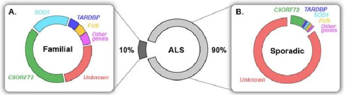
The idea of the involvement of genetic factors in the development of the disease has ancient origins, in fact pathological cases with family history and inheritance have been observed as early as 18508. Although most cases are classified as sporadic ALS (sALS), about 10% some cases of ALS are inherited, and therefore classified as familial cases (fALS), with multiple autosomal dominant and recessive forms. The sporadic and familial ALS shares clinical and neuropathological manifestations, and both types of patients, show a certain degree of heterogeneity regarding the symptoms, age of onset and duration of the disease. All the genes found mutated in fALS cases were found mutated in sALS2,9. Since fALS and sALS are clinically indistinguishable, with the exception of the time of the onset of the disease, that is found to be earlier in cases of family ALS1, several studies have been carried out in order to understand the role of genes associated with fALS in sporadic cases.
Figure 1. Known genetic causes in familial and sporadic ALS.Most ALS cases are sporadic (sALS) and only 10% are inherited, called familial (fALS). (A) 20% of fALS are caused by mutations in SOD1, which is the first known ALS-linked gene, identified in 19931.
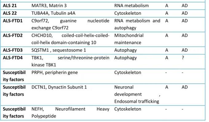
Over the years, a number of investigations have been carried out, leading to the identification of a large number of genetic factors associated with ALS (Table 1), which are categorized according to the risk of developing the disease in two large groups:
TARDBP, C9ORF72, UBQLN2, which are by no chance the ones that are most frequently mutated in pathologic subjects;
• low-risk sALS, so-called susceptibility factors such as NEFH11 (Table 1);
Up to now, about 20 genes explain most of the cases of familial sALS, but only a minority, about 10%, of sporadic cases of illness12,13.
LOCUS GENE, PROTEIN Disease Mechamism ONS ET
AD/AR ALS 1 SOD1, Cu/Zn SOD1 Oxidative stress A AD/AR
ALS 2 ALS2, Alsin Endosomal trafficking J AR
ALS 3 ? -- A AD
ALS 4 SETX, Senataxin RNA metabolism J AD
ALS 5 SPG11, Spatacsin DNA damage repair and axon growth
J AR
ALS 6 FUS, Fused in sarcoma RNA metabolism, DNA repair
J/A AD/AR
ALS 7 ? -- A AD/AR
ALS 8 VAPB, Vescicle-associated membrane Endoplasmatic reticulum stress
A AD
ALS 9 ANG, Angiogenin RNA metabolism A AD
ALS 10 TARDBP, TAR DNA-binding protein RNA metabolism A AD
ALS 11 Phosphoinositide-5-phosphatase Endosomal trafficking A AD
ALS 12 OPTN, Optineurin Autophagy A AD/AR
ALS 13 ATXN2, Ataxin 2 RNA metabolism A AD
ALS 14 VCP, valosin-containing protein Autophagy A AD/AR
ALS 15 UBQLN2, Ubiquilin 2 UPS
(ubiquitin-proteasome system) and Autophagy
J/A X-linked
ALS 16 SIGMAR1, sigma non-opioid intracellular receptor 1
UPS (ubiquitin-proteasome system) and Autophagy
J AR
ALS 17 CHMP2B, Charged multivesicular body protein 2B
Endosomal trafficking A AD
ALS 18 PFN1, Profilin 1 Cytoskeleton A AD
ALS 19 ERBB4, Neuregulin-ErbB4 Neuronal development
A AD
ribonucleoprotein A1
ALS 21 MATR3, Matrin 3 RNA metabolism A AD
ALS 22 TUBA4A, Tubulin a4A Cytoskeleton A AD
ALS-FTD1 C9orf72, guanine nucleotide exchange C9orf72
RNA metabolism and autophagy A AD ALS-FTD2 CHCHD10, coiled-coil-helix-coiled-coil-helix domain-containing 10 Mitochondrial maintenance A AD
ALS-FTD3 SQSTM1 , sequestosome 1 Autophagy A AD
ALS-FTD4 TBK1, serine/threonine-protein kinase TBK1
Autophagy A ?
Susceptibil ity factors
PRPH, peripherin gene Cytoskeleton - -
Susceptibil ity factors
DCTN1, Dynactin Subunit 1 Neuronal
development , Endosomal trafficking
A AD
Susceptibil ity factors
NEFH, Neurofilament Heavy Polypeptide
Cytoskeleton - -
Table 1.Principal gene locus involved in ALS and relative mutations:Genetic factors at the base of the SLA. Abbreviations: AD: dominant autosomal; AR: autosomal recessive; A: adult; J: youthful. (Data extracted from OMIM database: http://omim.org/phenotypiSeries/105400).
Superoxide dismutase 1 (SOD1) is the first ALS-linked gene that was identified in 199314 and, for almost fifteen years, ALS research has been focused on mutant forms of this protein15,16. Since 2008, starting with the discovery of ALS-linked mutations in DNA/RNA-binding proteins TDP-43 and FUS, an era of unprecedented genetic discoveries in ALS begun.
Although the genetic cause of most sALS is not known, recent studies have shown that mutations causing fALS are also able to cause illness in sporadic cases. In fact, only a small part of sALS patients showed that they had de novo mutations in known ALS-causing genes17-20. C9ORF72 repeat expansions, which were also found in a substantial fraction (~7%) of apparently sporadic ALS patients, most likely are not occurring de novo21,22, but rather represent cases with insufficient family history or incomplete penetrance. Taken together, ~10% of apparently sporadic ALS cases are caused by known genetic mutations, while the aetiology of the rest ~90% sALS remains unknown (figure 1).
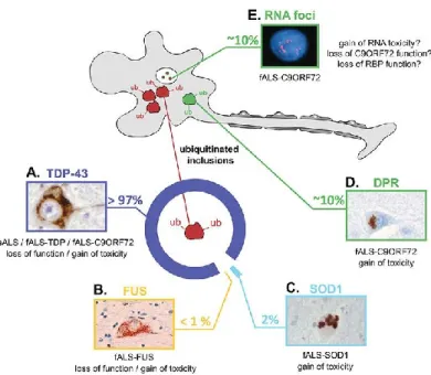
The pathological hallmark of ALS is the presence of ubiquitin-positive inclusions, consisting of misfolded protein aggregates in affected motor neurons and glial cells of the spinal cord and motor cortex. The most frequent protein component of these inclusions, in most ALS cases, is TDP-4323,24. Notable exceptions are patients with mutations in SOD1 and FUS, which lack TDP-43 inclusions, but show misfolded SOD125,26 or FUS27,28 protein accumulations. Moreover, patients with C9ORF72 expansions are affected by the most complex pathology form, characterized by typical TDP-43 inclusions, atypical TDP-43 negative inclusions29-31, consisting in abnormal dipeptide proteins32,33, as well as nuclear repeat RNA foci29,32,34 (figure 2 and 3).
ALS-associated genes code for proteins implicated in different cellular processes, and various mechanisms have been suggested as major contributors to neurodegeneration in fALS and sALS35.
There are two typical expressions of the onset of the disease: about 70% of patients present limb atrophy and muscular weakness at the trunk level at the initial stage of the disease , while the rest of the patients exhibit a symptomatology that ,during the first stage of the pathology, affects musculature of the tongue and swallowing36. Nearly 85% of patients with spinal-onset ALS, however, exhibit bulbar changes with disease progression. Approximately 50% of all patients diagnosed with ALS show cognitive and language impairment, while 10% of patients have clear signs of Frontotemporal dementia (FTD).
Some chromosomal loci containing ALS-causing genes are inherited as dominant while other as recessive autosomal traits, and it can be seen that there are at least 4 chromosome loci containing genes predisposing the pathology (Table 1). For the majority of ALS subtypes, the onset period is adulthood, with the exception of subtypes ALS2, ALS4, ALS5, whose period of onset is strictly juvenile.
1.3 Environmental and lifestyle factors
Environmental factors may play a decisive role in the onset of ALS disease. Among these risk factors the exposure to pesticides, fertilizers and heavy metals (such as selenium and mercury) has surely a significant role, especially for some specific occupational classes
(farmers, foundry workers)37. Exposure to secondary metabolites of cyanobacteria38, such as non-neurotoxic amino acid β-N-methylamino-L-alanine (BMAA), may also have a primary role as a possible causative agent of ALS39. Cigarette smoking would appear to be an etiologic agent, with a directly proportional association between exposure time and exposure intensity40. Exposure to this set of chemical substances would result in cellular oxidative stress phenomena, increasing levels of reactive oxygen (ROS) and blood species in cerebral tissue, yielding to progressive neurodegeneration41. It also seems that, among all environmental factors, alcohol abuse42, excessive physical activity and mechanical trauma associated with it43, self-immunity phenomena, as well as latent infections mediated by retrovirus and enterovirus44, play a key role in the onset of ALS. In fact, this disease, has been reported at a higher frequency among groups of athletes with respect to general population, although it is unclear whether physical activity is a risk factor or is simply a marker of underlying athletic prowess45.
2. Pathogenic mechanisms in ALS
ALS is a heterogeneous disease, to which great diversity in genetic and environmental causes correspond a complex network of molecular pathogenic mechanisms that haven’t been completely understood yet. Nevertheless neuropathological hallmark of disease is the aggregation and accumulation of ubiquitinated proteinaceous inclusions in motor neurons.
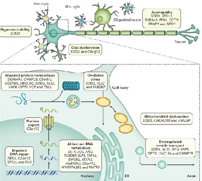
ALS pathogenic mechanisms, studied in many different experimental models and confirmed in ALS patients, outline a complex model in which non-competing mechanisms, including not only gene mutations and environmental factors but also impairment of protein homeostasis, aberrant RNA metabolism, impaired DNA repair, oxidative stress, mitochondrial dysfunction, dysregulation of nucleocytoplasmic and endosomal transport, neuroinflammation, excitotoxicity, axonopathy, are likely to converge in various unfortunate patterns to mediate selective motor neuron degeneration46 (Figure 2).
Figure 2. Molecular mechanisms of motor neuron injury in ALS. ALS is a complex disease involving activation of several cellular pathways in motor neurons, and dysregulated interaction with neighbouring glial cells2.
2.1 Impaired protein homeostasis
Pathological protein aggregates, identified as compact or skein-like ubiquitinated inclusions, are a cardinal feature of ALS48-50. The identification of TDP-43 as the most abundant protein constituent of these inclusions, initiated a major shift in our understanding of the pathobiology of ALS23. Under normal conditions, TDP-43 is predominantly localized in the nucleus, and loss of nuclear TDP-43 staining is seen in nearly all cells containing TDP-43-positive cytoplasmic inclusions23. TDP-43 inclusions are not restricted to motor neurons, and it seems that cytoplasmic redistribution of TDP-43 is an early pathogenic event in ALS. In 2008, when mutations in TARDBP, the gene encoding for TDP-43, were discovered in several fALS pedigrees, the evidences for TDP-43 dysfunction in ALS were consolidated, thus establishing this protein as a crucial player in both sporadic and familial disease51.
Although TDP-43 is the most abundant component of protein inclusions in 97% of ALS patients, different aggregates have been identified in various types of ALS, highlighting
the heterogeneity of the disease (Figure 3).
Figure 3: Pathogenic hallmarks of ALS and the underlying factors and mechanisms.
(A) In the majority of ALS cases, including all sporadic and most familial cases, the ubiquitinated inclusions contain TDP-43. When TDP-43 accumulates in the cytoplasm, it is excluded from the nucleus, where it normally resides in healthy cells. (B) In 0.5% of fALS that carry mutations in FUS, the cytoplasmic inclusions do not contain TDP-43, but they contain FUS protein. Similar observations to that of TDP-43, with partial nuclear clearance of FUS suggest that its neurotoxity may be the result of either loss of function or gain of toxicity. (C) In fALS patients with SOD1 mutations, misfolded SOD1 accumulates and becomes toxic to neurons. (D) In contrast, patients with hexanucleotide repeat expansions in C9ORF72 show typical TDP-43, similar to those found in sALS. (E) In addition to TDP-43 and DPR pathology, C9ORF72 patients accumulate nuclear RNA foci that contain sense and antisense hexanucleotide repeats in neuronal and glial cells. Insert images reproduced with permission from1.
SOD1 inclusions are found in motor neurons of patients with fALS, as well as in mouse and cellular models expressing SOD1 mutations52. Monoclonal antibodies that are specific for epitopes of misfolded SOD1, very common in inclusions motor neurons of patients with SOD1 fALS53, and seem to form similar structures in some patients with sALS54. Similarly, cytoplasmic inclusions containing mutant fused in sarcoma (FUS) protein have been observed in some patients with FUS-related fALS55,56. Proteins found in aggregates in ALS provide several important clues about the disease pathogenesis. Loss of nuclear
TDP-43 and/or aggregation of the protein in cytoplasmic inclusions, may be key of pathogenic processes in both sALS and fALS.
Protein misfolding and aggregation, in ALS, indicate both a defect in the activity of protein quality control (PQC) system, which includes molecular chaperons that lead protein folding and aggregation, and degradation systems (proteasome and autophagy). In fact, many ALS causing genes code for proteins that can promote dysfunction of the UPS (ubiquitin-proteasome system) such as SOD1, TDP-43 and VAPB, or proteins that are key regulators of both autophagy initiation, such as C9orf72 and optineurin, and execution, such as SQSTM1 and TBK1.
2.2 Oxidative stress
Several studies focused their attention on the role of mitochondria in the pathogenesis of neurodegenerative diseases such as ALS57. Mitochondrial dysfunctions, such as abnormal mitochondria and morphological defects, have been identified in skeletal muscles and intramuscular nerves of sALS patients58,59. In these cases, biochemical analysis revealed defects in muscular respiratory chain (complexes I and IV)60, which leads to/evolves/ends in (occhio a quale scegli per lasciare o togliere in) damage to motorneuron metabolism and alteration to mitochondrial membrane pores permeability, and high levels of mitochondrial calcium in muscle and spinal cord61. In transgenic mice, the main evidence of mitochondrial alteration is the presence of mitochondria with vacuoles. For example, in SOD1 G93A mice, there have been observed vacuous originated from the detachment between the internal and external mitochondrial membranes, leading to a remarkable increase in number and volume as pathology progresses62-64.
The expression of mutated SOD1 in neuronal cell lines or in primary motor neuron cultures, causes depolarization of mitochondria, alteration of calcium homeostasis and reduces ATP production, similarly as it happens in SOD1 G93A transgenic mice65-67.
Mitochondrial alterations may contribute to the generation of oxidative stress conditions, as they appear to mediate cell death by releasing calcium into the cytoplasm and by weakening the respiratory chain, leading to the release of cytochrome-c and pro-apoptotic factors that activate the cascade of caspases and trigger apoptosis68,69.
Oxygen is essential for life, but paradoxically, as a sub-product of its metabolism produces reactive oxygen species (ROS), which causes oxidative stress. Post-mortem analysis on brain tissues of patients with neurodegenerative diseases, such as ALS, clearly indicates an increase in the ROS index, in brain regions affected by the disease. The role of oxidative stress, and consequently of ROS in the onset of ALS, is still being discussed, since it is not clear yet whether they are the cause or a consequence of neuronal cell loss. However, ROS contribution to the events associated with neurodegeneration is evident70. Oxidative stress, in addition to mitochondrial damage, may be associated with an aberrant chemistry of SOD1. This appears to be a consequence of alterations in the shape of the active site that allow copper to interact with atypical substrates. In fact, modified SOD1, in contrast to normal activity, can act as peroxidase, resulting both in production of hydroxyl radicals and in inactivation of the enzyme itself, or can react with peroxynitrite causing tyrosine nitration71. Both peroxynitrite and hydroxyl radicals are strongly reactive species and can, therefore, cause oxidative damage to proteins, lipids, and DNA.
Such damage can cause alteration of protein conformation, destruction of the active site of enzymes, and modification of cell membrane properties by oxidation of unsaturated fatty acids and introduction of DNA mutations72. While earlier this mechanism was referable only to aberrant mutations of SOD1, recent studies investigate the possible involvement of FUS and TDP-43: it appears that TDP-43 mutations may be responsible for the production of reactive oxygen species and may alter the functioning of the mitochondrial respiratory chain73. The sum of these events seems to be directly correlated to motorneurons degenerative process in patients with ALS.
2.3 Aberrant RNA metabolism
Many different RNA-binding proteins (RBPs) have been implicated in human diseases, ranging from cancer to neurodegeneration, and alteration of mRNA processing is a key event in ALS pathogenesis. There are more than one hundred genes associated with ALS74, a handful of which encode proteins that control RNA processing; table 1 reports RBPs known to be mutated in ALS patients.
A2/B1 are structurally similar, as they contain two RNA recognition motifs (RRMs) and a Gly-rich C-terminal domain75. FUS, EWSR1 and TAF15, which form the FET family, share a similar structure: a zinc finger and RRM domain that facilitates DNA and RNA binding, respectively, an N-terminal low complexity, prion-like domain that mediates protein interactions and self-assembly76-78, multiple C-terminal Arg-Gly-Gly (RGG) domains that facilitate non-specific RNA binding and protein interactions, and an atypical Pro-Tyr nuclear localization signal (PY-NLS) that is recognized by transporters that control nuclear–cytoplasmic shuttling79. Those proteins are predominantly multifunctional nuclear proteins widely expressed in most cell and tissue types, since they are implicated in a broad range of cellular processes.
Whether motorneuron damage is caused by loss of normal nuclear function of TDP-43 and FUS in RNA processing, or by toxic gains of function, or both is unknown79. TDP-43 and FUS contain two RNA recognition domains—structures that are common to many RNA-interacting proteins, including those that are involved in mRNA transport. TDP-43 and FUS may form part of such RNA transport complexes and, when mutated, could thereby contribute to motor neuron injury through loss of axonal mRNA transport.
Fibroblast cell lines derived from patients with TARDBP-related ALS, showed the expected loss of nuclear expression of TDP-43, together with widespread changes in RNA splicing, including changes in transcripts of other RNA-processing genes and genes that have been previously implicated in ALS; hence, splicing disruption associated with the loss of nuclear TDP-43, is likely to contribute to ALS pathophysiology. Several in vivo models of TDP-43 dysfunction, including both knockout models and models overexpressing the wild-type and mutant forms of the protein, have recently been generated in Mouse, Rat, Drosophila melanogaster, Zebrafish and Caenorhabditis elegans80,81.
A note of caution relating to genetic studies is that, although gene structures are often similar between species, intronic sequences show great variability. Given the key role of TDP-43 in binding to long intronic sequences, the intraspecies variability in introns may hinder accurate modelling of human TDP-43 proteinopathies in other species, including rodents82,83.
2.4 DNA repair
Impaired DNA repair was suggested to have a role in ALS pathophysiology since the identification of FUS mutations, although the exact role of DNA repair failure in ALS remains to be clarified84. Interestingly both FUS and TDP-43 have been shown to be recruited by DNA damage; in fact, Wang et al. (2013) demonstrated that FUS is important in the mediation of DNA repair and DNA damage response (DDR) in post-mortem neurons, thus explaining how FUS mutations can alter the response to DNA damage leading to pathogenesis of neurodegenerative diseases84.
TDP-43, like FUS, plays a role in the prevention and repair of damages associated with DNA as well as damages associated with DNA loops, thus helping both DNA stabilization and damages related to its transcription. Specifically, TDP-43 is localized in foci before the damage of endogenous DNA and its presence increases even more following DNA damage, suggesting the existence of some shared functions for FUS and TDP-4385.
Mutations in NEK1 and C21orf2, which both encode for proteins involved in DNA repair, can cause ALS86-88, although the biological pathways associated with their causal role needs further confirmation.
2.5
Nucleocytoplasmic and endosomal transport
Perturbation of nucleocytoplasmic transport has been identified as a central mechanism underlying ALS-FTD, particularly those cases caused by mutations in C9ORF72, but is also involved in other neurodegenerative diseases as well as normal aging. The observation that most cases of ALS are associated with mislocalization of TDP-43 was, in hindsight, the first clue that the pathomechanism of ALS might involve an abnormality in nuclear transport. The temporal relationship among mislocalization of TDP-43, nuclear transport defects, and neurotoxicity in ALS remains unclear. It is possible that a primary defect in nuclear transport causes mislocalization and subsequent aggregation of TDP-43. Another hypothesis is that accumulation of cytoplasmic aggregates of TDP-43 drive a secondary defect in nuclear transport. Despite the accumulation of experimental data indicating impairment of nucleocytoplasmic transport through the nuclear pore complex (NPC) in disease models, evidence of such defects in patients with ALS and other related diseases
is thus far limited to redistribution of NPC components in end-stage disease. In addition, the relative pathogenic contribution of nucleocytoplasmic transport deficiency, compared to impairments in other cellular processes, has not been fully evaluated. Moreover, the relative roles of toxic species causing damage to nucleocytoplasmic transport machinery, whether toxic RNA or toxic DPRs, remain unresolved. While challenging, we anticipate that these issues will be addressed soon in this rapidly moving field89.
2.6 Neuroinflammation and Excitotoxicity
Activated microglia and infiltrating lymphocytes indicate an inflammatory component in CNS pathology of ALS90. Proinflammatory mediators, including monocyte chemoattractant protein 1 and IL‑891, are present in the (cerebrospinal fluid) CSF of patients with ALS, and biochemical indices of immune-response activation are present in the blood92. Reduced counts of CD4+CD25+ regulatory T (TREG) cells and monocytes (CD14+ cells) are detected early in ALS, suggesting recruitment of these cells to the CNS early in the neurodegenerative process. TREG cells interact with CNS microglia, attenuating neuroinflammation by stimulating secretion of anti-inflammatory cytokines93. Consistent with this scenario, double transgenic mice, carrying mSOD1 and lacking CD4, develop a more aggressive ALS phenotype, which is reversible by bone marrow transplantation94. Neuroinflammation is closely linked to activation of the immune response. A recent study identified CD40L — a ligand expressed by T cells that activates the immune response when bound by CD40 on antigen-presenting cells — as a promising therapeutic target95. Proinflammatory mediators, including monocyte chemoattractant protein 1 and IL‑891, are present in CSF of patients with ALS, and biochemical indices of immune-response activation are present in the blood92. Reduced counts of CD4+CD25+ regulatory T (TREG) cells and monocytes (CD14+ cells) are detected early in ALS, suggesting recruitment of these cells to CNS early in neurodegenerative process. Excitotoxicity is a particular neuroinflammation process that is due, at least in part, to the excessive activation of glutamate N-methyl-D-Aspartate receptors, which causes an increased calcium input through the ionic channel associated with the receptor, thus resulting in production of free radicals, such as NO and ROS, which trigger numerous
pathways related with the onset of ALS. A body of evidence implicates excitotoxicity as a mechanism contributing to motor neuron injury in ALS, although clear evidence that it is a primary disease mechanism is lacking. In some patients with ALS, levels of CSF glutamate are elevated96, and the expression and activity of EAAT2 are reduced in pathologically affected areas of the CNS97-99, although whether if this is a cause or a consequence of neuronal loss is unclear100. Electrophysiological studies in humans have shown hyperexcitability of the motor system in presymptomatic101 or early stages102 of ALS. Evidence suggests that the calcium permeability of AMPA receptors in the spinal ventral horn may be dysregulated by abnormal editing of the GluR2 AMPA receptor subunit103. In addition, a recently identified ALS-linked gene encodes d‑amino acid oxidase (DAO). This enzyme is responsible for the oxidative deamination of d‑amino acids, one of which — d-Serine — is an activator and co-agonist of N‑methyl‑d-Aspartate (NMDA) receptors104. Mutations in DAO could potentially contribute to excitotoxic motor neuron injury. Excitotoxicity in ALS pathology leads to many events, including altered electrophysiological properties and increased sensitivity of motor neurons to excitotoxicity105, altered AMPA receptor subunit expression, reduced expression and activity of EAAT2106, increased glutamate efflux from spinal cord nerve terminals107, a reduction in motor neuron inhibitory–excitatory synaptic ratio108, and loss of regulation by astrocytes of the expression of GluR2 by neighboring motor neurons109. Ameliorating excitotoxicity is the only strategy that has, so far, slowed disease progression in ALS. Riluzole, which has several effects, including inhibition of presynaptic glutamate release110, causes a modest increase in survival111.
3. Structure and function of TDP-43
3.1 TDP-43 genetics
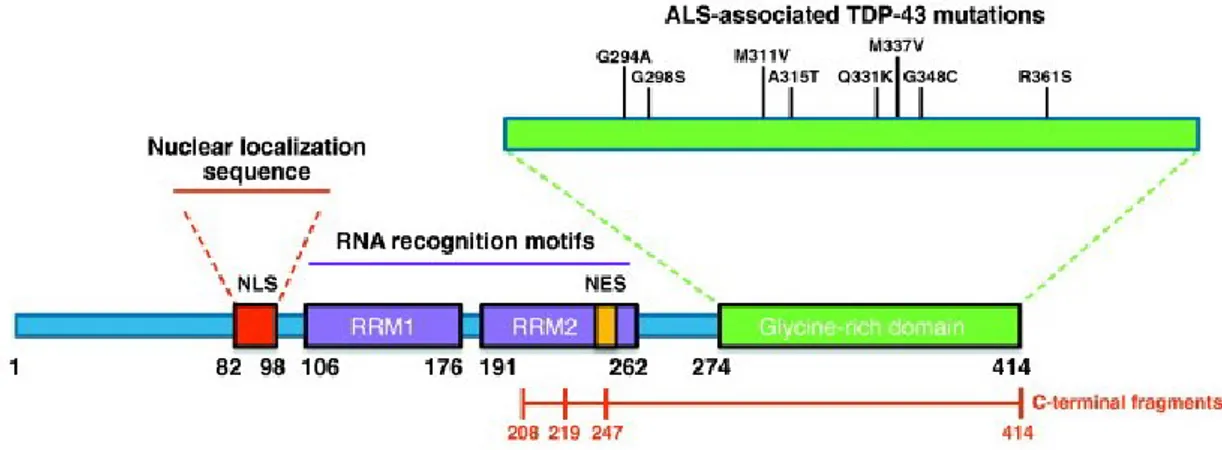
TDP-43 or TARDBP (TAR-DNA Binding Protein-43) is a 43 kDa ribonucleoprotein, originally identified as transcriptional repressor of the HIV-1 TAR-DNA. The gene coding for TDP-43 protein, consisting of 414 amino acids, is located along the short arm of chromosome 1 and consists of 6 exons (exon 1 is represented by a non-coding sequence). TDP-43 shares
with the heterogeneous ribonucleoprotein family a series of key protein domains essential for its activity:
● A Nuclear Localization Signal (NLS) domain at the N-terminal;
● Two domains "RRM1 and RRM2" (RNA-recognition motifs): recognition of DNA and RNA;
● A Nuclear Exportation Signal (NES) within the RRM2 domain;
● The "Glycine-rich region" located at the C-terminal end: Glycine-rich domain for interaction with other proteins and to promote the exon skipping of some pre-mRNAs 112,113 (figure 4).
Figure 4: Graphic representation of TDP-43 functional domains of the protein. TDP-43 have structural
similarities with both harboring a Prion-like domain, RNA recognition motif(s) and nuclear localization signal3.
TDP-43 has a predominantly nuclear localization and, as FUS, is involved in the regulation of many biological processes. In fact, TDP-43 is a DNA/RNA binding protein that participates in transcription regulation by promoting binding DNA transcription factors, is involved in the alternative splicing process, in biogenesis and maturation of microRNAs, and in transport and localization of some pre-mRNAs115. Additionally, TDP-43 is particularly important in regulating axonal transport and cytoskeletal integrity. Mutants and aberrant protein forms are therefore associated with a wide range of cellular toxicity phenomena116. Since 2006, studies on the composition of protein aggregates in neuronal brain cells of patients with neurodegenerative diseases such as ALS have shown that
TDP-43 protein is the major constituent of such inclusions23. As for FUS, more than 40 mutations were identified in TARDBP gene, and TARDBP pathogenic variants prevalence is:
● 1.6% in all individuals with ALS (fALS and sALS)
● 3.4%, but ranges from 0% to 12% across studies117,118 in fALS;
● 1.1% but ranges from 0% to 5% across studies in sALS119,120.
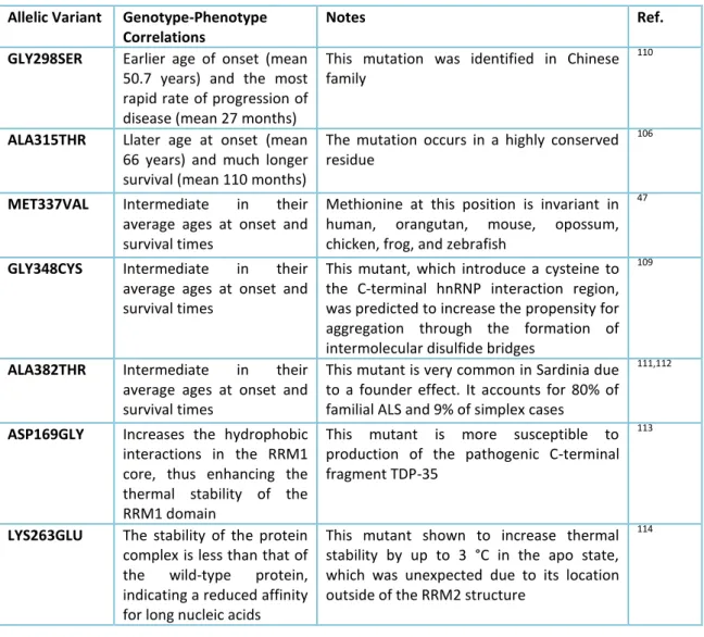
Most mutations fall within exon 6, that codes for the rich glycine region at the C-terminal end of the protein, and are inherited as dominant autosomal traits (Table 2).
Allelic Variant Genotype-Phenotype
Correlations
Notes Ref.
GLY298SER Earlier age of onset (mean
50.7 years) and the most rapid rate of progression of disease (mean 27 months)
This mutation was identified in Chinese family
110
ALA315THR Llater age at onset (mean
66 years) and much longer survival (mean 110 months)
The mutation occurs in a highly conserved residue
106
MET337VAL Intermediate in their
average ages at onset and survival times
Methionine at this position is invariant in human, orangutan, mouse, opossum, chicken, frog, and zebrafish
47
GLY348CYS Intermediate in their
average ages at onset and survival times
This mutant, which introduce a cysteine to the C-terminal hnRNP interaction region, was predicted to increase the propensity for aggregation through the formation of intermolecular disulfide bridges
109
ALA382THR Intermediate in their
average ages at onset and survival times
This mutant is very common in Sardinia due to a founder effect. It accounts for 80% of familial ALS and 9% of simplex cases
111,112
ASP169GLY Increases the hydrophobic
interactions in the RRM1 core, thus enhancing the thermal stability of the RRM1 domain
This mutant is more susceptible to production of the pathogenic C-terminal fragment TDP-35
113
LYS263GLU The stability of the protein
complex is less than that of the wild-type protein, indicating a reduced affinity for long nucleic acids
This mutant shown to increase thermal stability by up to 3 °C in the apo state, which was unexpected due to its location outside of the RRM2 structure
114
Table 2. Principal mutations of TDP-43 protein. TDP-43 has several pathological mutations, represented in small part in this table, mostly located in the domain rich in wiccans, although there are many discreetly discovered findings that resurface in the RRM1 and RRM2 domains, mainly due to lysine.
The pathological manifestation, in type 10 ALS patients, is characterized by a great heterogeneity in terms of age of onset, aggressiveness of the phenotype and geographical location. In fact, some pathological mutants are characteristic of delimited geographical areas: a clear example is represented by the mutant TDP-43 A382T (defined as the "Sardinian variant"), which accounts for approximately 30% of total ALS cases in Sardinia121,122.
3.2 TDP-43 protein aggregation
As described in paragraph 3.1, TDP-43 is present in the protein aggregates of almost all cases of ALS. Pathologic TDP-43 is hyperphosphorylated, ubiquitinated and undergoes an abnormal proteolytic process, from which a 25KDa carboxy terminal fragment is generated, resulting in function loss. Neo-formed fragments are transported from nucleus to cytoplasm of neuronal and glial cells, where they tend to aggregate and precipitate123. Immunoprecipitation and immunoblotting experiments demonstrated how the cytoplasmic inclusions are subjected to ubiquitination and phosphorylation processes. The ubiquitination of misfolded proteins that aggregate in the cytoplasm and nucleus of neuronal cells, seems to be one of the key phenomena involved in the pathogenesis of neurodegenerative diseases and proteinopathies: in fact this phenomenon is appreciable in the most advanced stages of the disease. The formation of cytoplasmic inclusion bodies, therefore, would appear to be due to the presence of aberrant mutations to the functional protein in wild-type form12.
3.3 TDP-43 as transcriptional modulator
TDP-43 was originally identified as a transcriptional repressor that binds to TAR DNA of human immunodeficiency virus type 1 (HIV-1) 124. Consistent with its role in transcription, TDP-43 was found, in human brain and in cell culture systems, to associate with euchromatin and to bind the promoter of mouse acrv1 gene, coding for SP-10 protein, which is required for spermatogenesis, acting as transcriptional repressor125. Recent evidences indicate that TDP-43 acts as a repressor of Acrv1 gene in spermatocytes and post-translational modifications, most likely ubiquitination, induce conformational change, leading to release of repression in round spermatids126. A second example that
illustrates TDP-43 activity as a transcriptional repressor in vivo came from a recent report that identified rat and human VPS4B gene (vacuolar protein sorting 4 homolog B) as a TDP-43 target in brain, via direct binding to a TG-rich region in the promoter127. Moreover TDP-43 is associated with proteins involved in transcription, including methyl CpG-binding protein 2 (MeCP2)128. Beside this role as transcriptional repressor, TDP-43 has been indicated also as transcriptional activator, by luciferase assay or mRNA level determination for Bim promoter129, NFB responsive elements130, p53 binding elements131
and CHOP promoter132.
3.4 TDP-43 as modulator of RNA metabolism
Splicing process is strictly regulated and is essential for an appropriate gene expression. It is not surprising, therefore, that dysfunctional spliceosome is closely linked to neurodegeneration. For example, when TDP-43 is removed from mouse embryonic stem cells, multiple cryptic exon have been coupled. This phenomenon has also been observed in cells from human patients with ALS and FTD133.
TDP-43 shows the ability to bind RNA in a very specific way through the presence of two RRM motifs within its sequence, preferably UG repeats and UG/GU-rich repeats interspersed by other nucleotides, and the acetylation of residues K145 and K192, located in these domains, leading to reduced binding activity134,135. These observations were subsequently confirmed by CLIP analyses136,137, and a high resolution TDP-43 nuclear magnetic resonance structure that binds one of these sequences has been recently solved85. It should also be considered that the presence of a potential binding site does not always mean that it will play a fundamental role in a particular process138.
In C. elegans, as well as in cell culture, TDP-43 loss induces the accumulation of double-stranded RNA (dsRNA) and the abnormal processing of ribosomal RNA. Since it remains to be clarified whether these changes in RNA metabolism play a role in ALS/FTD pathology, several research groups are currently examining the molecular mechanisms with which TDP-43 limits the dsRNA and are trying to determine whether the loss of FUS and MATR3 or the expression of hexanucleotide C9orf72 has similar effects on RNA metabolism.
Another important aspect is represented by cytoplasmic redistribution associated with TDP-43 disease, in particular in response to the expression of C9orf72 hexanucleotide expansion and the associated production of non-aggregated polypeptides139.
3.5 Transposable Elements and TDP-43
The transposable elements (TEs) are highly abundant genetic elements that constitute a great part of the eukaryotic genomes. Retrotransposons, which are transposed via an RNA intermediate, account for about 40% of the human genome140,141. Although most TE copies do not work, a subset has retained the ability to mobilize and even the stationary copies can be expressed142. Due to their potential to copy and paste into new genomic positions, TEs represent a huge endogenous reservoir of genomic instability and cellular toxicity141. The impacts of these genetic parasites are usually stifled by strong cellular mechanisms involving small interfering RNAs acting through induced RNA (RISC) to inhibit expression of transposon143. Although most surveys naturally focus on germs, where new insertions are inheritable and favoured from the evolution of the transposon, somatic tissues also have an active effect transposon mute mechanism whose functional meaning is less understood. As found in literature, TEs are normally active in brain144-147, and LINE and SINE (which are non-LTR retrotransponants) are even more expressed. LTR elements, on the other hand, have been associated with several neurodegenerative agents disturbances 148-154. With regard to this, it was evaluated by Wang group155 if TDP-43 RNA targets include transposon derivatives transcripts. Several recent studies used deep sequences for RNA analysis targets that co-purify with immunoprecipitated mouse, rat or human TDP-43, and also for profiling gene expression in mouse after TDP-43 abatement or excess expression156-159.
From data in literature, it can be stated that TDP-43 is generally destined to derive from TE transcripts, including many short interspersed nuclear elements (SINE), long interspersed nuclear elements (LINE) and long terminal repeat elements (LTR) classes as some DNA elements. Secondly, the association between TDP-43 and TE-derived RNA targets is reduced in patients with FTLD with respect to healthy subjects, consistent with the idea that loss of TE control could be a part of the onset of the disease160,161,162.
3.6 The role of TDP-43 in mitochondrial translation
Several research paper reported that, in ALS patients, TDP-43 co-localizes with various neuronal organelles in both human spinal cord and frontal cortex tissue samples, oppositely to what physiologically happens in age-matched normal individuals.
In fact, it was demonstrated that in control cases, both in spinal cord motor neurons and in cortical neurons, TDP-43 mainly localizes in nucleus, while both ALS motor neurons and FTD cortical neurons showed characteristically higher levels of cytoplasmic TDP-43 accumulation.
Notably, cytoplasmic TDP-43 co-localizes with mitochondrial markers in many ALS spinal cord motor neurons or FTD cortical neurons, but minimally overlaps with markers of Golgi, endoplasmic reticulum, lysosome, autophagosome, endosome or peroxisome. Despite its low abundance in cytoplasm, TDP-43 co-localizes with mitochondria in motor neurons and cortical neurons in human control samples. Consequent sub-mitochondrial fractionation analysis, detected that mitochondrial TDP-43 was exclusively present in the inner mitochondrial membrane but not in outer mitochondrial membrane, intermembrane-space or matrix. Also Immuno-electron microscopy analysis of isolated mitochondria and biopsied human cortex confirmed the principal localization of TDP-43 in inner mitochondrial membrane cristae.
Further studies on TDP-43 localization, showed that disease-associated mutations, such as A382T, influenced its mitochondrial localization too.
It remains to be clarified whether TDP-43 deficiency alone is sufficient to cause neuronal loss, or if, with an inactivated nuclear localization signal, it causes a significant co-localization of this protein with mitochondria, indicating mitochondrial co-localization as an intrinsic property of cytoplasmic TDP-43160.
Since mitochondrial dysfunction precedes TDP-43-induced neuronal death, and since mitochondria are involved in almost all types of cell death, including apoptosis and necrosis, it could be of great interest understanding whether mitochondrial dysfunction is induced by TDP-43 or not161,162.
4 Epigenetics and neurodegeneration
4.1 Epigenetic modifications
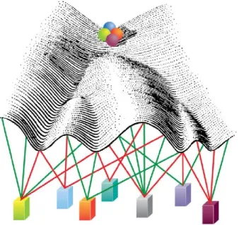
Epigenetics is defined as the set of potentially inheritable changes in gene expression that do not involve modifications in the nucleotide sequence of DNA137,163. The complexity of our organism clearly points out the existence in the genome of something more than DNA sequence that is capable of determining the great intra-inter-individual variability. The latter is also based on epigenetic modifications, which are responsible for the maintenance of chromatin stability and which are implicated in a wide range of neurodegenerative disorders164. To date, many molecular complexes are known to be responsible for chromatin remodelling, dependent enzymatic modification and DNA-related histone proteins, as well as substitution of the same histones. Epigenetics is, thus, the configuration by which regulation of transcription and, hence, gene expression within our cells is realized and, in particular, it explains how genome written modifications are read, interpreted and removed, thus contributing to the stability and plasticity of nerve cells164 (Figure 5).
Figure 5: Waddington's vision of an epigenetic landscape. This image shows that DNA provides the stable
base on which our individual details are written in the form of chemical marks. Most likely, the timing, location, and persistency of the marks are some of the variables that will determine how important they will
be in impacting such things as development and disease. Much remains to be elucidated, but the demonstration that external factors can alter the epigenome suggests that we can manipulate it, hopefully for good rather than evil.
Chromatin can be defined as a plastic substrate capable of responding to rapid,short-term and long-term changes within cells. Modulation and regulation of gene expression, at chromatin level, involve different epigenetic mechanisms, which can remodel or modify chromatin template164-166. Chromatin remodelling activities are ATP-dependent and increase DNA-binding proteins accessibility to genome by altering DNA–histone interaction non-covalently. Chromatin modifying activities are carried out by introducing covalent modifications on histone tails, histone core proteins or by DNA methylation. Histone modification complexes make post-translational changes of covalent nature at the N-terminal end of histone proteins, such as acetylation, methylation, phosphorylation, ubiquitation, sumoylation, glycosylation and ribosylation. These post-translational modifications, as well as the introduction of histone variants, co-operate in altering chromatin fiber167,168, changing the degree of chromatinic compaction, which closely correlates to transcriptionally active or inactive state169. The histone DNA-code interactions control the activation/repression of gene transcription; therefore, in case of transcriptional activation, compact and inaccessible DNA is made available to DNA binding proteins by chromatin modification170. For example, high levels of histones H3 and H4 acetylation and H3 methylation on lysine 4 (H3K4me) are generally found in promoter regions of transcriptionally active genes, while high levels of H3 methylation on lysine 27 (H3K27me) correlates with polycomb-mediated protein transcriptional repression171.
Of course, chromatin organization is designed to ensure that writing, reading and preservation of gene information is carried out within a well-defined spatial and temporal sequence, both during cell differentiation and during various stages of development164. It has long been suggested that histone modification patterns act as "recognition codes" for the recruitment of chromatin remodelling complexes and there are several experimental evidences that support the existence of the so-called histone code168,172. Histones N-terminal amino acids, when acetylated, are bound by proteins containing
well-characterized protein domains, such as bromodomain, found in many chromatin-associated proteins173,174. In addition, methylated lysine 9 of H3 is a specific recognition site for chromodomain, a domain typically involved in the formation of heterochromatin and in gene silencing; however, such residue may also be acetylated, and this modification is mutually exclusive with methylation175
. In addition, H3 acetylation on lysine 14 from GCN5 HAT complex, is preceded by phosphorylation of serine 10 on the same tail; this suggests the existence of a regulatory cascade that controls the pathway, thanks to complexes capable of recognizing specific amino acid residues on the N-terminal codons171.
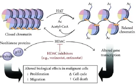
4.2 Acetylation and deacetylation
Lysine acetylation and deacetylation in N-terminal histone tail, play a prominent role in regulating neuronal plasticity and transcriptional regulation164,176. The ability to influence neuronal vitality by modulating levels and activity of enzymes regulating nuclear homeostasis, such as HATs (Histone acetyl transferase) and HDACs (Histone deacetylase), has been demonstrated. It is now clear that the pattern of acetylation is strongly damaged in case of degeneration of nerve cells. Not unexpectedly one of the major causes of dysfunction and toxicity in cells is the imbalance in the presence of HATs and HDACs177. Acetyltransferases and deacetylases, combined with large multiproteinic complexes, catalyse opposite reactions on protein substrates, including core histones and transcription factors; in addition to regulation of transcriptional machinery, HAT-HDAC system is also involved in the modulation of replication, in DNA repair and in site-specific recombination177. Acetyltransferases and deacetylases, combined with large multiproteinic complexes, catalyse opposite reactions on protein substrates including core histones and transcription factors; in addition to regulation of transcriptional machinery, the HAT-HDAC system is also involved in the modulation of replication, and of DNA repair and site-specific recombination177.
4.2.1 HATs
addition of acetyl groups derived from the Acetil-CoA. In particular, acetyl-transferase histones act on nucleosomal N-terminal tails and, in spite of the name, are also able to acetylate transcription factors such as p53, E2F and GATA1177. Acetylation of lysines neutralizes the positive charge of their ε-amine groups, thus reducing the affinity of these basic proteins to the acid DNA, altering histone core arrangement and relaxation of chromatin structure.
All core histones are subjected to acetylation, but the ones occurring on histones H3 and H4 are more characteristic; the main targets of HATs are lysine 9 and 14 on H3 and lysine 5, 8, 12 and 16 on H4-histone178. Acetylated lysine represent a surface on which many proteins with specific domains, such as bromodomain, can bind. Thus, chromatin is made more accessible to transcription factors. Acetylation of histones is consequently associated with transcriptional activation; on the contrary deacetylation closely correlates with gene silencing179.
4.2.2 HDACs: functions, location and classification
Histone deacetylation is related with the removal of acetyl groups from amino acid residues of N-terminal histone tails, leading to an increase in chromatin packaging, compatible with gene silencing.
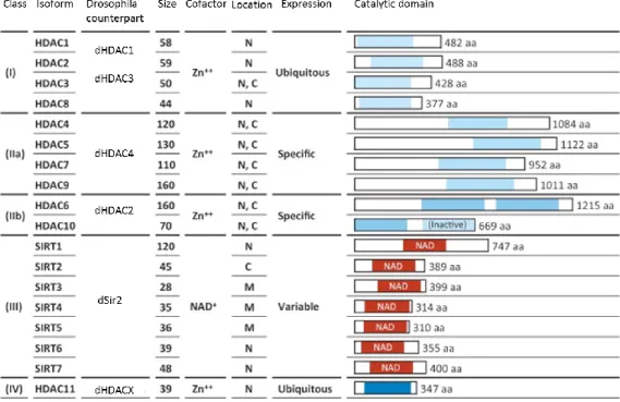
HDACs play a direct role in regulating transcription by deacetylation and regulation of TFs, or by interaction with transcriptional co-repressors. In Homo sapiens, 18 different HDACs have been identified, and have been divided into four categories based on their homology with yeast HDACs180: class I, class II, class III (sirtuine) and class IV HDACs (Figure 6).
Figure 6. Table representing Class I, II and IV HDACs. Class I (HDAC1, 2, 3 and 8), class IIa (HDAC4, 5, 7 and 9), class IIb (HDAC6 and 10), class III (sirtuin family) and class IV (HDAC11) HDACs with the Drosphila counterpart181,182.
Cellular localization, as well as site-specific expression, differs between various deacetylases. In order to carry out their biological activity, HDACs have to move to nucleus, where most of the target substrates are located.
Nuclear localization is guaranteed by the presence of a Nuclear Location Signal (NLS). Some HDACs are only nuclear, while others may be cytosolic, depending on the regulatory domains present. In particular, Class I HDACs, and a group of HDACs of class II (IIa), are the most expressed in cerebral areas associated with learning and memory183.
Class I, II and IV HDACs have zinc-dependent active sites, while sirtuins require NAD+ (nicotinamide adenin dinucleotide) cofactor to function. However, interaction between different classes of HDACs is necessary for their biological activity. All HDACs have a catalytic domain consisting in about 390 amino acids with a highly conserved amino acid sequence. The active site consists in a slightly curled tubular pocket with a wide bottom184. The removal of acetyl group occurs through a charge system, consisting in two adjacent histidine residues, two aspartate residues, located about 30 amino acids from
the histidines and separated by 6 amino acids, and a tyrosine residue, located 123 amino acids downstream of aspartate. A fundamental component of this system is zinc ion, retained by its binding site at the bottom of the pocket, although other cofactors are required for the activity of HDACs. The conserved tyrosine catalytic residue is substituted by a histidine in Class IIa HDACs. This substitution greatly reduces catalytic activity185
.
4.2.2.1 Class I HDACs
Deacetylases belonging to this class are expressed ubiquitously with a predominantly nuclear site. They show homology with the transduced yeast regulator RPD3 (Reduced Potassium Dependency 3) and are represented by HDACs 1, 2, 3 and 8186. These are largely expressed in brain, except for muscular-specific HDAC8, where they interact with key proteins that form part of large multi-unit complexes.
HDAC1 and 2, respectively constituted by 482 and 488 amino acids, exhibit high structural and functional similarity with a sequence homology equal to 82%, and participate in the formation of large transcriptional repression complexes consisting of SIN3A, NuRD and Co -REST187, thus inactivating the expression of neuronal genes in non-nervous tissues188. In particular, such complexes act on multiple aspects, such as cell cycle regulation, maintenance of pluripotency of stem cells and cell differentiation189. HDAC1 and HDAC2 are located in nucleus by acting not only on histones, but also on different substrates, including MyoD, E2F, p53 transcription factors and retinoblastoma protein. The two deacetylases carry out their biological activity only when incorporated into a multiproteic complex, which is, in fact, composed of proteins necessary for modulation of their deacetylase activity, DNA binding, and recruitment of HDACs on promoter regions. In addition, their activity, as well as the formation of repression complexes, is modulated by phosphorylation. Precisely, both deacetylases are phosphorylated by casein kinase 2 (CK2) and, in addition, HDAC1 represents the target of other kinases such as PKA (cAMP-dependent protein kinase A) and cGMP-(cAMP-dependent G protein kinase177. In resting cells HDAC1 and 2 are phosphorylated at low levels; when hyperphosphorylation occurs, there is a significant increase in deacetylase activity. Analysis of HDAC1 reveals two crucial sites for modulation of deacetylation by phosphorylation, represented by Ser421 and Ser423.
When these residues are mutated, the formation of protein complexes is hindered and HDAC1 biological activity is reduced190,191.
In ALS context, HDCA1 has been described as the interaction partner of FUS during DNA repair; in addition, ALS-causing FUS mutant displays a reduced binding activity84.
4.2.2.2 Class II HDACs
Class II HDACs are homologous to yeast histone deacetylase 1 (HDA1), and represent a class that is further subdivided in two subclasses:
• Class IIa HDACs, represented by HDACs 4, 5, 7 and 9; • Class IIb HDACs, represented by HDAC 6 and 10.
The former have, in addition to deacetylase domain, an extended N-terminal regulatory domain that regulates nucleotide-cytoplasmic shuttling and DNA-specific binding, while the latter have distinct C-terminal domains192. They both show cellular and tissue-specific distribution, with a narrower expression pattern than class I HDACs, suggesting their possible involvement in cell differentiation and development193, but they are expressed abundantly in brain194; in particular, almost all move from nucleus to cytoplasm because of the presence of nuclear signal, while cytosolic retention depends on phosphorylation and interactions with proteins 14-3-3195.
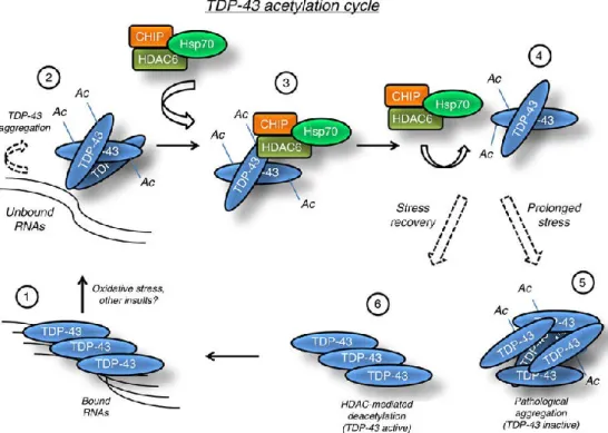
HDAC6, in particular, is expressed mainly in cytosol where it acts as an important constituent of cytoskeleton deacetylase, the α-tubulin196,197, but other substrates have been identified, such as TDP-43198. HDAC6 is the only enzyme with two deacetylase active domains organized in tandem and one zinc fingers (HDAC6-USP3 and Brap2-related zinc finger motif) domain in the carboxyl terminus. This latter domain is a signal for ubiquitination, suggesting, therefore, that this enzyme is particularly prone to degradation199. Although it is localized predominately in cytoplasm where, by inducing α-tubulin deacetylation, regulates cell motility and microtubule-dependent cell adhesion197, it was detected also at nuclear level in association with HDAC11. In addition, HDAC6 presents an activity independent from deacetylase functions200
; in fact, by tying the ubiquitin, it participates, for example, at the autophagy process regulation and activity of HSF1 protein (heat shock factor 1).
4.2.2.3 Class III HDACs
Commonly known as sirtuins, because of its homology with SIR2 regulator identified in yeast201, class III HDACs are represented by 7 different NAD+-dependent enzymes expressed in the mammalian brain. In particular, SIRT1, 2, 6 and 7 are localized both in nucleus and in cytosol, while SIRT3, 4 and 5 exhibit mitochondrial localization202,203. Sirtuins catalyse the deacetylation of other targets than histones: for example, SIRT1 acts on targets such as TAF68 (TBP-associated factor 68), p53 and p300; SIRT2 has the ability to deacetylate α-tubulin exactly as HDAC6204; SIRT3, 4 and 5 determine the global level of acetylation in mitochondria by regulating energy metabolism, but also lipid metabolism and apoptosis186.
In particular, many studies highlight the role of sirtuins in the maintenance of genomic integrity; in fact deficiency of their deacetylase activity is compatible with alterations in gene silencing, increased genomic instability and susceptibility to DNA damage. For example, it has been noted that SIRT6-deficient cells, accumulate a large number of chromosomal anomalies. Recent studies have also shown that this sirtuin is capable of affecting telomeric regions in human cells. Another important sirtuin, SIRT1, deacetylates factors associated with DNA repair mechanisms and is recruited on the same sites as a result of oxidative stress. Although this recruitment seems to have a protective role in genomic instability, it is accompanied by a depression of previously silenced genes, suggesting its implication in epigenetic silencing and chromatin modeling205. All sirtuins, except SIR2 and 5, have higher levels of expression in foetal brain with respect to adult, suggesting the crucial role of these molecules in initial brain development202.
4.2.2.4 Class IV HDAC
The only representative of this class, HDAC11, has common traits with class I and II HDACs but its function and substrates are still unknown. It can be found mainly in nucleus and it is expressed during the development of central nervous system of mammals, including oligodendrocytes, and plays a key role in maturation of this cell type206. It is also possible that it plays a role in regulating inflammation, through its inhibitory effect on interleukin 10 expression207,208.
4.3 HDACs regulation and deregulation in ALS
4.3.1 Zn-dependent HDACs in ALS
The implication of Class I HDACs in the onset of ALS was first shown by Janssen and colleagues209, which showed that in affected patients there is an over-regulation of HDAC2 in the motor cortex and in the grey matter of the spinal cord209, especially in the nuclei of motor neurons. Additionally, HDACs, in particular HDAC6, are increasingly implicated in ALS disease. A recent study reported that FUS and TDP-43 proteins, altered in many cases of ALS, interact with each other, forming a ribonucleoprotein complex that regulates the expression of HDAC6, by influencing the levels of its mRNA210. In addition, in nerve tissue of ALS patients, nucleus and cytoplasm of motor neurons present low levels of HDAC11 encoding mRNA209. In neurodegenerative context, HDAC6 plays a significant role in α-tubulin post-translational modification, which modifies the properties of cytoskeleton; HDACs strictly regulate the acetylation of a preserved lysine residue (K40) on α-tubulin protein, to regulate the movement of organelles within cells, mediated by motor proteins appearance211; this is especially important for those neurons who have to carry a large load for long distances.
The increase in acetylation levels in α-tubulin, following inhibition of the above-mentioned deacetylase activity, improves transport in primary neurons, thus preventing axonal degeneration212,213. Considering that reduction of acetylation levels in α-tubulin is a characteristic pathological marker, an approach based on the use of HDACs inhibitors for the treatment of neurodegenerative disorders with neurons with particularly long axons, has been attempted. In this respect, ALS is a significant example because axonal transport efficiency loss, in both upper and lower motorneurons, is significant in the onset of the disease214. A lot of experimental work on the role of α-tubulin acetylation in ALS was conducted in SOD1 G93A mice, where it has been observed that HDAC6 genetic ablation positively influences acetylated α-tubulin levels in the central and peripheral nervous system, and maintains integral axons. Moreover the potential for muscle activity and the number of neurons in ventral spinal cord horns are increased, suggesting that these events that are associated with increased cell survival215.