UNIVERSITÀ DEGLI STUDI DI SASSARI
Scuola di Dottorato di Ricerca in
Scienze Biomolecolari e Biotecnologiche
Indirizzo: Biochimica e Biologia molecolare
XXVI ciclo
Role of stromal fibroblasts in prostate
carcinoma progression and metabolic
reprogramming of cancer cells
Direttore della Scuola:
Prof.ssa Claudia Crosio
Tutor:
Prof. Gianfranco Pintus
Co-Tutor:
Prof.ssa Paola Chiarugi
Tesi di dottorato:
Dott. Alberto Marini
2
Dott. Alberto Marini
"Role of stromal fibroblasts in prostate carcinoma progression and metabolic reprogramming of cancer cells"
Tesi di Dottorato in Scienze Biomolecolari e Biotecnologiche-Università degli Studi di Sassari
INDEX
INTRODUCTION...5
TUMOR MICROENVIRONMENT...5
FIBROBLASTS AND CANCER ASSOCIATED FIBROBLASTS...6
ROLE OF CAFs IN TUMOR PROGRESSION...11
ROLE OF OTHER MICROENVIRONMENTAL COMPONENTS IN TUMOR PROGRESSION...16
Hypoxia...16
Acidity...21
TUMOR METABOLISM...23
REGULATION OF CANCER CELL METABOLISM: THE WARBURG EFFECT...26
Glucose transporters and glycolytic enzymes...26
Molecular pathways in aerobic glycolysis...32
REGULATION OF CANCER CELL METABOLISM: THE TCA CYCLE...36
Aconitase...36
Isocitrate dehydrogenase...37
Succinate dehydrogenase...37
Fumarate hydratase...38
REGULATION OF CANCER CELL METABOLISM: GLUTAMINOLYSIS...39
ROLE OF TUMOR MICROENVIRONMENT IN REGULATION OF CANCER CELL METABOLISM...42
TUMOR METABOLISM AND CHEMORESISTANCE...49
Glycolysis...52
PDK...56
Fatty acid biosynthesis...57
Glutaminolysis...58
Adaptations to oxidative stress and PPP...59
TUMOR MICROENVIRONMENT AND RESISTANCE TO CANCER THERAPY.62 CAFs...62
Hypoxia...63
OBJECTIVES...66
MATERIALS AND METHODS...67
3
Dott. Alberto Marini
"Role of stromal fibroblasts in prostate carcinoma progression and metabolic reprogramming of cancer cells"
Tesi di Dottorato in Scienze Biomolecolari e Biotecnologiche-Università degli Studi di Sassari
Common use solutions...68
METHODS...69
Cell cultures and treatments...69
Isolation of prostate fibroblasts...70
Fibroblasts and PCa cells activation...70
Cell transfection with lipofectamine...71
Proliferation assays...71
Invasion assay...72
Prostasphere formation...72
Isolation of PCa cells from co-culture...72
Annexin V/Iodidium propide cytofluorimetric staining...73
Cell lysis and protein quantification...73
Nuclear fractionation...74 SDS-PAGE analysis...74 Western blotting...75 Real-time PCR...76 ROS evaluation...76 Lactate assay...77
Glucose and lactate uptake...77
Incorporation of lactate into proteins...77
Detection of released CO2 by radioactive glucose and lactate...78
Measurement of PPP activity...78
Analysis of CA IX activity...78
Metalloproteinase analysis...79
Analysis of MMP-9 inhibitors by fluorimetric assays...80
Xenograft experiments...80
Statistical analysis...80
EXPERIMENTAL PART I...81
AIM OF STUDY...81
RESULTS...83
Analysis of HPFs and CAFs from human patients...83
Prostate HPFs undergo Warburg effect in response to activation...86
The Warburg metabolic shift in CAFs is redox- and HIF-1-dependent...87
SIRT3 is involved in ROS production and HIF-1 stabilization in PCa-AFs...89
PCa cells up-load lactate produced by CAFs...90
Natural antioxidant kaempferol impairs HIF-1 stabilization in CAFs/PCa co-culture...93
4
Dott. Alberto Marini
"Role of stromal fibroblasts in prostate carcinoma progression and metabolic reprogramming of cancer cells"
Tesi di Dottorato in Scienze Biomolecolari e Biotecnologiche-Università degli Studi di Sassari
EXPERIMENTAL PART II...99
AIM OF STUDY...99
RESULTS...101
CA IX is expressed in both PCa cells and CAFs...101
HIF-1 activation is mandatory for CA IX expression in both CAFs and PCa cells...102
CA IX expression leads to acidification of cancer-stromal environment...103
CA IX activity of CAFs enhances MMP-2 and MMP-9 secretion...104
CA IX activity of CAFs is mandatory to drive activation of EMT in PCa cells...105
MMP-9 from CAFs is a key player to drive activation of EMT in PCa cells...107
EXPERIMENTAL PART III...111
AIM OF STUDY...111
RESULTS...113
Phenotypic characterization of docetaxel-resistant PCa cells...113
Evaluation of antioxidant response in DoceRes cell line...114
Antioxidant response in DoceRes is not associated with increased PPP activity...116
Metabolic characterization of DoceRes cell line...118
Metformin impairs invasiveness of resistant cells...121
CAFs reduce sensitivity to docetaxel...122
DISCUSSION...124
5
Dott. Alberto Marini
"Role of stromal fibroblasts in prostate carcinoma progression and metabolic reprogramming of cancer cells"
Tesi di Dottorato in Scienze Biomolecolari e Biotecnologiche-Università degli Studi di Sassari
INTRODUCTION
TUMOR MICROENVIRONMENT
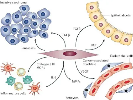
It is now well established that tumor growth, progression and metastasis is not just determined by malignant cells themselves, but also require the tumor microenvironment. The latter includes extra-cellular matrix (ECM) components, hypoxia and stromal cells, either resident or recruited from the circulation, represented by fibroblasts, pro-inflammatory cells (e.g. macrophages), endothelial cells and pericytes (Fig.1).
Fig. 1.Tumor as complex tissue. Cancer is not single cell disease and tumor initiation and progression is
highly influenced by tumor stroma that includes fibroblasts, immune cells and endothelial cells.
Tumor stroma is similar to that observed during wound healing and it is commonly referred to as "reactive stroma", which is associated with an increased number of fibroblasts, enhanced capillary density and collagen and fibrin deposition (Kalluri and Zeisberg, 2006). Cancer cells create a complex and continuative “cross-talk” with surrounding, non-malignant cells and/or with the extracellular architecture, made of direct cell-to-cell contacts and paracrine/exocrine signals. Tumor-stroma interactions at both the primary and secondary tumor sites allow and support tumor survival and outgrowth, organ homing and invasion. The role of tumor support played by stromal
6
Dott. Alberto Marini
"Role of stromal fibroblasts in prostate carcinoma progression and metabolic reprogramming of cancer cells"
Tesi di Dottorato in Scienze Biomolecolari e Biotecnologiche-Università degli Studi di Sassari
cells spans from growing of new vessels, with the recruitment of endothelial progenitors and their activation to form functional vessels, to secretion of a large amount of cytokines and soluble factors affecting cancer cell behavior (Taddei et al., 2013).
Neoplastic cells and stromal cells around them change progressively during the multistep transformation of normal tissues into high-grade malignancies and tumor-stroma interactions evolve along with tumor progression. Incipient neoplasia begin the interplay by recruiting and activating stromal cell types that assemble into an initial preneoplastic stroma, which in turn responds reciprocally by enhancing the neoplastic phenotypes of the nearby cancer cells. The cancer cells, which may further evolve genetically, again feed signals back to the stroma, continuing the reprogramming of normal stromal cells to serve the budding neoplasm; ultimately signals originating in the tumor stroma enable cancer cells to invade normal adjacent tissues and disseminate (Hanahan and Weinberg, 2011). In this context, several evidences have identified fibroblasts as key mediators in promoting tumor progression.
Fibroblasts and Cancer Associated Fibroblasts
Fibroblasts are elongated cells, characterized by extensive cellular processes, with fusiform and tapered shape (Tarin and Croft, 1969). They can be easily isolated from tissues and cultured in vitro. Their fusiform morphology makes them identifiable and, despite the paucity of specific markers, some molecules can be related to a fibroblastic phenotype, although none of these is exclusive of fibroblasts and/or expressed in all fibroblasts. Among these markers, the fibroblasts specific protein-1 (FSP-1) seems to provide the best specificity for the identifications of fibroblasts in vivo, but other markers can be considered site-specific, like desmin, a specific marker for skin fibroblasts.
The important functions of fibroblasts include the deposition of ECM, regulation of epithelial differentiation, regulation of inflammation and involvement in wound healing (Kalluri and Zeisberg, 2006). Fibroblasts synthesize many of the constituents of ECM, such as type I, III and V collagen and fibonectin. They also contribute to the formation of basement membranes by secreting type IV collagen and laminin. Fibroblasts are also
7
Dott. Alberto Marini
"Role of stromal fibroblasts in prostate carcinoma progression and metabolic reprogramming of cancer cells"
Tesi di Dottorato in Scienze Biomolecolari e Biotecnologiche-Università degli Studi di Sassari
an important source of ECM-degrading proteases such as matrix metalloproteinases (MMPs), which highlights their crucial role in maintaining an ECM homeostasis by regulating ECM turnover. In addition, fibroblasts are important in maintaining the homeostasis of adjacent epithelia through the secretion of growth factors and direct mesenchymal-epithelial cell interactions (Kalluri and Zeisberg, 2006).
As well as their function in healthy organs, fibroblasts have a prominent role in wound repair. They invade lesions, generate ECM to serve as a scaffold for other cells, and possess cytoskeletal elements that facilitate contractions of healing wounds (Kalluri and Zeisberg, 2006). When tissue injury occurs, fibroblasts undergo an activate state named "myofibroblast", characterized by the de novo expression of α-SMA protein, the actin isoform typical of smooth muscle cells, and the ability to synthesize increased levels of ECM components and ECM-degrading proteases such as matrix metalloproteinase-2 (MMP-2), MMP-3 and MMP-9, thus facilitating increased ECM turnover and altering ECM composition (Rodemann H.P. and Muller G.A., 1991) (Fig.2). Activated fibroblasts often secrete increased amounts of growth factors such as Hepatocyte Growth Factor (HGF), Insulin-like Growth Factor (IGF), Nerve Growth Factor (NGF), WNT1, Epidermal Growth Factor (EGF) and Fibroblast Growth Factor-2 (FGF2), which can induce proliferative signals within adjacent epithelial cells (Bhowmick et al., 2004). Activated fibroblasts also have an important role as modulators of the immune response following tissue injury, through the secretion of cytokines such as interleukin-1 and chemokines such as monocyte chemotactic protein interleukin-1 (MCPinterleukin-1) (Strieter et al., 1989; Rollins et al., 1989). Activated fibroblasts are found in healing wounds and sclerosing tissues, and are also associated with tumors. Different stimuli induce this activation, including growth factors such as Transforming Growth Factor-β (TGF-β), EGF, Platelet-Derived Growth Factor (PDGF) and FGF2, which are released from injured epithelial cells and infiltrating mononuclear cells such as monocytes and macrophages. In addition, fibroblasts are activated by direct cell-cell communication and contacts with leukocytes through adhesion molecules such as Intercellular-Adhesion Molecule-1 (ICAM1) or Vascular-Cell Intercellular-Adhesion Molecule-1 (VCAM1) (Clayton et al., 1998). Fibroblast activation can also be achieved through reactive oxygen species, complement factor C1 or altered ECM composition (Zeisberg et al., 2000).
8
Dott. Alberto Marini
"Role of stromal fibroblasts in prostate carcinoma progression and metabolic reprogramming of cancer cells"
Tesi di Dottorato in Scienze Biomolecolari e Biotecnologiche-Università degli Studi di Sassari
Fig.2. Fibroblasts activation. Fibroblasts can acquire an activated phenotype, which is associated with
an increased proliferative activity and enhanced secretion of ECM. Phenotypically, activated fibroblasts are often characterized as expressing α-smooth-muscle actin.
Most of knowledge about tumor stromal cells stem from the studies of carcinomas. In the early growth of tumors, cancer cells form a neoplastic lesion that is embedded in the microenvironment of a given tissue (Hanahan and Weinberg, 2000) but separated from the surrounding tissue and contained within the boundary of a basement membrane. This is called the carcinoma in situ (CIS). CIS is associated with "reactive stroma", similar to that observed during wound healing. Vascular Endothelial Growth Factor (VEGF), produced mainly by fibroblasts and inflammatory cells, represents a key molecule for the development of the stroma (Brown et al., 1999). VEGF induces microvascular permeability, thus allowing the extravasation of plasma proteins such as fibrin, which attracts fibroblasts, endothelial cells and inflammatory cells (Senger et al. 1983; Dvorak et al., 1984; Brown et al., 1999). These cells produce ECM that is rich in fibronectin and type I collagen, both implicated in the development of tumour angiogenesis (Leung et al., 1989; Brown et al., 1999; Feng et al., 2000). During tumor progression from carcinoma in situ to invasive carcinoma, tumor cells invade the reactive stroma (Dvorak et al., 2011; Ronnov-Jessen et al. 1996). Basement membrane and stroma are degraded, and myofibroblast come into direct contact with the tumor cells. Invasive cancer is usually associated with the expansion of tumor stroma and to an increase of the deposition of ECM, (Shekhar et al. 2003; Ronnov-Jessen et al. 1996; Van Kempen et al. 2003). This phenomenon appears to be very similar to the changes that take place during fibrosis, but while fibrosis is associated with a decrease of vascularization (Brown et al. 1999), solid tumors are more vascularized (Folkman 1971).
9
Dott. Alberto Marini
"Role of stromal fibroblasts in prostate carcinoma progression and metabolic reprogramming of cancer cells"
Tesi di Dottorato in Scienze Biomolecolari e Biotecnologiche-Università degli Studi di Sassari
Activated fibroblasts associated with reactive stroma are called Cancer Associated Fibroblasts (CAFs) and they are characterized by contractile and secretory features (Cirri and Chiarugi, 2012; Kalluri and Zeisberg, 2006) (Fig.3). CAFs become activated in response to tumor-delivered factors through a mesenchymal-mesenchymal transition (MMT). They were first identified by immunocytochemistry using a combination of different markers such as α-SMA (α-smooth-muscle-actin), vimentin, desmin and Fibroblast Activation Protein (FAP), a serine protease located on the cell surface of tumor stromal fiboblasts (Garin-Chesa et al., 1990; Lazard et al., 1993; Mueller and Fusenig, 2004). Unlike what happens for myofibroblasts, CAF activation is not reversed once the activating stimulus is attenuated, so their presence persists in tumor stroma (Tomasek et al., 2002; Hinz et al., 2001). Indeed, CAFs represent the most prominent cell type within the reactive stroma of many cancers, such as breast, prostate and pancreatic carcinoma (Pietras and Ostman, 2010; Kalluri and Zeisberg, 2006). CAFs are associated with cancer cells at all stages of cancer progression by promoting tumor growth, angiogenesis and the metastatic process (Tlsty and Coussens, 2006; Kalluri and Zeisberg, 2006; Orimo et al., 2005).
Fig. 3. Cross-talk between CAFs and different types of cells of tumor microenvironment.
CAFs originate through different ways. In primis, CAFs derive from resident fibroblast activated by cancer-derived growth factors such as TGF-β, PDGF and FGF2 (Elenbaas
10
Dott. Alberto Marini
"Role of stromal fibroblasts in prostate carcinoma progression and metabolic reprogramming of cancer cells"
Tesi di Dottorato in Scienze Biomolecolari e Biotecnologiche-Università degli Studi di Sassari
and Weinberg, 2001). TGF-β is associated with an increase of fibrotic tissue, tumor progression and fibroblasts recruitment (Siegel and Massague, 2003). In addition, TGF-β is the most important factor of the tumor microenvironment promoting Epithelial-Mesenchymal Transition (EMT) in tumor cells and leading fibroblasts to a CAF phenotype expressing α-SMA in vitro (Siegel and Massague 2003). EMT is an epigenetic program that leads epithelial cells to lose their cell–cell and cell-ECM interactions to undergo cytoskeleton reorganization and to gain morphological and functional characteristics of mesenchymal cells, thus generating an invasive cell, able to secrete proteases, to deeply change the surrounding ECM and to move away from the site of the primary tumor (Friedl, 2004; Kalluri and Weinberg, 2009; Nieto and Cano, 2012).
Several data indicate that CAFs activation is a redox dependent process (Cirri and Chiarugi, 2011); indeed, stimulation of CAFs with TGF-β elicits a burst of reactive oxygen species (ROS) which causes the achievement of the activated phenotype, the down regulation of gap junctions as well as their tumor promoting activity in skin tumor (Cat et al., 2006; Stuhlmann et al., 2004). In addition, the importance of oxidative stress in CAFs activation has been highlighted by Toullec et al.; in particular, ROS promote conversion of fibroblasts into highly migrating myofibroblasts through accumulation of the hypoxia-inducible factor (HIF)-1α transcription factor and the CXCL12 chemokine (Toullec et al., 2010) (Fig.4).
Fig.4. In carcinoma, chronic oxidative stress promotes the conversion of fibroblasts into myofibroblasts.
Recent studies conducted in our laboratory have shown that IL-6, secreted by prostate carcinoma PC3 cells isolated from a bone metastasis of prostate carcinoma (PCa) cells, promotes a particular phenotype named PCa-activated fibroblast (PCa-AF). In contrast
11
Dott. Alberto Marini
"Role of stromal fibroblasts in prostate carcinoma progression and metabolic reprogramming of cancer cells"
Tesi di Dottorato in Scienze Biomolecolari e Biotecnologiche-Università degli Studi di Sassari
to the TGF-β-dependent phenotype, these cells do not express α-SMA, but their activated state is confirmed by the expression of the FAP protein and production of ECM. The PCa-AFs strongly activate the process of EMT and therefore PC3 cells invasiveness (Giannoni et al., 2010) (Fig. 5). According to our observations, production of IL-6 by tumor cells has been correlated to higher carcinomas aggressiveness (Royuela et al., 2004; Chung et al., 2005; Niu et al., 2009).
A second source of CAFs are represented by bone marrow-derived mesenchymal stem cells (MSCs) which are recruited at tumor site by cytokines and growth factors produced by tumor cells (Dwyer et al., 2007; Spaeth et al., 2008; Feng and Chen, 2009). Moreover, emerging evidence indicates that also EMT involving normal epithelial cells flanking to the tumor, is a source of activated fibroblasts in both fibrosis and cancer (Selman and Pardo, 2006). In addition, CAFs may originate directly from carcinoma cells through EMT (Kalluri and Zeisberg, 2006; Radisky et al., 2007), allowing cancer cells to adopt a mesenchymal cell phenotype, with increased migratory and invasive capacities (Kalluri and Weinberg, 2009). Indeed, mainly in breast cancers, it has been reported CAF somatic mutations in p53 and PTEN, as well as gene copy number alteration at other loci in tumor stroma (Kurose et al., 2002; Moinfar et al., 2000). Finally, CAFs may derive from proliferating endothelial cells via endothelial to mesenchymal transition (EndMT) under TGF-β stimulation (Zeisberg et al., 2007).
Role of CAFs in tumor progression
Cancer cells and CAFs establish a close cross-talk based on mutual stimulation: carcinoma cells elicits a reactive response in the stroma and ,conversely, the activated fibroblasts located in the microenvironment affect cancer cell responses, thus influencing tumor progression.
Several findings underline the role of CAF in tumor growth. It has long been known that only human prostatic CAFs co-cultured with initiated human prostatic epithelial cells strongly stimulate growth and altere histology of the epithelial population, while normal prostatic fibroblasts are unable to elicit this effect (Olumi et al., 1999). Accordingly, it has been demonstrated that only tumor prostate fibroblasts, but not
12
Dott. Alberto Marini
"Role of stromal fibroblasts in prostate carcinoma progression and metabolic reprogramming of cancer cells"
Tesi di Dottorato in Scienze Biomolecolari e Biotecnologiche-Università degli Studi di Sassari
normal prostatic fibroblasts, stimulate proliferation and malignant transformation of epithelial cells derived from SV40-T antigen immortalized benign prostate hyperplasia (Hayward et al., 2001). Beside, a direct involvement of resident fibroblasts in the initiation of cancer has been elucidated: in a mouse model, the overexpression of TGF-β and/or HGF in fibroblasts induces the initiation of breast cancer within the normal epithelium (Kuperwasser et al., 2004).
Alongside the role of CAFs in the initiation of neoplasia, CAFs play a mandatory role in progression of tumors towards malignancy by the regulation of motility and invasion of cancer cells, thus positively affecting their metastatic spread towards distant organs (Joyce et al., 2009) (Fig. 5). The contribution of CAFs to the invasive process of cancer cells is mainly due to the induction of EMT (Brahimi-Horn et al., 2011; Pani et al., 2010). This feature is primarily dependent on CAFs’ ability to remodel the ECM. Indeed, in addition to secreting growth factors that directly affect cell motility, CAFs are sources of ECM degrading proteases as MMPs, which allow cancer cells to escape the primary tumor site (Kalluri and Zeisberg, 2006). It has been demonstrated that treatment of mammary epithelial cells with MMP3, which is highly expressed in fibroblasts, results in cleavage of E-cadherin and EMT induction, thus sustaining a progressive phenotypic conversion from normal mammary epithelial cells to an invasive mesenchymal phenotype (Lochter et al., 1997). In addition, EMT has been correlated with the induction of a cancer stem phenotype both in breast and in prostate cancer (Giannoni et al., 2010; Mani et al., 2008; Blick et al., 2010). This ability confers to cancer cells the self renewal capability that is crucial for clonal expansion during metastatic dissemination (Polyak and Weinberg, 2009). In particular, recent studies conducted by our research group showed that CAFs exposure promotes EMT of prostate carcinoma cells associated with enhanced expression of stem cell markers, ability to form prostaspheres and to self-renew, leading to an increase of aggressiveness and metastatic spread (Giannoni et al., 2010) (Fig. 5).
13
Dott. Alberto Marini
"Role of stromal fibroblasts in prostate carcinoma progression and metabolic reprogramming of cancer cells"
Tesi di Dottorato in Scienze Biomolecolari e Biotecnologiche-Università degli Studi di Sassari
Fig. 5 Reciprocal interplay between stromal fibroblasts and PCa cells through secretion of IL-6 (efferent
pathway) and MMP-dependent EMT in PCa cells (afferent pathway).
Moreover, in pancreatic cancer it has been highlighted a key role of the microenvironment in the induction of stemness: indeed, co-cultures of tumor and pancreatic stellate cells, the major profibrogenic cell type in the pancreas, enhance the cancer stem cell-like phenotypes of tumor cells (Hamada et al., 2012). Beside, the chemokine (C-C motif) ligand 2 (CCL2) secreted by CAFs, stimulates sphere formation and cancer stem cells self-renewal in breast cancer cells (Tsuyada et al., 2012). Finally, myofibroblasts, through hepatocyte growth factor secretion, activate β-catenin-dependent transcription and subsequently colon cancer cells clonogenicity (Vermeulen et al., 2010).
MicroRNAs are emerging as potential regulators of the relationship between EMT and stemness. Indeed, breast cancer stem cells show a miRNA expression profile very similar with respect to breast cancer cells undergoing EMT (i.e., high levels of miR-155 and low levels of miR-200 family) (Blick et al., 2010; Shimono et al., 2009). In agreement, manipulation of miRNAs able to revert the EMT phenotype, suppresses cancer stem cell properties, as demonstrated by the lower tumorigenic potential of miR-200c-expressing CD44high/CD24low cells. Furthermore, the achievement of EMT correlates to an increase in the resistance to apoptosis, allowing cells to survive along their route from the primary tumor to the site of dissemination (Gal et al., 2008).
Recent evidences suggests that EMT is a redox-dependent phenomenon. In mammary epithelial cells, exposure to MMP3 induces EMT, through a Rac1b-mediated release of
14
Dott. Alberto Marini
"Role of stromal fibroblasts in prostate carcinoma progression and metabolic reprogramming of cancer cells"
Tesi di Dottorato in Scienze Biomolecolari e Biotecnologiche-Università degli Studi di Sassari
mitochondrial ROS (Radisky et al., 2008). Accordingly, CAFs promote EMT of cancer cells by exploiting the intrinsic or extrinsic oxidative stress and producing cytokines and proteases (Cirri and Chiarugi, 2011). In keeping, our research group demonstrated that prostate CAFs release cytokines and MMP2 and MMP9, which in turn activate the small GTPase Rac1, cyclooxygenase-2 (COX-2) and a consequent COX-2-mediated ROS production (Giannoni et al., 2011). The establishment of a pro-oxidant environment in prostate cancer cells is also necessary for the redox dependent stabilization of HIF-1α and NF-kB (Comito et al., 2011; Gloire and Piette, 2009; Hamanaka and Chandel, 2009). These factors sustain then the activation of the transcription factors Snail and Twist, promoting the EMT program in prostate carcinoma cells (Fig. 5).
Furthermore, it is useful to mention that even senescent fibroblasts are able to induce EMT in nearby epithelial cells, thus influencing tumor progression (Laberge et al., 2012). Indeed, cellular senescence is associated with an increased oxidative stress and secretion of several pro-inflammatory cytokines which collectively generate the so-called SASP (Senescence Activated Secretory Pathway) phenotype (Bavik et al., 2006). Induction of EMT by SASP has been reported for breast cancer where the SASP component factors IL-6 and IL-8 enhance the invasiveness of a panel of cancer cell lines in culture (Badache and Hynes, 2001; Coppe et al., 2008; Yuan et al., 2005).
CAFs have a primary role in the guidance of tumor cell movement. Indeed, it has been demonstrated that stromal fibroblasts co-cultured with squamous cell carcinoma collectively move across the ECM; in particular cancer cells use Cdc42 and MRCK (Myotonic Dystrophy Kinase-Related CDC42-binding protein kinases) mediated regulation of myosin light chain (MLC) activity, exploiting the tracks generated by fibroblasts (Gaggioli et al. 2007). The contractile force in stromal fibroblasts to remodel the ECM for the creation of tracks for the collective migration is dependent on the activation of the kinase JAK1 (Sanz-Moreno et al., 2011). The same pathway is also responsible for the actomyosin contractility of melanoma cells that migrate with an amoeboid motility style (Sanz-Moreno et al., 2011).
The pro-invasive role of CAFs during tumor progression has been shown in several types of tumors, such as breast cancer, prostate cancer (Giannoni et al., 2010; Orimo et al., 2005) and head and neck squamous carcinoma (Hinsley et al., 2012). In ovarian
15
Dott. Alberto Marini
"Role of stromal fibroblasts in prostate carcinoma progression and metabolic reprogramming of cancer cells"
Tesi di Dottorato in Scienze Biomolecolari e Biotecnologiche-Università degli Studi di Sassari
carcinoma has been underlined that CAFs play a vital role in promoting angiogenesis, lymphangiogenesis and cancer cell invasion with respect to normal fibroblasts (Giannoni et al., 2010; Orimo et al., 2005; Zhang et al., 2011). Moreover, CAFs expressing FAP increase the migration and invasion of colorectal cancer cells through a Fibroblasts Growth Factor-1/Fibroblast Growth Factor Receptor 3 (FGF1/FGFR-3) signaling (Henriksson et al., 2011). Finally, in a model of human pancreatic cancer, the overexpression of FAP in fibroblasts directly modifies the stromal ECM through its enzymatic activity. The authors show that FAP alters the architecture and the composition of the ECM promoting tumor invasion along characteristic parallel fiber orientation (Lee et al., 2011).
Alongside their pro-invasive role, CAFs are able to recruit endothelial precursor cells (EPCs) from bone marrow, thereby inducing de novo angiogenesis (Orimo et al., 2005) and participate in the preparation of the metastatic site in which the secondary tumor will grow up. Recent data demonstrated that the metastatic cells can bring their stromal components from tumor primary site to the metastatic niche in the lungs. The stromal cells, co-traveling with metastatic cancer cells, provide them an early growth advantage and protect them in the bloodstream from anoikis (the apoptotic cell death due to lack or improper cell adhesion to ECM), thus ensuring long-term survival and proliferation in the metastatic sites (Duda et al., 2010).
Beside MMPs, which remodel ECM, activated fibroblasts produce several growth factors and cytokines that sustain tumor progression. Indeed, CAFs produce paracrine diffusible signals, including TGF-β, HGF, VEGF, FGF, Stromal cell-Derived Factor-1 (SDF-1) as well as cathepsins and plasminogen activators (Joyce and Pollard, 2009; De et al., 2008). In particular, it has been shown that CAFs promote the growth of breast carcinoma cells significantly more with respect to normal mammary fibroblasts derived from the same patients. These CAFs, play a central role in promoting directly the growth of tumor cells through their ability to secrete SDF-1 as well as in promoting angiogenesis through the recruitment of EPCs (Orimo et al., 2005). Moreover, fibroblast-derived SDF-1 enhanced the invasion of pancreatic cancer cells as well as their production of CXCL8. The cooperation between CXCL8 and SDF-1 enhances also the proliferation/invasion of human umbilical vein endothelial cells, thus sustaining tumor progression (Matsuo et al., 2009). Furthermore, in the same cellular model,
16
Dott. Alberto Marini
"Role of stromal fibroblasts in prostate carcinoma progression and metabolic reprogramming of cancer cells"
Tesi di Dottorato in Scienze Biomolecolari e Biotecnologiche-Università degli Studi di Sassari
cancer cell-derived IL-1α significantly promotes HGF expression by fibroblasts. As a consequence, HGF enhances not only the invasiveness and proliferation of pancreatic cancer cells, but also enhances migration and proliferation of human umbilical vein endothelial cells, thus positively influencing the metastatic potential of pancreatic cancer cells (Xu et al., 2010).
Anyway, the role of CAFs in tumor progression goes beyond the engagement of pro-invasive features in cancer cells and embrace a real metabolic reprogramming of both cell types. This topic will be extensively described later.
Role of other microenvironmental components in
tumor progression
Among components of tumor microenvironment, both hypoxia and acidity require adaptive response that influence cancer cells behaviors and involve metabolic pathway and invasive properties.
HYPOXIA
Solid tumors frequently outgrow the blood supply, resulting in nutrient and oxygen insufficiency. In this state, commonly referred as hypoxia, tumors can experience decreased oxygen pressure to 1% or below. Hypoxia is considered as an independent negative prognostic indicator and contributes to cancer progression affecting the behavior of both cancer and stromal cells (Taddei et al., 2013). Intratumoral hypoxia causes:
activation of a glycolytic metabolism to circumvent lack of oxygen;
acquisition of invasive features, mainly through EMT, in order to escape from the hostile environment;
activation of pathways for survival to stressful conditions;
secretion of soluble growth factors eliciting de novo angiogenesis allowing nutrient/oxygen supply.
The ability of cells to sense and adapt to hypoxia is mainly mediated by hypoxia inducible factor (HIF) family of transcription factors. Targets of HIFs span from
17
Dott. Alberto Marini
"Role of stromal fibroblasts in prostate carcinoma progression and metabolic reprogramming of cancer cells"
Tesi di Dottorato in Scienze Biomolecolari e Biotecnologiche-Università degli Studi di Sassari
glucose, glutamine and fat metabolism, oxygen homeostasis, tissue remodeling and motility, angiogenesis, erythropoiesis, proliferation, and survival to stress (Keith et al., 2012). HIF1 and HIF2 complexes are the major responsible from gene expression changes during hypoxia. They are heterodimers composed of the constitutively expressed HIF-1β (or ARNT) subunit and either the HIF-1α or HIF-2α subunits. Under normoxic conditions, the HIF-1α subunits undergo oxygen-dependent hydroxylation by prolyl-hydroxylases (PHDs) on Pro402 and Pro564, which results in their recognition by von Hippel-Lindau tumor suppressor (VHL), an E3 ubiquitin ligase, and subsequent degradation (Chan et al., 2002) (Fig. 6). Of note, PHDs catalyze the hydroxylation reaction requiring oxygen and α-ketoglutarate as substrates, iron and ascorbate as cofactors. When hypoxia is occurring, HIFα are stabilized by lack of hydroxylation and functionally dimerizes with its partner HIF-1β to bind to the core sequence 5'-RCGTG-3' and enhancing the transcription of target genes involved in promoting all adaptations to hypoxia (Fig. 7). Levels of HIF-1α are also influenced in normoxic conditions by genetic alterations, including mutations of VHL gene, succinate dehydrogenase and fumarate hydratase, or growth factors, hormones and cytokines produced by both tumor and stromal cells (Dery et al., 2005) (Fig. 6).
Fig. 6. Regulation of HIF-1 stabilization. HRE: Hypoxia response element.
Hypoxic cells undergo a glycolytic switch from aerobic to anaerobic metabolism (Wheaton et al., 2011), allowing maintenance of metabolic activities under limited oxygen avaibility. Other adaptive metabolic features include, engagement of pentose
18
Dott. Alberto Marini
"Role of stromal fibroblasts in prostate carcinoma progression and metabolic reprogramming of cancer cells"
Tesi di Dottorato in Scienze Biomolecolari e Biotecnologiche-Università degli Studi di Sassari
phosphate pathway (PPP) and addiction to glutamine use. Of note, several of these adaptations have been acknowledged as the biochemical basis for resistance to chemotherapy and radiation (DeClerck and Elble, 2010; Heddleston et al., 2010; Rohwer and Cramer, 2011). Several genes controlling glucose uptake (e.g, glucose transporters GLUT1 and GLUT3), glycolysis (e.g., hexokinase-1/2, enolase, phosphoglycerate kinase-1, aldolase, pyruvate kinase M), lactate fermentation (lactate dehydronase A), inhibition of mitochondrial respiration (e.g., pyruvate dehydrogenase kinase) are directly targeted by HIFs (Stubbs and Griffiths, 2010) (Fig. 7).
Fig. 7. HIF target genes.
Increased glucose uptake is associated with inhibition of pyruvate kinase. Indeed, all tumors analyzed to date express the M2 variant of pyruvate kinase (PKM2), which is a redox sensitive enzyme undergoing inhibition following cysteine oxidation (Anastasiou et al., 2011; Bayle and Devilee, 2012; Chen et al., 2010). Hypoxia has been correlated with a state of oxidative stress, both due to mitochondrial or NADPH oxidase delivery of ROS causing ROS-mediated inhibition of PHDs through a Fenton reaction, and leading to HIF-1 stabilization. Hypoxic oxidative stress causes oxidation and inhibition
19
Dott. Alberto Marini
"Role of stromal fibroblasts in prostate carcinoma progression and metabolic reprogramming of cancer cells"
Tesi di Dottorato in Scienze Biomolecolari e Biotecnologiche-Università degli Studi di Sassari
of PKM2, with the consequence of accumulation of glycolysis intermediates, rapidly fueling anabolism of aminoacids and proteins, as well as PPP. The latter increases the pool of NADPH, leads to synthesis of ribose and DNA, allowing to safely handle with oxidative stress and to overcome DNA damage and chemotherapy stress. In keeping with this, hypoxia and PPP reprogramming have been correlated with increased resistance to therapy in several cancer models (Anastasiou et al., 2011; Brahimi-Horn et al., 2011; Gruning and Ralser, 2011).
In hypoxia, cancer cells are unable to catabolize pyruvate into acetyl-CoA, stimulating lipid biosynthesis. This is mainly due to HIF-1-mediated expression of pyruvate dehydrogenase kinase (PDK), which totally blocks decarboxylation of pyruvate through pyruvate dehydrogenase (PDH) (Wheaton et al., 2011). Hypoxic cells solve this problem becoming addicted to glutamine uptake (Dang, 2012). Using a reductive carboxylation, glutamine is converted into isocitric acid by isocitric dehydrogenase-2 (IDH-2), leading to citrate exportation from mitochondria. Citrate is converted by ATP-citrate lyase to acetyl-CoA that is used to lipid synthesis. IDH1/IDH2 acknowledged to produce α-hydroxy-glutarate which is a strong inhibitor of PHDs due to its competition with their cofactor α-ketoglutarate, have been correlated to activation of HIF-1-dependent transcriptional response (Chowdhury et al., 2011; Metellus et al., 2011). A latere with metabolic changes, during hypoxia tumor cells activate a specific escaping program to run away this hostile environment. In addition, the products of glycolysis cause acidification of the extracellular space and the resulting cellular toxicity acts as a further selective pressure for cells that are resistant to acidic conditions (Pani et al., 2010). Moreover, hypoxic cells release angiogenic factors that stimulate de novo vascularisation, which restores O2 and nutrient supply in order to meet the growing
metabolic demands of proliferating cells.
To escape the hypoxic environment, the first strategy is to activate an invasive program and the primary driver engaged by hypoxia is the hepatocyte growth factor-MET receptor (Pennacchietti et al., 2003). In particular, mitochondrial ROS lead to stabilization of HIF-1α and expression and activation of MET, resulting in cell migration towards the blood or lymphatic microcirculation (Comito et al., 2011). Moreover, hypoxia also promotes tumor cell motility and invasion by triggering the EMT. Indeed, HIF-1α is a potent activator of Twist and other EMT inducers (Giannoni
20
Dott. Alberto Marini
"Role of stromal fibroblasts in prostate carcinoma progression and metabolic reprogramming of cancer cells"
Tesi di Dottorato in Scienze Biomolecolari e Biotecnologiche-Università degli Studi di Sassari
et al., 2011; Cannito et al., 2008; Jiang et al., 2011; Yang et al., 2008). Twist1/2 cause downregulation of E-cadherin, that is concomitant with de novo expression of N-cadherin, a trait leading to up-regulation of polarized motility through the small GTPase Rac1. Through both MMPs produced during EMT program and up-regulation of urokinase-type plasminogen activator receptor expression, hypoxia enhances proteolytic activity at the invasive front of cancer cells, stimulating motility by altering integrin/ECM interactions (Sullivan and Graham, 2007; Del Rosso et al., 2002). In addition, hypoxia induced EMT has been correlated to a supplementary adaptation, i.e. resistance to anoikis (Sullivan and Graham, 2007; Nieto, 2011; Gort et al., 2008; Whelan et al., 2010). This adaptation allows survival in the bloodstream of metastatic cancer cells which have experienced activation of the HIF-1 transcriptional program. Hypoxia-driven EMT is reinforced by a reciprocal activation between HIF-1α and NF-kB, underscoring a further link between hypoxia and inflammation (Giannoni et al., 2011; Rius et al., 2008).
In order to permit successful metastases, hypoxia elicits adaptations useful to permit successful metastases, such as de novo angiogenesis and lymphangiogenesis (Sullivan and Graham, 2007). HIFs target several angiogenic factors, the masterpiece of which is VEGF, reportedly correlated with sprouting angiogenesis, lymphangiogenesis, as well as with the dynamic tumor-stromal interactions required for the subsequent stages of metastasis (Carmeliet and Jain, 2011). Angiogenesis and lymphangiogenesis within the primary tumor provide the necessary routes for dissemination, and VEGF-induced changes in vascular permeability promote both intravasation and extravasation. Through instruction of these critical pathways and adaptations, hypoxia promotes several step of the metastatic cascade, selecting tumor cell populations able to escape the hostile microenvironment of the primary tumor. In addition, HIFs transcription factors have been involved in the regulation of stemness, both in normal and in cancer cells, through up-regulation of pluripotency genes (Simon and Keith, 2008). Indeed, HIF-2 is an upstream regulator of Oct4, one of the mandatory transcription factors to reprogram differentiated cells towards stemness (the so called induced-stem cells) (Iida et al., 2012; Li et al., 2009). Recent data have demonstrated that even HIF-1 shows a mandatory role in the regulation of stemness of cancer cells induced by hypoxia. Indeed, at least in lymphomas, the HIF-1α inhibitor echinomycin selectively kills
c-21
Dott. Alberto Marini
"Role of stromal fibroblasts in prostate carcinoma progression and metabolic reprogramming of cancer cells"
Tesi di Dottorato in Scienze Biomolecolari e Biotecnologiche-Università degli Studi di Sassari
Kit+Sca-1+ cancer stem cells, and the ablation of HIF-1α in these cancer stem cells severely impedes their self-renewing and tumor-initiating activity (Wang et al., 2011).
ACIDITY
Deregulated pH is emerging as another adaptive response of most cancers. Tumor cells show a ‘reversed’ pH gradient with a constitutively increased intracellular pH (pHi) that is higher than the extracellular pH (pHe) (Webb et al., 2011). In normal adult cells, pHi is generally ~7.2, while pHe is ~7.4. On the contrary, cancer cells have a higher pHi of >7.4 and a lower pHe of ~6.7–7.1 (Gallagher et al., 2008).
The acid-outside pH of tumor cells is a consequence of the adaptation towards glycolytic metabolism induced by hypoxia; indeed, enhanced glucose uptake and his conversion to lactic acid, as well as the inefficient removal of lactate and CO2 as a
consequence of a poor vasculature within the tumor mass, result in the increase of acid production (Horn and Pouyssegur, 2007; Horn et al., 2007a; Brahimi-Horn et al., 2007b).
Intracellular pH is crucial to normal cell function. Therefore, hypoxic tumor cells have developed key strategies to protect the cytosol from acidosis and allow cells to survive in hypoxia. HIF-1 regulates proton extrusion and pH homeostasis by enhancing expression of plasma membrane ion pumps and transporters (Brahimi-Horn and Pouyssegur, 2007). The most notable pHi regulatory systems of tumor cells are:
the Na+/H+ exchanger (NHE-1), known to play a key role in vivo in tumor
development, in particular when highly glycolytic cells produce large amounts of lactate (Cardone et al., 2005; Pouyssegur et al., 2001; Shimoda et al., 2006); the V-ATPase, which mainly contributes to the maintenance of an aberrant pH
gradient between the alkaline cytosol and the acidic extracellular environment (Nishi and Forgac, 2002);
the monocarboxylate transporters (MCTs), whose increased expression confers a selective advantage to cancer cells owing to the high affinity of these transporters for lactate (Ullah et al., 2006);
the bicarbonate transporters that import HCO3- into the cells through Cl-/HCO3
22
Dott. Alberto Marini
"Role of stromal fibroblasts in prostate carcinoma progression and metabolic reprogramming of cancer cells"
Tesi di Dottorato in Scienze Biomolecolari e Biotecnologiche-Università degli Studi di Sassari
imported HCO3- traps intracellular H+ and thus maintains a permissive pHi that
favors cell survival (Izumi et al., 2003; Karumanchi et al., 2001).
Another contribution to intracellular alkalinization is provided by a family of proteins, the carbonic anhydrases (CAs), which have extracellular catalytic domains that accelerate the hydration of extracellular CO2 to HCO3- and H+. In particular, expression
of the membrane-associated CA IX and CA XII isoforms is substantially increased under hypoxic conditions in HIF-1-dependent manner (Chiche et al., 2009; Chiche et al., 2010; Ilie et al., 2010; Loncaster et al., 2001; Pastorekova et al., 2006; Swietach et al., 2009; Wykoff et al., 2000; Kaluz et al., 2009; Supuran, 2008).
Deregulation of pH confers adaptive advantages to cancer cells, allowing the achievement of specific features, such as growth-factor independent proliferation, evasion of apoptosis, metabolic adaptation, migration and invasion and thus facilitating the metastatic dissemination of tumor cells (Cardone et al., 2005; Gatenby et al., 2006; Stock and Schwab, 2009). For example, the polarity of migrating cells, the de novo assembly of actin filaments and the dynamics of integrin-ECM attachments are processes facilitated by high pHi. Moreover, the low pH of tumor extracellular microenvironment may promote the degradation and remodeling of ECM through the activation of proteolytic enzymes, including MMPs and tissue serine proteases (Busco et al., 2010; Rofstad et al., 2006). Of note, the activity of MMP3, is higher at acidic pH (Johnson et al., 2000) and the expression and secretion of MMP9 increases at lower pHe and higher pHi (Bourguignon et al., 2004; Putney and Barber, 2004). In addition, there is strong evidence indicating a pivotal role of lysosomal-like vesicles in the degradation of ECM, cell invasion and cell migration (Glunde et al., 2003; Montcourrier et al., 1994; Montcourrier et al., 1997). For example, in breast cancer, acidic pHe causes a significant redistribution of lysosomes from the perinuclear region to the cell periphery (Glunde et al., 2003). Lysosomes displacement to the cell periphery may be a mechanism to facilitate increased secretion of degradative enzymes (Glunde et al., 2003). In malignant tumor cells, vesicles containing cathepsin B also redistributed toward the cell periphery at acidic pHe and constitutive secretion of active cathepsin B allowed for the cleavage of secreted, latent MMPs into active enzymes (Giusti et al., 2008; Rozhin et al., 1994).
23
Dott. Alberto Marini
"Role of stromal fibroblasts in prostate carcinoma progression and metabolic reprogramming of cancer cells"
Tesi di Dottorato in Scienze Biomolecolari e Biotecnologiche-Università degli Studi di Sassari
TUMOR METABOLISM
The uncontrolled cell proliferation, that represents the essence of neoplastic disease, involves not only deregulated control of cell growth but also corresponding adjustments of energy metabolism, in order to fuel anabolic pathway. One important characteristic of cancer cell metabolism is the consistent switch of the energy production pathway from oxidative phosphorylation to glycolysis (Kondoh et al., 2007) (Fig.8).
24
Dott. Alberto Marini
"Role of stromal fibroblasts in prostate carcinoma progression and metabolic reprogramming of cancer cells"
Tesi di Dottorato in Scienze Biomolecolari e Biotecnologiche-Università degli Studi di Sassari
In 1924, Otto Warburg observed that cancer cells consume much larger quantities of glucose than their normal counterparts and metabolize it predominantly through glycolysis, thus producing high levels of lactate even in oxygen-rich conditions (Warburg, 1956, Warburg et al., 1924). This process is called aerobic glycolysis, or "Warburg effect". In general, through the glycolysis process, one molecule of glucose generates two molecules of pyruvate and produces two molecules of ATP. In mammals, pyruvate has diverse fates. In an environment with limited oxygen, pyruvate is converted into lactate by lactate dehydrogenase (LDH), a process which is called anaerobic glycolysis. In the presence of oxygen, pyruvate is oxidized into acetyl-CoA by the pyruvate dehydrogenase (PDH) complex (Fig. 9).
Fig. 9. Fates of pyruvate.
Acetyl-CoA is oxidized in the citric acid cycle (also known as Krebs cycle or trocarboxylic acid cycle) to generate electrons -in the form of NADH and FADH2- and
CO2 in the mitochondria. The electron transport chain generates a pH gradient across
the inner mitochondrial membrane. The force motive produced by the pH gradient generates ATP by ATP synthase. This process is called oxidative phosphorylation. In the latter, one molecule of glucose can be completed degraded into H2O and CO2 to
produce 36 molecules of ATP. Overall, the oxidative phosphorylation process is more efficient in producing ATP than glycolysis. The reason why cancer cells prefer consuming more glucose by aerobic glycolysys to produce ATP is not well defined yet.
25
Dott. Alberto Marini
"Role of stromal fibroblasts in prostate carcinoma progression and metabolic reprogramming of cancer cells"
Tesi di Dottorato in Scienze Biomolecolari e Biotecnologiche-Università degli Studi di Sassari
Further studies by Warburg itself hypothesized that the Warburg effect was mainly related to a mitochondrial deficiency developed by tumor cells, for example, through genetic mutations, thereby leading to a low energy delivery from mitochondrial respiration. Finally, aerobic glycolysis had to be the only possible metabolic mechanism that allowed cells to survive (Warburg, 1956). Anyway, subsequent studies demonstrated that mitochondrial deficiency was a rare condition in cancer cells (Weinhouse, 1956). Today, several authors highlight that the importance of aerobic glycolysis for tumor cells, as well as for proliferating cells in multicellular organisms, extends beyond ATP production, to allow nutrient assimilation into biosynthetic precursor and facilitate biomass accumulation (Lunt and Vander Heiden, 2011). Therefore, the main function of up-regulated glycolysis is to maintain the levels of glycolytic intermediates needed to support biosynthesis. Indeed, glucose provide the precursors for the chemical constituents (nucleotides, amino acids, lipids) that are used to build macromolecules essential for cell division. For example:
glucose-6-phosphate can be oxidized into PPP in order to produce NADPH and ribose-5-phosphate, which in turn are used to synthesize nucleotides for DNA and RNA;
dihydroxyacetone phosphate is the precursor to glycerol-3-phosphate, that is crucial for the biosynthesis of the phospholipids and triacylglycerols that serve as major structural lipids in cell membranes;
3-phosphoglycerate provides the carbons for cysteine, glycine and serine, whereas pyruvate provides the carbons for alanine.
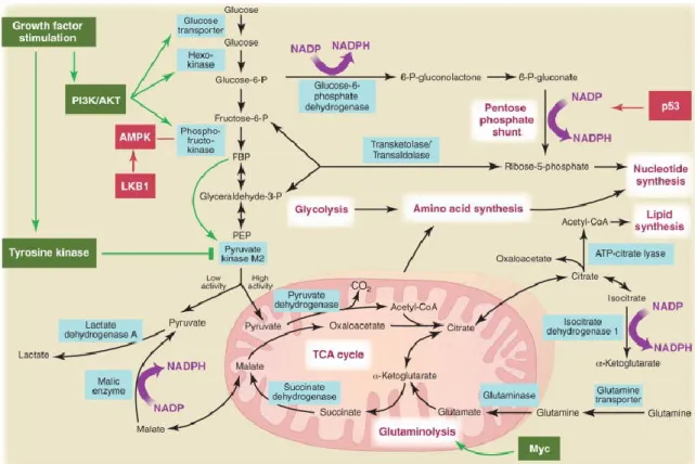
Besides aerobic glycolysis, cancer cells exhibit substantial alterations in several metabolic pathway including tricarboxylic acid (TCA) cycle, glutaminolysis, mitochondrial respiratory chain and PPP. The metabolic changes can be attributed to the activation and/or malfunction of oncogenes, and/or loss of tumor suppressors (Chen and Russo, 2012) (Fig. 10).
26
Dott. Alberto Marini
"Role of stromal fibroblasts in prostate carcinoma progression and metabolic reprogramming of cancer cells"
Tesi di Dottorato in Scienze Biomolecolari e Biotecnologiche-Università degli Studi di Sassari
Fig. 10. Metabolic pathways active in proliferating and cancer cells are directly controlled by signaling
pathways involving known oncogenes (green) and tumor suppressor genes (red).
Regulation of cancer cell metabolism: the Warburg
effect
Aerobic glycolysis has been observed in a wide variety of tumors that originate from different cell types, but most normal cells in adult tissues from which cancer cells arise generally do not utilize this kind of metabolism. Thus, cancer cells revert to a metabolic phenotype that is characteristic of rapidly dividing cells, which suggests that aerobic glycolysis must provide advantages during proliferation.
GLUCOSE TRANSPORTERS AND GLYCOLYTIC ENZYMES
Glucose import into mammalian cells is facilitated primarily by five transmembrane transporters, GLUT1–5. Although GLUT2, GLUT4, and GLUT5 are found only in specific tissues, GLUT1 and GLUT3 are expressed in nearly all mammalian cells and have the lowest KM values (1mM) of the five, which allow them to transport glucose
27
Dott. Alberto Marini
"Role of stromal fibroblasts in prostate carcinoma progression and metabolic reprogramming of cancer cells"
Tesi di Dottorato in Scienze Biomolecolari e Biotecnologiche-Università degli Studi di Sassari
into the cell at a nearly constant rate from serum in which the glucose concentration ranges from 4mM to 8mM. Tumor cells frequently overexpress GLUT1 and GLUT3 and have an increased capacity for glucose uptake (Au et al., 1997; Suzuki et al., 1999; Yamamoto et al., 1990; Younes et al., 1996). Suppression of GLUT1 expression in a human gastric cancer cell line can decrease the number of cells in S phase and inhibit tumor growth (Noguchi et al., 2000).
Once in the cell, glucose must be converted to glucose-6-phosphate by hexokinase (HK) to prevent its transport out of the cell and to prime it for metabolism in subsequent reactions (Berg et al., 2007). Four mammalian HK isoforms (HKI–IV) are known, and HKI is expressed in most normal cells and at particularly high levels in brain tissue (Wilson, 2003). HKII expression is more limited and is normally found mainly in skeletal muscle and adipose tissue. However, cancer cells frequently overexpress HKII (Mathupala et al., 2006), and at least some glioblastoma cells are specifically dependent on HKII over other isoforms of the enzyme (Wolf et al., 2011). Both HKI and HKII are associated with the voltage-dependent anion channel (VDAC) on the cytosolic side of the outer mitochondrial membrane. This VDAC-HK association may inhibit mitochondria-induced apoptosis (Majewski et al., 2004) and give HK preferential access to mitochondria generated ATP (Arora and Pedersen, 1988). Why mitochondria-bound HKII appears to be selected for in cancer cells remains unclear.
Although HK traps glucose inside the cell, phosphofructokinase-1 (PFK1) controls its commitment to glycolysis and is therefore highly regulated. PFK1, which irreversibly converts fructose-6-phosphate to fructose-1,6-bisphosphate (FBP), is overexpressed in various human cancer cell lines (Vora et al., 1985). PFK1 is allosterically inhibited by high levels of ATP (Berg et al., 2007), and relieving this ATP inhibition is an important means to increase glucose metabolism in proliferating cells (Fang et al., 2010; Israelsen and Vander Heiden, 2010; Scholnick et al., 1973). PFK1 inhibition by ATP is diminished by fructose-2,6-bisphosphate, a metabolite synthesized from fructose-6-phosphate by PFK2. Regulation of PFK2 expression or activity has been proposed as an important way to couple growth signals with regulation of glucose metabolism in proliferating cells (Christofk et al., 2008; Marsin et al., 2000; Telang et al., 2006); moreover, PFK2 is expressed constitutively in several human cancer cell lines and is found to be required for tumor cell growth (Chen and Russo, 2012). In human tissues,
28
Dott. Alberto Marini
"Role of stromal fibroblasts in prostate carcinoma progression and metabolic reprogramming of cancer cells"
Tesi di Dottorato in Scienze Biomolecolari e Biotecnologiche-Università degli Studi di Sassari
PFK1 subunit composition, a complex mixture of homotetramers or heterotetramers composed of up to three different subunits, can vary depending on tissue type. Each subunit (C, L, M) differs in sensitivity to allosteric effectors; thus, the kinetic and regulatory properties of PFK1 are determined by subunit composition (Dunaway et al., 1988). PFK1 subunits overexpressed in rat thyroid carcinomas and human gliomas are less sensitive to the allosteric inhibitors ATP and citrate (Meldolesi et al. 1976, Oskam et al. 1985, Staal et al. 1987). In addition to controlling glucose commitment to glycolysis, PFK1 may regulate the amounts of glucose-6-phosphate available for nucleotide biosynthesis (Lunt and Vander Heiden, 2011).
Among glycolytic enzymes, pyruvate kinase (PK) has received particular attention during the last few years. Indeed, recent findings have shown that many cancer cells exclusively express the M2 isoform of PK (Mazurek et al., 2005), and PKM2 expression is important for tumor growth (Christofk et al., 2008). PK catalyzes the reaction generating pyruvate and ATP from phosphoenolpyruvate (PEP) and ADP. Four isoforms of PK (L, R, M1, and M2) are present in mammals. The L and R isotypes are encoded by the PKLR gene. Their expression is tissue specific and is regulated by different promoters. The L isotype is expressed in the liver, kidney, and intestine, and the R isotype is expressed in red blood cells (Mazurek et al., 2005; Mazurek, 2011; Clower et al., 2010; Noguchi et al., 1987). PKM1 and PKM2 are encoded by the PKM gene and are the products of two mutually exclusive alternatively spliced exons (exon 9 and exon 10, respectively) (Clower et al., 2010; David et al., 2010; Noguchi et al., 1986): M1 is expressed in most adult differentiated tissues such as brain and muscle, whereas M2 is expressed in embryonic cells, adult stem cells, and cancer cells (Christofk et al., 2008, Mazurek et al., 2005; Mazurek, 2011; Bluemlein et al., 2011; Lee et al., 2008; Cairns et al., 2011). Splicing of PKM is controlled by the splicing repressors, heterogeneous nuclear ribonucleoprotein (hnRNP) A1 and A2, as well as polypyrimidine tract binding protein (PTB, also known as hnRNPI), and the expression of those repressors is up-regulated by c-Myc (Clower et al., 2010; David et al., 2010). These proteins bind to exon 9 and repress PKM1 mRNA splicing, resulting in the inclusion of exon 10 and thereby contributing to the high levels of PKM2 expression (Clower et al., 2010; David et al., 2010; Chen et al., 2012) (Fig. 11).
29
Dott. Alberto Marini
"Role of stromal fibroblasts in prostate carcinoma progression and metabolic reprogramming of cancer cells"
Tesi di Dottorato in Scienze Biomolecolari e Biotecnologiche-Università degli Studi di Sassari Fig. 11. PKM1 and PKM2 expression after alternative splicing.
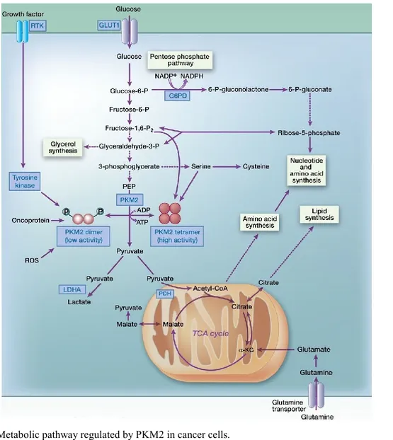
A number of regulators of PKM2 expression have been reported (Clower et al., 2010; Mazurek, 2011; David et al., 2010; Chen et al., 2012; Luo et al., 2011; Lv et al., 2011; Panasyuk et al., 2012): in particular, a recent study showed that PKM2 expression is induced by activated mTOR (mammalian target of rapamycin), which transactivates HIF-1 and promotes the c-Myc-hnRNPs–mediated alternative splicing, leading to the aerobic glycolysis in tumor cells (Sun et al., 2011). The expression and lower glycolytic enzyme activity of PKM2 are necessary for the Warburg effect, which provides cancer cells with selective advantages, including tumor growth and suppression of ROS (Christofk et al., 2008; Vander Heiden and Cantley, 2009; Cairns et al., 2011; Anastasiou et al., 2011) for the following reasons. First is that the glycolytic pathway generates ATP more rapidly than the oxidative phosphorylation (Pfeiffer et al., 2011), allowing faster incorporation of carbon into its biomass (Vander Heiden and Cantley, 2009; Hamanaka and Chandel, 2012). Between yield and rate of ATP production, a trade-off has been reported to be present in sugar degradation by glycolysis and mitochondrial respiration. Then, glycolysis generates ATP at a high rate but low yield via massive consumption of glucose (Pfeiffer et al., 2011; Vazquez et al., 2010). The second reason is that lower activity of PKM2 facilitates the production of glycolytic intermediates to enter the glycolysis branch pathways, such as glycerol synthesis, amino acid syntesis and the pentose phosphate pathway, which generates NADPH to suppress ROS production and is also involved in nucleotide synthesis (Vander Heiden and
30
Dott. Alberto Marini
"Role of stromal fibroblasts in prostate carcinoma progression and metabolic reprogramming of cancer cells"
Tesi di Dottorato in Scienze Biomolecolari e Biotecnologiche-Università degli Studi di Sassari
Cantley, 2009; Cairns et al., 2011; Hamanaka and Chandel, 2012; Jiang et al., 2010; Boxer et al., 2010). In other words, the increase in glycolysis induced by the lower activity of PKM2 can supply cancer cells with varied resources of substrates necessary for their rapid proliferation (Fig 12).
PKM2 exists as either a low-activity dimeric or high-activity tetrameric form, whereas PKM1 constantly exists as a high-activity tetrameric form (Mazurek, 2011; Mazurek et al., 2005; Dang, 2009). Cancer cells predominantly express the low-activity dimeric form of PKM2 (Christofk et al., 2008; Christofk et al., 2008a; Hitosugi et al., 2009). Christofk and colleagues (Christofk et al., 2008) and Vander Heiden and colleagues (Vander Heiden et al., 2010) reported that PKM1-expressing cells showed much higher PK activity than PKM2-expressing cells; these cells consumed more oxygen, produced less lactate, and were highly sensitive to the mitochondrial ATP synthesis inhibitor, oligomycin (Christofk et al., 2008). In addition, Hitosugi and colleagues reported that tyrosine phosphorylation (Tyr 105) of PKM2 disrupts the active tetrameric form of PKM2, leading to the suppression of its activity. Furthermore, PKM2-mutated cells, in which tyrosine residue 105 is replaced with a phenylalanine, had increased PK activity as observed in PKM1-expressing cells (Hitosugi et al., 2009). Therefore, the low activity of dimeric PKM2 is a very important driver for glycolysis. In contrast, the high activity of PKM2 and PKM1 tetramers drives the TCA cycle (Christofk et al., 2008; Christofk et al., 2008a; Hitosugi et al., 2009) (Fig. 11, Fig. 12).
Various factors have been reported to control the switch between the dimeric and tetrameric forms of PKM2 (Fig. 11, Fig. 12). For example, fructose-1,6-bisphosphate binds allosterically to PKM2 and facilitates the formation of the active tetramer (Tamada et al., 2012). Serine is also a positive regulator of PKM2 (Mazurek, 2011; Ward and Thompson, 2012; Ashizawa et al., 1991; Eigenbrodt et al., 1983; Ye et al., 2012). In contrast, tyrosine phosphorylation of PKM2 induces the release of fructose-1,6-bisphosphate, which causes PKM2 to convert from tetrameric form to less active dimeric form (Christofk et al., 2008a; Hitosugi et al., 2009). In addition, oncoproteins such as HPV-16 E7 and activated pp60v-src kinase dissociate the tetrameric form to yield the dimeric form (Mazurek et al., 2002). Furthermore, recent studies show that oxidative stress causes dissociation of the tetramer and a subsequent reduction in PKM2 activity (Anastasiou et al., 2011), and that acetylation of lysine residue within PKM2
31
Dott. Alberto Marini
"Role of stromal fibroblasts in prostate carcinoma progression and metabolic reprogramming of cancer cells"
Tesi di Dottorato in Scienze Biomolecolari e Biotecnologiche-Università degli Studi di Sassari
suppresses its catalytic activity and induces the degradation by chaperone-mediated autophagy (Lv et al., 2011). In addition, it has been reported that mucin 1 phosphorylated by EGF receptor (EGFR) interacts with PKM2 and suppresses its activity (Kosugi et al., 2011). As previously described, PKM2 activity is inhibited by oxidative stress as well as tyrosine phosphorylation (Anastasiou et al., 2011). Oxidative stress induces the oxidization of Cys358 within PKM2, which promotes glycolysis and PPP flux, leading to the production of glutathione (GSH) and consequent ROS depletion (Anastasiou et al., 2011; Gruning and Ralser, 2011; Hamanaka and Chandel, 2011). Thus, cancer cells have multiple mechanisms for avoiding ROS accumulation, which gives them a survival advantage in terms of tumor growth and therapeutic resistance (Anastasiou et al., 2011; Tamada et al., 2012; Ishimoto et al., 2011).
32
Dott. Alberto Marini
"Role of stromal fibroblasts in prostate carcinoma progression and metabolic reprogramming of cancer cells"
Tesi di Dottorato in Scienze Biomolecolari e Biotecnologiche-Università degli Studi di Sassari
Finally, an increasing number of reports document the non-glycolytic functions of dimeric PKM2. In particular, the role of PKM2 in transcription is attracting attention. It has been reported that PKM2 interacts directly with the HIF-1 subunit and promotes transactivation of HIF-1 target genes (Luo et al., 2011). As HIF-1 also activates the transcription of the genes encoding PKM2, cancer cells may have the positive feedback loop between PKM2 and HIF-1, which contributes to the characteristic metabolism in cancer cells.
Nuclear PKM2 has been shown to activate gene transcriptions and cell proliferation (Lee et al., 2008; Hoshino et al., 2007; Gao et al., 2012; Luo et al., 2011; Yang et al., 2011; Ignacak and Stachurska, 2003). Translocation of PKM2 into the nucleus induced by EGFR activation was reported to promote β-catenin transactivation, leading to expression of cyclinD1 and c-Myc (Yang et al., 2011). Given that c-Myc upregulates transcription of hnRNPs contributing to the high PKM2/PKM1 ratio (Clower et al., 2010; David et al., 2010), and that c-Myc promotes glycolysis by driving the expression of GLUT1 and LDHA (Munoz-Pinedo et al., 2012; Dang et al., 2009), the events induced by the translocation of PKM2 into the nucleus may be connected with a feed-forward loop to drive glycolysis.
MOLECULAR PATHWAYS IN AEROBIC GLYCOLYSIS
The metabolic changes induced by cell growth signals are largely conserved between normal and cancer cells; however, cancer cells activate signaling pathways in the absence of normal extracellular stimuli, thus promoting a metabolic phenotype that allows inappropriate cell proliferation (Fig. 10, Fig. 13).
33
Dott. Alberto Marini
"Role of stromal fibroblasts in prostate carcinoma progression and metabolic reprogramming of cancer cells"
Tesi di Dottorato in Scienze Biomolecolari e Biotecnologiche-Università degli Studi di Sassari Fig. 13. Regulation of glucose transporters and glycolysis by c-Myc, HIF-1α and p53.
A major regulator of glucose metabolism is the phosphoinositide 3-kinase (PI3K) signaling pathway. PI3K signaling through the protein kinases AKT and mTOR can increase uptake of glucose by increasing expression of the glucose transporter GLUT1 (Barthel et al., 1999; Frauwirth et al., 2002; Vander Heiden et al., 2001) and maintaining GLUT1 levels on the cell surface by preventing internalization (Wieman et al., 2007) (Fig. 13). AKT activation enhances flux through glycolysis (Elstrom et al. 2004) in part by maintaining HK association with mitochondria (Gottlob et al., 2001) and PFK2 through phosphorylation, which generates the allosteric activator of PFK1 fructose-2,6-bisphosphate (Deprez et al., 1997). In normal cells, the PI3K pathway is tightly controlled to increase glucose uptake and metabolism in response to growth signals (Cantley, 2002). However, in cancer cells, various mutations activate PI3K in the absence of growth signals, which suggests that inappropriate activation of this pathway may be a major driver of aerobic glycolysis in cancer cells (DeBerardinis et al., 2008).