ALMA MATER STUDIORUM - UNIVERSITÀ DI BOLOGNA
CAMPUS DI CESENA
SCUOLA DI INGEGNERIA E ARCHITETTURA
CORSO DI LAUREA MAGISTRALE IN INGEGNERIA BIOMEDICA
Engineering the synthesis of lantibiotics in
E. coli by combining the cinnamycin and
nisin modification systems
Tesi in
BIOINGEGNERIA MOLECOLARE E CELLULARE LM
Relatore: Presentato da:
Prof. Emanuele D. Giordano Michele Bellancini
Correlatori:
Prof. Oscar P. Kuipers
Dr. Yoshimitsu Masuda
Sessione III
Keywords
Lantibiotic,
Synthetic Biology
nisin
cinnamycin
chimeric leader-peptide
CONTENTS
INTRODUCTION ... 12
CHAPTER 1: Lantibiotic definition and mode of action
... 16
1.1 Definition of Lantibiotic ... 16
1.2 Clinical use of lantibiotics ... 18
1.3 Mode of action of lantibiotics ... 20
1.3.1 Effect of the presence of thioether bridges in lantibiotics .... 25
1.4 Strategies to develop new lantibiotics. ... 27
1.4.1 Traditional approach: discover new lantibiotics using
wild-type producers ... 27
1.4.2 In silico genomic mining-tools to discover new lantibiotics 29
1.4.3 Improve the bioactivity through mutagenesis... 30
1.4.4 The Synthetic Biology approach ... 31
CHAPTER 2: Analysis of the lantibiotics Nisin and
Cinnamycin ... 36
2.1 Structure, biosynthesis, modification machinery of nisin. Design of
new lantibiotics through a Synthetic Biology approach... 36
2.2.1 Biosynthesis of nisin. ... 37
2.2.2 Analysis of nisin structure ... 40
2.2.3 Nisin modification machinery ... 42
2.2.3 Use of nisin and nisin modification enzymes in synthetic
biology ... 46
2.2 Structure, mode of action and modification machinery of cinnamycin
... 48
2.2.1 Analysis of the cinnamycin gene cluster ... 50
2.2.3 Mode of action of cinnamycin ... 58
CHAPTER 3: Material & Methods ... 59
3.1 Heterologous expression of cinnamycin in Escherichia Coli ... 61
3.1.1 Cloning of genes involved in expression and modification .. 61
3.1.2 Expression and detection of the cinnamycin peptide... 70
3.1.3 Purification and mass spectrometry analysis ... 73
3.2 Test of nisin modification system in E.coli on nisin with chimeric
leader peptide ... 79
3.2.1 Design and cloning of the synthetic design encoding nisin
with chimeric leader peptide. ... 79
3.2.2 Expression and activity test ... 81
3.2.3 Purification and mass spectrometry analysis ... 84
3.3 Modification of nisin with chimeric leader peptide by CinM ... 86
CHAPTER 4: Results and Discussion ... 92
CONCLUSIONS ... 96
BIBLIOGRAPHY ... 99
INTRODUCTION
The following project was carried out at MolGen research group of the Rijksuniversiteit Groningen, NL.
The aim of this work is to provide new tools to engineer lantibiotic peptides. Lantibiotics are post-translationally modified antimicrobial peptides that show antimicrobial activity against a wide group of Gram-positive bacteria.
To obtain antimicrobial activity lantibiotics are processed by a post-translational modification machiney formed by a set of enzymes.
Lantibiotics represent a potential solution to figth multi-drugs resistant pathogens. Apromising way in the use of lantibiotics as new antibiotics involves a Synthetic Biology approach.
Synthetic Biology aims to apply the principles of modularity and orthogonality of engineering to Biology.
On lantibiotics, this means standardization of parts of the peptide or the biosynthetic machinery in such a way that they can be predictably combined to produce a new antimicrobial molecule.
Lantibiotics structure involves two regions, a leader-peptide and a core-peptide. Core-peptide is the region that harbour the modifications and that possess antimicrobial activity.
A leader peptide is necessary for recognition of the molecule by modification enzymes.
The cleavage of the leader peptide is requested to have antimicrobial activity. Structure, classification and mode of action of lantibiotics are described in the first chapter.
This project involved two lantibiotics and their modification machinery: nisin and cinnamycin.
Nisin is a lantibiotic with a rod-like structure that after modification possesses lanthionine and metylanthionine rings. These rings are fundamentals for its antimicrobial activity.
The modification machinery of nisin involves the enzymes NisB and NisC,
responsible for the lanthionine and methylantionine formation; NisP, necessary for the cleavage of the leader peptide; and NisT that transports the modified nisin out of the cell.
Unlike nisin, cinnamycin has a globular structure and in addition to lanthionine and methylanthione ring possess others modifications, a hydroxylated aspartic acid and a lysinoalanine bridge.
The modification enzymes are: CinM, the single enzyme responsible for lanthionine and methylantionine formation; CinX the hydroxylase; Orf7 that form the
lysinoalanine bridge.
The second chapter of this thesis focuses on nisin and cinnamycin structure and describes their modification machinery.
The main goal of this work is to prove that cinnamycin enzyme CinM can modify nisin to obtain a fully modified molecule that show antimicrobial activity.
To do that, a cinnamycin heterologous expression system was implemented in E.
coli, confirming that cinnamycin can be produced and modified in a heterologous
producer.
The activity of the peptide was confirmed by the modifications on core peptide demonstrated by mass spectrometry analysis, and by activity tests.
These result confirmed that CinM can be produced and work in E. coli.
To allow modification of nisin by CinM was necessary to design a specific molecule formed by a chimeric leader peptide and nisin core peptide.
The chimeric leader peptide was formed by fusion of cinnamycin leader peptide and nisin leader peptide, in this way CinM could recognize the cinnamycin leader and modify nisin.
The production of this chimeric nisin and its modification by nisin system was tested beforehead.
The results proved that the presence of cinnamycin leader does not affect the nisin production and modification, as well its activity.
Finally, the modification of nisin by CinM was tested on the same chimeric molecule.
The result showed that CinM can modify nisin, this means that a single enzyme as CinM can act in the same way as the combination of NisC and NisB.
This result proves that CinM can be used to modify lantibiotics using a chimeric leader system, even if optimal modification conditions are to be found.
In addition, the use of CinM open the way to the introduction of lysinoalanine bridge formation by Orf7 in nisin or designed substrates, because this last enzyme require the presence of CinM to work.
The experiments carried out and the results are described and discussed in chapters three and four.
CHAPTER 1: Lantibiotic definition and mode of action
1.1 Definition of lantibiotic
Lantipeptides are polycyclic peptides characterized by the presence of the thioether-cross-linked amino acids meso-lanthionine (Lan) and (2S, 3S, 6R)-3-methyllanthionine (Melan) [1].
Lantipeptides that shows antimicrobial activity are called lantibiotics.
Small antimicrobial peptides produced by Gram-positive bacteria are named bacteriocins.
Bacteriocins that do not require post-traslational modifications for their antimicrobial activity are classified as non-lantibiotics, whereas members of the lantibiotic group need modifications to show antimicrobial activity.
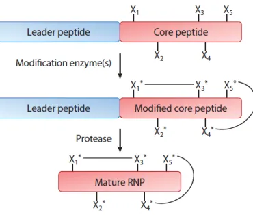
Lantibiotics are produced by ribosomes as inactive pre-peptides, consisting in an N-terminal leader-peptide and a C-N-terminal core-peptide part [2].
The modifications are carried out by lantibiotic-specific modification enzymes, that requires the presence of a leader peptide to recognize the target molecule.
The modifications involves only the core-peptide part, in which most of the serine (Ser) and threonine (Thr) residues are dehydrated to dehydroalanine (Dha) and dehydrobutyrine (Dhb) respectively.
Subsequently this dehydroresidues are coupled to cysteines (Cys) residues, this leads to the formation of (methyl)lanthionine rings. This step is generally called cyclization. In the end, the cleavage of the leader peptide by dedicated protease is necessary to obtain the active compound.
Fig 1: General representation of ribosomally synthetsized natural product (RNP) biosynthesis.Xn* represent modified amino acidic residues
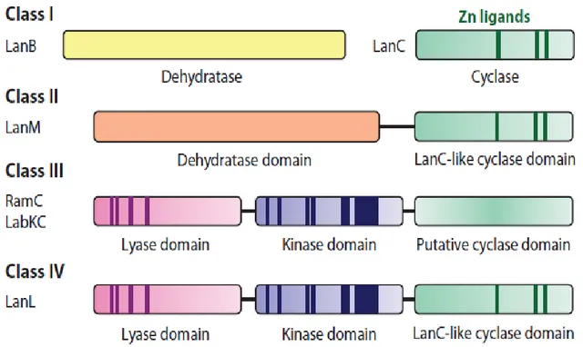
On the basis of the biosynthetic machinery involved in the formation of (methyl)lanthionine rings, is possible to define four classes of lantipeptides.
For class I, two distinct enzymes, the dehydratase LanB and the cyclase LanC, perform Ser/Thr dehydration and cyclization respectively.
The term “Lan” is a general abbreviation for proteins involved in lantibiotic biosynthesis.
A member of Class I lantibiotic is nisin.
For class II, these reactions are carried out by a single bifunctional enzyme, LanM, containing both the dehydratase (N-terminal part) and cyclase (C-terminal part) domains.
This is the case of cinnamycin.
Class III lantibiotics are formed by two subunits called α-peptide and β-peptide that are linked together.
Lantipeptides that are members of Class III, are modified by a trifunctional synthetase, containing an N-terminal lyase domain, a central kinase domain, and a putative cyclase domain in the C-terminal part.
For this last domain is used the term putative because it lacks many of the conserved active-site motif found in LanM and LanC ezymes.
In the end, recently identified Class IV lantipeptides, are modified by a single enzyme similar as Class III synthetase, LanL. This enzyme contains the lyase domain and kinase domain as the Class III enzyme, but its C-terminal cyclase domain is analogous to LanC
Fig. 2: Schematic representation of the four classes of lantipeptides modification enzymes. Highlighted part represent conserved motifs.
1.2 Clinical use of lantibiotics
Due to an increasing resistance of bacteria to available antibiotics, there is an urgent need to search for substances active against multidrugs resistant pathogens [2]. In the last years, the increase of multidrug resistant bacteria, especially in hospital environments, has caused an increasing concern in medicine.
Some of this multidrug resistant bacteria are: methicillin-resistant Staphilococcus
aureus (MRSA), vancomycin-resistant Enterococcus faecalis (VRE), third generation
cephalosporin-resistant E. coli.
This topic has become of major interest in antibacterial therapy, highlighting the need of new ways to treat infections.
In literature is possible to find several approaches to tackle down this problem, such as isolation of infected patients in hospitals, reduction of non-human antibiotics, rational use of antimicrobials, or phage therapy [6-8].
Another way to fight this growing threat is to find new antibiotics with a new mechanism of action.
Under this respect a new approach in the research of antibacterial agents is needed, because the research in traditional antibiotics did not provide good results.
According to a recent report of European Medicines Agency, only 15 compounds of 167, investigated in clinical trial, shows a new mechanism of action or involve a new target.
The situation is even worse for Gram-negative bacteria, where only 2 of the 167 compounds had a novel target or mechanism of action.
This results lead to a reduced interest from pharmaceutical sector (1.6% of the total molecules under development in 2004), and should be compensated by investments from public funds [9].
It has already been shown that some lantibiotics exhibit activity against antibiotic-resistant pathogens at nanomolar concentrations, so at concentration similar to commercial antibiotics, targeting a different compound of bacterial membrane compared with traditional antibodies.
Clinical applications of lantibiotics are currently of great interest.
Lantibiotics possess potent activity against several clinically relevant Gram-positive bacteria, such as Staphylococcus, Streptococcus, Enterococcus, but also against Gram-negative bacteria as Neisseria and Helicobacter.
Currently there are several lantibiotics in clinical and preclinical development such as duramicycin for the treatment of cystic fibrosis-associated pneumonias, mersacidin, that shows activity against methicillin-resistant Streptococcus aureus strains, and lacticin 3147 proved to be a successful antimicrobial agent against vancomycin-resistant Enterococcus faecalis andpenicillin-vancomycin-resistant Streptococcus pneumonia.
1.3 Mode of action of lantibiotics
The mechanism of action by which lantibiotics exert bactericidal activity has been studied only in few cases, but most of this bactericidal peptides are believed to inhibit cell wall biosynthesis and/or form pores through the membrane, leading to cell disruption.
Target of lantibiotics seems to be Lipid II, an amphipathic peptidoglycan precursor molecule involved in the synthesis of the cell wall of bacteria.
Lipid II is responsible to deliver the complete peptidoglycan,formed at cytoplasmic side of the membrane, to the extracellular side (Fig 3) [4].
Fig 3: Schematic representation of cell wall biosynthesis steps. GLCNAc (N-acetylglucosamine) and MurNAc (N-acetylmuramic acid)are compounds of peptidoglycan involved in cell wall synthesis
Binding of Lipid II leads to inhibition of transglycosylation reaction, necessary in the synthesis of peptidioglycan and therefore in the synthesis of the cell wall.
Bound Lipid II are sequestered from the cell wall division site (septum) and translocate in a non-functional locations, in this way the cell wall biosynthesis is stopped.[3]
As mentioned above, the other mechanism of action for lantibiotics involves the formation of pores through the membrane of bacteria.
This mechanism has been extensively studied for the Class I lantibiotic nisin.
The first two rings in nisin, called A and B, and formed by a Lan and a MeLan respectively, can bind the pyrophosphate portion of Lipid II (Fig 4).
Fig 4: Structure of Lipid II. The red bars indicate the minimal binding sites in Lipid II of glycopeptide antibiotics (1), nisin (2), ramoplanin (3) and mersacidin (4).
GlcNAc (N-acetylglucosamine); MurNAc
(N-acetylmuramic acid).
As the A and B rings are conserved in most of Class I lantibiotics, this mode of action is credited to other class I lantipeptides.
Once the two rings bind Lipid II, nisin is able to insert its tail into the membrane. A group of eight nisin, and four Lipid II, form a stable pore that leads to the loss of intracellular compounds in the extracellular environment and consequently to the death of the bacteria (Fig 6) [3].
Fig 6: Schematic representation of pore-formation nisin mechanism. (a): nisin reach the extracellular side of the membrane; (b): nisin rings A and B bind the pyrophosphates of LipidII; (c): transmembrane insertion of nisin; (d): four nisin bind four Lipid II, and four additional nisin are recruited for pore formation.
As mentioned above, and shown in Fig. 5, nisin can bind the Lipid II at pyrophosphate level. General glycopeptide antibiotics, such as vancomycin, can bind Lipid II at level of the pentapeptide. This kind of binding is related to the specific sequence of this oligopeptide.
This means that changes in the amino acidic sequence can compromise the binding. Bacteria can change this sequence and thus develop resistance to general glycopeptide antibiotics. On the other hand, the pyrophosphates are a fundamental mojeties of Lipid II and difficult to replace. This could represent a potential advantage for using lantibiotics, as bacteria cannot easily develop resistance. This is proved from the fact that in 50 years of use of nisin as food preservative no significant resistance has been observed.
Some kind of resistance mechanism has been detected, as an increising in the cell wall thickness in an attempt to reduce the access to the Lipid II, but this does not lead to a significant decreasing of nisin activity.
As nisin, classII lantibiotics (e.s. mersacidin) kill bacteria with a mechanism that inhibits cell wall replication by binding Lipid II, but this mechanismdoes not involve pore formation.
The binding site of mersacidin involves the GLcNAc sugar, but cannot bind it in LipidII.
The mechanism of Lipid II binding seems to be related with positive charge, in particular seems that this mechanism is dependent to the bivalent ion Ca2+ that is
responsible for the formation of a bridge between the lantibiotic and the sugar of Lipid II [4].
Not all the class II lantibiotics act in this way, cinnamycin e.g. does not target Lipid II.
Mode of action of Class III lantibiotics involves both the subunits.
The α-peptide behave like mersacidin, binding the LipidII then, the β-peptide is recruited to form pores.
An example of this lantibiotics is haloduracin.
In this case, a complex formed by a Lipid II, two α-peptide and two β-peptide is necessary for the pore formation.
This mode of action explains why a single subunit of a two component lantibiotic shows low or no activity. [1].
The literaturereports of other lantibiotics, such as Pep5 and epilacin K7, that form pores without the binding of Lipid II, suggesting a specific but alternative molecular target [10].
Another advantage in the use of antibiotics targeting Lipid II is the low toxicity in human, due to the fact that Lipid II production is restricted to bacteria.
However, there are still obstacles that must be overcome before lantibiotics can be introduced in clinical applications.
For example, nisin shows poor pharmacokinetic that represents the main impediment to its clinical development.
Anyway, the development of nisin as therapeutic molecule can benefit of the large amount of information collected from more 50 years of usage of nisin as food preservative, for example, this information can be used on issues relating stability and conditions of use [4].
In conclusion, the available data support the great capabilities of lantibiotics as potent antimicrobials with a novel mechanism of action [5].
1.3.1 Effect of the presence of thioether bridges in lantibiotics
As mentioned before, lantibiotics have still to overcome some obstacles before their clinical use is confirmed.
On the other hand, lantibiotics have a characteristic that is very important for a therapeutic peptide: the presence of thioether-bridges.
The main limitation in the application of therapeutic peptides is their rapid degradation by proteases.
A strategy to prolong the clearance of these peptides is to bind them to a protein as albumin.
This problem does not affect lantibiotics, because of the presence of thioether-bridges that confer strong resistance against proteolitic degradation [6].
In lantibiotics, thioether-bridges are present in the amino acids lanthionine and methylanthionine, introduced as post-traslational modifications.
Therefore, Lan and MeLan rings are useful both for the activity and for the resistance against proteolitic degradation.
In lantibiotics the Lan and MeLan formation follow this steps: 1) dehydratation of serines (Ser) and threonines (Thr); 2) resulting dehydroalanines and dehydrobutyrines are coupled to cysteines. This process is catalized by a cyclase enzyme (Fig 7) [6].
Fig 7: Schematic representation of thioether-bridge formation.
Resistance against proteases gives the advantage that a higher concentration of therapeutic peptide can be reached in the body and can be kept for longer time. More important, proteolitic stability prevent the formation of toxic compounds from lysate peptides.
The presence of thioether-bridges seems to open the way to the production of oral delivery peptide drugs.
This kind of delivery is very difficult to reach in peptide drugs that do not contain thioethers links, but seems to be possible in those that have these bridges, as shown
for thioether-bridged angiotensin, that could be successfully delivered both orally and airborne [5].
1.4 Strategies to develop new lantibiotics.
As stated in the previous paragraph, the use of lantibiotics represents a valid and novel strategy to fight the growing problem of multi-resistant pathogens.
To increase the success of lantibiotics as new drugs several strategies can be used. For example exploiting the possibility to realize fusion leader-peptide and the substrate tolerance for modification enzymes, the design of novel lantibiotics or an improved activity of bioactive compounds is possible.
In this paragraph possible strategies in the discover of new lantibiotics are described, including both a natural and a synthetic biology approach. Furthermore, methods to improve activity of these lantibiotics, as additional modifications or mutagenesis, are illustrated.
1.4.1 Traditional approach: discover new lantibiotics using wild-type producers
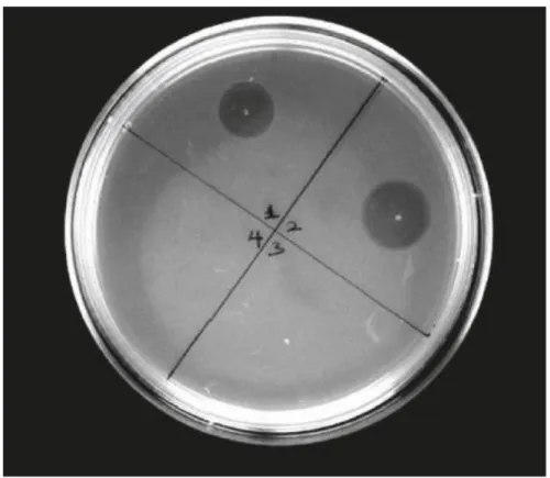
The traditional approach to search new potential antibiotic compounds is based on the isolation of producer organisms from diverse habitats.
After isolation of the producer, the next step is to check the antimicrobial activity. The most common way to do that is an agar diffusion assay (or activity test).
With this assay is possible to check if the producer actually can produce antimicrobial molecules (lantipeptides in our case), and this is confirmed by an inhibition of growth of the indicator strain on the plate (Fig 7).
For this kind of assay two variable are to be taken into account: the indicator strain and the conditions for the growth of the producer.
The number and diversity of indicator strains that can be tested is a limitation, but is also true that we can limit this number selecting only strain suitable for certain application.
Also, finding the growth condition in which the producer can express the lantibiotic is not an easy task. [5]
If areas of growth inhibitions are detected is necessary to check if the inhibition actually depends from the antimicrobial peptide or if it is related to others substances, for example catabolities as organic acids.
Therefore, additional steps are necessary to check the antimicrobial molecule, but anyway this method is still a good strategy to screen potential producers.
The main disadvantage of this kind of approach is the wide screening area that has to be covered, which lead to a high demand of resources. This point can be partially overcome by automatic high throughput screening systems [5].
On the other hand, the advantage is that identified antimicrobial compound are already been selected in the appropriate growth conditions.
Fig 7: Example of activity test. In this case the activity of cinnamycin samples against Bacillus subtilis LH45 (indicator strain) is shown on LB-agar plate.
1.4.2In silico genomic mining-tools to discover new lantibiotics
An alternative way to discover new lantibiotics involve the analysis of the genome of potential producers.
Anyway, lantibiotic structural genes (genes that codify LanA) are too small and show low homology to use a similarity oriented search.
Instead of searching for homology of structural genes, a better strategy is search for homology for modification enzymes, because of their larger size and conserved domains.
Literature report that structural genes for lantibiotics are surrounded by “Open Reading Frames” (ORFs) for modification enzymes.
That means that searching in the genome of a potential producer for a modification enzyme gene can lead to the entire gene cluster for the expression of the lantibiotic. The use ofin silico mining tools, such as “Bagel2” and “Bagel3” softwares developed by “Molgen” group of the Groningen University, is necessary to this aim.
This searching engine allow the identification in the genome of a gene cluster related to the production of lantibiotics , considering the structure of the putative structural gene, the surrounding ORFs and their homology to modification enzymes.
This kind of strategy allow to check exotic and non-culturable bacteria, and if a positive result is found is possible to try heterologous expression.
Other application of in silicoanalysis, oriented to microbial ecology, permit to restrict the environments to screen to find new potential producers.
1.4.3 Improve the bioactivity through mutagenesis
Once a novel lantibiotic has been defined it’s possible to improve its properties as stability, potency or spectrum using mutagenesis.
That means the replacement one or more amino acids in the peptide sequence.
Mutagenesis can be also use to better understand the lantibiotic’s mechanism of action, its maturation process and the bacterial immunity system.
Experiments carried out on the leader peptide highlight the role of this portion on the maturation step, because after mutation the modification enzymes could not more recognize the leader and modify the core peptide.
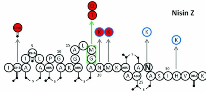
However, point mutation can improve molecules, this is the case of nisin Z, in which after a substitution was observed an improved stability and solubility without decreasing the activity. This was possible by an insertion of a positively charge amino acids in the first ring (Fig 8).
Nowadays, it is possible obtain libraries of mutants lantibiotics, with all the possible point mutation, or even with several mutation at the same time, and to test
thesecompounds with high throughput screening systems to identify variants with higher expression level, better stability or specific bioactivity.
Of course this strategy can be used on wild type lantibiotics as well as on new synthetized ones.
Fig 8: Nisin Z with possible mutations. Red circles indicates improved activity, blue circle indicates improved physiochemical proprieties.
1.4.4 Synthetic Biology approach
Synthetic Biology aims to apply the principles of modularity and orthogonallity of engineering to Biology.
Thus, the isolation of parts of the peptide or the biosynthetic machinery should be standardized in such a way that they can be predictably combined to produce a new antimicrobial molecule [5].
As mentioned above, modification enzymes of lantibiotics can modify a wide range of substrate peptides as long as they are linked to the leader peptide that is necessary for the recognition from the enzymes.
In the chapter 3 of this thesis the modification of nisin with a chimeric leader peptide is reported. The synthetic leader is formed by the fusion of cinnamycin and nisin leaders by the class LanM modification enzyme CinM, from cinnamycin modification machinery.
Indeed, it has been proved that “silent” lantibiotics from S. pneumoniae,can be produced and modified by nisBCT in L.lactis, thusin a heterologous host, if the core-peptide is linked to nisin leader.
This means that the enzymes are able not only to dehydrate and cyclize the peptide, but also to match dehydroamino acids and cysteines in the correct way.
This is a clear example of modularity applied to lantibiotics production.
Heterologous expression allow to produce lantibiotics of non-culturable bacteria without engineering the original producer.
In addition, heterologous expression of lantibiotics modified by nisin system in E.coli has been reported.
In addition to the characteristic lanthionine and methilantionine, lantibiotics carry additional post-translational modification important for stability and modification, such as lysinoalanine bridge in cinnamycin.
Exploiting the concept of modularity it is possible to introduce theseadditional modifications of some lantibiotics to hypermodify existing molecules, and confer to them improved bioactivity or new proprieties.
However,it is necessary to characterize this modification enzymes in order to use them in a rational and predictable way.
Modularity in lantibiotics is not only referred to leader peptide, but also to the core peptide sequence.
Three different regions can be described in nisin: 1)a leader peptide that leads the modification enzymes; 2) a Lipid II binding domain, formed by the first two rings of nisin; 3) a membrane insertion tail.
That means that is theoretically possible to design a lantibiotic with the Lipid II binding domain from nisin and a C-terminal tail to introduce additional post-traslational modification to create a novel molecule.
Modularity is present also in Class III lantibiotics, in which one subunit is necessary to bind Lipid II and the other is necessary for pore formation.
Moreover, combination of lantibiotics with traditional antibiotic could represent a valid strategy.
In vitrocombination of nisin with vancomycin via chemical semi-synthesis has been
reported, and improved activity was detected for vancomycin[11].
The possibilities in design of novel lantibiotics with a synthetic biology approach are enormous because the number of combinations is very high, and can be raised with the discover of new lantibiotics and the modification by mutagenesis method.
This is particularly true if we consider the possibility to introduce non-canonical amino acids in the sequence of lantibiotics.
These non-canonical amino acids can allow introduction of new properties for the peptides.
In conclusion, all the strategies described above should be combined to obtain a considerable improvement in the research of new lantibiotics (Fig8).
CHAPTER 2:Analysis of the lantibiotics Nisin and
Cinnamycin
The project reported in this work involves two lantipeptides, the class I lantibiotic nisin, and the class II lantibiotic cinnamycin.
In this chapter, structure, modification machinery and mode of action of these two lantibiotics are described.
Furthermore, some examples of application of synthetic biology approach on nisin are reported.
2.1 Structure, biosynthesis, modification machinery of nisin. Design of
new lantibiotics through a Synthetic Biology approach
Nisin is a class I lantibiotic produced by Lactococcus lactis, is one of the oldest know antibiotic compounds, and shows antimicrobial activity against Gram-positive bacteria.
The genes necessary for nisin biosynthesis process - that involves production, maturation, immunity and regulation - are located in a conjugative transposon [20]. Nisin was used as food preservative for 50 years, and its high efficiency makes it one of only few commercial applied lantibiotics.
Target for nisin antimicrobial activity are Gram-positive bacteria such as Bacillus
cerus, Listeria monocytogenes, Enterococci, Staphylococci and Streptococci.
For this very high antimicrobial activity, nisin has been proposed to be used as model to develop new antibiotics.
Belonging to class I lantibiotics, nisin harbours thioether bridges in the Lan and MeLan resiudues, introduced after the action of the modification enzymes NisB and NisC.
In the following paragraphs biosynthesis, modification machinery and nisin role in developing of new lantibiotics through a Synthetic Biology approach are described.
2.2.1 Biosynthesis of nisin.
Nisin biosynthetic genes are organized in four operons, nisABTCPIRK, nisI, nisRK and nisFEG.
Nisin precursor peptide is encoded in the gene nisA, the result of the translation of this gene is a peptide of 57 aminoacidic(a.a.) residues.
To obtain the mature lantibiotic is necessary that the modification machinery processes the precursor peptide.
The modification system is encoded in the four genes nisB,nisC,nisT,nisP.
The first one encodes the dehydratase NisB, responsible for the dehydration of three serine and five threonine.
Some of this dehydrated residues are coupled with cysteins by the cyclase NisC. This result in the formation of one lanthionine and four methyllantionine, and also one dehydrobutyrine and two dehydroalanines that are not coupled with cysteins. The next step in the biosynthesis of nisin is the exportation of the modified molecule through the membrane.
This process is carred out by the transporter NisT, an ABC-type transporter [21]. Both for the modification and for transportation the presence of the leader peptide is necessary for the enzymes to recognize the substrate molecule.
The leader peptide is a peptide of 23 a.a., and until it is binding the modified nisin the molecule does not show any activity.
Thus, the last step involves the cleavage of the leader peptide. This process is carried out by the membrane protein NisP.
The mature nisin is also capable to induce the complex NisRK, the system that regulates the transcription of nisin gene cluster [20].
The analyis of nisin transposon (Fig. 9) highlights the presence of a regulation operon, formed by nisRK, and an immunity operon formed by nisI and nisFEG.
Fig 9: Transcriptional organization of nisin biosynthetic gene cluster.
The regulatory system for nisin biosynthesis is a two-component system, formed by NisR and NisK.
NisK is a histidine sensor kinase that is located in the extracellular side of the membrane and act as receptor for fully matured nisin.
The binding of NisK with nisin initiate a signal cascade, that starts with the auto-phosphorylation of NisK.
Subsequently, the phosphate is transferred to NisR, which is a transcriptional activator that binds the promoter regions of nisABCPRK and nisFEG. This induces the transcription of genes required for nisin biosynthesis and immunity.
The promoter of nisRK was shown to be independent from nisin regulation and the two genes to be constitutively expressed.
This system provide a tight regulation of gene expression, for this reason nisin-controlled expression system (NICE) [22] were developed and used to control overexpression of protein in heterologous expression systems.
Lactococcus lactishas developed an immunity system against the bactericidal activity
The immunity system consist in a two-component complex made by the lipoprotein NisI and the ABC-type transporter NisFEG [23].
The two components strongly interact to confer immunity as proved by the fact that each component alone provide 5-20% of the total immunity level.
NisI is a lipoprotein anchored on the extracellular side of the membrane, but also free NisI was detected.
The function of NisI is to interact with nisin by binding it and avoid that nisin itself can target the Lipid II of the membrane.
NisFEG form a transporter system that has the role to export nisin from the cytoplasmatic side of the membrane.
The two systems interact because NisFEG can provide high local concentration region for nisin, so that NisI can easily intercept nisin and avoid its movement in the extracellular environment [20].
Fig 10: Representation of nisin biosynthetic system. P in the blue circle indicates the phosphorylation of NisK and NisR.
2.2.2 Analysis of nisin structure
After modification nisin is a peptide of 31 amino acids containing one lanthionine ring and four methyllanthionine rings, plus two dehydroalanine and one dehydrobutyrine. The five rings on nisin molecule are named as A, B, C, D, and E starting from the N-terminal side to the C-N-terminal side of the molecule.
The main part of nisin consist of the lanthionine ring A, and the two methyllanthionine rings B, C. This part of the molecule is linked by a “hinge” region to two intertwined double methyllanthione rings D and E.
Thus, nisin shows as a rod-like molecule with a hinge region that confer flexibility to the total structure, and also the two parts are both quite flexible.
It is remarkable that rings A, B and C share the feature that hydrophobic part is situated opposite to the thioether bonds. The hydrophobic part is composed of the residues Ile4, Leu6, Pro9, Leu16 and Mey17 whereas the hydrophilic Lys12 is located at the opposite face [20].
Taken together, the molecule is amphipathic in two ways: first, most of the residues in the N-terminal part are hydrophobic and only a single charged residue is present (Lys12), whereas the charged and hydrophilic amino acids are mainly located in the C-terminal half of the molecule.
Secondly, both the N-sided domain, with rings A, B and C as well the C-sided domain, containing rings D and E, have a hydrophobic and hydrophilic side [20].
Nisin amino acidic sequence and structure is shown in the figure below.
(a)
(b)
Figure 11: (a) Structure of fully modified nisin. Red regions represent dehydrobutyrines and dehydroalanines involved in Lan and MeLan formation. Blue regions represent cysteines involved in Lan and MeLan formation. Green regions are dehydroalanines. Purple region is dehydrobutyrine.
2.2.3 Nisin modification machinery
Nisin modification machinery is composed by four enzymes: the dehydratase NisB, the cyclase NisC, the ABC-transporter type NisT and the protease NisP.
All the modification steps seems to be carried out in proximity of the membrane. This was dimostrated by immunoprecipitation assay [24], furthermore it was demonstrated that physical interaction between the modification enzymes is requested for the modification of nisin pre-peptide.
However, in vitrostudies show that the modification enzymes can work separately, this means that the lanthionine sintetase complex, formed by all four modification enzymes, is not a prerequisite for functioning of any of the modification and transport enzymes, and it is likely highly unstable and transient in nature [20].
This means that the single modification enzymes canbe use separately to modify lantibiotics using a synthetic biology approach, exploiting the not strict specificity of these enzymes for their substrate.
It was demonstrated that the overexpression of nisBCT gene leads to insertion of Lan and MeLan rings in non-lantibiotic peptides if they are linked to nisin leader.
This proves that nisin modification enzymes do not have highly specificity and that the main prerequisite for modification is the presence of the leader peptide upstream the substrate.
Figure 12 show nisin modification steps and lanthionine sintetase complex located in the cytoplasmic side of the membrane.
NisB, a protein of 117.5 kDa, is the dehydratase responsible for the dehydratation of Ser and Thr residues in the nisin core-peptide.
This was proved by the inhibition of nisBgene. This operation leads to a complete loss of nisin production. Furthermore, overexpression of this enzyme rises the efficiency of dehydratation in nisin.
To carry out its function NisB requires the presence of the leader peptide.
Furthermore, it was proved that NisB could dehydrate Ser and Thr also in non-lantibiotic peptides, showing a relaxed substrate specificity.
Apparently the substrate specificity is related to the amino acidic residues near Ser and Thr residues.
Studies demonstrates that the nature of residues close to dehydrated serine and dehydrated threonine may influence the dehydratation step [20].
These residues should be surrounded by hydrophobic residues to obtain the dehydratation of Ser residues.This is the main limitation for NisB substrates.
NisC is the cyclase that carry out the coupling of dehydrate residues with free cysteine, it is a 47.9 kDa protein.
Thus, the ring formation is lead by NisC and all the rings are oriented in the same manner, from N-terminal side, where the dehydro residue is located, to C-terminal side where the cysteine is positioned.
The role of NisC was confirmed by knoking out this gene in nisin gene cluster. This operation leads to the production of nisin with dehydro- residues without lanthionine and menthyllanthionine rings.
This proves that NisC is fundamental in cyclization.
It was also proved that NisC can work separately from the other modification enzymes.
It was demonstrated that NisC need the presence of the leader peptide to carry out its function.
So, as for NisB the leader peptide is fundamental, and seems that NisC can recognize the leader by a specific region formed by hydrophobic and negatively charges residues that form a channel in which the leader peptide is supposed to be trapped.
NisT is an ABC half-transporter that consist of putative α-helices that crosses the cytoplasmic membrane five times and a hydrophilic nucleotide-binding domain that binds ATP needed to energize the transport.
A typical ABC transporter includes two modules, i.e., two trans-membrane segments and two nucleotide-binding domains.
Therefore, NisT is an half ABC transporter and most likely requires another half-transporter to form an active unit[20].
This putative partner is likely another NisT.
The function of NisT was confirmed bythe knock out of the gene nisT.As a result the secretion of nisin was inhibit.
It was also proved that NisT can transport unmodified or partially modified nisin, but also non-lantibiotic peptides if this are fused with the nisin leader sequence.
NisP is the protease that carry out the cleavage of nisin leader peptide. This was proved by knocking out the gene encoding NisP.
As a result, no active compound was detected because the cleavage of the leader is necessaryto obtain active nisin.
The activity was restored by incubation with cells expressing NisP.
NisP contain a Sec-signal in the N-terminal side, that is likely responsible for targeting and transporting NisP out of the cell via Sec pathway.
NisP contains also a C-terminally located LPXTG sequence, that suggests its anchoring to the cell surface [20].
NisP can cleave only the nisin leader peptide, furthermore neither unmodified nor dehydrated pre-nisin could be cleaved by NisP, indicating that one or more thioether rings are required for its activity [25].
2.2.3 Use of nisin and nisin modification enzymes in synthetic biology
As mentioned in the previous chapter, Synthetic Biology represent a valid strategy in the design of new lantibiotics.
The Synthetic Biology approach is exploiting the relaxed substrate specificity of enzymes involved in lantibiotic modification, and also the modular structure of lantibiotics themselves.
At least two separate modules are detectable in latibiotic, a leader peptide and a core peptide. The first one is involved in the recognition of the molecule from modification enzymes.
Using this peculiarity it is possible design non-lantibiotic peptides fused with a leader peptide that can be modified by modification enzymes.
It is also possible to introduce in a given lantibiotic post-translational modifications that belong to other lantipeptides.
The following are example of use of Synthetic Biology in the modification of lantibiotics.
The first one describe the use of nisin modification machinery in the production of a two-component lantibiotic of S.pneumoniae, whereas the second one shows the possibility of using modification enzymes from different lantibiotics to design novel molecules.
The first example is reported in [2], and reports the expression of a class II two-component lantibiotic from S.pneumoniaeusing the nisin modification machinery.
To do that it was necessary toisolate the gene encoding for the lantibiotic peptide and to modifyit to obtain a gene that encodes the fusion of nisin leader peptide with the two-component lantibiotic peptide.
Then the overexpression of this novel gene and of the nisBCTgenes was performed. The peptide was secreted out and found in the supernatant of the culture, meaning that the transporter nisT can export the peptide.
Mass spectrometry analysis dimostrated the presence and partial localization of multiple dehydratated serines and/or threonines and (methyl)lanthionines in both peptides.
Furthermore, after the cleavage of the leader peptide from both the components antimicrobial activity against Micrococcus flavuswas detected.
These results proved that the nisin modification machinery can be succefully used to modify and produce other antimicrobially active lantibiotics, and confirm the validity of the synthetic biology approach.
The second example is reported in [26] and is related with the design and production of novel lantibiotics using a combination of different lantibiotic modification enzymes.
The specific results highlight:i)the modification of the lantibiotic gallidermin by nisin modification enzymes NisBTC and gallidermin modification enzyme GdmD ; ii) the design and production of a novel lantibiotic formed by the nisin region containing rings ABC and a gallidermin tail. This hybrid molecule was modified by the combination of NisBTC and GdmD; iii) the design and production of modified nisin by introduction of a modification carried out by lacticin modification enzyme LntJ. GdmD is responsible for C-terminal decarboxylation, and LntJ is a reductase that stereospecifically converts dehydroalanine in D-alanine.
The modification and transport of this molecule was proved by heterologous expression in L.lactis, this results provide a plug and play system that can be used to
select different sets of modification enzymes to work on diverse, specifically designed substrates, with the aim to obtain new to nature molecules with antimicrobial activity and/or new physicochemical proprieties.
The representation of the system is shown in figure 13.
Figure 13: Schematic representation of the synthetic biology plug and play system for the design and production of new to nature lantibiotic.
2.2 Structure, mode of action and modification machinery of the class
II lantibiotic cinnamycin
Cinnamycin is an antibiotic peptide produced by several Streptomyces strains, including Streptomyces cinnamoneus DSM 40005 [13].
Cinnamycin belongs to lantibiotic group as it carries lanthionine and methyllanthioninebridges.
As in other lantibiotics, these bridges are formed from dehydro serine and threonine residues, coupled with cysteines.
These modifications are carried out by a LanM modification enzyme, called CinM, given that cinnamycin belongs to class II lantibiotics.
Unlike nisin or others class I lantibiotics, cinnamycin possesses other post-translational modification.
Cinnamycin contains a β-hydroxy-aspartate residue and a lysinoalanine bridge (Lal). The hydroxylation of the aspartic acid residue (Asp) is carried out by the modification enzyme CinX and the formation of the Lal bridge is made by the small enzyme Orf7. Class II lantibiotics are less studied and usually have globular and inflexible tertiary structure (Fig 14), unlike the class I lantibiotics that are more flexible and have linear structure [14].
Another difference between class I molecules as nisin and class II lantibiotics as cinnamycin is in the mode of action.
As mentioned in the previous chapter, class I lantibiotics exercise their antimicrobial activity interacting with Lipid II of the cell wall, this generally lead to pore formation. Some class II lantibiotics as cinnamycin and duramycin seems to exercise their activity binding enzymes involved in the cell wall biosynthesis.
Similar to other lantibiotics the non-mature peptide is formed by the fusion of a leader peptide and a core peptide.
The core peptide is a 19a.a.-long peptide, and it is the portion that harbour the lanthionine and methyllantiones residues, and the other post-translational modification.
The leader peptide is a 59a.a.-sequence, a very long sequence if compared to leader peptide of others lantibiotics, especially in class I.
The leader peptide is important for the modification enzymes to recognize their target. To obtain the active compound the cleavage of the leader is necessary.
Unlike nisin, it seems that there is not a dedicated protease for this taskin the cinnamycin gene cluster.
Therefore, it seems that the last three amino acids, “AFA”, of the leader are involved in the cleavage, fitting with the “AXA” motif recognized by type I signal peptidases of the general secretory pathway (Sec) [13].
This “AFA” sequence will be taken into account in the heterologous expression of cinnamycin in E.coli (described in chapter 3), to engineer the cleavage of the leader peptide.
Fig 14: Schematic structure of cinnamycin. MeLan bridges are shown in green. Lan bridge is shown in pink. Blue is used to label the Lal bridge. Red is used to label the hydroxylation of Asp.
2.2.1 Analysis of the cinnamycin gene cluster
The study of the gene cluster involved in the expression of cinnamycin in S.
cinnamoneumwas possiblethanks to genomic analysis tools[13].
The genome contains 21 genes, but it is not yet clear which is the functionfor some of them.
The function of each gene was studied through a homology approach, thus searching proteins similar to those encoded in the cin gene cluster.
Most lantibiotic gene clusters contain the following genes: i) lanA, the lantibiotic prepeptide structural gene; ii) lanBC or lanM, the genes that encodes the enzyme for dehydratation an cyclization of dehydrated serine and threonine residues with cysteines; iii) lanT, which encodes a transporter enzyme for export the lantibiotic; iv)
lanP, the gene encoding the protease for the cleavage of the leader peptide; v) lanRK,
which encodes a two-component regulatory system; vi) lanFEG, encoding a transporter enzyme responsible for the immunity of the producer bacteria to mature lantibiotic [13].
This are the most common genes in a general lantibiotic gene cluster.
Proceeding from left to right on the cluster represented in Fig 15, the genes in cinnamycin gene cluster are the following.
Fig15: Representation of the cinnamycin gene cluster
cinorf3: encodes an N-terminal signal peptide. It shares 78% homology with a
cinorf4: seems to encode a regulatory protein with a helix-turn-helix DNA-binding
motif similar for the 67% to an S.coelicolor protein [15].
cinorf7: is a 360 bp long gene. It seems to encode the enzyme responsible to
synthesize the lysinoalanine bridge, as reported in [12].
cinA: is the gene encoding the cinnamycin prepeptide. It has a length of 234 bp.
cinM: encodes the modification enzyme cinM, member of LanM family, thus
responsible both for the dehydratation and cyclization of Ser and Thr residues. This gene contains the rare leucine codon TTA, for this reason in this project the cinM gene was modified to replace this rare codon. The gene has a length of 3276 bp [16].
cinX: this gene has a length of 968 bp. It encodes the protein for the hydroxylation of
Asp [12].
cinT and cinH: cinT share homology with a transporter membrane protein of ABC
family transporter. CinH seems to be similar to a protein involved in drug efflux and resistance, suggesting that it could have a role in the immunologic system [13].
cinY: encodes a product that may possess an N-terminal signal peptide, suggesting
that it may be exported.
cinZ: has a length of 627 bp. The function in unknown.
cinorf8: has a length of 420 bp and seems to be a member of the CoA-binding protein.
cinorf9: has a length of 321 bp. The encoded protein may have a possible N-terminal
signal peptide suggesting that could be exported [13].
cinR and cinK: Encodes a two-component regulatory system and shares homology
with two protein in the regulatory system of Bacillus subtilis [13].
cinorf10 and cinorf11: does not show significant similarity with other proteins.
cinR1: encodes a protein involved in Streptomyces regulatory system. [13].
cinorf12, cinorf13 and cinorf14: does not show similarity with other proteins [13].
In conclusion, the genes involved in the modification and expression of cinnamycin are cinA, cinM, cinX and cinorf7. Thus these genes were the ones selected for the heterologous expression in E.coli described in chapter 3.
2.2.2 Modification machinery of cinnamycin
Only the genes cinM, cinX and cinorf7 encode a modification enzyme in the entire gene cluster of the lantibiotic cinnamycin.
These modification enzymes act on the pre-peptide encoded by the gene cinA.
As in others lantibiotics the modification enzymes recognize the target molecule thanks to the presence of a leader peptide.
The cinnamycin leader peptide has a length of 59 a.a., whereas the core peptide is long 19 a.a.
The cleavage of the leader peptide is necessary to obtain the active molecule after the modification of the core peptide.
Mature cinnamycin has a globular structure, unlike the rod-like structure of nisin, and carries several kind of post-traslational modification.
Cinnamycin belongs to class II lantibiotics: this means that the introduction of Lan and MeLan rings depends on a single LanM enzyme, CinM.
Unlike nisin, the introduction of the thioether-bridge containing a.a is not the only modification.
Cinnamycin harbour an erythro-3-hydroxy-L.aspartic acid resulting from the hydroxylation of L-aspartate by CinX [12].
The last modification in cinnamycin core peptide is the lysinoalanine bridge, carried out by enzyme Orf7.
This modification is fundamental to confer antimicrobial activityto cinnamycin, because this bridge can bind the target of cinnamycin in the membrane of bacteria. The two modifications described above are illustrated in figure 16.
(a) (b)
Fig 16: Representation of lisynoalanine bridge (a) and of hydroxylated aspartic acid (b)
The modification enzyme CinM requires the presence of a leader peptide to modify the substrate.
This class LanM enzyme is responsible for the dehydratation of Ser and Thr residues in cinnamycin core peptide.
In total four residues are modified, two serine at positions 4 and 6 and two threonine at positions 11 and 18.
This process leads to the formation of two dehydroalanine resulting from dehydration of Ser residues, and two dehydrobutyrines, resulting from dehydration of Thr residues. LanM enzymes as CinM carry out also the cyclization of these dehydro-residues matching them with cysteine.
Unlike the case of nisin, where the cyclization is driven by the single enzyme nisC, in cinnamycin the Lan and MeLan rings are generated in a bidirectional way, such that the MeLan rings are formed using cysteines that are located N-terminal to the dehydrobutyrines, and the Lan bridge is formed using a cysteine located C-terminal to dehydroalanine with which it reacts [12].
In total the action of CinM introduces one lanthionine ring and two methillanthionine rings, and also an additional dehydroalanine involved in lysinoalanine bridge formation.
Fig 17: Schematic representation of the action of CinM. It is possible to note the different orientation of Lan and MeLan bridges. The link between the second Dha and the last lysine is the lysinoalanine bridge.
Analysis on cyclase enzymes (ex. Subtilin SpaC and nisin nisC) dimostrated that this protein contains zinc.
These results were confirmed from the anlaysis of the crystal structure of NisC. The zinc seems to be involved in the formation of thioether rings [17]].
LanM enzyme share sequence homology at the C-terminus with LanC cyclases, including two Cys and one His that coordinate the Zn2+ in LanC proteins.
Thus, it was hypothesized that LanM cyclase domains might act through a similar zinc-involved mechanism.
This hypothesis was confirmed with a spectrophotometric assay using metallochromic indicator [12].
The peculiarity of CinM to drive the formation of Lan and MeLan bridge in a bidirectional manner makes this enzyme interesting from the Synthetic Biology point of view, because it could be used to introduce Lan and MeLan ring in a different manner.
CinX is the enzyme responsible for the hydroxylation of Asp in position 15 of core peptide.
It was demonstrated [12] that the action of CinX can happen both after or before the formation of Lan and Melan rings.
Furthermore, it was demonstrated that CinX does not need the presence of a leader peptide to carry out its action, unlike most lantibiotics modification enzymes.
Orf7 is the protein encoded in the gene cinorf7. This small protein (13 kDa) carries out the formation of the lysinoalanine bridge, matching the last lysine of the core peptide with the dehydroalanine in position 4.
Activity test for cinnamycin missing lysinoalanine bridge gives negative result, showing that this modification is fundamental in the antimicrobial activity.
Therefore, in vitro modification of partially modified cinnamycin (modified by CinM and CinX) with Orf7 shows again negative result.
Thus, this could mean that Orf7 requires the presence of the other modification enzymes to carry out its own function, and not only a dehydratated serines and a lysine.
Fig 18: Schematic representation of the biosynthesis of cinnamycin by S.cinnamoneus.
2.2.3 Mode of action of cinnamycin
Unlike nisin and others class I lantibiotics, or class II lantibiotic mersacidin, cinnamycin does not target Lipid II, but interacts with membrane proteins such as phospholipase A2.
Phospholipase A2 has a major role in the release ofarachidonic acid from phospholipids in the cell membranes. Further oxidative metabolism of free
arachidonic acid leads to prostaglandins and leukotrienes. Several of these
eicosanoids are potent mediators of diseases, such as inflammation and allergy [18]. In addition, A2 phospholipase is involved in atherosclerosis.
Inhibition of the enzymatic activity of phospholipase A2 may therefore be therapeutically beneficial.
Cinnamycin shows activity against Gram-positive bacteria as Clostridium botilinum and Mycobacterium [14].
This lantibiotic also causes the trans-bilayer phospholipid movement in the membrane of mammalian cells to access phosphotidylethanolamine (PE) residing predominantly in the inner side of the membrane [19].
PE is also a substrate for the phospholipase A2.
The lantibiotic duramycin, a close structural analogus of cinnamycin, promotes chloride secretion in lung ephitelial cells by binding to PE.
This secretion lead to a mucus clearance from the lung, as result this compound I is in phase II clinical trials for the treatment of cystic fibrosis [12].
For this reason cinnamycin has been suggested as an alternative treatment for atherosclerosis, because can inhibit phospholipase A2 by binding its substrate PE.
CHAPTER 3: Material & Methods
This research project can be divided in three modules.
The first one involves the heterologous expression of cinnamycin in E. coli.
Ti do that was necessary to introduce in the bacteria the gene encoding for the LanA peptide and the genes encoding for the modification enzymes.
tested.
The second module involves the modification of nisin fused with a chimeric leader peptide formed by the fusion of cinnamycin leader peptide and nisin leader peptide. Nisin was modified by nisin system (NisBC) and the leader peptide was cleaved by NisP.
The aim of this module was to check if the presence of the cinnamycin leader peptide upstream nisin leader could affect nisin modification and activity.
The last part of the project aimsto prove the modification of nisin by a single class LanM enzyme, CinM.
Therefore, the modification of CinM on nisin was tested with the previous chimeric leader peptide.
Figure 19 illustrates a summary of the project.
3.1 Heterologous expression of cinnamycin in E. coli
The heterologous expression of cinnamycin in E.coliinvolves several steps.
Firstit was necessary to select the correct genes for the expression of the cinnamycin pre-peptide and for the expression of the modification enzymes.
In particular it was necessary to use a mutated variant of the structure gene cinA to bypass the absence of a dedicated protease for the cleavage of the leader peptide in cinnamycin gene cluster.
Once the genes were selected, the next step was the cloning of these genes in plasmid vectors.
Then the plasmid carrying the genes were introduced in the bacteria by electroporation.
After that, fresh-transformed cells were used to start a culture and obtain the expression of the cinnamycin.
After that, the cells werelysated and the peptide molecules purified.
A digestion step with protease LysC was necessary for the cleavage of the leader peptide.
Finally, the activity of fully modified cinnamycin was tested against Gram-positive bacteria B.subtilis and M.luteus.
3.1.1 Cloning of genes involved in expression and modification
To obtain cinnamycin expression are necessary the structure gene cinA, encoding the cinnamycin pre-petide, and the genes encoding the modification enzymes CinM, CinX and Orf7.
The aim of these cloning steps is to obtain a pACYC plasmid that harbors the two modification enzymes orf7 and cinX, and a pRSF plasmid with cinA and cinM.
The genes used for this project are not the wild type genes, but are modified to increase the expression level for E.coli.
This genes were provided by Lanthiopharma, a company which collaborate with Molgen research group of the Groningen University.
In particular,cinMwas modified to replace the rare codon “TTA” that encodes for leucine with a more common codon. In this way the expression of CinM is
optimized for an heterologous producer.
The ribosome binding site (RBS) or orf7was removedbecause an RBS was already present in the first cloning site of pACYC plasmid, in this way a double RBS situation was prevented.
The modified Orf7 is called Orf7b.
The most important modification involves the structure gene cinA. This gene encodes for the cinnamycin pre-peptide, formed by cinnamycin leader peptide and core peptide.
As reported in chapter 2 there is not a specific protease for the cleavage of the leader peptide in cinnamycin gene cluster. It seems that in the natural producer strain this is obtained by peptidase of the general secretory pathway Sec.
In E.colithe cleavage cannot be obtained in this way, so itis necessary to use a specific protease.
For this reason,cinA was mutated to replace the alanine at the last position of the leader peptide (position -1) with a lysine. After this mutation the gene is named
cinA(A-1K).
In this way is possible to obtain the cleavage by protease LysC that can break the peptide bond at the C-terminal side of lysine.
Only two lysine are present In cinnamycin sequence after this mutation, one at the last position of the leader peptide and the second at the last position of the core peptide.
Therefore, using LysC lead to a single cut that allow the separation of the leader peptide from the core peptide.
The genes were cloned into a pRSF and a pACYC plasmid.
Both these plasmids possess two separate cloning site, each one controlled by an IPTG inducible promoter.
For this reason a gene encoding lactose inhibitor is present in both plasmids under the regulation of a constitutive promoter.
Kanamycin resistance gene is present in pRSF plasmid, whereas a gene encoding for chloramphenicol resistance is present in pACYC. A constitutive promoter regulates these two genes.
These two plasmids possess the same origin of replication (ori), this mean the will be present in the same number inside the host.
The first cloning site of the pRF plasmid possess a his-tag sequence.
Therefore, cloning of a gene downstream this sequence to obtain the protein expressed in the gene with a sequence of histidine at N-teeminal side is possible. In this case the gene in the first cloning site of the pRSF plasmid was cinA(A-1K). The his-tag (formed by six histidine residues) is necessary to detect the expression of the pre-peptide and for its purification by affinity chromatography.
The structure of the two plasmids used in this project are reported tIn the figure below.
Fig 20: Structure of pACYC and pRSF plasmids
The final expression system consist in a pACYC plasmid carrying in the first cloning site orf7b and in the second cloning site cinX (pACYC orf7b-cinX), and a pRSF plasmid carrying cinA(A-1K) in the first cloning site and cinM in the second (pRSF cinA(A-1K)-cinM).
The project started from a pACYC orf7b and a pRSF cinA(A-1K) already built by Lanthiopharma.
To obtain the final expression system the following steps were performed:
Digestion
Ligation
Transformation
Colony PCR
Sequencing
The first step was to amplify the gene by PCR.
To do that it was necessary to design primers to amplify cinX, whereas for cinM case was possible to use the general primers for pACYC plasmid.
In fact, a cinM gene was contained in the second cloning site of a pACYC
plasmid,thus was possible to amplify it using the couple of primers pACYC 2nd-Fw
and pACYC 2nd-RV, that allow to amplify the second cloning site of the plasmid
when used together in a PCR step. (Primers sequences are reported in supporting information section) [SI 1].
CinX was contained in a pET plasmid, so it was necessary to design the correct
primers to amplify the gene.
The primers were designed using the software Clone Manager.
PCR reaction was carried out using phusion polymerase enzyme by Thermo scientific, and result analyzed by electrophoresis.
According to Clone Manager, PCR product for cinX case is 1030 bp, whereas for
cinM case is 3456 bp.
Fig 21: Analysis by electrophoresis of (a) PCR products for cinX case, (b) PCR products for cinM case.
The PCR product contains the gene sequence plus additional DNA, whichharbors restriction sites for digestion enzymes.
Using the correct combination of digestions enzyme it is possible to introduce the gene sequence into the plasmid.
For cinM PCR product was possible to cut the DNA sequence by enzymes NdeI and KpnI (the same enzyme cut the pRSF plasmid).