Restoration of peripheral blood natural killer and B cell levels in
patients affected by rheumatoid and psoriatic arthritis during
etanercept treatment
P. Conigliaro,* P. Triggianese,* C. Perricone,†M. S. Chimenti,*
G. Di Muzio,* E. Ballanti,* M. D. Guarino,* B. Kroegler,* G. Gigliucci,* S. Grelli‡and
R. Perricone*
Departments of *Medicina dei Sistemi, Rheumatology, Allergology and Clinical Immunology and‡Experimental Medicine and Biochemical Science, University of Rome Tor Vergata, and†Reumatologia, Dipartimento di Clinica e Terapia Medica, Sapienza Università di Roma, Rome, Italy
Summary
Etanercept (ETN) is an anti-tumour necrosis factor (TNF)-α agent used in rheumatoid arthritis (RA) and psoriatic arthritis (PsA). Few studies focused
on the effects of anti-TNF-α on peripheral blood cells. We aimed to evaluate
peripheral blood cells in RA and PsA patients during ETN treatment and to
explore their relationships with disease activity. RA (n= 82) and PsA (n = 32)
patients who started ETN were included into the study and evaluated pro-spectively before the beginning of ETN therapy and after 14, 22, 54 and 102 weeks. Patients were studied in terms of disease activity score on 28 joints (DAS28), clinical response and laboratory findings. Natural killer (NK) cells, B cells and T cells were characterized by immunophenotyping. Both the RA and the PsA patients showed reduced NK and B cell count before ETN treat-ment compared with controls. A negative correlation was demonstrated between DAS28 and B cell count in RA patients at baseline. Sustained signifi-cant increase of NK and B cells up to normal levels was observed in RA and PsA patients along ETN treatment. Increase of NK cell count was associated with a good–moderate clinical response to ETN in both RA and PsA patients. During ETN treatment peripheral blood NK and B cells levels were restored in RA and PsA patients. Correlations between NK and B cells with disease activity were observed, suggesting that those effects could be mediated by ETN treatment.
Keywords: arthritis, autoimmunity, B cell, natural killer cells Accepted for publication 18 March 2014
Correspondence: P. Conigliaro, University of Rome Tor Vergata, Via Montpellier 1, 00133 Rome, Italy.
E-mail: [email protected]
Introduction
Rheumatoid arthritis (RA) is a chronic autoimmune inflammatory disease characterized by synovitis that leads to destruction of cartilage and bone, functional limitation and disability [1]. Psoriatic arthritis (PsA) is defined as a hyperproliferative and inflammatory arthritis associated closely with psoriasis [2]. PsA is distinguished from RA by infrequent seropositivity for rheumatoid factor (RF) and anti-citrullinated peptide antibodies (ACPA), as well as by the presence of distinctive clinical features [3]. Elevated levels of tumour necrosis factor (TNF)-α have been detected in both the blood and the synovial fluid of RA and PsA patients together with the tissue targets [3–5]. Treat-ment with anti-TNF-α agents represents a significant advance in the management of these patients [3–5]. Etanercept (ETN) is the first biological to be approved for use in RA and PsA. It is able to reduce disease signs and symptoms, to improve physical function and to inhibit the
progression of structural damage in patients with moderate to severe active RA and in patients with active PsA [2,6–9]. ETN is the only fully human TNF-α receptor p75-Fc fusion protein that binds both the soluble and the cell-surface transmembrane form of TNF-α [10,11]. Its mechanism of action concerns the competitive inhibition of the binding of both TNF-α and TNF-β [lymphotoxin-α (LT- α)] to cell surface TNF receptors modulating a wide range of immune and inflammatory pathways [12–20]. Few lines of evidence focus on the effects of ETN on the regulation of different cell populations in RA and PsA, and there is no consensus in the literature about the effects exerted by ETN on periph-eral blood cells [21,22].
Thus, we aimed to evaluate the peripheral blood cells, in particular natural killer (NK) and B cells, in a cohort of RA and PsA patients treated with ETN and to explore the rela-tionship with disease activity reaching novel markers that can potentially be related with the response to the treatment.
Materials and methods Patients
This study is a longitudinal cohort study that involves repeated observations over a long time (102 weeks). Obser-vation times of repeated measurements occurred every 3 months in order to assess therapeutic efficacy and disease control. Written informed consent was obtained from patients and healthy controls (HC) according to the Decla-ration of Helsinki (updated 2008) and the study was approved by the scientific ethic committee of the University of Tor Vergata, Rome, Italy. Blood samples were obtained from 82 patients affected by RA, diagnosed according to the American College of Rheumatology revised criteria [23], and 32 patients affected by PsA diagnosed according to the CASPAR (ClASsification criteria for Psoriatic Arthritis) cri-teria [24]. Blood samples from 45 HC, matched for sex and age, were also analysed. Sera were collected using standard protocols and stored at−70°C until they were tested. Clini-cal and demographic data of all subjects are summarized in Table 1. Disease activity was assessed using the disease activ-ity score on 28 joints (DAS28) with the evaluation of the erythrocyte sedimentation rate (ESR, mm/h) [25]. All RA and PsA patients had active disease (DAS28> 3·2), and for this reason they started treatment with ETN (50 mg/week subcutaneously). Twenty-six RA (31·7%) and 19 (59·4%)
PsA patients did not take disease-modifying anti-rheumatic drugs (DMARDs) or steroid treatment in the last month before recruitment since they failed to respond to those drugs (referred to in the text as ‘DMARDs-free’). This DMARDs-free group of patients started ETN in monotherapy; the other patients added ETN to their thera-peutic regimen (Table 1). The clinical and laboratory find-ings were evaluated at baseline (T0) and after 14, 22, 54 and 102 weeks (T14, T22, T54, T102) from the start of ETN therapy. Clinical response was evaluated according to DAS28 remission and European League Against Rheuma-tism (EULAR) response criteria (classified as good, moder-ate or no-response). Patients were divided into ‘responders’, which included good and moderate response, and ‘no-responders’ [26]. The clinical response and the dropouts during the follow-up are indicated in Table 2. During the follow-up DMARDs and/or steroid were added if necessary in those patients who resulted as no-responders. Dropouts were registered when patients did not reach disease control and/or experienced side effects.
Laboratory assays
All patients were evaluated for serum levels of C-reactive protein (CRP, mg/l) and ESR. Anti-nuclear antibodies (ANA) were evaluated by indirect immunofluorescence using Hep2 cells as substrate (Medica, Bedford, MA, USA). An ANA titre ≥ 1:160 was considered positive. RF was tested using immunonephelometry (Behring, Marburg, Germany). Results were expressed as international units (IU)/ml according to the manufacturer’s instructions and values above 20 IU/ml were considered positive. ACPA were detected by second-generation anti-CCP-2 antibody QUANTA-Lite CCP immunoglobulin (Ig)G enzyme-linked immunosorbent assay (ELISA) (Medical Technology Promedt Consulting GmBH, Ingbert, Germany). Results were expressed in IU/ml and values above 20 IU/ml were considered positive. Anti-cardiolipin (aCL) IgG and IgM antibodies were detected by means of standard ELISA (Diamedix, Miami, FL, USA). Results were expressed as
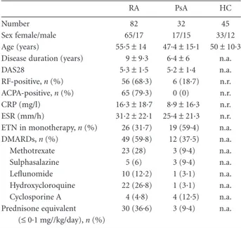
Table 1. Clinical and demographic data of enrolled patients at baseline
and healthy controls.
RA PsA HC
Number 82 32 45
Sex female/male 65/17 17/15 33/12
Age (years) 55·5± 14 47·4± 15·1 50 ± 10·3
Disease duration (years) 9± 9·3 6·4± 6 n.a.
DAS28 5·3± 1·5 5·2± 1·4 n.a.
RF-positive, n (%) 56 (68·3) 6 (18·7) n.r.
ACPA-positive, n (%) 65 (79·3) 0 (0) n.r.
CRP (mg/l) 16·3± 18·7 8·9± 16·3 n.r.
ESR (mm/h) 31·2± 22·1 25·4 ± 21·3 n.r.
ETN in monotherapy, n (%) 26 (31·7) 19 (59·4) n.a.
DMARDs, n (%) 49 (59·8) 12 (37·5) n.a. Methotrexate 23 (28) 3 (9·4) n.a. Sulphasalazine 5 (6) 3 (9·4) n.a. Leflunomide 10 (12·2) 1 (3·1) n.a. Hydroxycloroquine 22 (26·8) 1 (3·1) n.a. Cyclosporine A 4 (4·8) 4 (12·5) n.a. Prednisone equivalent (≤ 0·1 mg//kg/day), n (%) 30 (36·6) 3 (9·4) n.a.
Data are reported as mean± standard deviation. ACPA = anti-citrullinated peptide antibodies; CRP= C-reactive protein; DAS28 = disease activity score on 28 joints; DMARDs= disease modifying anti-rheumatic drugs; ESR= erythrosedimentation rate; ETN = etanercept; HC= healthy controls; n.a. = not applicable; n.r. = not reported; PsA= psoriatic arthritis; RA = rheumatoid arthritis; RF = rheumatoid factor.
Table 2. Clinical response and dropouts in rheumatoid arthritis and
psoriatic arthritis patients treated with etanercept during the follow-up.
Timing T14 weeks T22 weeks T54 weeks T102 weeks RA patients (n) 82 81 70 66 Responders (n/%) 60/73·2 58/71·6 49/70 43/65·2 No-responders (n/%) 22/26·8 23/28·4 21/30 23/34·8 Dropouts (n/%) 1/1·2 11/13·6 4/5·7 8/12·1 PsA patients (n) 32 32 29 27 Responders (n/%) 21/65·6 21/65·6 20/68·9 19/70·3 No-responders (n/%) 11/34·4 11/34·4 9/31·1 9/29·7 Dropouts (n/%) 0 3/9·4 2/6·9 2/7·4
PsA= psoriatic arthritis; RA = rheumatoid arthritis; T = time-point.
IU/ml according to the manufacturer’s instructions, and values above 20 IU/ml were considered positive. Extractable nuclear antigen (ENA) autoantibodies were measured by standard ELISA using the ENA-6 screen Enzyme Immuno-assay test kit (Diamedix) to detect IgG antibodies against the following antigens: Sm, Sm/nRNP, SS-A, SS-B, Scl-70 and Jo-1. Values above 20 IU/ml were considered positive according to the manufacturer’s instructions.
Peripheral blood cells
Peripheral blood mononuclear cells (PBMC) were isolated from heparinized blood samples of patients and HC by density-gradient centrifugation on Ficoll-Hypaque (Nycomed Pharma, Oslo, Norway) and resuspended in 1·5 ml phosphate-buffered saline (PBS)/1% bovine serum albumin (BSA) (1× 10⁶cells/100μl).
Immunophenotyping
Freshly isolated PBMC were stained with fluorescein isothiocyanate (FITC), phycoerythrin (PE), peridinin-chlorophyll protein (PerCP) or allophycocyanin (APC)-conjugated monoclonal antibodies (mAbs) specific for the following human cell surface markers: anti-CD3 (PerCP), CD56 (PE), CD16 (FITC), CD19 (PE), anti-CD45 (APC), anti-CD4 (FITC) and anti-CD8 (PE) or the fluorescence-conjugated isotype-matched controls (all pur-chased from BD Biosciences, Mountain View, CA, USA). Cells were incubated with a combination of mAb in PBS/1% BSA at+4°C for 20 min. Stained cells were washed twice in PBS/BSA and analysed using a fluorescence acti-vated cell sorter (FACS) Calibur flow cytometer (BD Biosciences, Oxford, UK) with FACS diva software (BD Biosciences). A total of 50 000–80 000 events were acquired for each sample. Peripheral blood NK cells were identified as CD45+CD56+CD16+cells, B cells as CD45+CD19+cells, T cells as CD45+CD3+CD4+cells and CD45+CD3+CD8+cells. Results were expressed as a percentage of total lymphocyte count. Cell counting was provided by the Laboratory Medi-cine service at the University of Tor Vergata by using auto-mated counts through flow cytometer. Blind cell counting of the blood samples of patients and controls was per-formed at each time-point.
Statistics
Normally distributed variables were expressed as the mean± standard deviation, and non-normally distributed variables were summarized using median, interquartile ranges (IQR) and confidence intervals (CI), unless specified differently. Statistical comparisons in patients and HC were performed using a non-parametric unpaired Mann– Whitney U-test. Median group values of different cell popu-lations were compared before and after the start of ETN in
RA and PsA patients using the non-parametric paired Wilcoxon’s signed-rank test. Univariate comparisons between nominal variables were performed by χ2 test.
Spearman’s rank order correlation test was used to assess the relationship between NK and B cell populations and disease activity scores. P-values< 0·05 were considered sig-nificant. All statistical analyses were performed using GraphPad Prism software version 4.
Results
NK cells in patients with RA and PsA
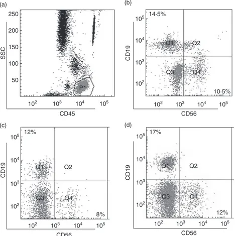
Figure 1 shows representative dot-plots of NK cells in a control subject (Fig. 1a,b) and in a patient with RA (Fig. 1c,d) before and after ETN treatment. The influence of gender and age on the number of NK cells was first ana-lysed. Neither the gender nor the age affected the number of NK cells in RA and PsA patients and in HC (data not shown). NK cell count at T0 was reduced significantly in RA patients (median 230 cells/μl, IQR 186–373·5 cells/μl) com-pared with HC (median 309·5 cells/μl, IQR 230–406·5 cells/ μl) (P = 0·01) (Fig. 2a). A significant increase in NK cell number was observed in RA patients during ETN treatment at T14 (P= 0·01), T22 (P < 0·01), T54 (P < 0·01) and T102 (P< 0·01) compared with T0 (Fig. 2a). During ETN treat-ment, the NK cell percentage increased at T22 (median 17%, IQR 12–20%) and T54 (median 17%, IQR 12·7– 22·2%) compared with T0 (median 14%, IQR 9·5–17%) (P= 0·04 and P = 0·01, respectively). Conversely, the NK cell percentage did not change at T14 (median 14%, IQR 10–21%) and T102 (median 17%, IQR 10–22%) compared with the baseline values. NK cell count was reduced signifi-cantly in PsA patients (median 210 cells/μl, IQR 145·5– 290·5 cells/μl) compared with HC (median 309·5 cells/μl, IQR 230–406·5 cells/μl) (P = 0·008) (Fig. 2b). During ETN treatment, PsA patients showed a significant increase in the NK cell number at T54 (P< 0·05) and T102 (P < 0·01) com-pared with T0 (Fig. 2b). Conversely, NK cell percentage did not change compared with T0 (T0 median 11·5%, IQR 8–17·7%; T14 median 15·5%, IQR 10·7–20·2%; T22 median 15%, IQR 10–20·5%; T54 median 14%, IQR 11–20·5%; T102 median 12%, IQR 12–22%).
There were no significant differences in the NK cell per-centage between HC (median 13·5%, IQR 10–18·2%) and both RA and PsA patients at T0.
DMARDs-free RA and PsA patients were analysed sepa-rately. NK cell number was reduced significantly in RA (median 223 cells/μl, IQR 202·5–235·5 cells/μl) and PsA patients (median 191 cells/μl, IQR 145·5–232 cells/μl) com-pared with HC (P= 0·002 and P = 0·004, respectively) (Fig. 2c,d). Moreover, in patients treated with ETN in monotherapy, a significant increase in NK cell number was observed in RA at T22 (P= 0·03), T54 (P < 0·0006) and
T102 (P= 0·005) (Fig. 2c) and in PsA compared with T0 (T0 versus T14 and T0 versus T54: P= 0·01, T0 versus T22:
P= 0·008, T0 versus T102: P = 0·004) (Fig. 2d). There was
no significant difference in the baseline NK cell percentage in DMARDs-free RA (median 14%, IQR 9–19%) and PsA patients (median 11·5%, IQR 8–16%) compared with HC
(median 13·5%, IQR 10–18·2%). Similarly, NK cell percent-age did not change in DMARDs-free RA and PsA patients during the follow-up (RA: T14 median 15·5%, IQR 12–18·7%; T22 median 17%, IQR 13–20%; T54 median 16% IQR 12·7–20·7%; T102 median 17%, IQR 14–21%; PsA: T14 median 17%, IQR 12–21·2%; T22 median 15·5%,
102 103 104 105 102 103 104 105 8% 12% Q2 Q4 CD56 CD19 102 103 104 105 14·5% 10·5% Q1 Q2 Q3 Q4 102 103 104 105 CD56 CD19 102 103 104 105 102 103 104 105 12% 17% Q2 Q1 Q3 Q1 Q3 Q4 CD56 CD19 50 102 103 104 105 P1 100 150 200 250 SSC CD45 (a) (b) (c) (d)
Fig. 1. Circulating CD16+CD56+natural killer (NK) and CD19+B cells in a healthy control (HC) and a rheumatoid arthritis (RA) patient. Representative examples of dot-plots from one HC (a,b) and one RA patient (c,d). (a) The gating strategy with the lymphocyte population identified by CD45 (indicated with P1). CD45+CD56+cells identified NK cells and CD45+CD19+cells showed B cells in a HC (b). (c) CD45+CD56+NK cells and CD45+CD19+B cells are shown in a representative RA patient before etanercept treatment. (d) CD45+CD56+ and CD45+CD19+cells of the same RA patient are shown after 102 weeks of etanercept treatment. 0 200 400 600 800 1000 1200 NK cells
(absolute number per
µ l) *** * ** ** 0 200 400 600 800 1000 1200 * * ** * ** NK cells
(absolute number per
µ l) 0 200 400 600 800 1000 1200 NK cells
(absolute number per
µ l) * * ** (a) (b) (c) (d) HC T0 T14 T22 T54 T102 HC T0 T14 T22 T54 T102 HC T0 T14 T22 T54 T102 HC T0 T14 T22 T54 T102 0 200 400 600 800 1000 1200 NK cells
(absolute number per
µ l) * ** ** * **
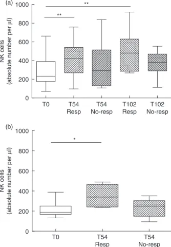
Fig. 2. Circulating natural killer (NK) cells
during etanercept treatment. Circulating NK cell numbers in treated rheumatoid arthritis (RA) (n= 82) (a), treated psoriatic arthritis (PsA) patients (n= 32) (b), disease-modifying anti-rheumatic drugs (DMARDs)-free RA (n= 26) (c) and DMARDs-free PsA (n = 19) (d) patients before and after etanercept at different time-points. Data registered in healthy controls (n= 45) are reported in each panel. Data are shown as box-plots; each box represents the 25th–75th percentiles; lines inside the box represent the median. The whiskers represent the 95% confidence interval (CI). Statistical analyses in patients and controls were performed using the Mann–Whitney U-test, while Wilcoxon’s signed-rank test was used to compare cells pre- and post-treatment. *P< 0·05; **P < 0·01; ***P < 0·001.
IQR 13·7–20·5%; T54 median 14·5% IQR 12–23·5%; T102 median 13·5%, IQR 12–25%).
Relationship between NK cells and disease activity during ETN treatment
A significant reduction of the disease activity evaluated with DAS28 was demonstrated in both RA (P< 0·0001 at all time-points) and PsA patients (P< 0·0001 at all time-points) during ETN treatment, together with the progres-sive increase of NK cell number (Fig. 3). No correlation was demonstrated between DAS28 and NK cell number at T0 in RA and PsA patients (data not shown).
A significant increase in NK cell number was observed in RA patients who reached a good–moderate EULAR response at T54 and T102 compared with those patients who did not reach a clinical response (T0 versus T54:
P= 0·002; T0 versus T102: P = 0·003) (Fig. 4a). In the same
manner, an increase in NK cell number was observed in PsA patients who reached a good–moderate response at T54 compared with that in PsA patients who did not reach a clinical response (T0 versus T54: P= 0·01) (Fig. 4b). No cor-relations were detected between NK cell number and CRP, ESR and DAS28 remission during ETN treatment in both RA and PsA patients (data not shown).
B cells in patients with RA and PsA
Figure 1 shows representative dot-plots of B cells in a control subject (Fig. 1b) and in a patient with RA (Fig. 1c,d) before and after ETN treatment. The influence of gender and age on the number of B cells was investigated. Neither gender nor age affected the number of B cells in RA and PsA patients and HC (data not shown). A significant reduction in B cell number was observed in RA patients at T0 (median 149 cells/μl, IQR 114–282 cells/μl) compared with HC (median 265·5 cells/μl, IQR 193·5–354 cells/μl) (P= 0·0003) (Fig. 5a). During the follow-up, an increase in B cell number was observed in RA patients at T14, T54 and T102 compared with T0 (P= 0·04 for all the considered time-points) (Fig. 5a). A significant reduction in B cell number was also detected in PsA patients at T0 (median 183 cells/μl, IQR 147·5–224 cells/μl) compared with HC
0 1 2 3 4 5 6 7 8 9 0 50 100 150 200 250 300 350 400 450 500 B NK DAS28 0 1 2 3 4 5 6 7 8 9 0 50 100 150 200 250 300 350 400 450 500 B NK DAS28 (a) (b) T0 T14 T22 T54 T102 T0 T14 T22 T54 T102 Cells µ I DAS28 Cells µ I DAS28
Fig. 3. Disease activity score on 28 joints (DAS28), circulating natural
killer (NK) and B cell numbers in patients treated with etanercept. DAS28 levels, circulating NK and B cell number in rheumatoid arthritis (a) and psoriatic arthritis patients (b) before and after 14, 22, 54 and 102 weeks of treatment with etanercept. Each value represents mean± standard error.
0 200 400 600 800 1000 NK cells
(absolute number per
µ l) ** ** 0 200 400 600 800 1000 NK cells
(absolute number per
µ l) * (a) (b) T0 T54 Resp T54 No-resp T102 Resp T102 No-resp T0 T54 Resp T54 No-resp
Fig. 4. Circulating natural killer (NK) cells in rheumatoid arthritis
(RA) and psoriatic arthritis (PsA) patients according to the clinical response. NK cell numbers in RA (a) and PsA (b) patients classified as responders (Resp) and no-responders (No-resp) after 54 and 102 weeks of treatment with etanercept. Data are shown as box-plots, where each box represents the 25th–75th percentiles; lines inside the box represent the median. The whiskers represent the 95% confidence interval (CI). Statistical analyses were performed using Wilcoxon’s signed-rank test. *P< 0·05; **P< 0·01.
(median 265·5 cells/μl, IQR 193·5–354 cells/μl) (P = 0·003) (Fig. 5b). PsA patients showed a significant increase in B cell number at T14 compared with T0 (P= 0·01) (Fig. 5b).
DMARDs-free RA and PsA patients were evaluated sepa-rately. A significant reduction in B cell count was detected in RA (median 189 cells/μl, IQR 135·5–314·5 cells/μl) and PsA patients (median 153 cells/μl, IQR 120–170 cells/μl) compared with HC (P= 0·03, P = 0·001, respectively) (Fig. 5c,d). In patients treated with ETN in monotherapy, a significant increase in B cell number was detected in RA (Fig. 5c) at all time-points (P< 0·05 for all time-points), as well as in PsA at T14 and T22 compared with T0 (P= 0·01 and P= 0·03, respectively) (Fig. 5d).
There were no significant differences in the percentage of B cells between HC (median 12%, IQR 8·7–15%) and the groups of patients at T0 (RA: median 9%, IQR 7–11·5%; PsA: median 9%, IQR 7–10%). Moreover, the percentage of B cells did not change during ETN treatment in all patients (RA: T14 median 10·5%, IQR 9·5–18·2%; T22 median 11·5%, IQR 8–14·5%; T54 median 12% IQR 9·2–15·7%; T102 median 11%, IQR 6·5–15%; PsA: T14 median 10·5%, IQR 9–19·5%; T22 median 16·5%, IQR 8·7–20·2%; T54 median 10% IQR 8·2–15·5%; T102 median 11·5%, IQR 6·5–16·5%).
Relationship between B cells and disease activity during ETN treatment
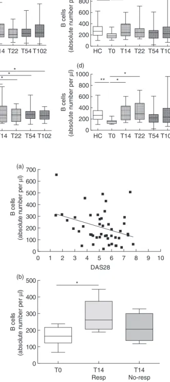
A negative correlation was detected between B cell number and DAS28 in RA patients at T0 (r= −0·34, P = 0·01) (Fig. 6a). In contrast, no correlation was demonstrated between DAS28 and B cell number in PsA patients (data not shown). There was no significant difference in B cell number when RA patients were stratified according to EULAR response criteria. Conversely, a significant increase
0 200 400 600 800 1000 B cells
(absolute number per
µ l) * * * * * 0 200 400 600 800 1000 B cells
(absolute number per
µ l) *** * * * 0 200 400 600 800 1000 B cells
(absolute number per
µ l) ** * (a) (b) (c) (d) HC T0 T14 T22 T54 T102 HC T0 T14 T22 T54 T102 HC T0 T14 T22 T54 T102 HC T0 T14 T22 T54 T102 0 200 400 600 800 1000 B cells
(absolute number per
µ
l)
* * **
Fig. 5. Circulating B cell numbers during
etanercept treatment. Circulating B cell numbers in treated rheumatoid arthritis (RA) (n= 82) (a), treated psoriatic arthritis (PsA) (n= 32) (b), disease-modifying anti-rheumatic drugs (DMARDs)-free RA (n= 26) (c) and DMARDs-free PsA patients (n= 19) (d) during etanercept treatment. Data registered in healthy controls (n= 45) are reported in each panel. Data are shown as box-plots, where each box represents the 25th–75th percentiles; lines inside the box represent the median. Whiskers represent the 95% confidence interval (CI). Statistical analyses in patients and controls were performed using the Mann–Whitney U-test, while Wilcoxon’s signed-rank test was used to compare cell population pre- and
post-treatment. *P< 0·05; **P < 0·01; ***P< 0·001. T0 T14 Resp T14 No-resp 0 100 200 300 400 500 B ce lls (a bs o lut e n u m b e r pe r µ l) * 0 1 2 3 4 5 6 7 8 9 10 0 100 200 300 400 500 600 700 DAS28 B ce lls (a bs ol ut e n u m be r p e r µ l) (a) (b)
Fig. 6. Circulating B cells and clinical outcome in rheumatoid
arthritis (RA) and psoriatic arthritis (PsA) patients. (a) Negative correlation between disease activity score on 28 joints (DAS28) levels and B cell numbers in RA patients (n= 82) before etanercept treatment. The correlation was assessed by Spearman’s rank order correlation test. (b) B cell numbers in responders and no-responders PsA patients after 14 weeks of treatment with etanercept. Data are shown as box-plots, where each box represents the 25th–75th percentiles; lines inside the box represent the median. Whiskers represent the 95% confidence interval (CI). Statistical analyses were performed using Wilcoxon’s signed-rank test. *P< 0·05.
in B cell number was detected in responder PsA patients compared with no-responders at T14 (T0 versus T14:
P= 0·01) (Fig. 6b). No correlations were detected between B
cell number and CRP, ESR and DAS28 remission during ETN treatment in both RA and PsA patients (data not shown).
NK and B cells in RA patients according to RF and ACPA status
RA patients were stratified into groups according to their RF status: RF-positive [n= 56, 43 women, 13 men, median age 55 (range 33–75) years, median disease duration 7 (range 1–40) years] and RF-negative patients [n= 26, 21 women, five men, median age 58 (range 19–76) years, median disease duration 5 (range 1–49) years]. The obser-vations described above concerning NK and B cell number/ percentage in RA patients at baseline and during the ETN treatment were confirmed in both RF-positive and -negative patients (data not shown). No differences occurred in both NK and B cell number/percentage between RF-positive and -negative patients (data not shown). RA patients were also stratified into groups accord-ing to their ACPA status: ACPA-negative patients [n= 17, 16 women, one man, median age 55 (range 19–76) years, median disease duration 8 (range 1–49) years] and ACPA-positive patients (n= 65, 49 women, 16 men, median age 58 (range 33–76) years, median disease duration 7 (range 1–40) years]. NK and B cell number/percentage were not affected by ACPA status at baseline and during the ETN treatment (data not shown).
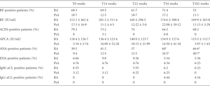
Autoantibodies
During the ETN treatment, a significant increase in the per-centage of ANA-positive RA patients was observed in both RA and PsA patients at T54 and T102 with respect to T0 (P< 0·05). There was no change in RF and ACPA serum levels in RA and PsA patients during ETN treatment, despite a trend towards a reduction in RF levels in RA patients. No modifications in aCL IgG /IgM and ENA anti-bodies were reported in ETN-treated RA and PsA patients during the follow-up (Table 3).
Peripheral blood T cells
There was no significant change in the count and percent-age of CD4+T cells, CD8+T cells and total CD3+T cells in both RA and PsA patients during ETN treatment. An increase in CD45+lymphocyte number was detected in RA patients at T102 (median 2449 cells/μl, IQR 1890–2700 cells/μl) compared with T0 (median 1890 cells/μl, IQR 1420–2325 cells/μl) (P = 0·006). Conversely, no significant change in CD45+lymphocyte number was observed in PsA patients at all time-points compared with baseline (data not shown).
Discussion
This is the first study, to our knowledge, where peripheral blood NK and B cells have been analysed prospectively in a long-term follow-up in RA and PsA patients during ETN treatment. We have demonstrated that RA and PsA patients,
Table 3. Autoantibody profile in rheumatoid arthritis and psoriatic arthritis patients during etanercept treatment
T0 weeks T14 weeks T22 weeks T54 weeks T102 weeks
RF-positive patients (%) RA 68·3 69·5 61·7 71·4 53 PsA 18·7 12·5 18·7 17·2 11·1 RF (IU/ml) RA 212·1± 461·4 201·2± 311·4 160± 296·5 174·6± 308·4 169·9± 263·8 PsA 17·3± 16·9 11·2± 4·5 12·22± 5·0 22·08± 39·12 11·13± 3·29 ACPA-positive patients (%) RA 79·3 73·2 74 64·2 68·2 PsA 0 3·1 0 6·8 0 APCA (IU/ml) RA 118·4± 126·7 136·4± 123·4 140·9± 123·7 134·9± 127·6 115·5± 112·7 PsA 3·18± 3·74 16·09± 52·28 10·15± 21·99 14·50± 41·18 3·97± 1·43 ANA-positive patients (%) RA 30·5 41·2 37 60a 66·6a PsA 9·4 12·5 12·5 34·5a 40·7a ENA-positive patients (%) RA 6·66 9·8 9·26 5·34 5·36 PsA 4·76 4·76 4·76 4·34 6·25
IgM–aCL positive patients (%) RA 6·35 6 5·55 6·2 6·23
PsA 3·12 3·12 6·25 6·25 0
IgG–aCL-positive patients (%) RA 0 0 0 4·44 4·16
PsA 0 0 0 0 0
Data are reported as mean± standard deviation or the percentage of positive patients. Statistical analyses between nominal variables in rheuma-toid arthritis and psoriatic arthritis patients were performed at all different time-points compared with T0 usingχ2test. aP< 0·05. aCL =
anti-cardiolipin antibodies; ACP= anti-citrullinated protein antibodies; ANA = anti-nuclear antibodies; ENA = extractable nuclear antigens; Ig= immunoglobulin; PsA = psoriatic arthritis; RA = rheumatoid arthritis; RF = rheumatoid factor; T = time-point.
both treated and DMARDs-free, showed lower levels of cir-culating NK and B cells compared with HC. This imbalance was restored after ETN treatment.
NK cells are prominent components of the innate immune response that can exert a disease-promoting or a disease-controlling role in immune-mediated diseases including RA and PsA [27–32]. An impaired NK activity has been demonstrated in RA, while peripheral blood NK cell number can be normal or reduced compared with healthy subjects [28,29,32]. Moreover, a positive correlation between NK cell cytotoxicity in vitro and disease activity has been described in PsA [33,34]. An abnormal distribution of peripheral B cell subsets has also been reported in both RA and PsA. Evidence has been reported that RA patients exhibit a reduction in both naive and memory B cell number compared with controls [35,36]. Different mecha-nisms may explain the reduction of peripheral NK cells and B lymphocytes observed in the present study, such as their recruitment into the inflamed target tissues (synovium and skin) and altered apoptosis [35–37]. Indeed, NK cells enrich RA synovial membrane and secrete proinflammatory cytokines such as TNF-α, interleukin (IL)-12, IL-18 and IL-15 [28]. In this study the reduction of peripheral blood NK and B cells seems to be restored during ETN treatment. We report an early and permanent increase up to normali-zation of NK and B cells in both DMARDs-free and treated RA patients. The same results occur in NK cell number in both DMARDs-free and treated PsA patients, while the modification in B cell number appears to be early, but tran-sient. These data may suggest the existence of a mechanism connecting the TNF-α pathway and NK and B cells in the peripheral blood. Previous evidence showed debated data about the effects exerted by TNF-α inhibitors on the immune cellular network in RA and PsA patients [38]. A recent study supports that ETN decreases CD69 expression on peripheral blood NK cells from healthy donors in vitro [39]. A significant reduction in CD27+memory B cell count has been detected in the peripheral blood from RA patients treated with ETN compared with patients treated with methotrexate and HC [40]. Furthermore, ETN neutralizes both TNF-α and LT-α that is crucial for B cell proliferation [41]. Thus, in our study, the effect of ETN on peripheral blood B cells may be partially associated with these interac-tions. According to our data, modification of NK and B cell number appears to be independent from the RF and the ACPA status in RA patients. This result is partially in con-trast with previous findings showing that memory B cells increased significantly only in RF-negative patients treated with anti-TNF-α [42]. However, it should be noted that we characterized all the CD19+cells, but did not characterize the memory B cell compartment. It is well known that treatment with anti-TNF-α agents may affect the white blood cell count in RA and PsA patients [5]. Thus, the modifications in NK and B cell number that we reported may be related to this issue, rather than a specific change in
these cell subsets. However, according to our data, CD4+T cells, CD8+T cells, CD3+T cells and CD45+cells did not change in both RA and PsA patients during treatment, sug-gesting that ETN could act directly on the NK and B cell subsets. Indeed, we reported an intriguing correlation between peripheral blood NK and B cells and disease activ-ity in RA and PsA patients. The increase of NK cell number occurs only in those patients who achieved a good– moderate clinical response, suggesting a relationship between peripheral blood cells levels and clinical outcome. Moreover, a negative correlation was demonstrated between B cell number and DAS28 in RA patients at baseline. Previ-ous study observed that DAS28 was correlated negatively with the number of regulatory B cells (Breg) in anti-TNF-
α-treated RA patients, indicating that Bregcells have a
poten-tially crucial role in controlling disease activity. Thus, the increase of the peripheral blood B cell number that we reg-istered might be associated with modification of the Breg
subset, as reported in the literature [43,44]. It is well estab-lished that anti-TNF drugs may stimulate the production of autoantibodies such as ANA, although the effect of ETN on RF, ACPA and aCL antibodies is debated [18,45–49]. A high prevalence of ANA-positive patients was detected during ETN treatment in both RA and PsA, while we did not find significant modification in RF, ACPA and aCL levels.
The present study suffers from several limitations. First, analysis of the NK and B cells relies on basic cell surface markers that should be further improved by using specific immunophenotyping and activity assays. Secondly, we ana-lysed only healthy subjects as controls, and did not include patients who were not treated with ETN; thus, the observed changes in cell populations could not be ascribed defini-tively to ETN itself.
Acknowledgements None.
Disclosures
The authors declare that there are no conflicting financial interests and they received no financial support for this study.
References
1 Lee DM, Weinblatt ME. Rheumatoid arthritis. Lancet 2001;
358:903–11.
2 Mease PJ, Goffe BS, Metz J, VanderStoep A, Finck B, Burge DJ. Etanercept in the treatment of psoriatic arthritis and psoriasis: a randomised trial. Lancet 2000; 356:385–90.
3 van Kuijk AW, Tak PP. Synovitis in psoriatic arthritis: immunohistochemistry, comparisons with rheumatoid arthritis, and effects of therapy. Curr Rheumatol Rep 2011; 13:353–9. 4 Takeuchi T, Miyasaka N, Tatsuki Y et al. Baseline tumour necrosis
infliximab therapy in patients with rheumatoid arthritis. Ann Rheum Dis 2011; 70:1208–15.
5 Feldmann M. What is the mechanism of action of anti-tumour necrosis factor-alpha antibody in rheumatoid arthritis? Int Arch Allergy Immunol 1996; 111:62–5.
6 Moreland LW, Baumgartner SW, Schiff MH et al. Treatment of rheumatoid arthritis with a recombinant human tumor necrosis factor receptor (p75)-Fc fusion protein. N Engl J Med 1997;
337:141–7.
7 Weinblatt ME, Kremer JM, Bankhurst AD et al. A trial of etanercept, a recombinant tumor necrosis factor receptor:Fc fusion protein, in patients with rheumatoid arthritis receiving methotrexate. N Engl J Med 1999; 340:253–9.
8 Jarvis B, Faulds D. Etanercept: a review of its use in rheumatoid arthritis. Drugs 1999; 57:945–66.
9 Chimenti MS, Perricone C, Graceffa D et al. Complement system in psoriatic arthritis: a useful marker in response prediction and monitoring of anti-TNF treatment. Clin Exp Rheumatol 2012;
30:23–30.
10 Mitoma H, Horiuchi T, Tsukamoto H et al. Mechanisms for cytotoxic effects of anti-tumor necrosis factor agents on transmembrane tumor necrosis factor alpha-expressing cells: comparison among infliximab, etanercept, and adalimumab. Arthritis Rheum 2008; 58:1248–57.
11 Horiuchi T, Mitoma H, Harashima SI, Tsukamoto H, Shimoda T. Transmembrane TNF-α: structure, function and interaction with anti-TNF agents. Rheumatology (Oxf) 2010; 49:1215–28. 12 Harashima S, Horiuchi T, Hatta N et al. Outside-to-inside signal
through the membrane TNF-alpha induces E-selectin (CD62E) expression on activated human CD4+ T cells. J Immunol 2001;
166:130–6.
13 Klimiuk PA, Sierakowski S, Domyslawska I, Chwiecko J. Effect of etanercept on serum levels of soluble cell adhesion molecules (sICAM-1, sVCAM-1, and sE-selectin) and vascular endothelial growth factor in patients with rheumatoid arthritis. Scand J Rheumatol 2009; 38:439–44.
14 Zivojinovic SM, Pejnovic NN, Sefik-Bukilica MN, Kovacevic LV, Soldatovic II, Damjanov LF. Tumor necrosis factor blockade dif-ferentially affects innate inflammatory and Th17 cytokines in rheumatoid arthritis. J Rheumatol 2012; 39:18–21.
15 Caproni M, Antiga E, Melani L, Volpi W, Del Bianco E, Fabbri P. Serum levels of IL-17 and IL-22 are reduced by etanercept, but not by acitretin, in patients with psoriasis: a randomized-controlled trial. J Clin Immunol 2009; 29:210–4.
16 Wang F, Smith N, Maier L et al. Etanercept suppresses regenerative hyperplasia in psoriasis by acutely downregulating epidermal expression of interleukin (IL)-19, IL-20 and IL-24. Br J Dermatol 2012; 167:92–102.
17 Esposito M, Giunta A, Mazzotta A et al. Efficacy and safety of sub-cutaneous anti-tumor necrosis factor-alpha agents, etanercept and adalimumab, in elderly patients affected by psoriasis and psoriatic arthritis: an observational long-term study. Dermatology 2012;
225:312–9.
18 Di Muzio G, Perricone C, Ballanti E et al. Complement system and rheumatoid arthritis: relationships with autoantibodies, serologi-cal, clinical features, and anti-TNF treatment. Int J Immunopathol Pharmacol 2011; 24:357–66.
19 Ballanti E, Perricone C, Di Muzio G et al. Role of the complement system in rheumatoid arthritis and psoriatic arthritis: relationship with anti-TNF inhibitors. Autoimmun Rev 2011; 10:617–23.
20 Catrina AI, Klint EA, Ernestam S et al. Anti-tumor necrosis factor therapy increases synovial osteoprotegerin expression in rheuma-toid arthritis. Arthritis Rheum 2006; 54:76–81.
21 Leandro MJ. Anti-tumor necrosis factor therapy and B cells in rheumatoid arthritis. Arthritis Res Ther 2009; 11:128.
22 Moreland LW, Bucy RP, Weinblatt ME, Mohler KM, Spencer-Green GT, Chatham WW. Immune function in patients with rheumatoid arthritis treated with etanercept. Clin Immunol 2002; 103:13–21.
23 Arnett FC, Edworthy SM, Bloch DA et al. The American Rheumatism Association 1987 revised criteria for the classifi-cation of rheumatoid arthritis. Arthritis Rheum 1988; 31: 315–24.
24 Taylor W, Gladman D, Helliwell P, Marchesoni A, Mease P, Mielants H, CASPAR Study Group. Classification criteria for pso-riatic arthritis: development of new criteria from a large interna-tional study. Arthritis Rheum 2006; 54:2665–73.
25 Prevoo ML, van’t Hof MA, Kuper HH, van Leeuwen MA, van de Putte LB, van Riel PL. Modified disease activity scores that include twenty-eight-joint counts. Development and validation in a pro-spective longitudinal study of patients with rheumatoid arthritis. Arthritis Rheum 1995; 38:44–8.
26 van Gestel AM, Haagsma CJ, van Riel PL. Validation of rheuma-toid arthritis improvement criteria that include simplified joint counts. Arthritis Rheum 1998; 41:1845–50.
27 Manda G, Neagu M, Livescu A, Constantin C, Codreanu C, Radulescu A. Imbalance of peripheral B lymphocytes and NK cells in rheumatoid arthritis. J Cell Mol Med 2003; 7:79–88.
28 Conigliaro P, Scrivo R, Valesini G, Perricone R. Emerging role for NK cells in the pathogenesis of inflammatory arthropathies. Autoimmun Rev 2011; 10:577–81.
29 Perricone R, Perricone C, De Carolis C, Shoenfeld Y. NK cells in autoimmunity: a two-edged weapon of the immune system. Autoimmun Rev 2008; 7:384–90.
30 Perricone R, Perricone C, Shoenfeld Y. Autoimmunity: when the immune system becomes the self-ish giant. Autoimmun Rev 2011;
10:575–6.
31 Mukai E, Nagashima M, Hirano D, Yoshino S. Comparative study of symptoms and neuroendocrine–immune network mediator levels between rheumatoid arthritis patients and healthy subjects. Clin Exp Rheumatol 2000; 18:585–90.
32 Aramaki T, Ida H, Izumi Y et al. A significantly impaired natural killer cell activity due to a low activity on a per-cell basis in rheu-matoid arthritis. Mod Rheumatol 2009; 19:245–52.
33 Massari D, Prpic-Massari L, Kehler T et al. Analysis of granulysin-mediated cytotoxicity in peripheral blood of patients with psori-atic arthritis. Rheumatol Int 2012; 32:2777–84.
34 Spadaro A, Scrivo R, Moretti T et al. Natural killer cells and gamma/delta T cells in synovial fluid and in peripheral blood of patients with psoriatic arthritis. Clin Exp Rheumatol 2004;
22:389–94.
35 Sellam J, Rouanet S, Hendel-Chavez H et al. Blood memory B cells are disturbed and predict the response to rituximab in patients with rheumatoid arthritis. Arthritis Rheum 2011; 63:3692–701. 36 Souto-Carneiro MM, Mahadevan V, Takada K et al. Alterations in
peripheral blood memory B cells in patients with active rheuma-toid arthritis are dependent on the action of tumour necrosis factor. Arthritis Res Ther 2009; 11:R84.
37 Eggleton P, Harries LW, Alberigo G et al. Changes in apoptotic gene expression in lymphocytes from rheumatoid arthritis and
systemic lupus erythematosus patients compared with healthy lymphocytes. J Clin Immunol 2010; 30:649–58.
38 Benucci M, Saviola G, Manfredi M, Sarzi-Puttini P, Atzeni F. Tumor necrosis factors blocking agents: analogies and differences. Acta Biomed 2012; 83:72–80.
39 Thaher F, Plankenhorn S, Klein R. Differential effects of the tumor necrosis factor alpha-blocker infliximab and etanercept on immunocompetent cells in vitro. Int Immunopharmacol 2011;
11:1724–31.
40 Anolik JH, Ravikumar R, Barnard J et al. Cutting edge: anti-tumor necrosis factor therapy in rheumatoid arthritis inhibits memory B lymphocytes via effects on lymphoid germinal centers and follicular dendritic cell networks. J Immunol 2008; 180:688–92. 41 Chiang EY, Kolumam G, McCutcheon KM et al. In vivo depletion
of lymphotoxin-alpha expressing lymphocytes inhibits xenogeneic graft-versus-host-disease. PLOS ONE 2012; 7:e33106.
42 Roll P, Muhammad K, Schumann M, Kleinert S, Tony HP. RF positivity has substantial influence on the peripheral memory B-cell compartment and its modulation by TNF inhibition. Scand J Rheumatol 2012; 41:180–5.
43 Ma L, Liu B, Jiang Z, Jiang Y. Reduced numbers of regulatory B cells are negatively correlated with disease activity in patients with new-onset rheumatoid arthritis. Clin Rheumatol 2014; 33:187–95.
44 Karampetsou MP, Andonopoulos AP, Liossis SN. Treatment with TNFα blockers induces phenotypical and functional aberrations in peripheral B cells. Clin Immunol 2011; 140:8–17.
45 Valesini G, Iannuccelli C, Marocchi E, Pascoli L, Scalzi V, Di Franco M. Biological and clinical effects of anti-TNFalpha treatment. Autoimmun Rev 2007; 7:35–41.
46 De Rycke L, Baeten D, Kruithof E, Van den Bosch F, Veys EM, De Keyser F. The effect of TNFalpha blockade on the antinuclear antibody profile in patients with chronic arthritis: biological and clinical implications. Lupus 2005; 14:931–7.
47 Yazdani-Biuki B, Stadlmaier E, Mulabecirovic A et al. Blockade of tumor necrosis factor alpha significantly alters the serum level of IgG- and IgA-rheumatoid factor in patients with rheumatoid arthritis. Ann Rheum Dis 2005; 64:1224–6.
48 Chen HA, Lin KC, Chen CH et al. The effect of etanercept on anti-cyclic citrullinated peptide antibodies and rheumatoid factor in patients with rheumatoid arthritis. Ann Rheum Dis 2006;
65:35–9.
49 Jonsdottir T, Forslid J, van Vollenhoven A et al. Treatment with tumour necrosis factor alpha antagonists in patients with rheuma-toid arthritis induces anticardiolipin antibodies. Ann Rheum Dis 2004; 63:1075–8.