Facoltà di Scienze matematiche, fisiche e naturali Corso di Laurea Specialistica in Neurobiologia
Post translational modifications in the GDNF molecule:
functional characterization
Candidata Relatori Correlatori
Elisa Piccinini Prof. Mart Saarma Prof.ssa Marcella Camici Prof.ssa Mercedes Garcia Gil Dott. Matteo Caleo
-
Anno accademico 2009/2010 Luglio 2010
To two of the simplest and yet most wonderful people I've ever known. Unfortunately, you can no longer read these words.
INDEX
Abbreviations used page i
Riassunto page iii
Abstract page v
Introduction page 1
Parkinson's disease page 2
Locations of Parkinson's disease-related lesions page 2 Organization of the motor system and the limbic system page 5
Symptoms page 7
Aetiology page 9
Therapy page 14
Drug treatment page 14
Stem cells page 17
Gene therapy page 18
Neurotrophic factors page 19
GDNF, its receptor complex and signalling page 22
What are neurotrophic factors? page 22
Neurotrophins page 22
Neurokines page 24
MANF family page 24
GFLs page 25 Artemin page 28 Persephin page 28 Neurturin page 29 GDNF page 29 GFRαs page 34 RET page 36
GFL-GFRα-RET complex page 40
Signalling page 42
Related diseases page 45
Hirschsprung’s disease page 46
Medullary thyroid carcinoma and papillary thyroid carcinoma page 47
Parkinson’s disease page 48
Aims of the study page 52
Materials and methods page 54
Computational analysis page 55
Production of GDNF variants page 55
Mutagenic PCR page 55
Plasmid phosphorylation and ligation page 57
Transformation of E. coli cells page 57
Plasmid purification page 58
Verifying the presence of the mutation page 58
Cell culture page 58
CHO cells page 58
MG87-RET cells page 58
Transfection page 59
Transfection of CHO cells, 60 mm plates page 59
Transfection of CHO cells, 24-well plate page 59
Cotransfection page 59
Glycosylation of GDNF page 59
Treatment with PNGase F page 59
Treatment with tunicamycin page 60
Cell lysates page 60
60 mm plates page 60
24-well plate page 60
RET phosphorylation assay page 60
Starvation page 61
Induction page 61
Lysis page 61
Immunoprecipitation page 61
Western blot page 61
Detection conditions page 61
Antibodies page 62
GDNF detection page 62
FLAG tag detection page 62
RET detection page 62
Phosphorylated RET detection page 63
Stripping page 63
Treatment with commercial furin page 63
Incubation of GDNF at 37 oC page 63
Results page 64
Computational analysis page 65
Glycosylation of GDNF page 67
Treatment with PNGase F page 67
Treatment with tunicamycin page 68
GDNF variants: size comparison page 69
Specificity for α receptors page 71
Mutant on the 1st glycosylation site with prosequence page 71
Mutant on the 1st glycosylation site without prosequence page 71
α1 compared with α2 page 72
α1 compared with α3 page 73
α1 compared with α4 page 73
Stability of GDNF page 74
Stability at 37 oC page 74
Furin cleavage page 75
Coexpression with furin page 76
Treatment with commercial furin page 77
Activity of GDNF variant with mutation on the furin cleavage site page 77
Affinity for GFRα1 page 77
Duration of RET phosphorylation page 78
Interpretation of banding pattern page 78
Discussion page 81
Glycosylation of GDNF page 82
Furin cleavage site page 84
i
ABBREVIATIONS USED
6-OHDA 6-hydroxydopamine
11C-DTBZ 11C-(+)-α-dihydrotetrabenazine
18F-DOPA 3.4-dihydroxyphenylalanine marked with radioactive fluorine 125I-GDNF glial cell line-derived neurotrophic factor marked with
radioactive iodine
AADC aromatic L-amino acid decarboxylase
ALDH aldehyd dehydrogenase
ARTN artemin
BDNF brain derived neurotrophic factor
BH4 6-tetrahydrobiopterin
bHLH basic helix-loop-helix
cAMP cyclic adenosine monophosphate
CDNF conserved dopamine neurotrophic factor
CNS central nervous system
COMT catechol-O-methyltransferase
DA dopamine
DOPAC 3.4-dihydroxyphenylacetic acid EGFR epidermal growth factor receptor
En-1 engrailed-1
ER endoplasmic reticulum
ES embryonic stem cells
ETF epidermal growth factor receptor transcription factor
Fgf8 fibroblast growth factor 8
FMTC familial medullary thyroid carcinoma
GABA gamma-amino butyric acid
GAG glycosaminoglycan
GDNF glial cell line-derived neurotrophic factor
GFL GDNF family ligand
GFRα GFL receptor α
gp130 glycoprotein 130
GPe external pallidum
GPi internal pallidum
GPI glycophospatidylinositol
GRB2 growth factor receptor-bound protein 2
IL-6 interleukin 6
HS heparan sulphate
HVA homovanillic acid
JNK Jun N-terminal kinase
L-DOPA L-3.4-dihydroxyphenylalanine
LAR leukocyte common antigen-related protein
LIF leukemia inhibitory factor
LIFR LIF receptor
LRRK2 leucine-rich repeat kinase 2
MANF mesencephalic astrocyte-derived neurotrophic factor
MAO monoamine oxidase
Mash-1 mammalian homologues of achaete-scute 1
MEN multiple endocrine neoplasia
MPDP+ 1-methyl-4-phenyl-2.3-dihydropyridinium
ii
NAG N-acetyl glucosamine
NCAM neural cell adhesion molecule
NEC1/NEC2 proprotein convertase 1 / proprotein convertase 2
NGF nerve growth factor
NMDA N-methyl-D-aspartate
NT-3 neurotrophin-3
NT-4/5 neurotrophin-4/5
NRTN neurturin
Nurr1 Nuclear receptor related 1
PC7 proprotein convertase 7
PD Parkinson's disease
PDGFs platelet derived-growth factors
PET positron emission tomography
PINK-1 PTEN-induced putative kinase 1
PKA protein kinase A
PNGase F peptide N-glycosidase F
PSPN persephin
PTPRJ receptor-type protein tyrosine phosphatise J
RET rearranged during transfection
ROS reactive oxygen species
SHC Src homology 2 domain-containing protein
SKI1 subtilisin/kexin isozyme 1
Shh sonic hedgehog
SNc substantia nigra pars compacta
SNr substantia nigra pars reticolata
SOS sucrose octasulphate
Sox Sry-related HMG box
SPCs subtilisin-like proprotein converetases
STN subthalamic nucleus
TGFβ transforming growth factor β
TH tyrosine hydroxylase
Trk tropomyosin-related kinase
UCH-L1 ubiquitin carboxy-terminal hydrolase L1 UDP-GlcNAc uridine diphosphate N-acetyl glucosamine
iii
Post translational modifications in the GDNF molecule: functional characterization
La malattia di Parkinson è caratterizzata dalla degenerazione dei neuroni dopaminergici. Il GDNF (Glial cell line Derived Neurotrophic Factor) è stato scoperto inizialmente per le sue proprietà neurotrofiche su neuroni dopaminergici e, nonostante le sue funzioni anche esterne al sistema nervoso centrale, l’interesse clinico si è concentrato sul suo potenziale nel trattamento della mallatia di Parkinson. È stato dimostrato che il GDNF ha effetti protettivi e neurorestorativi in diversi modelli animali di Parkinson e questa molecola ha inoltre mostrati risultati promettenti in due trial clinici su tre.
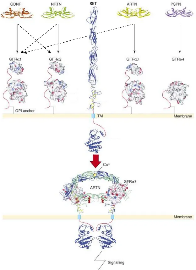
Il GDNF appartiene alla famiglia dei GFL (GDNF Family Ligands). Gli altri membri della famiglia sono neurturina, artemina e persefina. In presenza dei corecettori GFRα1-4 (GDNF Family Receptor), i GFL possono attivare il recettore tirosin chinasico RET (Rearranged during Transfection). Il GDNF segnala preferenzialmente attraverso il corecettore GFRα1.
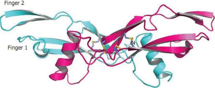
A causa dell’interesse terapeutico, gran parte della ricerca si è focalizzata su come viene formato il complesso recettoriale, dove sono espresse le varie componenti e quali cascate intracellulari vengono attivate da RET. Al contrario, si sa molto poco su come il GDNF è sintetizzato, trasportato e degradato. Il GDNF è sintetizzato come una preproproteina, ed è stato determinato l’N-terminale della proteina matura, che viene secreta. La molecola è anche stata cristallizzata: è dimerica, con sette ponti cisteinici. È anche ben documentato che il GDNF è N-glycosylato, ma i dettagli e il ruolo di questa glycosylazione non sono stati studiati.
In questo lavoro ho eseguito un’analisi computazionale delle potenziali modifice post traduzionali nel GDNF in diverse specie, e ho anche comparato i quattro membri della famiglia dei GFL. I due potenziali siti di N-glycosylazione sono conservati nei mammiferi, negli uccelli, in Xenopus e nei pesci. Il primo sito di N-glycosylazione si sul primo “dito” della molecola, vicino a residui coinvolti nel legame al recettore, e perciò potrebbe essere coinvolto nella specificità della molecola per il GFRα1. Il secondo sito si trova vicino al “polso”, fra le due “dita”. È interessante notare come tutti i GFL (tranne la persefina che è il membro più divergente della famiglia) abbiano un potenziale sito di taglio da parte della furina nella stessa regione.
Ho preparato mutanti a cui mancava il primo sito di glycosylazione o sia il primo che il secondo sito, mentre varianti con la mutazione del solo secondo sito, del sito di taglio della furina o di entrambi questi due ultimi siti erano già stati sintetizzati.
Ho analizzato le diverse varianti dopo averle fatte esprimere in cellule CHO, usando Western blot con anticorpi anti-GDNF. L’analisi dell’espressione del GDN in presenza o assenza di tunicamycina rivela che solo il primo dei due potenziali siti di N-glycosylazione è in uso. Comunque saggi di fosforilazione di RET mostrano che questo sito non cambia la specificità del GDNF nei confronti dei diversi GFRα: la glycosylazione su questo sito sembra influire maggiormente sul processamento della proteina, che è apparentemente secreta come proGDNF e in quantità minore (mentre l’ammontare di proteina intracellulare è comparabile con quello del GDNF wild type). In realtà, in questo caso il livello di fosforilazione di RET è più basso di quello indotto dal wild type, ma questo non è causato dalla mancanza di glycosylazione. Ho anche prodotto infatti un mutante sul primo sito di glycosylazione che manca della sequenza pro: in questo caso la fosforilazione di RET era allo stesso livello di quella indotta dal wild type, nonostante l’ammontare di questa variante di GDNF nel medium fosse ancora minore rispetto a quella del mutante sul primo sito di N-glycosylazione con la sequenza pro.
È difficile dire quanto la mancanza di glycosylazione influenzi la stabilità di questi mutanti, a causa del fatto le la loro quantità nel medium è minore rispetto a quella del wild type, ma è comunque possibile vedere che questi mutanti sono più stabili rispetto
iv al GDNF ricombinante prodotto in E. coli, che manca sia di glycosylazione che di sequenza pro, che è stato usato nei trials clinici.
Il sito di taglio da parte della furina si trova immediatamente dopo l’α-elica e secondo le analisi computazionali è conservato nei mammiferi, uccelli e nello Xenopus. Comunque questo sito sembra non essere in uso durante la secrezione della molecola, poiché quando il GDNF è coespresso in cellule CHO insieme alla furina (sia full-lenght che solubile), solo le bande che si pensa corrispondano al proGDNF (glycosylato e non); inoltre non si osservano differenze fra il GDNF wild type e il mutante sul sito previsto di taglio della furina. Gli stessi risultati sono stati ottenuti quando il GDNF è stato digerito con furina commerciale.
Il sito sembra non essere in uso anche quando il GDNF si lega al complesso recettoriale, infatti sia la durata che i livelli di fosforilazione indotti dal mutante sul sito furinico sono gli stessi di quelli indotti dal wild type.
v
Post translational modifications in the GDNF molecule: functional characterization
Parkinson’s disease is characterised by a degeneration of dopaminergic neurons. The glial-cell-line-derived neurotrophic factor (GDNF) was first discovered as a neurotrophic factor for dopaminergic neurons and, in spite of its functions also outside the central nervous system, the clinical interest in this protein has focused on the potential of GDNF in the treatment of Parkinson’s disease. GDNF has been shown to have protective and neurorestorative effects in several animal models of the Parkinson’s disease, and GDNF has also showed promising results in two out of three clinical trials.
GDNF belongs to the GDNF family ligands (GFLs). The other members of this family are the highly homologous neurturin, artemin and persephin. In the presence of the co-receptors GDNF family receptors α 1-4 (GFRα1-4) the GFLs can activate the receptor tyrosine kinase RET (REarranged during Transfection). GDNF signals preferentially through GFRα1.
Due to the clear clinical interest in GDNF, a lot of research has focused on how the receptor complex is formed, where the ligand-receptor components are expressed, and what intracellular signalling cascades as well as biological functions RET mediates. On the contrary, very little is known on how GDNF is synthesised, transported and degraded. GDNF is synthesised as a preproprotein, and the N-terminus of the secreted mature protein has been determined. The crystal structure of GDNF shows that the protein is dimeric, with seven cystein bridges. It is also well documented that GDNF is N-linked glycosylated, but the details and role of this glycosylation has not been studied.
In this study I have made a computational mapping of the potential post-translational modifications in GDNF from different species. I have also compared the four human GFLs. I have found that the two potential N-linked glycosylation sites in GDNF are conserved in mammalian, birds and frog. The first of the glycosylation sites is located close to the receptor-binding finger tip, and may therefore be involved in restricting the specificity of GDNF to GFRα1. The second glycosylation site is located adjacent to the “hinge” region, between the two receptor-binding “finger structures”. Interestingly all the human GFLs (except persephin, which is however the most divergent member of the family) harbour a potential furin cleavage site in the same hinge region.
I prepared GDNF mutants which lack the first predicted glycosylation site or both the first and the second sites, while variants lacking the second glycosylation site, the furin cleavage site, or both the second glycosylation site and the furin site had already been synthesized.
I have analyzed the variants after expression in CHO cells, using Western blotting with anti-GDNF antibodies. Expression analysis in the presence or absence of tunicamycin shows that only one of the two potential glycosylation sites is in use. This site is close to the receptor binding finger tip. However, the results from RET phosphorylation assays show that this glycosylation site does not change the specificity of GDNF with regard to the different GFRα receptors: glycosylation on the first site seems to affect mainly the processing of the protein which is apparently secreted in its proform and in a lower amount (while its quantity inside the cell is comparable to the one of wild type GDNF). In this case, actually, the phosphorylation level of RET is lower than the one obtained with wild type, but this is not caused by lack of glycosylation or amount of GDNF in the medium: when a mutant on the first glycosylation site lacking the prosequence was created, levels of RET phosphorylation were the same as the ones induced by wild type, despite the amount of this GDNF variant in the medium being even lower than the amount of the mutant on the first glycosylation site.
It is difficult to tell how much the stability of these mutants is affected by the lack of glycosylation, since their amount in the medium is much lower than the amount of wild type, but still it is possible to see that these variants are still more stable than
vi recombinant GDNF produced in E. coli which lacks both glycosylation and prosequence and that has been used in clinical trials.
The putative furin cleavage site is located immediately after the α-helix and according to computational analysis it is conserved in mammals, birds and X. laevis. However this site seems not to be in use during secretion, since when wild type GDNF was coexpressed with either full-length or soluble furin only the bands that are thought to correspond to proGDNF (either glycosylated or not) were affected; moreover no differences were observed between wild type GDNF and a variant with mutation of the putative furin cleavage site. The same results were obtained when digestion with commercial furin was performed.
The furin site in not in use even when GDNF binds to the GFRα-RET complex, since both the duration and the amount of phosphorylation induced by this mutant are the same as the ones induced by wild type.
2
Parkinson's disease
Locations of Parkinson's disease-related lesionsParkinson's disease (PD) is a neurodegenerative disease that is, according to the classical view, characterized by the loss of dopaminergic neurons projecting from the substantia nigra pars compacta to the striatum. It affects the human central, peripheral and enteric nervous system, and progresses during time. Its diagnosis is very difficult and usually requires a post-mortem verification, despite the characteristic symptoms of the disease.1 Moreover, these symptoms appear only in a late stage, when the majority of the dopaminergic neurons have deteriorated.
PD progression can be divided in six stages, and in each of them there is the de-velopment of characteristic inclusions named Lewy bodies and of Lewy neurites. Lewy neurites are composed by abnormally phosporylated neurofilaments, while Lewy bodies are aggregates of proteins containing misfolded α-synuclein fibrils together with other proteins such as ubiquitin. α-synuclein is usually located in the axon and in the presinap-tic buttons, it is soluble but usually binds to membranes with a high affinity. Sometimes, and the reason why is still under research, α-synuclein loses its affinity with the mem-branes and takes on a β-sheet conformation and aggregates with similarly misfolded α-synuclein molecules, ubiquitin, syniphilin-1 and phosporylated neurofilaments, forming Lewy bodies. It remains unclear why cells fail to eliminate the misfolded α-synuclein with ubiquitination and proteasome recycling.2
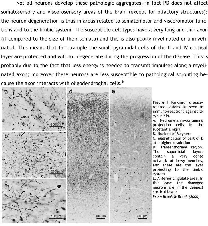
Lewy bodies can be found not only in PD, but also in amyotrophic lateral sclerosis, Hallervorden-Spatz disease and multiple system atrophy. However, in PD, Lewy bodies and Lewy neurites are markers present in specific cell types at predisposed sites.3 Some cells affected by PD are the neuromelanin-containing projection cells in the pars com-pacta of the subtantia nigra, the neurons of the basal nucleus of Meynert, of the tran-senthorinal region, and in the deep layers of the anterior cingulate areas (see Figure 1).4 Later post mortem studies have shown that the earliest Lewy bodies form in the olfac-tory bulb, in the anterior olfacolfac-tory nucleus, lipofuscin-containing projection cells of the dorsal IX/X motor nucleus and dorsal IX/X pigmented nucleus, of the intermedial
1 Braak H. & Braak E., Pathoanatomy of Parkinson's disease, J Neurol (2000) 247: II/3-II/10
2 Braak H. et al., Stages in the development of Parkinson's disease-related pathology, Cell Tissue Res (2004) 318: 121-134
3 Del Tredici K. et al., Where does Parkinson Desease Pathology Begin in the Brain?, Journal of Neuropa-thology and Experimental Neurology (2002) 61: 413-426
3 late zone and in melanoneurons of the coeruleus-subcoeruleus complex. These are fol-lowed by the lipofuscin-containing nerve cells of the dorsomedial nucleus of the solitary tract, gigantocellular nucleus of the reticular formation, and caudal Raphe group. This study also showed that the affection of the A6/A7 melanin-laden nerve cells not only ex-ceeds but also precedes that of the substantia nigra (see Figure 2), therefore we can say that the classical view is somehow incomplete. 5
Not all neurons develop these pathologic aggregates, in fact PD does not affect somatosensory and viscerosensory areas of the brain (except for olfactory structures): the neuron degeneration is thus in areas related to somatomotor and visceromotor func-tions and to the limbic system. The susceptible cell types have a very long and thin axon (if compared to the size of their somata) and this is also poorly myelinated or unmyeli-nated. This means that for example the small pyramidal cells of the II and IV cortical layer are protected and will not degenerate during the progression of the disease. This is probably due to the fact that less energy is needed to transmit impulses along a myeli-nated axon; moreover these neurons are less susceptible to pathological sprouting be-cause the axon interacts with oligodendroglial cells.6
5 Del Tredici K. et al., Where does Parkinson Disease Pathology Begin in the Brain?, Journal of Neuropa-thology and Experimental Neurology (2002) 61: 413-426
6 Braak H. et al., Stages in the development of Parkinson's disease-related pathology, Cell Tissue Res (2004) 318: 121-134
Figure 1. Parkinson disease-related lesions as seen in immuno-reactions against α-synuclein.
A. Neuromelanin-containing projection cells in the substantia nigra.
B. Nucleus of Meynert C. Magnification of part of B at a higher resolution D. Transenthorinal region. The superficial layers contain a very dense network of Lewy neurites, and these are the layer projecting to the limbic system.
E. Anterior cingulate area. In this case the damaged neurons are in the deepest cortical layers.
4 Figure 2. Lesions caused by Parkinson Disease detected using α-synuclein immunoreactions.
A. Overview of the dorsal IX/X complex . Arrowheads indicate Lewy neurites, arrows indicate Lewy bodies and open arrowheads indicate Lewy neurites within intramedullary axons of the vagus nerve.
B. Large visceromotor neuron of the dorsal motor nucleus containing both Lewy neurites (arrowhead) and Lewy bodies (arrow).
C. The inferior salivatory nucleus constitutes the rostral continuation of the dorsal motor nucleus and displays isolated Lewy neurites (arrowheads) and/or Lewy bodies (arrow).
D. α-synuclein inclusion bodies (arrowheads) in the lateral fringe subnucleus of the dorsal IX/X pigmented nucleus. E. The cells in the area postrema are free of lesions. Arrows indicate Lewy bodies and arrowheads Lewy neurites. F, G. In the mildest cases, the lesions in the dorsal IX/X motor nucleus are asymmetrical and can consist of a single Lewy neurite.
H. Lewy neurites (arrowheads) in the intermediate reticular zone.
J, K. The arrow indicates a Lewy body in a non-melanized projection cell of the intermeduate reticular zone. Arrowheads indicate Lewy neurites.
Scale bar in (B) applies to (G). Scale bar in (J) also is valid for (A,C, E, F, and H).
dmo = dorsal IX/X motor nucleus; lfr = lateral fringe subnucleus; isa = inferior salivatory nucleus; dpi = dorsal IX/X pigmented nucleus; arp = area postrema; irz = intermediate reticular zone.
5
Organization of the motor system and the limbic system
Every moment we receive visual, auditory and somatosensory inputs that reach our thalamus (respectively corpus geniculatum laterale, corpus geniculatum mediale and the ventral posterior complex). Thalamus sends these inputs to the primary sensory fields of the neocortex, and from there they reach, in order, secondary sensory fields, sensory association areas, prefrontal association areas, premotor fields and the primary motor field, giving eventually rise to a somatomotor output. We receive also viscerosen-sory inputs, and the visceromotor reactions generate from the processing of these inputs in the bulbar and spinal autonomic nuclei.7
These inputs are filtered, so that only the most important information (and not the “background noise”) can reach the cortex. The information is then processed not only by the areas listed above, but also by the limbic centres, the striatal centres and the cerebellum. Actually, from the sensory association areas the information reaches both the prefrontal association areas and the limbic centres, entering this loop through the enthorinal territory and the lateral nucleus of the amygdala. This nucleus, together with the central amygdala, integrates exteroceptive sensory data with enteroceptive stimuli from autonomic centres. From the limbic loop the inputs are sent to the hypo-thalamus (and from there to the endocrine system), to diffusely projecting non-thalamic nuclei, to the bulbar and spinal autonomic nuclei and to the prefrontal association ar-eas. Thus, the limbic loop regulates the way our body reacts to viscerosensorial inputs and affects the prefrontal association areas. It is important to notice that these prefron-tal areas are connected to the sensory association areas both directly and indirectly (via the limbic loop).8
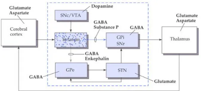
In order to perform correctly a movement the striatal loop and the cerebellar loop are very important. The informations enter the striatal loop at the dorsal striatum and exit from the internal pallidum and the substantia nigra pars reticulata, reaching the ventral anterior thalamic nucleus that projects to areas of the cortex involved in movements. The route from the afferent nuclei to the efferent ones can be both direct and indirect. The direct route is facilitatory, whereas the indirect route is inhibitory: the cortex projects to the striatum that is GABAergic and projects to the internal pallidum and to the substantia nigra pars reticulata (both GABAergic). Internal pallidum and sub-stantia nigra pars reticulata are efferent nuclei and project to the thalamus that is exci-tatory on the cortex. The result of these two inhibitions is facilitation on thalamus, so
7 Braak H. & Braak E., Pathoanatomy of Parkinson's disease, J Neurol (2000) 247: II/3-II/10 8 Ibid.
6 the direct route is facilitatory. The striatum projects also to the external pallidum which is GABAergic and projects to the subthalamic nucleus (glutamatergic), projecting to the efferent nuclei. This time there is a resulting facilitation on the subthalamic nucleus, which activates the efferent nuclei, eventually inhibiting the thalamus (see Figure 3).
It is very important to maintain the correct balance between these two routes in order to perform correct movements. In PD this balance is lost and this causes problems to the movement.
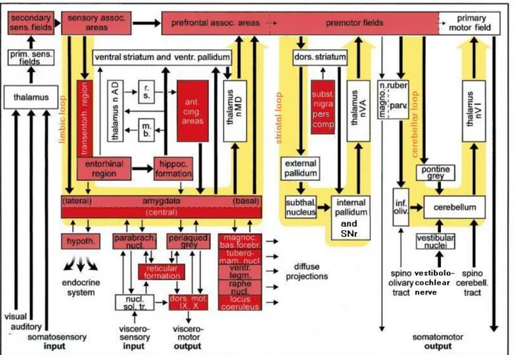
Also cerebellum plays an important role in controlling the movements. It receives informations from the spinocerebellar tract, the vestibular nerve and the inferior olive; these are processed so that the cerebellum can control equilibrium, eye and head movements and also regulate movements so that they are commensurate with their aim. It is then possible to summarize the importance of these three loops in a scheme such as the one in Figure 4. The damaged regions are coloured in red: the darker is the colour, the more severe is the lesion.
Figure 3. Organization of the basal ganglia. The cortex projects to the striatum that is GABAergic and projects to the internal pallidum and to the substantia nigra pars reticolata (both GABAergic). GPi and SNr are efferent and project to the thalamus that is excitatory on the cortex. Since we have two inhibitions there is a facilitation on thalamus, so the direct route is facilitatory.
The striatum projects also to the external pallidum (GABAergic) that projects to the subthalamic nucleus (glutamatergic), projecting to the efferent nuclei. This time we have two inhibitions (striatum and GPe) that are facilitatory on the subthalamic nucleus, that activates the efferent nuclei, eventually inhibiting the thalamus. Abbreviations: GPe = external pallidus, GPi = internal pallidus, SNc = substantia nigra pars compacta, SNr = substantia
nigra pars reticolata, VTA = ventral tegmental area.
7
Symptoms
PD was described for the first time by James Parkinson in 1817 in his essay “An essay on the Shaking Palsy”. The symptoms appear when the greater part of the dopa-minergic neurons is lost. This loss results in a decreased stimulation of the motor cortex and causes muscle rigidity, tremor, bradykinesia, a slowing of physical movements, and postural instability. 9
The tremor is usually the first appearing symptom, however 30% of patients do
9 Jankovic J., Parkinson's disease: clinical features and diagnosis, J. Neurol. Neurosurg. Psychiatr. (2008) 79: 368–76
Figure 4. Importance of limbic, striatal and cerebellar loop in regulating viscero- and somatomotor outputs. The damaged lesions are coloured in red (dark red = severe lesions, light red = less severe lesions), while the yellow big arrows represent in order the limbic loop, the striatal loop and the cerebellar loop.
Abbreviations: ant. cing. areas = anterior cingulate areas, dors. mot. IX, X = dorsal motor area of the glossopharyngeal and vagal nerves, dors. striatum = dorsal striatum, hippoc. formation = hippocampal formation, hypoth. = hypothalamus, inf. oliv. = inferior olive, m. b. = mamillary body, magnoc. bas. forebr. = Magnocellular nuclei of the basal forebrain, n. ruber magno. = magnocellular portion of the nucleus ruber, n. ruber parv. = parvocellular portion of the nucleus ruber, nucl. sol. tr. = nuclei of the solitary tract, parabrach. nucl. = parabrachial nuclei, periaqued. Grey = periaqueductal gray, prim. sens. fields = primary sensory fields, r.s. = retrosplenial region, raphe nucl. = raphe nuclei, SNr = substantia nigra pars reticolata, subst. nigra pars comp. = substantia nigra pars
compacta, secondary sens. fields = secondary sensory fields, sensory assoc. areas = sensory association areas,
spinocerebell. tract = spinocerebellar tract, subthal. nucleus = subthalamic nucleus, thalamus nAD = anterodorsal nucleus of the thalamus, thalamus nMD = mediodorsal nuclei of the thalamus, thalamus nVA = ventral anterior nuclei of the thalamus, thalamus nVI = ventral intermediate nuclei of the thalamus, transentorh. region = transentorhinal region, tuberomam. nucl. = tuberomamillary nucleus, ventr. pallidum = ventral pallidum, ventr. tegm. = ventral tegmentum
8 not show tremor in the beginning and it appears only later on. It affects the most distal parts, it's usually unilateral and it is a rest tremor: it is maximal when the limb is at rest and disappears during voluntary movements and sleep.10 Thus, this kind of tremor is very different from the one that appears, for example, in cerebellar tumour, which is maxi-mal during movements, especially when a high coordination is needed.
Rigidity comes from joint stiffness and increased muscle tone and is often associ-ated with pain. The result is a so-called “cogwheel rigidity” when the muscle is passively moved and is due to the loss of harmony between flexor muscles and antagonist mus-cles. In normal conditions gamma circuit feels muscle tone and the extrapyramidal cir-cuit (composed by basal nuclei) regulates its activity. In PD this regulation is lost and the subsequent increase of muscle tone causes movement problems. For example, another symptom is micrography and it is the consequence of increased muscle tone.11
Patients with PD show also slowness of movement, this is called bradykinesia; in the worst cases also akinesia (absence of movement) can be observed. The problem here is not only the execution of the movements but also its planning and initiation. In pa-tients affected by PD there is also a phenomenon called kinesia paradoxica: it is possible to see that sometimes immobile patients are capable of doing rapid movements if they get excited. This suggests that their motor programmes could be intact, but patients have problems in accessing them. It has been suggested that bradykinesia is the result of the disruption in normal motor cortex activity caused by reduced dopaminergic function. In about 50% of the cases also freezing can be observed. This most commonly affects the legs, and the result is that the patient suddenly cannot move; freezing state is transient since usually the motor block lasts for less than 10 s. There are different kinds of freez-ing: start hesitation, turn hesitation, hesitation in tight quarters, destination hesitation and open space hesitation. Episodes are mitigated by L-DOPA therapy and sometimes pa-tients can use tricks to avoid freezing, such as marching to command, walking to music, stepping over objects, and so on.12
Postural instability is also shown. Patients tend to walk with short steps, with their feet barely leaving the ground, and with a reduced arm swinging. The trunk is flexed forward. There is also the failure of postural reflexes which, together with or-thostatic hypotension, leads to falls due to impaired balance.13
10 Jankovic J., Parkinson's disease: clinical features and diagnosis, J. Neurol. Neurosurg. Psychiatr. (2008) 79: 368–76
11 Ibid. 12 Ibid. 13 Ibid.
9 Secondary symptoms include language problems (for example monotonic speech or dysarthria), facial hypomimia, dysphagia, drooling, cognitive problems (for example slowed reaction time, dementia, and short term memory loss). Sleep disturbances, im-paired proprioception, reduction of sense of smell, urinary incontinence, altered sexual function, constipation, hallucinations and decreased
blink rate are also shown.14
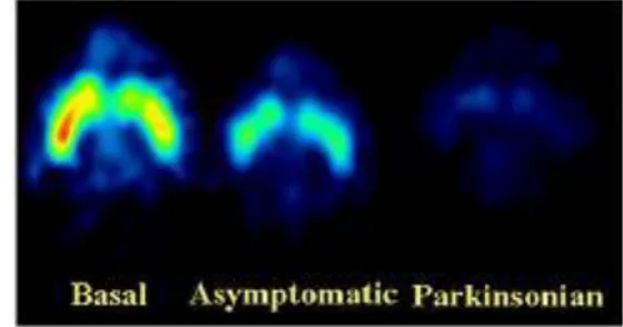
At the present time, there aren't known bio-markers or tests that can help us to recognize PD, since the presence of Lewy bodies and Lewy neu-rites in specific regions of the nervous system can be observed only post-mortem. The only other test available is a PET that shows reduced uptake of 18 F-DOPA or 11C-DTBZ in the striatum (see Figure 5).
Aetiology
PD is a multifactorial disease. This means that it may have different causes. The most common is idiopathic (without a known cause) PD, but it can be also caused by tox-ins, traumas, cerebral anoxia, drugs and genetic factors (so in some cases it can be an inherited disease). There is also the possibility of developing an iatrogenic form of PD, for example some patients can develop a reversible form of PD because of the drugs used to cure schizophrenia. This eventually disappears when the drug treatment is sus-pended.
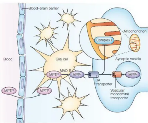
During 1980s some heroin addicts in California consumed a street drug and devel-oped a parkinsonism. The altered synthesis conditions of this drug lead to pH alterations and caused the formation of MPTP, a contaminative. å MPTP can cross the blood brain barrier and in the glial cells is converted by monoamine oxidase B in MPP+. This can en-ter the dopaminergic neurons using the DA transporen-ter and blocks the complex I of the mitochondrial respiratory chain leading the cell to death (see Figures 6 and 7).15
14 Jankovic J., Parkinson's disease: clinical features and diagnosis, J. Neurol. Neurosurg. Psychiatr. (2008) 79: 368–76.
15 Chiueh C. et al., In vivo generation of hydroxyl radicals and MPTP-induced dopaminergic toxicity in the basal ganglia, Ann N Y Acad Sci (2004) 738: 25–36
Figure 5. Reduced uptake of 11C-DTBZ can be seen in the brain of a non-human primate model of PD (right) if compared to a normal brain (left). A reduced uptake is also visible in the asymptomatic model, which isn’t showing the symptoms yet. The areas where the uptake is lower are blue and green whereas the ones with a higher uptake are yellow and red. (Modified from Blesa et al., 2010)
10 Figure 7. The increased produc-tion of reactive oxygen species can be due to mitochondrial im-pairment but also to the distribu-tion of vesicular dopamine into cytosol (also caused by MPP+). in the cytosol DA auto-oxydizes and generates ROS. These damage DNA and probably modify the ex-pression of redox-sensitive tran-scription factors. The damage is sensed by p53 that activates and upregulates Bax. This is translo-cated into the mitochondrion (probably by mediation of JNK) and induces cytochrome C release into the cytosol. Cytochrome C, with the adaptor molecule apaf1, activates caspase 9 that activates caspase 3, leading the cell to death.
Modified from Vila M. & Przed-borski S., 2003
Figure 6. MPTP can cross the brain barrier and in non-dopaminergic cells it is metabo-lized in MPDP+ by the enzyme MAO-B. Then, probably by spon-taneous oxidation, it generates MPP+, which has a high affinity for the DA transporter and enters dopaminergic neurons. Here, it is stored with an active process into the mitochondrion, where inter-feres with complex I of the elec-tron transport chain. This im-pedes the flow of electrons and results in an increased production of free radical which causes oxi-dative stress and activation of programmed cell death molecular pathways.
From Vila M. & Przedborski S., 2003
11 Since then one of the most common toxin-induced models for PD is the MPTP model. Other toxin-induced models are made with 6-hydroxydopamine (a toxin that kills dopa-minergic neurons and that has been used to evaluate potential therapies such as cell transplantation and neurotrophic factors) or rotenone (a pesticide and herbicide).16
In other cases PD can derive from other illnesses, for example one of the conse-quences of the 1918 flu pandemic was post-encephalitic parkinsonism.
As said before, also genetic factors may play a role in PD. Understanding which are the mutated genes and their functions can help also in understanding the mecha-nisms underlying the pathogenesis of idiopathic PD. A big difference between genetic and sporadic PD, however, is that the onsets of the genetic forms of the disease are usu-ally at an earlier age that the ones related to the sporadic forms.
The first mutation related to PD was found in a large family with Italian origins (known as Contursi family). In this case the disease is autosomal dominant. This muta-tion is on the chromosome 4 and affects the gene for α-synuclein (see Figure 8), an abundant neuronal protein implied in the synaptic mechanisms. In this family a missense mutation (G209A) could be found, then other mutations on this gene were found in other families in Germany and Spain, and some of these mutations are responsible not only for PD but also for dementia with Lewy bodies and multiple system atrophy. Human α-synuclein is a monomeric protein of 140 amino acids with a high homology grade with the same protein in other species. It is one of the members of the synuclein family, to-gether with β-synuclein, γ-synuclein and synoretin and is expressed not only in neuronal cells but also in endothelial cell and platelets. Its biological function is still unclear: some hypotheses are lipid binding, inhibition of phospholipase D2, interaction with other proteins such as syniphilin, chaperone activity, regulation of dopamine release, inhibi-tion of tyrosine-hydroxylase activity, control of dopamine transporter, synaptic vesicle transport, plasticity and/or regulation of vesicle pool. Structural changes can be caused by oxidative stress and/or genetic mutations, and in vitro studies have shown that mu-tated α-synuclein increases its tendency to aggregate and generate fibrils, favours mito-chondrial depolarization, favours apoptosis activation and inhibits proteasome activity. 17 There is also another type of autosomal dominant PD caused by duplication or triplica-tion of the α-synuclein gene (see Figure 8). The increase of repetitriplica-tions is associated with
16 Hattori N. & Sato S, Animal models of Parkinson’s disease: Similarities and differences between the disease and models, Neuropathology (2007) 27: 479–483
17 Farrer MJ., Genetics of Parkinson disease: paradigm shifts and future prospects, Nature (2006) 7: 306-318
12 a more severe phenotype. 18
A mutation can occur also on the ubiquitin carboxy-terminal hydrolase L1 (see Figure 8). UCH-L1 is very abundant in the brain (2% of the soluble protein total) and the missense mutation C277G decreases its activity. 19
Another mutation can be on the parkin gene (see Figure 8). In this case PD is in-herited as an autosomal recessive disease. Parkin gene is on chromosome 6 and the mu-tations can be deletions, exonic duplications or triplications, point mumu-tations, etcetera. Parkin protein has an ubiquitin-like domain at its N-terminus and two RING-finger do-mains flanking a cystein enriched domain at the C-terminus. It belongs to the ubiquitin E3 ligase family, and is probably important for the degradation of anomalous proteins, but when it is mutated loses its activity and this causes an accumulation of non-ubiquitinated proteins and this can lead to Lewy bodies formation. 20
Other mutations may affect the mitochondrial function and can be involved in oxidative stress. For example a mutation on the PTEN-induced putative kinase 1 (PINK-1)
18 Farrer MJ., Genetics of Parkinson disease: paradigm shifts and future prospects, Nature (2006) 7: 306-318
19 Ibid. 20 Ibid.
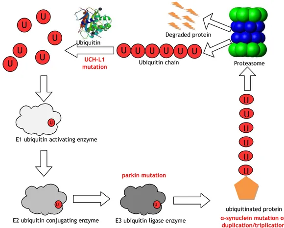
Figure 8. The mutations described above influence the proteasome pathway. In red: mutated genes affecting the various steps of the pathway.
Abbreviations: U = ubiquitin, UCH-L1 = ubiquitin carboxy-terminal hydrolase L1 U b Proteasome Degraded protein U b Ub Ubiquitin chain Ub Ub Ub Ub U b U b U b U b U b Ubiquitin hydrolase U
E1 ubiquitin activating enzyme
U
E2 ubiquitin conjugating enzyme E3 ubiquitin ligase enzyme U ubiquitinated protein UCH-L1 mutation parkin mutation α-synuclein mutation or duplication/triplication U b U b U b U b U b U b
13 locus on chromosome 1 makes the neuron become more susceptible to oxidative stress. PINK-1 is a ubiquitous mitochondrial protein with a highly conserved kinase domain and is homologous to kinases of the calmodulin family. Its function is still unknown, but it seems to protect neurons from stress-induced mitochondrial dysfunction and apoptosis.21
Other mutations can occur on the DJ-1 locus, which is still on chromosome 1, very near to PINK-1 locus. The mutations can be both point mutations and deletions, also in the splicing sites. This protein has been found also in cytoplasmic inclusions in tau-pathies, such as multiple system atrophy, and its role is still unclear, even if it is thought that it could be important for the oxidative stress response because it is localized both in cytoplasm and nucleus, but when oxidized translocates to mitochondria.22
An autosomal dominant variant of PD with incomplete penetrance is caused by a mutation on the leucine-rich repeat kinase 2 (LRRK2) which transcript generates a pro-tein called dardarin. This propro-tein has two domains: a tyrosine kinase catalytic domain and a GTPase domain. It has been hypothesised that the mutation can cause a gain of function. Dardarin is a member of the ROCO protein family, is widely expressed and has a cytosolic location. About 10% of LRRK2 can be found in mitochondria and its suggested role is in cell signalling, cytoskeletal reorganization, vesicle trafficking and nucleo-cytoplasmic transport.23
Also α-synuclein and parkin mutation may interfere with mitochondrial function: in transgenic mice, α-synuclein over expression increases oxidative stress and enhances the toxicity of MPTP.24 Parkin can associate with the outer mitochondrial membrane, protecting the cell against cytochrome C release and the consequent caspase activa-tion.25 It may also associate with mitochondrial transcription factor A and enhance mito-chondrial biogenesis.26 Moreover, physical association have been reported between DJ-1 and α-synuclein, DJ-1 and parkin, and DJ-1 and PINK-1 and there is evidence that DJ-1, PINK-1 and parkin function sequentially in the same pathway.27
21 Farrer MJ., Genetics of Parkinson disease: paradigm shifts and future prospects, Nature (2006) 7: 306-318
22 Ibid. 23 Ibid.
24 Song DD. et. al, Enhanced substantia nigra mitochondrial pathology in human alpha-synuclein trans-genic mice after treatment with MPTP, Exp Neurol. (2004) 186(2):109-11
25 Darios F. et al., Parkin prevents mitochondrial swelling and cytochrome c release in mitochondria-dependent cell death, Hum Mol Genet. (2003) 12(5): 517-26
26 Kuroda Y. et al., Parkin enhances mitochondrial biogenesis in proliferating cells, Hum Mol Genet. (2006) 15(6): 883-95
27 Lin MT. & Flint Beal M., Mitochondrial dysfunction and oxidative stress in neurodegenerative diseases, Nature (2006) 443: 787-795
14
Therapy
Currently, all therapies available for PD are just symptomatic therapies; this means that they do not stop PD progression, but reduce signs and symptoms for the com-fort of the patient.
Drug treatment
Pharmacological approach includes dopaminergic drugs, dopaminergic agonists, MAOB and COMT inhibitor, amantadine, and anticholinergic drugs.
Dopaminergic drugs aim to maintain normal dopamine levels. Unfortunately do-pamine itself is not able to cross the blood brain barrier, so what is normally used is its precursor L-DOPA. L-DOPA causes sudden improvements, but has many side effects re-sulting from the increase of dopamine levels: nausea, vomit, postural hypotension, hal-lucinations and dyskinesias. Moreover, this drug is administered per os and is metabo-lized in peripheral districts, so that only a small part reaches CNS (1-3%). Usually other drugs are used together with L-DOPA, so that its side effects are lowered and also its dose can be decreased. For example the result of inhibiting peripheral dopadecarboxy-lases is that L-DOPA remains intact for a longer time, and thus more drug reaches the brain (10%). Also nausea and vomit can be reduced since these are the result of the stimulation of the chemoreceptor trigger zone and they can be controlled with domperi-done, an antidopaminergic drug that does not cross the blood brain barrier. However, after some years, the effects of L-DOPA decrease in half of the patients: they have a shorter duration and dyskinesias are more frequent, so, in order to delay this phase, the lowest possible amount of L-DOPA is always used. These complications are due to the fact that when the therapy begins there is still a certain percentage of dopaminergic neurons left, but with time the number of neurons decreases so the storage of dopamine becomes impossible.28
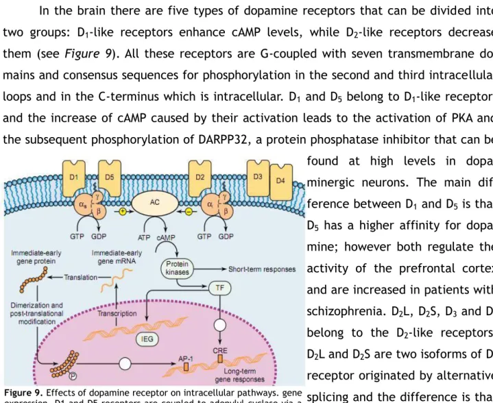
15 In the brain there are five types of dopamine receptors that can be divided into two groups: D1-like receptors enhance cAMP levels, while D2-like receptors decrease them (see Figure 9). All these receptors are G-coupled with seven transmembrane do-mains and consensus sequences for phosphorylation in the second and third intracellular loops and in the C-terminus which is intracellular. D1 and D5 belong to D1-like receptors and the increase of cAMP caused by their activation leads to the activation of PKA and the subsequent phosphorylation of DARPP32, a protein phosphatase inhibitor that can be found at high levels in dopa-minergic neurons. The main dif-ference between D1 and D5 is that D5 has a higher affinity for dopa-mine; however both regulate the activity of the prefrontal cortex and are increased in patients with schizophrenia. D2L, D2S, D3 and D4 belong to the D2-like receptors. D2L and D2S are two isoforms of D2 receptor originated by alternative splicing and the difference is that D2L is postsynaptic while D2S is a presynaptic autoreceptor. These receptors decrease cAMP levels and Ca++ channels permeability, and increase K+ channels perme-ability; the difference between D2, D3 and D4 is in their localiza-tion and their affinity for differ-ent drugs. 29 Dopamine agonists are less effective than L-DOPA, but they can be used to reduce the dose administered. They can derive from ergot or not. Ergot derivatives are less used now because of their side effects, like fibrosis affecting cardiac valves. Non-ergot drugs act on D3 receptors, determining however an excessive stimulation of other areas that causes hypersexuality, hallucinations, psychosis, leg edemas and sleepiness.30
29 Siegel GJ. et al., Basic Neurochemistry, molecular, cellular and medical aspects, Elsevier, 2006 30 Rang HP., Dale MM., Pharmachology, Elsevier, 2007
Figure 9. Effects of dopamine receptor on intracellular pathways. gene expression. D1 and D5 receptors are coupled to adenylyl cyclase via a stimulatory G protein, while D2 receptor inhibits cyclase activity via coupling to an inhibitory G protein. Also the activation of D3 and D4 receptors inhibits cyclase activity. Adenylyl cyclase catalyzes the conversion of ATP into cAMP, which causes dissociation of the regulatory and catalytic
subunits of protein kinase A. This for example can phosphorylate and activate DARPP-32 that inhibits protein phosphatase-1. The phosphoproteins activated by the kinase in turn can lead to short-term responses within the cell or activate transcription factors which enter the nucleus and alter gene expression acting either directly on the target gene or via an immediatly early gene.
Abbreviations: AC = adenylyl cyclase, AP-1 = adaptor protein 1, CRE = cAMP response element, IEG = immediately early gene, TF = transcription factor.
From Siegel et al., Basic Neurochemistry, molecular, cellular and medical aspects, Elsevier, 2006
16 Dopamine, like norepinephrine and epinephrine, is a catecholamine. Its synthesis in the brain starts from tyrosine that is converted into DOPA by tyrosine hydroxylase, an enzyme whose activity is the limiting step for dopamine synthesis (tyrosine hydroxylase receives a negative feedback from dopamine: there are autoreceptors synaptic level that sense dopamine levels in the synaptic cleft and inhibit TH). DOPA is then trans-formed into dopamine by DOPA decarboxylase and, in noradrenergic cells, dopamine-β-hydroxylase turns it into norepinephrine, which can be eventually converted into epi-nephrine by phenilethanolamine-N-methyltransferase. Dopamine is stored in acid vesi-cles, in which is transported with the vesicle transporter V-MAT. The acidity of these vesicles is achieved with proton pumps and it's necessary for the catechol ring of dopa-mine.31
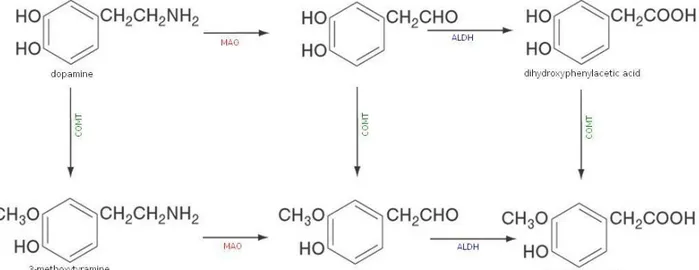
Dopamine is degraded by monoamine oxidases and catechol-O-methyltransferase (see Figure 10). When it is degraded by COMT, 3-methoxytyramine is formed and this is then converted into homovanillic acid by MAO and aldehyde dehydrogenase. The other possible way is that dopamine is degraded by MAO and then converted into dihydrox-phenylacetic acid by aldehyde dehydrogenase. Eventually, COMT can turn DOPAC into HVA. Is it possible to measure HVA levels in urine, but it is important to consider that these are not the result of the CNS production only but also of the peripheral produc-tion. 32
There are many drugs interacting with the catabolic pathway of the dopamine,
31 Siegel GJ. et al., Basic Neurochemistry, molecular, cellular and medical aspects, Elsevier, 2006 32 Ibid.
Figure 10. Dopamine catabolism. DA can be converted into dihydroxyphenilacetic acid by monoamine oxydase and aldehyd dehydrogenase, or into 3-methoxytyramine by catechol-O-metyltransferase. This can be then converted indo homovanillic acid by MAO and ALDH, and also dihydrxyphenylacetic acid can be transformed into HVA by COMT. Abbreviations: ALDH = aldehyd dehydrogenase, COMT = catechol-O-methyltransferase, MAO = monoamine oxydase.
17 some of them interfere with MAOB and some others with COMT and all of them make possible to diminish the quantity of L-DOPA administered. 33
The degeneration of dopaminergic neurons leads also to an unbalance between dopamine and acetylcholine levels in the basal nuclei, thus also anticholinergic drugs are used. These are not very effective if compared to L-DOPA and dopaminergic agonist, but can be used especially in iatrogenic parkinsonism. It is a good rule not administering these drugs to people with cognitive problems, because they can be worsened, and other side effects are decreased mucus production in nose and throat (and consequent dry throat), urinary retention, confusion with possible hallucinations.34
Another drug used is the antiviral amantadine. It is used in the advanced phases of the disease and probably blocks glutamatergic receptors NMDA. This drug has an ef-fect on dyskinesias caused by chronic use of L-DOPA and its side efef-fects are confusion, livedo reticularis, hallucinations and nightmares.35
Stem cells
Since the drug treatment is only symptomatic and there is the real necessity of blocking the disease progression (or at least interfere with it), also stem cell therapy has been considered. This could occur through endogenous cell recruitment or transplant.
In 1980 both transplant of developing dopaminergic neurons (from abortions of 7-9 weeks) and xenotransplants (from pig) have been made. The results were interesting since the disease progression seemed to stop, but ten years later other studies carried out also with placebos have reopened the discussion: midbrain stem cells can modify the course of the disease, but their ability to produce dopaminergic cells is unstable and thus they are not and adequate source of this type of cells.36
In 1998 Ye W. et al. found out that DA neurons are induced by Fgf8 and Shh, and two years later the first TH positive mouse neurons were produced. First of all neural precursor cells must be produced and then these have to differentiate into dopaminergic neurons. In order to differentiate ES to a neural fate is it possible to use embryoid bod-ies’ formation, retinoic acid, or other protocols. The cells are then evaluated by the presence of neural precursor markers such as Sox1, Sox2, βtubulin3, MAPs and neuro-filament proteins. Then Fgf8, Shh and ascorbic acid are added to the culture and in this
33 Rang HP., Dale MM., Pharmachology, Elsevier, 2007 34 Ibid.
35 Ibid.
36 Yong VW. et al., Transplantation of human sympathetic neurons and adrenal chromaffin cells into parkinsonian monkeys: no reversal of clinical symptoms, J Neurol Sci. (1989) 94:51-67
18 way is it possible to detect TH in over 30% of the neurons. Later this proportion has been increased using a cocktail containing survival promoting factors such as interlukin-1β, GDNF, neurturin, TGFβ3 and dibutyryl-cAMP: these factors enhance RNA expression levels of En-1, Mash-1, dopamine receptor 2, TH and Nurr1.37
However, transplantation of human DA neurons obtained from embryonic stem cells in animal models has also shown poor survival levels; these need to be increased for a clinical application.38
Another of the observations that can be made is that transplanted cells can re-place the degenerated ones, but do not solve the problem at its roots since the disease progression is not stopped.39
Gene therapy
Another approach can be transplant cells modified to produce higher levels of neurotrophic factors with neuroprotective effects, such as GDNF, or more enzymes in-volved in dopamine metabolism. However, as said earlier, this approach presents the problem of cell survival and of their stability.40
An alternative approach could be the use of viral vectors to deliver those genes directly into neurons. The delivered genes could be involved in DA metabolism, in neu-roprotection or in inhibition of brain nuclei that become overactive in PD.41
Viral vectors encoding for TH have been used, resulting in a long term expression of tyrosine hydroxylase and some corrections of behavioural deficits. However it was used a partial-lesion 6-OHDA model, meaning that there was still some endogenous TH activity left, so it is impossible to determine the exact amount of dopamine produced by the delivered tyrosine hydroxylase. Nevertheless these studies provided proof-of-principle that the use of TH delivered with viral vectors improved the symptoms.42
Tyrosine hydroxylase needs a cofactor called BH4, so also the cotransfection with TH and guanosine triphosphate cyclohydrolase I (the limiting enzyme for the synthesis of BH4) has been tested. The results were better than the ones of transfection with
37 Maxwell SL. & Li M., Midbrain dopaminergic development in vivo and in vitro from embryonic stem cells, J Anat. (2005) 207(3):209-18
38 Lindvall O., Kokaia Z., Stem cells for the treatment of neurological disorders, Nature. (2006) 441(7097):1094-6
39 Ibid.
40 Lawlor PA. & During MJ., Gene therapy for Parkinson’s disease, Expert Rev Mol Med. (2004) 6:1-18 41 Ibid.
19 sine hydroxylase only.43
Also viral vectors encoding AADC have been used to increase dopamine levels. Transduction of these vectors together with exogenous L-DOPA lead to an increase in do-pamine levels higher than the one due to only L-DOPA administering. This is interesting as it makes possible a reduction of L-DOPA used in therapy, and a consequent diminution of side effects.44
Moreover, loss of dopaminergic neuron leads to an increased activity of the exci-tatory neurons of the subthalamic nucleus resulting in an increase of inhibition on thalamus and downstream motor pathways. It has been proposed that silencing of STN could result in an improvement of symptoms, and actually electrical inhibition, ablation or pharmacological silencing of the subthalamic nucleus have all been reported to con-trol PD symptoms. So delivery into the STN of glutamic acid decarboxylase, the enzyme involved in the synthesis of GABA, of parkinsonian rats has been tested and it has given proof-of-principle of the improvement of the symptoms.45
One of the main issues of gene delivery, however, is regulation of gene expression, as over-expression of delivered genes may result in a series of undesired side effects. Inducible systems are being developed, but at the present time they are still leaky or poorly inducible in vivo.46
Neurotrophic factors
Neurotrophic factors could be very useful for PD, in different ways. First of all they can promote survival of subpopulations of neurons exposed to MPTP or 6-OHDA in models of PD; so, if toxin induced death shares any mechanism with the pathogenesis of PD, neurotrophic factors could slow or stop dopaminergic degeneration. Second, they could restore functions of neurons that are losing their phonotype and are losing their capacity of synthesizing dopamine. Third, some neurotrophic factors have effects on cell excitability and could stimulate the dopaminergic system. Last, it can be that in some cases PD is caused by loss of neurotrophic factors or disruption in their signalling cas-cades, thus a therapy could be replacing the missing factors.47
Hence the idea of using neurotrophic factors for therapy. GDNF was administered
43 Ibid.
44 Lawlor PA. & During MJ., Gene therapy for Parkinson’s disease, Expert Rev Mol Med. (2004) 6:1-18 45 Ibid.
46 Ibid.
47 Peterson AL. & Nutt JG., Treatment of Parkinson's disease with trophic factors, Neurotherapeutics. (2008) 5(2):270-80
20 by monthly intraventricular injections, but, while the results in animal models were good, in humans it had no effect. This was probably due to the route of delivery and to the fact that switching from an animal model to a human the tissue volume increases, so the diffusion of the neurotrophic factor is affected. Also relevant side effects were shown, probably for the diffusion of GDNF in the cerebrospinal fluid that caused its act-ing on structures with GDNF receptors but not involved in movement. Thus GDNF was administered by chronic infusion using pumps in the postero-dorsal putamen. The side effects were limited, and no nausea and vomit were observed. After three months of GDNF infusion the symptoms improved, and after six months periods of severe immobil-ity were completely eliminated and also 18F-DOPA uptake was increased.48 This was a phase I trial, but sadly a phase II trial did not confirm the results since there weren't sig-nificant differences between patients receiving the neurotrophic factor and the control group.49 However, other studies have hypothesized that this discrepancy in results may be explained in terms of bioavailability of GDNF: applying the protocol from trial phase II, 125I-GDNF was unevenly distributed in the putamen of parkinsonian monkeys. It was found mainly around the catheter tip with its concentration decreasing exponentially as the distance from the tip increased and as a result its bioavailability was limited to 2-9% of the putamen. Thus, even if further studies are required, probably the different re-sults may be explained with the different protocol and catheter used in phase II trial.50
Also neurotrophic factors can be delivered with viral vectors: adenovirus, lenti-virus and adenoassociated lenti-virus have been used to introduce GDNF into brain tissue and it affected positively both behaviour and nigrostriatal innervation levels.51
Other studies have been carried out with neurturin on cell cultures. This molecule is from the same family as GDNF and was delivered with lentiviral vectors. Wild type neurturin had no effect because it was secreted in its pro-form, so other variants of neurturin were considered. Deletion of the pro-region resulted in significantly higher se-cretion of active NRTN, which was further increased when the signal peptide was substi-tuted with the immunoglobulin heavy-chain signal peptide. This was reproduced in vivo in lentiviral-transduced rat striatal cells and enabled efficient neuroprotection of dam-aged nigral DA neurons, similar to GDNF. So this is an interesting option that should be
48 Gill SS. et al., Direct brain infusion of glial cell line-derived neurotrophic factor in Parkinson disease, Nat Med. (2003) 9(5):589-95
49 Lang AE. et al., Randomized Controlled Trial of Intraputamenal Glial Cell Line–Derived Neurotrophic Factor Infusion in Parkinson Disease, Ann Neurol (2006) 59: 459-466
50 Salvatore MF. et al., Point source concentration of GDNF may explain failure of phase II clinical trial, Exp Neurol. (2006) 202(2):497-505
21 considered.52
In other studies BDNF, which is required for the developing of the correct number of neurons in the substantia nigra, has been used. It is expressed by DA neurons both in the SNc and VTA and, at lower concentrations, in the striatum. In post-mortem samples both BNDF and BDNF mRNA levels were lower in patients with PD than in controls. Fibro-blasts secreting BDNF have been implanted near the substantia nigra of rats that were later treated with MPP+, and death of neurons was 86% lower than in controls. Con-versely, the knock-down of BDNF receptor turned out in a decreased number of neurons in the SNc, accumulation of α-synuclein, and reduced TH activity in the striatum.53
52 Fjord-Larsen L. et al., Efficient in vivo protection of nigral dopaminergic neurons by lentiviral gene transfer of a modified Neurturin construct, Exp Neurol. (2005) 195(1):49-60
53 Peterson AL. & Nutt JG., Treatment of Parkinson’s Disease with Trophic Factors, Neurotherapeutics. (2008) 5(2):270-80
22
GDNF, its receptor complex and signalling
What are neurotrophic factors?A short definition could be that neurotrophic factors are molecules involved in neuron growth and survival, however it is not easy to tell exactly what a neurotrophic factor is: there are also some inorganic molecules that promote neuron survival, such as potassium at high concentrations.54 Moreover many molecules known as neurotrophic factors act also outside the CNS, so this definition is somehow incomplete. In 1988 some criteria to define a neurotrophic factor were proposed. First of all it keeps alive neurons that would die in its absence, second it has to be present in its active form, synthesized and secreted by the target tissue of the those neurons, third its amount in the target tis-sue must be very low, and fourth it affects development and maintenance of neurons in vivo.55
At the beginning of the development of the central nervous system there are more neurons than in the end. According to the neurotrophic factor hypothesis, post-synaptic cells produce neurotrophic factors, which are needed for neuron survival, in li-miting quantity. The consequence is that only neurons that synapse correctly (and con-sequently have access to more quantities of neurotrophic factor) will survive, while the others will die for programmed cell death.56 Each sub-population of neurons depends on the actions of different neurotrophic factors.57
The first factor that was described as important for growth and survival of neu-rons was the nerve growth factor.58 Today, we know four families of neurotrophic fac-tors: neurotrophins, neurokines, glial cell line-derived neurotrophic factors (GFLs) and mesencephalic astrocyte-derived neurotrophic factor family (MANF).
Neurotrophins
Neurotrophins belong to a superfamily of growth factors with a cysteine knot: this knot consists of three disulfide bonds formed by cysteine residues. Neurotrophins act as head-to-head dimers. To this cysteine knot-family belong also neurotrophic factors such
54 Por SB. & Huttner WB., A Mr 70000 phosphoprotein of sympathetic neurons regulated by nerve growth factor and by depolarization, J Biol Chem. (1984) 259:6526-33
55 Barde YA., What, if anything, is a neurotrophic factor?, Trends Neurosci. (1988) 11:343-6 56 Thoenen H.& Barde YA., Physiology of nerve growth factor, Physiol Rev. (1980) 60:1284-335
57 Davies AM., The neurotrophic hypothesis: where does it stand?, Philos Trans R Soc Lond B Biol Sci. (1996)351:389-94
58 Levi-Montalcini R. & Hamburger V, Selective growth factor stimulating effects of mouse sarcoma on the sensory and sympathetic nervous system of the chick embryo, J Exp Zool. (1951) 116:321-61