UNIVERSITÀ DELLA TUSCIA
FACOLTÀ DI SCIENZE MATEMATICHE FISICHE E NATURALI
DECOS - Dipartimento di Ecologia e Sviluppo Economico Sostenibile
Dottorato in Ecologia e Gestione delle Risorse Biologiche XXII ciclo
“Bacterial and archaeal community composition along the water
column and in the sediment of Averno lake as revealed by 16S rRNA
and [FeFe]-hydrogenases genes-based approaches”
(SSD BIO/07)Coordinatore: D.ssa Roberta Cimmaruta
Tutor: Dott. Luigi Chiarini D.ssa Silvia Tabacchioni
3
Abbreviations. 6
PART 1
BACKGROUND, OBJECTIVES AND OVERVIEW OF THE THESIS 7
PART 2
OVERVIEW OF THE LITERATURE 11
Chapter I: Lake microbial ecology 12
I.1. General introduction 12
I.2. Phototrophic prokaryotes 15
I.3. Aerobic methylotrophic prokaryotes 17
I.4. The methanogenic bacteria 20
I.5. Sulfate- and sulfur-reducing prokaryotes 23
I.6. The colorless sulfur bacteria 27
I.7. The denitrifying prokaryotes 29
I.8. Dinitrogen-fixing prokaryotes 33
I.9. The inorganic nitrogen compounds oxidizing prokaryotes 34
I.10. The hydrogen-metabolizing prokaryotes 36
I.10.1. Hydrogen-producing bacteria and hydrogenases 43
I.10.1.1. [NiFe]-hydrogenases 45
I.10.1.2. Iron–sulfur cluster-free hydrogenase (Hmd) 46
I.10.1.3. [FeFe]-hydrogenases 46
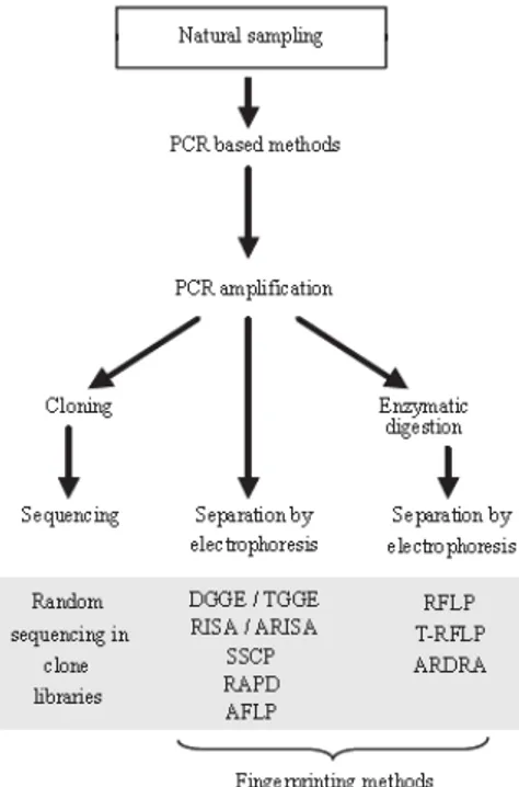
Chapter II: Molecular approaches to assess the biodiversity in lake microbial
communities 56
I.I.1. General introduction 56
I.I.2. Bacterial community diversity based on 16S rDNA 57
I.I.3. Single-strand conformation polymorphism 60
I.I.4. Terminal restriction fragment length polymorphism 61
I.I.5. Ribosomal intergenic spacer analysis and automated ribosomal intergenic
spacer analysis 62
I.I.6. Denaturant gradient gel electrophoresys 63
4
I.I.6.2. Microbial community diversity analysis using DGGE banding patterns 64
I.I.6.3. Advantages, limitations, and complementarity 65
I.I.7. Random sequencing in clone libraries 66
I.I.7.1. Advantages, limitations 67
I.I.8. Use of functional genes to target populations involved in specific metabolic
processes 67
I.I.8.1 Use of [FeFe]-hydrogenases gene to investigate the diversity of
H2-producers communities 69
PART 3
EXPERIMENTAL WORK 75
Chapter III: Vertical distribution of bacterioplankton in Lake Averno in relation to
water chemistry 76
III.1. Introduction 76
III.2. Material and Methods 77
III.2.1. Site and sampling 77
III.2.2. Physico-chemical analyses of water column 78
III.2.3. DNA extraction and PCR amplification 78
III.2.4. DGGE 81
III.2.5. Cluster analysis and diversity indices 81
III.2.6. Extraction, sequencing and phylogenetic analysis of DGGE bands 82
III.3. Results 83
III.3.1. Physico-chemical parameters of the lake 83
III.3.2. DGGE fingerprints and PCA 85
III.3.3. Phylogenetic analysis 91
III.4. Discussion 96
Chapter IV: Microbial community composition of surface sediment from Lake Averno
revealed by 16S rDNA and [FeFe]-hydrogenase genes -based approaches 101
IV.1. Introduction 101
IV.2. Material and Methods 102
5
IV.2.2. DNA extraction 102
IV.2.3. PCR amplification and DGGE 102
IV.2.4. Recovery, cloning and sequencing of excised DGGE bands 103
IV.2.5. Phylogenetic analysis of sequenced DGGE bands 103
IV.2.6. Primer design and PCR hydrogenase gene amplification 103
IV.2.7. Cloning, sequencing and phylogenetic analysis of FHG fragments 104
IV.3. Results and discussion 104
IV.3.1. Eubacterial DGGE profile and sequence analysis of excised bands 104 IV.3.2. Archaeal DGGE profile and sequence analysis of excised bands 109 IV.3.3. Diversity of FHGs in sediment sample and phylogenetic analysis 111
6
Abbreviations
ARISA – automated ribosomal intergenic spacer analysis BCC – bacterioplankton community composition
DGGE – Denaturing Gradient Gel Electrophoresis
DG-DGGE – double gradient denaturing gradient gel electrophoresis DO – dissolved oxygen
Eh – redox potential
FHGs – [FeFe]-hydrogenase genes
FISH – fluorescence in situ hybridization H’ – Shannon Weaver index
Hmd – methylentetrahydromethanopterin dehydrogenase HPB – H2-producing bacteria
IGS – ribosomal intergenic spacers ITS – Internal transcribed spacers
LSU rRNA – large subunit of ribosomal RNA OTU – operational taxonomic unit
PCA – principal component analysis PCR – Polymerase Chain Reaction
PPFM – pink-pigmented facultatively methylotrophic R – richness index
rDNA – ribosomal DNA
RDP – Ribosomal Database Project
RISA – ribosomal intergenic spacer analysis rRNA – ribosomal RNA
S – Simpson index
SSCP – single strand conformation polymorphism SSU rRNA – small subunit of ribosomal RNA T – temperature
T-RF – terminal restriction fragment
T-RFLP – Terminal restriction fragment length polymorphism V – hypervariable region of 16S rRNA gene
PART 1
BACKGROUND, OBJECTIVES AND
OVERVIEW OF THE THESIS
8
Background
Prokaryotes are not only the most abundant organisms in lake habitats, but they are also key players in biogeochemical processes (e.g. the metabolization of dissolved organic carbon or nitrogen cycling). Furthermore, these microorganisms have a relevant role in the processes controlling the water quality, because they are involved in the biodegradation of pollutants deriving from urban, industrial and agricultural activities. Detailed knowledge of the diversity, specific functions and ecology of prokaryotes dwelling in lakes is an essential prerequisite for the sustainable management of these freshwater resources. Moreover, since the academic and industrial interest in the discover of novel microorganisms that could produce undescribed secondary metabolites or that could have a potential biotechnological application is on the rise, the knowledge of the microbial diversity of lakes, representing a rich source of bacteria with different metabolic pathways, could provide important information in that regard.
During the last two decades, methods based on direct Polymerase Chain Reaction (PCR) and analysis of ribosomal RNA (rRNA) genes were developed and allowed a more comprensive analysis of microbial communities in comparison with cultivation based techniques. Among the techniques which no longer require the isolation and cultivation of bacteria, Denaturing Gradient Gel Electrophoresis (DGGE) analysis of 16S rRNA gene fragments has been widely used to assess the diversity of complex microbial communities in sediments and water column of lakes. Recent microbial ecology studies have begun to explore the links between diversity and function or identity and activity of microbial assemblages using not only rRNA-based primers, but also primers specific for determined functional groups, such as ammonia-oxidizing bacteria, sulphate-reducing bacteria, denitrifiers, methanotrophs and methanogens. Recently, primers specific for [FeFe]-hydrogenase genes have been developed to identify hydrogen producing bacteria occurring in anaerobic H2 biofermentation systems, but no studies have been performed to detect them in natural samples.
9
Aims of the work
In this respect, the aims of this thesis were:
to investigate the in situ distribution, abundance, and diversity of prokaryotes along the water column of Lake Averno, in relation to the depth gradient of physicochemical properties; to identify predominant populations constituting the microbial community of Averno lake
superficial sediment and to detect hydrogen-producing bacteria naturally occurring in this habitat.
Overview of the thesis
PART 2 of the thesis presents an overview of the literature related to the content of this work. Chapter I starts with a general introduction to the microbial ecology of lakes. This chapter provides general information about the microbial functional groups involved in the main metabolic processes occurring in lakes, giving a particular attention to the hydrogen-producing bacterial community. Chapter II deals with the molecular tools mainly used to investigate lake bacterial community composition with particular emphasis on DGGE and cloning techniques and the use of functional genes as markers to study microbial diversity. Moreover, it gives a short overview on the use of [FeFe]-hydrogenases gene to investigate the diversity of H2-producers communities.
PART 3 of the thesis presents the experimental work performed in this Ph.D. study. Chapter III describes the methods used to investigate the depth-related changes in entire microbial communities of the Lake Averno water column. In the same chapter, the obtained molecular results are shown and discussed in relation to the depth gradient of physicochemical properties of the lake. Chapter IV is a description of the overall microbial community found in superficial sediments of Lake Averno using the molecular methods described in Chapter III with the addition of a cloning step of DGGE bands, necessary to overcome the limit of the technique for the complex microbial community of sediment. Moreover, this chapter describes the composition of H2–producing bacteria using as molecular target the gene encoding [FeFe]-hydrogenase enzyme. Therefore, this chapter reviews the conclusions drawn from results obtained developing a new set of PCR primers
10
targeting the functional Clostridium iron-only [FeFe]-hydrogenase genes, known as the most indispensable element for hydrogen-evolving microorganisms.
PART 2
Chapter I: Lake microbial ecology
I.1. General introduction
Prokaryotes are a important component of lake ecosystem because of their abundance, their species diversity and the multiplicity of their metabolic activities (Miskin et al., 1998; Wetzel, 2001; Wei et al., 2008). They play a key role in the decomposition of organic matter, in the biogeochemical cycles of the main elements (carbon, nitrogen, phosphorus, etc) and of trace elements (iron, nickel, mercury, etc), as well as in biodegradation of pollutants deriving from anthropic activities. Therefore, a detailed knowledge of the diversity, specific functions and ecology of microorganisms inhabiting lakes is needed for the sustainable management of these ecosystems. Several lakes have been studied as model systems to analyze the inter-relationships between microorganisms and their living and non living environments (Clark and Walsby, 1978; Gorlenko et al., 1977; Kuznezow, 1977). In particular, stratified lakes, characterized by zonation and gradient forming processes (Overbeck, 1972; Pfennig, 1979; Sorokin, 1970), represent an important topic in the microbial ecology.
In a stratified lake (Fig. 1), the warming of the surface layers yields a less dense upper layer (epilimnion) of circulating, aerated water and a dense, cold, stagnant bottom layer (hypolimnion). The intermediate layer (metalimnion) is characterized by a temperature gradient (the thermocline) and, after oxygen deprivation and anaerobic decomposition of organic matter in the hypolimnion, by a chemical gradient (chemocline) also.
The upper part of the epilimnion is the zone of primary production by the oxygenic phototrophic organisms, such as photosynthetic higher plants, algae, diatoms, flagellates, green algae, and cyanobacteria. These primary producers are accompanied by bacteria, protozoa, and metazoa that consume photosynthetized biomass, which results in secondary production and release of cellulosic detritus (Fenchel and Jøorgensen, 1977). Part of the biomass usually sinks to the hypolimnion and to the bottom of the lake where it is degraded. Degradation is accompanied by oxygen consumption, resulting in a decrease in oxygen concentration that leads to anoxic conditions in the bottom layer. Continued anaerobic degradation results in the production of organic fermentation products as well as hydrogen, methane, hydrogen sulfide, and carbon dioxide, which diffuse upwards forming concentration gradients. Methane, produced by methanogenic bacteria using H2 + CO2, formate and acetate as main substrates is released from the sediment and discharged in the form of gas bubbles.
Part of the methane is dissolved in the water as it moves upwards and is oxidized by methane-utilizing aerobic bacteria. The quick removal of oxygen from the hypolimnion is primarily due to the quick dispersal of methane and the growth of methane-oxidizing bacteria. Soluble fermentation products, hydrogen gas included, are used not only to produce methane but also to reduce sulfate to hydrogen sulfide. In the water column, the activity of sulfate reduction has two maxima: one in the hypolimnion bottom layers and one below the chemocline (Sorokin, 1970). While the reduction power for the first process originates from the sediment, sulfate reduction below the chemocline depends on the presence of purple and green sulfur bacteria that release sulfate during their anoxygenic photosynthesis. Simultaneously, nitrate is reduced to nitrite, which transiently accumulates and forms a distinct layer, and then to nitrogen.
The metalimnion is a layer of high biological activity. Because of its richness in inorganic nutrients as compared to the epilimnion, it is inhabited by a few aerobic photosynthetic prokaryotes that can tolerate hydrogen sulfide and anaerobic conditions. Because of the oxidation of hydrogen sulfide by the purple sulfur bacteria in the light and diffusion of oxygen from the epilimnion, the chemocline moves downward several meters during the daytime. Diurnal vertical fluctuations of the redox discontinuity layer obviously represent a major selective factor for the organisms occupying this habitat.
Figure 1. Diagram of production, consumption, and decomposition in an aquatic ecosystem with a chemocline
The groups of bacteria involved in the above mentioned metabolic processes of lake are included into different taxonomic groups commonly retrieved in plankton and sediment of lakes.
Bacterioplankton community composition has been studied extensively, and research has shown that it is highly variable among different lakes (Lindstrom et al., 2000; Yannarell et al., 2005). The prevalent taxa detected belong to β-Proteobacteria, Actinobacteria, α-Proteobacteria, Bacteroidetes, Cyanobacteria, Verrucomicrobia, OP-10, and Planctomycetes (Zwart et al., 2002; Wei et al., 2008). Generally, β-Proteobacteria are the most dominant bacterial taxa detected in freshwater (Cottrell et al., 2005; Van der Gucht et al., 2005). In some freshwater systems, dominance by β-Proteobacteria is rivaled by Actinobacteria (Allgaier & Grossart, 2006; Newton et al., 2006) and Bacteroidetes (Schauer et al., 2005; Eiler & Bertilsson, 2007). Less is known about Archaea in the water column of freshwater habitat although several studies have investigated these communities (Casamayor, 2001; Percent et al., 2008). From previous culture-dependent studies, members of the Archaea found in freshwater plankton were restricted to methanogens and sulfur-metabolizing thermophiles (Aravalli et al., 1998) of the anoxic layer. On the contrary, using culture-independent molecular techniques, sequences related to methanogens were found in the anaerobic sulfide-rich waters of Lake Ciso´ and Lake Vilar as well as from the aerobic depths (Casamayor et al., 2000).
The composition of microbial communities in freshwater sediments, may be largely influenced by interactions with surrounding aquatic and terrestrial habitats. In this respect it is of great interest to compare the composition of bacterial populations in surface sediments and the overlying water column, in order to reveal what sort of bacteria thriving in freshwater sediments are indeed restricted to this habitat. For example, sequences affiliated to the β-subclass of Proteobacteria were frequently recovered both from freshwater sediments and the overlying water body, while some sequences of δ-Proteobacteria were retrieved only from sediments, indicating that the occurrence of several representatives of this group is restricted to benthic environments. Defining or quantitatively describing the lake microbial community diversity is often difficult and incomplete, expecially for sediment that probably represents one of the most complex microbial habitat on earth, containing thousands of bacterial species in one gram (Urakawa et al., 2000). It has also been observed that the presence of Eubacteria and Archaea that are not affiliated to known phylogenetic groups is quite high in freshwater sediments (Spring et al., 2000).
In the following paragraphs the prokaryotes involved in the main metabolic processes occurring in lakes are described.
I.2. Phototrophic prokaryotes
The capacity for chlorophyll-based photosynthetic energy conversion is found in five of the 36 currently recognized bacterial lineages (Hugenholtz et al., 1998) that include the Chloroflexus subgroup, the green sulfur bacteria, the Proteobacteria, the Cyanobacteria, and the Heliobacteriaceae. With the exception of the Cyanobacteria, phototrophic bacteria perform anoxygenic photosynthesis, which is not accompanied by photochemical cleavage of water and does not lead to the formation of molecular oxygen.
Among the phototrophic prokaryotes, Chloroflexus subgroup and Heliobacteriaceae species have been mainly retrieved from geothermal springs and soil, respectively (Overmann and Garcia-Pichel, 2006; Madigan and Ormerod, 1995). More widespread occurrence in freshwater lakes was observed for the other groups. Van Gemerden and Mas, (1995) reported on the presence of phototrophic sulfur bacteria in 63 different lakes and 7 sediment ecosystems. The phototrophic sulfur bacteria (green and purple sulfur bacteria) grow preferentially by photolithoautotrophic oxidation of reduced sulfur compounds. Therefore, they are limited to those environments where light reaches anoxic, sulfide-containing bottom layers. Because light and sulfide occur in opposing gradients, growth of phototrophic sulfur bacteria is confined to a narrow zone of overlap and is only possible if the chemical gradient of sulfide is stabilized against vertical mixing.
Green sulfur bacteria (Chlorobiaceae family) represent a coherent and isolated group within the domain Bacteria. They are strict photolithotrophs and contain chlorosomes. During the oxidation of sulfide, elemental sulfur is deposited extracellularly. Another typical feature of this group is the very limited physiological flexibility.
Purple sulfur bacteria belong to the γ-subclass of Proteobacteria. This group includes two families of phototrophic species, the Chromatiaceae and Ectothiorhodospiraceae, typically found in freshwater environments and in hypersaline waters, respectively. Chromatiaceae accumulate sulfur globules within the cells, whereas members of the Ectothiorhodospiraceae deposit elemental sulfur extracellularly. The purple sulfur bacteria contain bacteriochlorophylls a and b, and all components of the photosynthetic apparatus are located in the intracellular membrane.
Purple nonsulfur bacteria, belonging to the α- and β-Proteobacteria groups, do not form separate phylogenetic clusters but are highly intermixed with various other phenotypes. Characteristically, members of these two groups exhibit a high metabolic versatility and are capable of photoorganotrophic, photolithoautotrophic and chemoorganotrophic growth. Photosynthetic pigments are bacteriochlorophyll a or b and a variety of carotenoids. Light-harvesting complexes, reaction centers, and the components of the electron transport chain are located in intracellular membrane systems of species-specific architecture. Purple nonsulfur bacteria include members of Rhodospirillales (e.g. Rhodospirillum) and Rhizobiales, (e.g. Rhodopseudomonas palustris) orders and members of Rhodobacteraceae (e.g. Rhodobacter), Rhodocyclaceae (e.g. Rhodocyclus) and Comamonadaceae (e.g. Rhodoferax) families. In contrast to the phototrophic members of the γ-Proteobacteria that form dense layers in pelagic environment, purple nonsulfur bacteria of the α- and β-subclasses of Proteobacteria do not accumulate under natural conditions (Biebl and Drews, 1969; Swoager and Lindstrom, 1971; Steenbergen and Korthals, 1982). However, they have been isolated from a wide variety of lacustrine environments (Imhoff and Trüper, 1989).
Oxygenic photosynthesis is only found in members of Cyanobacteria. The Cyanobacteria (= oxyphotobacteria) are defined by their ability to carry out oxygenic photosynthesis (water-oxidizing, oxygen-evolving, plant-like photosynthesis) based on the coordinated work of two photosystems. All of them are able to grow using CO2 as the sole source of carbon, which they fix using primarily the reductive pentose phosphate pathway. Their chemoorganotrophic potential typically is restricted to the mobilization of reserve polymers (mainly starch but also polyhydroxyalkanoates) during dark periods, although some strains are known to grow chemoorganotrophically in the dark at the expense of external sugars.
Cyanobacteria as a group exhibit the widest range of habitats of all phototrophic prokaryotes due to the ubiquity of water, their preferred electron donor for the reduction of CO2. Cyanobacteria are found in the plankton of coastal and open oceans and in freshwater and saline inland lakes. They thrive in the benthos of marine, intertidal, lacustrine and fluvial waters as well as in a large variety of terrestrial habitats (soils, rocks, trees).
I.3. Aerobic methylotrophic prokaryotes
Methylotrophic bacteria are those organisms with the ability to utilize (as their sole source of carbon and energy) reduced carbon substrates with no carbon-carbon bonds. By this definition the group includes bacteria that can grow on substrates such as methane, methanol, methylated amines, halogenated methanes and methylated sulfur species. Methylotrophic bacteria are quite widespread in nature, being found in a variety of aquatic and terrestrial habitats (King, 1992). They appear to play an important role in the cycling of carbon in specific habitats (King, 1992), and they comprise the principal biological sink for methane and other methylated greenhouse gases, highlighting an important role in global warming (King, 1992; Oremland and Culbertson, 1992). Although many anaerobic methylotrophic bacteria are known, especially among the methanogens, this paragraph will cover only the aerobic and facultatively anaerobic methylotrophs (for convenience, termed “aerobic methylotrophs”). Table 1 lists the major groups of aerobic methylotrophs with examples of the genera that have been described to contain methylotrophs.
Table 1. Characteristics of aerobic methylotrophic bacteria (from Lidstrom, 2006).
Aerobic methylotrophic bacteria are phylogenetically diverse, with representatives found among the Proteobacteria as well as the high and low G+C Gram-positive bacteria (Table 1). Two functional groups of methylotrophs may be distinguished: those capable of growth on methane and methanol, called “methanotrophs,” and those capable of growth on methanol and/or other methylated compounds but not on methane.
Methanotrophs are found in most environments in which both methane and O2 are
present, and have been isolated from a variety of environments including those with extremes values of pH and temperature (Hanson and Hanson, 1996; Bodrossy et al., 1997; Bowman et al., 1997; Dedysh et al., 2000). They are an integral part of the natural ecosystem, consuming much of the methane that is biogenically (through methanogenesis) and non-biogenically
(e.g., from hydrocarbon seeps, natural gas fields and coal mines) derived. This interception of methane helps maintain a balance of atmospheric methane. Methanotrophs can utilize methane as they possess an enzyme called methane monooxygenase (MMO). There are two major groups of methanotrophs named Type I and II. Type I methanotrophs belong in the γ subdivision of the Proteobacteria and are split into two more groups (Type I and Type X). The biology of Type I and Type II methanotrophs differs in phylogeny, chemotaxonomy, internal ultrastructure, carbon assimilation pathways, and certain other biochemical aspects. These differences are summarized in Table 1. The Type I methanotrophs are included in the Methylococcaceae family and are organized in six genera: Methylococcus (the type genus), Methylocaldum, Methylomonas, Methylobacter, Methylomicrobium and Methylosphaera. The first two genera are also referred to as Type X methanotrophs, and this group is distinguished by certain physiological, biochemical and phylogenetic characteristics. The Type II methanotrophs are grouped in a family called the Methylocystaceae with Methylocystis (type genus) and Methylosinus as the member genera assigned to the subdivision of the α-Proteobacteria (Bratina et al., 1992; Brusseau et al., 1994). Methanotrophs make up a high proportion of the total bacterial biomass in many freshwater aquatic environments, mostly within surface sediments (Boon et al., 1996; Ross et al., 1997). Indirect immunofluoresence studies indicate that both Type I and Type II methanotrophs are common in surface sediments and in the water column of various fresh and brackish water environments (Reed and Dugan, 1978; Saralov et al., 1984; Ambramochkina et al., 1987; Malashenko et al., 1987; Galchenko et al., 1988; Galchenko, 1994).
The bacteria capable of growth on methanol and other methylated compounds but not on methane are more diverse than those capable of growing on methane. So far, all of the Gram-positive and α-proteobacterial strains are facultative methylotrophs, whereas the β-proteobacterial and γ-β-proteobacterial strains are either obligate methylotrophs or restricted facultative methylotrophs (Anthony, 1982).
Many but not all of the methylotrophs also can use N2 as a nitrogen source and, therefore,
are considered to be diazotrophs (Table 1). In addition, several of the methylotrophs also affect the nitrogen cycle by carrying out transformations of ammonia and nitrate (Anthony, 1982). Some methylotrophs are known that can use methylated sulfur species, and these appear to play an important role in sulfur cycling (DeBont et al., 1981; Kelly and Murrell, 1999). A number of methylotrophs can grow on halogenated methanes (Leisinger and Braus-Stromeyer, 1995) and have the potential to play an important role in the detoxification of these pollutants.
Because the natural diversity of methylotrophs is still under investigation, the current clustering of phylogenetic and physiological groups may not hold up, as new strains are identified and characterized.
Among the known non-methane utilizing Methylotrophs, the genus Methylobacterium, composed of a variety of pink-pigmented facultatively methylotrophic (PPFM) bacteria, is widely distributed in a variety of habitats (Green and Bousfield, 1981; Green and Bousfield, 1983), including also stratified lake systems in which some dissolved oxygen exists (Hanson, 1980), where they occupy a special niche.
I.4.
The methanogenic bacteria
The methanogenic bacteria are strictly anaerobic Archaea that produce methane as the major product of their energy metabolism. They obtain energy for their growth from the conversion of a limited number of substrates to methane gas. The major substrates are H2 +
CO2, formate, and acetate. In addition, some other C-1 compounds such as methanol,
trimethylamine, and dimethylsulfide and some alcohols such as isopropanol, isobutanol, cyclopentanol and ethanol are substrates for some methanogens. All of these substrates are converted stoichiometrically to methane. In this regard, the metabolism of the methanogens is strikingly different from that of the so-called “minimethane” producers, which are other anaerobic microorganisms that produce very small amounts of methane as a consequence of side reactions of their normal metabolism.
The list of substrates for growth of methanogens may be divided into three groups. In the first group, the energy substrate (electron donor) is H2, formate, or certain alcohols and the
electron acceptor is CO2, which is reduced to methane. The ability to utilize H2 as an electron
donor for CO2 reduction is almost universal among methanogens. Likewise, many
methanogens also utilize formate, but the ability to utilize alcohols is less common (Bleicher et al., 1989; Zellner and Winter, 1987). Some methanogens also utilize carbon monoxide as an electron donor, but growth is very slow (Daniels et al., 1977). In the sediments of lakes, only about one-third of the methane is formed from CO2 reduction. However, this reaction is
very important for maintaining the very low concentrations of H2 and formate typical of these
anaerobic habitats and facilitating the process of interspecies electron transfer.
In the second group, the energy substrate is one of a variety of methyl-containing C-1 compounds, which can serve as substrates for a few taxa of methanogens. Usually these
compounds are disproportionated. Some molecules of the substrate are oxidized to CO2. The
electron acceptors are the remaining methyl groups, which are reduced directly to methane. In the third group, acetate is the major source of methane, but the ability to catabolize this substrate is limited to species of Methanosarcina and Methanosaeta (“Methanothrix”). Methane synthesis proceeds through an aceticlastic reaction, in which the methyl carbon of acetate is reduced to methane and the carboxyl carbon is oxidized to CO2. Methanogenesis
from acetate is common in anoxic freshwater sediments where the catabolism of acetate by other anaerobes is limited by the availability of alternate electron acceptors such as sulfate or nitrate.
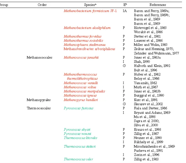
Table 2. Taxonomy of the methane-producing Archaea a . From Hedderich and Whitman, 2006
Methanogenic bacteria are abundant in habitats where electron acceptors such as O2,
NO3-, Fe3+ and SO42- are limiting. Methanogens are generally absent from the water column
of unstratified lakes and rivers because convection currents rapidly aerate the deep waters. However, the diffusion of O2 between the layers of stratified lakes is often too slow to
maintain oxic conditions in the lower layers. Similarly, the physical structure of sediments limits dispersive mechanisms, so deeper sediments are often anoxic and harbor methanogens.
In anoxic environments, the presence of NO3-, Fe3+ and SO42- inhibits methanogenesis by
the presence of sulfate, sulfate-reducing bacteria utilize H2 at concentrations lower than the
minimum concentration which can be utilized by methanogens (Kristjansson et al., 1982; Lovley, 1985). Presumably, the ability of the sulfate reducing bacteria to outcompete the methanogens is a direct consequence of the more-positive reduction potential of SO42−
compared to that of CO2. In environments with sufficient quantities of sulfate, hydrogen
sulfide is the predominant reduced product, and the major fate of biodegradable organic carbon is oxidation to CO2. If sulfate becomes limiting, methane replaces hydrogen sulfide as the reduced product, and the organic carbon is disproportionated to CO2 and methane.
Sulfidogenesis normally dominates in estuarine, marine, and hypersaline sediments, where sulfate diffuses from overlying water, whereas methanogenesis is the dominant process in anaerobic environments that contain large amounts of easily degradable organic matter such as the anoxic compartment of lakes.
I.5.
Sulfate- and sulfur-reducing prokaryotes
Sulfate-reducing bacteria gain energy for growth by coupling the oxidation of organic compounds or molecular hydrogen (H2) to the reduction of sulfate (SO42-) to sulfide (H2S,
HS-). Hence, sulfate-reducing bacteria are easily recognized by the production of high sulfide concentrations (with non-limiting electron donor and sulfate, usually in the range of several millimolar) concomitantly with growth and the strict dependence of this process on the presence of free sulfate. This process is also termed “dissimilatory sulfate reduction,” to allow clear differentiation from assimilatory sulfate reduction. Assimilatory sulfate reduction generates reduced sulfur for biosynthesis (e.g., of cysteine) and is a widespread biochemical capacity in prokaryotes and plants. Assimilatory sulfate reduction does not lead to the excretion of sulfide. The assimilated reduced sulfur is released as sulfide only through decay (putrefaction) of the biomass. The amounts of sulfide produced by dissimilatory sulfate reduction with a given amount of biomass is by orders of magnitude higher than the amount of sulfide liberated from the organic sulfur during putrefaction of the same amount of biomass.
The production of high concentrations of H2S often indicates the activity and presence of
sulfate-reducing microorganisms in natural habitats. The presence of H2S is obvious by its
characteristic smell, black precipitation of ferrous sulfide when iron minerals are present, and white patches of elemental sulfur as an oxidation product formed in contact with air. Such
signs for the activity of sulfate reducers are often encountered if organic substances accumulate in the presence of sulfate under anoxic conditions. Growth conditions for sulfate-reducing microorganisms prevail in sediments of virtually all aquatic habitats, which may be cold, moderate or geothermally heated up to approximately 105°C. But also flooded soils such as rice paddies and technical aqueous systems (as for instance sludge digestors, oil tanks or vats in the paper-making industry) may offer suitable growth conditions for sulfate-reducing microorganisms. From such habitats, in particular marine sediments, a great variety of sulfate-reducing microorganisms has been isolated.
Bacterial sulfate reducers fall into three major branches, the δ-subclass of Proteobacteria with more than twenty-five genera, the Gram-positive bacteria with the genera
Desulfotomaculum and Desulfosporosinus, and branches formed by Thermodesulfobacterium
and Thermodesulfovibrio. Sulfate reducers in the latter branch are thermophilic, whereas the two other branches comprise psychrophilic, mesophilic as well as thermophilic species.
Recently, also archaeal sulfate reducers have been detected. However, in comparison to bacterial species, fewer archaeal sulfate reducers are known. Moreover, they are less ubiquituous than their bacterial counterparts. Indeed, archaeal sulfate reducers appear to be restricted to habitats like hydrothermal vents, hot springs and deep, warm oil reservoirs.
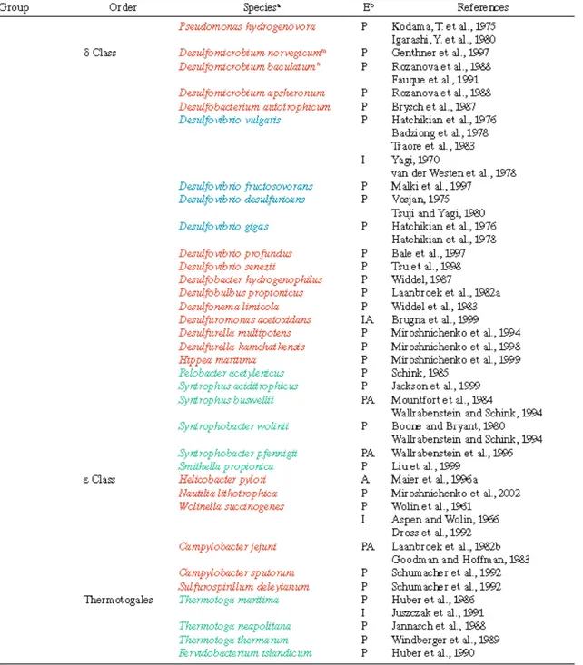
Currently recognized genera of sulfate-reducing prokaryotes and their main morphological and physiological properties are summarized in Table 3.
Table 3. Morphological and physiological properties of the genera of sulfate-reducing Bacteria and Archaea. From Rabus et al., 2006.
In addition to sulfate-reducing microorganisms, a variety of prokaryotes exists that reduce elemental sulfur but not sulfate. Among the lower oxidation states, the elemental sulfur is probably the most widespread sulfur species in sediments and geological deposits. Many chemical and biological oxidation processes of H2S do not directly lead to sulfate (the highest
that reduce elemental sulfur are widespread among different phylogenetic groups of Bacteria and Archaea.
Bacterial sulfur reducers may be mesophilic or moderately thermophilic, whereas archaeal sulfur reducers are all extremely thermophilic. Typical habitats of the hyperthermophilic sulfur reducers are solfataric fields, hot springs and hydrothermal systems in the deep sea, whereas mesophilic bacterial sulfur reducers can be isolated from almost every freshwater or marine sediment, or even from wet soil. Moreover, bacterial sulfur reducers have been found both in oxic and anoxic environment. Indeed, in contrast to dissimilatory sulfate reduction, the capacity for sulfur reduction has also been observed in bacteria that grow with O2 and which are, therefore, facultative anaerobes. However, many
sulfur-reducing microorganisms are strictly anaerobic.
An overview of the morphological and physiological properties of Bacteria and Archaea capable of S0-respiration is provided in Table 4.
Table 4. Morphological and physiological properties of Bacteria and Archaea capable of respiratory reduction of elemental sulfur. From Rabus et al., 2006.
I.6.
The Colorless Sulfur Bacteria
The name “colorless sulfur bacteria” has been used since the time of Winogradsky to designate prokaryotes that are either able, or believed to be able, to use reduced sulfur compounds (e.g., sulfide, sulfur and organic sulfides) as sources of energy for growth. Today, it is known that this group comprises a very heterogeneous collection of bacteria, many of which have little or no taxonomic relationship to each other. The colorless sulfur bacteria play an essential role in the oxidative side of the sulfur cycle. Like all of the element cycles, the sulfur cycle has an oxidative and a reductive side, which, in most ecosystems, are in balance. However, this balance does not always exist, and accumulations of intermediates such as sulfur, iron sulfides, and hydrogen sulfide are often found. On the reductive side, sulfate (and sometimes elemental sulfur) functions as an electron acceptor in the metabolic pathways used by a wide range of anaerobic bacteria, leading to the production of sulfide. Conversely, on the oxidative side of the cycle, reduced sulfur compounds serve as electron donors for anaerobic, phototrophic bacteria or provide growth energy for the extremely diverse group of (generally) respiratory colorless sulfur bacteria. Common oxidation products of sulfide are elemental sulfur and sulfate. The adjective “colorless” is used because of the lack of photopigments in these bacteria, although it should be noted that colonies and dense cultures can actually be pink or brown because of their high cytochrome content.
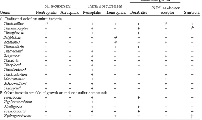
Table 5 lists the genera that have traditionally been regarded as colorless sulfur bacteria, as well as genera containing species originally not classified as such that have now been shown to be able to obtain energy from the oxidation of reduced sulphur compounds.
Table 5. Genera of the colorless bacteria traditionally recognized as being capable of growth on reduced sulfur compounds and their environmental parameters. From Robertson and Kuenen, 2006.
In addition to inorganic sulfur compounds, some species can also gain energy from the oxidation of other inorganic compounds such as hydrogen or ferrous iron. As well as differences in substrate range, there are also some variations in electron acceptor usage. Although most colorless sulfur-oxidizing bacteria require oxygen, a few are able to grow anaerobically using nitrogen oxides (e.g., nitrate) as their terminal electron acceptor during denitrification. One or two species (of the genus Acidianus) are capable of anaerobic metabolism by the reduction of sulfur (Segerer and Stetter, 1989), during which organic compounds or hydrogen serve as electron donors. Thiobacillus ferrooxidans is known to be able to reduce ferric iron under anaerobic conditions (Sugio et al., 1985). A somewhat exotic example of a sulfate reducer that might also be considered to be a colorless sulfur bacterium is
Desulfovibrio sulfodismutans, which can grow anaerobically by the disproportionation of
thiosulfate to sulfate and sulfide (Bak and Pfennig, 1987).
The heterogeneous group of colorless sulfur bacteria occurs in almost every life-supporting environment where reduced sulfur compounds are present.
In natural habitats, the reduced sulfur compounds available tend to be either sulfides (including metallic ores) or sulfur. Due to the activities of sulfate-reducing bacteria, especially
in anoxic sediments, hydrogen sulfide is very commonly available. One of the main factors that bacteria growing on hydrogen sulfide have to contend with is the chemical reaction between sulfide and oxygen. Therefore, the colorless sulfur bacteria are frequently found in the gradients at the interface between anoxic, sulfide-containing areas and aerobic waters and sediments where, at very low oxygen and sulfide concentrations, they can effectively compete with the spontaneous chemical oxidation reaction.
I.7. The denitrifying prokaryotes
The denitrifying prokaryotes reduce nitrate to gaseous nitrogen oxides, principally nitrogen gas.
The initial step in this process is the reduction of nitrate to nitrite, as occurs in ammonification. In the next step, nitrite is reduced to nitric oxide, a gaseous nitrogen oxide. This conversion of a fixed, non-gaseous form of nitrogen to gaseous forms has led this reaction to be termed “denitrification” because biologically preferred forms of nitrogen are lost.
In prokaryotes, reduction of nitrate is a respiratory process. That is, it is coupled to ATP synthesis via electron transport chains. However, with one or two exceptions, denitrifiers can also respire with oxygen as the terminal electron acceptor and, because it is usually available at higher concentrations, oxygen is typically the preferred electron acceptor. When oxygen becomes limiting, the capacity to utilize nitrate as a terminal oxidant allows denitrifying bacteria to continue respiration using an alternative electron acceptor.
Because each nitrogen oxide reduction has a positive redox couple, every step in denitrification need not be carried out to achieve a net conservation of energy. In fact, it is quite common to isolate bacteria that express only portions of the denitrification electron transport chain. Those prokaryotes that contain partial denitrification chains will be included in this paragraph and a list of prokaryotic genera suggested to include dentrifying strains is shown in Table 6.
Denitrification ability is widespread among Eubacteria, and almost exclusively in those strains that are capable of aerobic growth. Rarely it occurs in anaerobic bacteria. The majority of currently characterized denitrifiers are found in α β γ and ε subdivisions of the
Proteobacteria group. The δ subdivision, which contains a number of strict anaerobes, has not
been found to contain any strains that denitrify, as yet.
The α subdivision of the Proteobacteria contains a number of well characterized denitrifiers including Paracoccus denitrificans, various Rhodobacter strains, and several rhizobia. Important denitrifiers in the α subdivision include Hyphomicrobium, budding bacteria often found in waste-water treatment facilities (Fesefeldt, 1998). Denitrification among the rhizobia, which are known for their roles as nitrogen-fixing symbionts, is also noteworthy because these strains have the seemingly contradictory capacity for both nitrogen fixation and denitrification (O’Hara, 1985). This ability to both fix and “unfix” nitrogen is fairly widespread among denitrifiers: Rhodobacter, Hyphomicrobium, Frankia, Azospirrilum,
Azoarcus and some pseudomonad strains can both fix nitrogen and reduce nitrates to nitrogen
gas. Some members of the genus Nitrobacter, which are nitrifying α-Proteobacteria, produce nitric oxide from nitrite. Even though a putative nitrite reductase has been purified from
Nitrobacter vulgaris (Ahlers, 1990), other studies did not reveal production of nitric oxide or
nitrous oxide in cultures of Nitrobacter species (Baumgartner, 1991; Goreau, 1980). Further studies are required to determine if these nitrifying bacteria are also denitrifiers.
Although a number of members of the β-Proteobacteria have the capability to denitrify, they are not as well studied, on the whole, as members of the α or γ subdivisions. Among these, a number of aromatic compound-degrading denitrifiers have been isolated, and many belong to the genera Azoarcus and Thauera (Fries, 1994; Springer, 1998; van Schie, 1998; Anders, 1995). Moreover it has been well documented that several species of Neisseria can denitrify (in fact, one species is named Neisseria denitrificans). Also some species of the genus Nitrosomonas, that are included into the β-Proteobacteria subdivision, have been described for their ability to oxidize ammonia to nitrite. The finding of a copper-containing nitrite reductase from Nitrosomonas europaea (Miller, 1985; Dispirito, 1985) strongly indicates that Nitrosomonas species are nitrifying denitrifiers. However, the role of denitrification in Nitrosomonas, whether for detoxification or for energy conservation, has not been established as yet.
Denitrification in members of γ-Proteobacteria subclass has been well studied in
Pseudomonas stutzeri and Pseudomonas aeruginosa. Also Beggiatoa and Thioploca are
internal vacuoles (McHatton, 1996; Fossing, 1995). Then they use this accumulated nitrate as an oxidant and sulfide in the surrounding environment as a reductant. The use of reduced sulfur compounds as electron donors is known to occur in several denitrifiers including
Aquifex (Huber, 1992), Paracoccus (Friedrich, 1981), and Thiobacillus (Schedel, 1980).
A few denitrifiers have been found in the ε subdivision of the Proteobacteria. Some exhibit truncated denitrification chains. For example, both Wolinella succinogenes and some
Campylobacter species have the capacity to grow with nitrous oxide as sole terminal oxidant
(Yoshinari, 1980; Payne, 1982). However, neither bacterium is able to reduce nitrite to nitrous oxide. Thiomicrospira denitrificans, which is closely related to the Campylobacter group (Muyzer, 1995), can reduce nitrite to gaseous end products (Timmer-ten Hoor, 1975). Also, T.
denitrificans can use sulfur compounds as reductants.
Among the Gram-negative bacteria that are not proteobacteria, denitrifying strains are found in the genera Aquifex (Huber, 1992), Flexibacter (Jones, 1990), and Flavobacterium (Coyne, 1989). Even though the majority of denitrifiers are Gram-negative, denitrifying bacteria are well represented among Gram-positive bacteria. For example, there have been a number of denitrifying Bacillus species described. Other Gram-positive bacteria capable of denitrification include strains of Propionibacterium (Swartzlander, 1993) and Jonesia
denitrificans (originally Listeria denitrificans) (Rocourt, 1987). These strains are somewhat
unusual because they seem to be denitrifying strains in groups of bacteria that are primarily non-denitrifiers. A high number of denitrifying strains were found in Frankia genus (Lensi, 1990). Recently, it has been shown that a number of actinomycetes, including Streptomyces,
Dermatophilus and Nocardia, are capable of nitrous oxide evolution from nitrate or nitrite
(Shoun, 1998).
Only a few Archaea capable of denitrification have been isolated. Most of the denitrifying Archaea are halophilic bacteria capable of aerobic respiration but more recently, also denitrifying non-halophilic Archaea such as the hyperthermophile Pyrobaculum
I.8. Dinitrogen-fixing prokaryotes
Dinitrogen fixation, the biocatalytic conversion of gaseous nitrogen (N2) to ammonium, is
an exclusive property of prokaryotes. The enzyme catalyzing this reaction is the nitrogenase. Nitrogen fixation (N2 fixation) is the most important way by which N2 present in the
atmosphere enters biological systems. Many diazotrophs (di = two, azote = nitrogen; trophs = eaters: dinitrogen fixers) are found to be associated with the roots of plants where they exchange fixed nitrogen for the products of photosynthesis. Biological N2 fixation is the main
source of nitrogen in soil, marine environments such as oligotrophic oceanic waters (where dissolved fixed-nitrogen content is extremely low; Zehr et al., 1998; Staal et al., 2003), subtropical and tropical open ocean habitats (Karl et al., 2002), and hydrothermal vent ecosystems (Mehta et al., 2003). N2-fixing prokaryotes inhabit a wide range of environments
including soils, seas, oceans, insects, cow rumena, human intestines, and feces (Bergersen and Hipsley, 1970). Nevertheless, the presence of a N2-fixing bacterium is not evidence for the
occurrence of N2 fixation. This is the case of many lakes, such as Mono Lake, where no significant nitrogen fixation rate was detected, although potential nitrogen fixers were present (Steward et al., 2004). Also the N2 fixation rates measured in lake Cadagno were moderate, but the detection of nitrogenase gene and of its expression in both oxic and anoxic waters, indicated that N2 fixation might be occurring throughout the water column (Halm et al., 2009). Anyway, at the present time, little is known about N2 fixation occurring in lakes.
Diazotrophs are encountered in Bacteria and in some groups of Archaea. Nowadays, only 6 out of 53 currently identified major lineages or phyla within the domain Bacteria (Proteobacteria, Cyanobacteria, Chlorobi (green nonsulfur), Spirochetes and the Gram-positives (Firmicutes and Actinobacteria) have nitrogen-fixing members. The N2-fixing
capability is unevenly distributed throughout these prokaryotic taxa, and N2-fixing bacteria
are in restricted clusters among species of non-N2-fixing bacteria.
Among Eubacteria, N2-fixing species seem to be dominant in Rhodospirillaceae
(Madigan et al., 1984) whereas only a subset of cyanobacterial species are able to fix N2.
Gluconacetobacter diazotrophicus and a couple of other N2-fixing species are the only
diazotrophs in a larger group comprising Acetobacter, Gluconacetobacter and Gluconobacter (Fuentes-Ramírez et al., 2001). Similarly, among aerobic endospore-forming Firmicutes (Gram-positive bacteria), N2 fixers are encountered mainly in a discrete group (defined by
al., 1999). Among the Actinomycetes, N2-fixing Frankia, represented by a diversity of
phenotypes from different habitats, are grouped on the basis of their 16S rRNA gene sequences (Normand et al., 1996).
In Archaea, N2-fixing organisms are found in the methanogen group and in the halophile
group within the Euryarchaeota but not in the sulfur-dependent Crenarchaeota (Young, 1992).
I.9. The inorganic nitrogen compounds oxidizing prokaryotes
Nitrifying bacteria are separated into two groups, the ammonia- and the nitrite oxidizers, on the basis of their ability to lithotrophically oxidize ammonia to nitrite and nitrite to nitrate, respectively. These two groups possess very different key enzyme systems: among the main enzymes involved in oxidation of ammonia and nitrite, the enzyme ammonia monooxygenase
(AMO) catalyzes the conversion of ammonia to hydroxylamine, the first intermediate of ammonia oxidation, whereas the enzyme nitrite oxidoreductase (NO2-OR) catalyzes the oxidation of nitrite to nitrate.
Nitrifying bacteria are present in oxic and even anoxic environments. They are widely distributed in freshwater, seawater and soils. Nitrifiers also could be enriched or isolated from extreme habitats like heating systems with temperatures of up to 47 °C (Ehrich et al., 1995) and permafrost soils up to a depth of 60 m at a temperature of down to -12 °C. Although the pH optimum for cell growth is 7.6–7.8, nitrifiers were frequently detected in environments with suboptimal pH (e.g., acid tea soils and forest soils at pH values below 4) but also in highly alkaline soda lakes showing pH values of 9.7–10.5 (Sorokin et al., 2001).
Lithotrophic nitrifiers are Gram-negative bacteria and conventionally have been placed in the family Nitrobacteriaceae (Buchanan, 1917; Watson, 1971; Watson et al., 1989). However, phylogenetically the lithoautotrophic ammonia oxidizers, characterized by the prefix Nitroso-, and nitrite oxidizers, characterized by the prefix Nitro-, are not closely related (Teske et al., 1996; Purkhold et al., 2000).
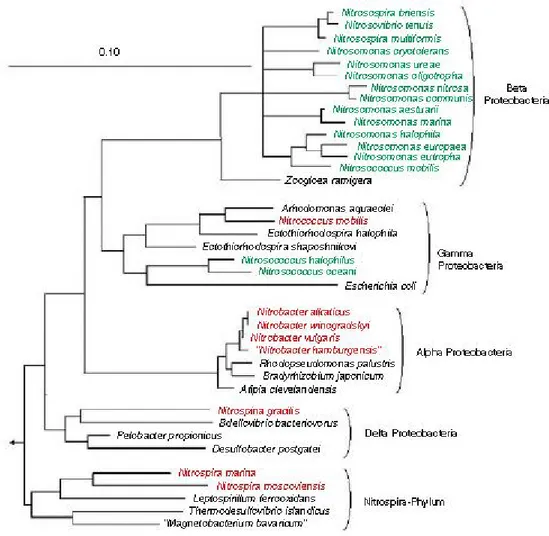
Comparative 16S rRNA sequence analysis demonstrated that all recognized ammonia oxidizers are either members of the β- or γ-subclass of Proteobacteria (Fig. 2). The genera
Nitrosomonas (including Nitrosococcus mobilis), Nitrosospira, Nitrosolobus and
Nitrosovibrio form a closely related monophyletic assemblage within the β-subclass of Proteobacteria (Head et al., 1993; Woese et al., 1984; Teske et al., 1994; Utåker et al., 1995;
constitutes a separate branch within the γ-subclass of Proteobacteria (Woese et al. 1985; Purkhold et al., 2000). Among the nitrite oxidizers, the genera Nitrobacter, Nitrococcus and
Nitrospina were assigned to the α-, γ-, and δ subclass of Proteobacteria, respectively (Orso et
al., 1994; Teske et al., 1994). Nitrite oxidizers of the genus Nitrospira are affiliated with the recently described Nitrospira-phylum, which represents an independent line of descent within the domain Bacteria (Ehrich et al., 1995).
Figure 2. 16S rRNA-based tree reflecting the phylogenetic relationship of ammonia- and nitrite-oxidizing bacteria. The consensus tree is based on the results of a maximum likelihood analysis of the 16S rRNA primary structure data from the nitrifying bacteria shown in the tree and a selection of reference sequences. Only homologous positions that share identical residues in at least 50% of all available almost complete bacterial 16S rRNA sequences were included for tree reconstruction. In the tree, ammonia oxidizers are labeled green, and nitrite oxidizers are depicted in red. It should be noted that the assignment of the genus Nitrospina to the δ-Proteobacteria is tentative and might change if additional reference sequences become available. Multifurcations connect lineages for which no unambiguous branching order could be retrieved using different treeing methods. Bar represents 10% estimated sequence divergence. From Bock and Wagner (2006)
In addition to lithotrophic nitrifiers, various heterotrophic bacteria, (Focht and Verstraete, 1977; Killham, 1986; Papen et al., 1989) are capable of oxidizing ammonia to nitrate. However, in contrast to lithotrophic nitrification, heterotrophic nitrification is not coupled to energy generation. Consequently, heterotrophic nitrifiers are dependent on the oxidation of
organic substrates (Focht and Verstraete, 1977; Kuenen and Robertson, 1987). During heterotrophic nitrification, ammonia or reduced nitrogen from organic compounds (e.g., the amino group of amino acids) is co-oxidized to hydroxylamine, gaseous nitrogen oxides, nitrite or nitrate. For example, methane-oxidizing bacteria were shown to co-oxidize ammonia to nitrite (Anthony, 1982; Yoshinari, 1985; O’Neil and Wilkinson, 1977; Zahn et al., 1994; Bergmann et al., 2000).
The environmental importance of heterotrophic nitrifiers is controversial in the literature. Generally it is assumed that in most environments, the biological conversion of reduced forms of nitrogen to nitrite and nitrate is catalyzed mainly by the lithoautotrophic ammonia- and nitrite-oxidizing bacteria and not by heterotrophic nitrifiers. This reflects the fact that the nitrification rates of heterotrophic nitrifiers are small compared to those of autotroph ic nitrifiers (Robertson and Kuenen, 1988).
I.10. The hydrogen-metabolizing prokaryotes
Hydrogen is an important and widespread metabolite among prokaryotes living in oxic as well as in anoxic habitats. The physiological role of H2 in microbes is dual. Firstly, H2 is a
growth substrate, i.e., a source of energy and reductant. Prokaryotes of different metabolic types, such as methanogens, anoxygenic phototrophs, and aerobic knallgas bacteria, use H2.
Secondly, H2 production is a means of dispersing excess reductant from fermentative
metabolism. Hydrogen is among the fermentation products of both facultative fermenters, such as Escherichia coli, and obligate fermenters such as Clostridium pasteurianum.
The list of H2-metabolizing prokaryotes (Tables 7 and 8) shows as H2 metabolism is not
Table 8. H2-metabolizing Archaea. From Schwartz and Friedrich, 2006
The common denominator of the disparate taxonomic and physiological groups treated in this paragraph is the presence of hydrogenase, the main enzyme responsible both for hydrogen evolution and consumption. These reactions are not catalyzed by hydrogenases alone. Nitrogenase, the enzyme which catalyzes the production of NH3 from atmospheric N2,
produces H2 as a byproduct of N2 fixation and therefore also N2-fixing prokaryotes give an important contribution to the global H2 flux.
Both biogenic and abiogenic H2 production can support growth of H2-utilizing
prokaryotes. Many H2-utilizing species profit from fermentative H2 production in anoxic
sediments. The two most important groups of H2-consumers in such biotopes are the
that methanogens and sulfate reducers compete for H2 (Winfrey and Zeikus, 1977; Winfrey et
al., 1977; Abram and Nedwell, 1978; Oremland and Polcin, 1982; Lovley et al., 1982; Lovley and Klug, 1983). Sulfate reducers outcompete the methanogens in the presence of sulfate, because the former have a higher affinity for H2 and a higher growth yield (Kristjansson et al.,
1982; Schönheit et al., 1982). The key factor determining which of the two terminal degradation processes—sulfidogenesis or methanogenesis— prevails in a given habitat is SO42- concentration. In anoxic marine sediments, where there is an abundant supply of SO42-,
sulfate reduction is the dominant process consuming most of the available H2 and acetate. The
sulfate level in lakes varies depending on their trophic state, but in general is lower than in seawater. The thickness of the zone of sulfate reduction varies accordingly. In eutrophic lakes the sulfate concentration in the sediment drops sharply. Here the zone of sulfate reduction is only a few centimeters thick. In lakes with lower nutrient contents the zone of sulfate reduction extends deeper into the sediment (Lovley and Klug, 1983). Competition for H2 does
not mean that sulfate reduction and methanogenesis are mutually exclusive processes. Various methanogens can exploit substrates, e.g. methylamine, that are not utilized by sulfate reducers (Oremland and Polcin, 1982; Winfrey and Ward, 1983). Therefore, the two groups of organisms can coexist in the same biotope, as has been shown for instance for estuarine sediments. In sediment from an oligotrophic lake, it was reported that the total flux of electrons and carbon routed through sulfate reduction is between 30 and 81% of the total terminal metabolism (Lovley and Klug, 1983). Sulfate reduction is also an important process in extreme environments, such as the anaerobic sediments of soda lakes. Among the specialized, H2-utilizing, sulfate-reducing bacteria found in such sediments are the alkaliphilic
lithoheterotroph Desulfonatronovibrio hydrogenovorans and the alkaliphilic lithoautotroph
Desulfonatronum lacustre (Zhilina et al., 1997; Pikuta et al., 1998).
H2-evolving prokaryotes will be treated more exhaustively in the next paragraph.
I.10.1. Hydrogen-producing bacteria and hydrogenases
The decomposition of organic matter via fermentation is one of the major biotic energy-yielding processes in anaerobic habitats. Various types of fermentation result in the formation of H2 as a terminal product and hence constitute a substantial contribution to the global H2
balance. Both obligate and facultative anaerobic bacteria produce H2. These microorganisms
are limited to anoxic zones rich in organic substance, such as marine and lacustrine sediments. These habitats are fed by a constant influx of organic material derived from photosynthetic
primary producers and from the ensuing food chains. The upper, oxic layer of the sediment varies in depth both in marine and freshwater sediments. Below the upper, oxic layer is the zone of anoxic decomposition. In this layer, the H2 produced neither accumulates nor does it
escape in significant quantities to the oxic zone. If H2 were to accumulate, the fermentative
metabolic processes would soon come to a halt, since these are inhibited by relatively low concentrations of H2 in the environment. The inhibitory concentrations vary for the different
fermentative reactions, depending on their energetics. Fermentation of fatty acids such as butyrate and propionate to acetate, H2 and CO2, for instance, is more endergonic than the
fermentation of ethanol to acetate and H2 and the former process ceases at a much lower
concentration of external H2 than the latter.
Hydrogen is generated also as a byproduct of N2 fixation in both oxic and anoxic
environments. Cyanobacteria and prochlorophytes are probably the most widespread diazotrophs on earth. These organisms inhabit the upper, oxic zones of oceans and lakes. The anoxygenic photosynthetic bacteria, which occupy deeper zones depleted for O2, also engage
in N2 fixation. These and other diazotrophs contain uptake hydrogenases and, hence, are
capable of exploiting at least a part of the H2 generated by nitrogenase. However,
hydrogenase-free strains are widespread in nature and produce large quantities of H2.
Bacteria that possess the capability to produce hydrogen include strict anaerobes (Clostridia, and species belonging to the groups of methylotrophs, rumen bacteria, methanogenic bacteria and Archaea), facultative anaerobes (Escherichia coli, Enterobacter, Citrobacter), and even aerobes (Alcaligenes, Bacillus). A comprehensive review on the hydrogen-producing characteristics of these bacteria was published (Nandi and Sengupta, 1998). Among the hydrogen-producing bacteria, Clostridium sp. and Enterobacter are the most widely studied. Species of genus Clostridium are gram-positive, rod-shaped, strict anaerobes and endospore formers (Holt et al., 1994; Nandi and Sengupta, 1998), whereas Enterobacter species are gram-negative, rod-shaped, and facultative anaerobes (Holt et al., 1994). The hydrogen producing characteristics of these two genera and three thermophilic species were reviewed by de Vrije and Claassen (2003). Species of Clostridium and Enterobacter genera that have been studied for H2 production include Clostridium sp. (Taguchi et al., 1992, 1993, 1994, 1995), Clostridium parapatrificum (Evvyernie et al., 2000, 2001), Clostridium butyricum (Heyndrickx et al., 1986, 1990), Enterobacter aerogenes (Tanisho et al., 1983, 1987, 1989; Tanisho and Ishiwata, 1994; Rachman et al., 1998) and Enterobacter cloacae (Kumar and Das, 2000, 2001; Kumar et al., 2000). The thermophiles
include Thermotoga maritime (Schr¨oder et al., 1994), Thermotoga elfii (de Vrije et al., 2002), and Caldicellulosiruptor saccharolyticus (van Niel et al., 2002).
Hydrogenases, the enzymes that catalyze the reversible oxidation of molecular H2, are widespread in prokaryotic and lower eukaryotic biological systems (Stephenson and Stickland, 1931; Vignais et al., 2001). Hydrogenases are diverse in their protein structure and in the type of electron carrier they use (e.g. ferredoxins, rubredoxins and quinines etc.).
Early classification schemes, which had mainly biochemical data to go on, grouped hydrogenases on the basis of metal content or redox cofactors (Fauque et al., 1988; Przybyla et al., 1992). A rapidly growing base of nucleotide sequence data was used to attempt a classification on the basis of comparisons of deduced amino-acid sequences (Voordouw, 1992). In a similar study, Wu and Mandrand (1993) went a step further. These authors generated multiple alignments for full-length amino acid sequences and performed cluster analysis on the pairwise alignment scores. On the basis of the resulting dendrograms, 30 hydrogenases were grouped in six classes. Recently, Vignais and coworkers have refined and extended this classification system, including a greatly expanded database (Vignais et al., 2001; Vignais and Billoud, 2007). They carried out cluster analyses using both complete amino acid sequences and segments corresponding to functional domains.
The results of these important studies have led to the identification of three phylogenetically distinct classes of proteins, the [NiFe]-hydrogenases, the [FeFe]-hydrogenases, and the iron-sulfur-free [FeFe]-hydrogenases, initially called metalfree and now renamed [Fe]-hydrogenases.
I.10.1.1. [NiFe]-hydrogenases
The revised system of Vignais and coworkers subdivides the [NiFe]-hydrogenases into four groups (Vignais et al., 2001):
Group 1. Energy-transducing hydrogenases:
Enzymes which couple the oxidation of H2 to electron-transport phosphorylation. This group includes both membrane-bound hydrogenases attached to the periplasmic side of the cytoplasmic membrane and nonmembrane-bound, periplasmic hydrogenases. The group breaks down into two subclusters. One contains the membrane-bound hydrogenases of the Proteobacteria, and the other, archaeal membrane-bound hydrogenases.