Abstract. – OBJECTIVE: The approval of the anti-PD1 antibody nivolumab has provided a sig-nificant therapeutic opportunity in the landscape of metastatic melanoma. In pivotal clinical tri-als, nivolumab improved clinical outcomes with a great safety profile. However, in real-world prac-tice, the majority of the population with metastat-ic melanoma does meet one or more eligibility criteria of pivotal trials, since they have an ECOG-PS ≥ 2 or active/untreated known brain metasta-ses. Waiting for larger real-wold studies that are currently lacking, but would be crucial to confirm the efficacy of nivolumab in challenging patients and to detect rare adverse events that could not be noticed in pivotal trials, this review collects both literature and unpublished case reports on nivolumab treatment in metastatic melanoma.
PATIENTS AND METHODS: Case reports, pub-lished from 2016 to February 2018, and five, un-published case reports, representative of Italian clinical practice, were reported and potential is-sues that physicians could face with the use of nivolumab in the real world were discussed.
RESULTS: Among Italian cases, one patient had a huge retro-nuchal mass, which significantly de-creased with few cycles of nivolumab; two pa-tients were affected by cardiovascular comorbid-ities and one had brain metastasis; the last had a long history of disease, firstly diagnosed in 1997. A literature review was mainly focused on the ex-perience in the management of rare immune ad-verse events related to treatment.
CONCLUSIONS: Nivolumab confirmed its effi-cacy and safety in real-world; the decision-mak-ing process on startdecision-mak-ing and scheduldecision-mak-ing the
treatment, even in the management of adverse events, should consider multiple factors related to both patient (i.e., BRAF status, ECOG PS, co-morbidities) and disease (burden, metastasis).
Key Words:
Nivolumab, Metastatic melanoma, Case report, An-ti-PD1, Real world, Immunotherapy, Brain metastasis, Clinical practice.
Introduction
The approval of immune checkpoint blocking antibodies has provided a significant therapeu-tic opportunity in the landscape of many can-cers1. These drugs – ipilimumab, a fully human
IgG1 monoclonal antibody that targets CTLA4, nivolumab and pembrolizumab, humanized IgG4 monoclonal antibodies that target PD1 – can pre-vent the interaction between co-inhibitory mol-ecules and their receptors, thereby boosting the body’s natural defense against tumors2,3. FDA
has approved nivolumab for many indications, including advanced melanoma, advanced non-small cell lung cancer, advanced renal cell car-cinoma, classical Hodgkin lymphoma, advanced squamous cell carcinoma of the head and neck, urothelial carcinoma, MSI-H or dMMR metastat-ic colorectal cancer, and hepatocellular carcino-ma. The longest clinical experience on nivolumab
R. DEPENNI
1, C. DE ROSSI
2, M. DE TURSI
3, R. MARCONCINI
4, T. TROIANI
51Department of Oncology and Haematology, Division of Oncology, University Hospital of
Modena, Modena, Italy
2U.O.C. Medical Oncology, Ospedale dell’Angelo di Mestre, SS Giovanni and Paolo Hospital
Venice, Venice, Italy
3Department of Oral and Biotecnological Medical Sciences, G. D’Annunzio University, Chieti, Italy 4Unit of Medical Oncology 2, Azienda Ospedaliero-Universitaria Pisana, Department of Traslational
Research and New Technologies in Medicine and Surgery, University of Pisa, Italy
5Dipartimento di Internistica Clinica e Sperimentale, Università degli studi della Campania “Luigi
Vanvitelli”, Napoli, Italy
All authors equally contributed to this work
Real world treatment practice in patients with
advanced melanoma in the nivolumab era: five
novel Italian case reports and a literature review
use has been achieved in the setting of metastat-ic melanoma that histormetastat-ically has been one of the first cancers treated with immunotherapy. Melanoma cells express co-inhibitory molecules within the tumor microenvironment to escape the immune system and hamper an effective tumor clearance4,5. Therefore, it is the ideal disease to
target with checkpoints inhibitors.
The current World Health Organization (WHO) estimates are that 132,000 melanomas occur each year around the world, resulting in 65,000 deaths annually6. Early diagnosis and resection cure
almost 90% of cases of stage I melanoma7. By
contrast, the prognosis for regional and distant metastatic melanoma (stages III and IV, respec-tively) is variable and generally poor, with 5-year survival rates for stage III of 13%-69% and as low as 6% in stage IV8,9. An improvement of
clinical outcomes in patients with metastatic mel-anoma has been reported with the introduction of checkpoint inhibitors, which seem are more effective and yet no less tolerable than control interventions10. A recent metanalysis indicates
that targeting PD1 seems to offer greater efficacy than blocking CTLA410. Indeed, in clinical trials,
nivolumab showed a significant improvement in clinical outcomes, as compared with dacarbazine and ipilimumab11-14. The safety profile of this
drug was favorable, with pruritus, fatigue, diar-rhea, and nausea as the most common adverse events11-14.
However, in pivotal trials, patients were highly selected and only patients with ECOG-PS ≤ 1, without active brain metastasis, ocular melano-ma or autoimmune disease were included11-13. In
real-world practice, 55% of the total population with metastatic melanoma did not meet one or more eligibility criteria at first evaluation, and an ECOG-PS ≥ 2 or active/untreated known brain metastases accounted alone for 74% of non-eligi-bility cases15.
Furthermore, infrequent and rare adverse events could be more accurately described in a wider population than those included in pivotal trials, as in post-marketing surveillance and re-al-world study.
As per our knowledge, no data are currently available about real-world practice with nivolum-ab in metastatic melanoma. Therefore, in this minireview, we collected clinical case reports published from 2016 to February 2018 and 5 novel cases from Italian real-world experience, to better define the characteristics of patients who may have major benefits from nivolumab therapy.
Italian patients provided a written consent. Ethical requirements were fulfilled, accordingly to “Decreto Legge 196”, article 4 (2003) and all clinical cases were conducted according to cur-rent good clinical practices and local laws.
Case 1
In 2015, a 72-year-old woman was diagnosed of melanoma. The lesion was vascularized, der-mo-hypodermically located at the retro-nuchal level, and wild-type for BRAF mutation. Baseline Fdg-PET/CT scan showed increased uptake in the retro-nucal mass as well as in the omolateral nuchal and cervical lymph nodes, left upper pul-monary lobe, left pulpul-monary hilar and mediastinal lymph nodes. From May 2016 to June 2016, the patient received a first-line chemotherapy with carbo-platin plus paclitaxel for six courses, with-out obtaining any clinical response, whereas the burden of the lesion was progressively increasing (Figure 1A). In August 2016, when a second-line treatment with nivolumab (3 mg/kg, every two weeks) was started, the CT-scan described a 90 × 75 × 60 mm right nuchal mass infiltrating the ad-jacent muscles, a 60 × 60 × 40 mm left pulmonary lesion at the hilum without apparent cleavage plane with the pulmonary artery, and pathological lateral cervical and mediastinal lymph nodes.
LDH level was ≥ 5 × Upper Limit Normal. After two doses of nivolumab size reduction of the main lesion was visible, becoming more ev-ident in the next weeks; (Figure 1B) and lactate dehydrogenase (LDH) level returned within the normal range after the 5th cycle of therapy. The
CT scan performed after 11 cycles of nivolumab, January 2017, showed a markedly reduction in size of the retro-nuchal lesion (45 × 30 mm) and the left pulmonary lesion (20 mm), and the cervi-cal and mediastinal lymph-nodes returned within < 1 cm in short axis. Nivolumab was continued for further six months, when the patient report-ed pruritus (grade 2) at arms and trunk, with desquamation areas. Cetirizine treatment (10 mg twice daily) and emollient cream were prescribed and nivolumab was temporarly discontinued. The last radiological assessment (October 2017) con-firmed the excellent clinical response, and the main lesion is no more visible (Figure 1C). Treat-ment is still ongoing without significant toxicities
Case 2
In 2016, an 80-years-old man with severe car-diac comorbidities was diagnosed of metastatic melanoma. Previously, in 2008, he underwent
anterior resection with colorectal anastomosis for rectal carcinoma, treated with neoadjuvant chemo-radiation; in 2015, he underwent coro-nary artery bypass graft surgery for hyperten-sion and received an implantable cardioverter defibrillator. In addition, he took ASA, warfarin, digoxin, furosemide, atorvastatin, amiodarone, and carvedilol.
In January 2016, the CT scan showed a nod-ule 2.1 × 2.0 cm in the apical segment of the right lower lobe and 1.0 cm right hilar lymph node; the PET-CT analysis highlighted an uptake in the pulmonary nodule, but not in the hilar lymph node. Hyperactivity was also observed at the dermal-subcutaneous thickening in the left scapular region and in the right mammary region. In March 2016, the presence of multiple subcutaneous nodules in the shoulder and right sub-mammary was confirmed by soft tissue ul-trasound analysis and the histopathological re-port referred as melanoma metastasis, with a V600K mutation in B-Raf gene. Considering the existing cardiovascular condition, a thorough cardiovascular evaluation and echocardiography were performed: the patient presented a dilated hypokinetic cardiomyopathy, mitral failure, ejec-tion fracejec-tion at 38%, and congestive heart class III NIHA. In April 2016, in addition to previously observed pulmonary lesions (Figure 2A), the CT scan showed a further solid lesion (1.8 × 1.5 cm) in the pararenal adipose tissue and in the objec-tive examination three subcutaneous nodules in the left axilla, left scapula and perineal area were detected. In May 2016, the patient started a ther-apy with nivolumab (3 mg/kg, every two weeks). At the first tumor assessment, CT scan showed complete regression of pulmonary metastasis (Figure 2B), significant regression of the
posteri-or pararenal metastatic localization, and complete regression of subcutaneous metastatic melanoma. The patient continued nivolumab, with excellent tolerance and a good clinical condition (ECOG PS=0). The cardiac evaluation confirmed that the pre-existing dilated hypokinetic cardiomyopathy and the severely reduced contractile function (EF 36%) were unchanged. No drug-related ad-verse events were noted. In August 2017, at the last tumour assessment, CT scan confirmed the complete regression of pulmonary, pararenal, and subcutaneous metastases. After the 14th cycle of
nivolumab, asymptomatic hypothyroidism was reported (TSH level < 0.1 mU/ml, FT4 and FT3 in the normal range) and none therapy was pre-scribed. After the 18th cycle, the THS value
returned within the normal range (0.38 mU/ml, range 0.27-4.20). Currently, the patient’s clinical status is excellent; he is still receiving therapy with nivolumab in complete remission, with a good tolerance after 33 cycles.
Case 3
In November 2011, a 72-year old man affect-ed by hypertension and asymptomatic ischemic disease underwent surgery to excise a cutaneous pigmented lesion in the xiphoid region. The histo-pathological report referred as epithelioid nodular melanoma, not ulcerated (Breslow 2.35 mm; pT3a). The sentinel lymph node biopsy in the right axilla was positive for multiple sub-capsular metastases, which occupy 30% of parenchyma. However, the histological report after axillary dissection did not refer any lymph node as positive. In January 2015, several nodules of malignant melanoma in transit, with melanomatous cells in reticular derma and in the vascular-adipose tissue were detected. The histological examination indicated the presence
Figure 1. Main lesion size decreasing during nivolumab treatment. (A) Before treatment; (B) after two doses of nivolumab;
of BRAF mutation V600E, while NRAS was
wild-type; LDH level was within the normal range
(531 U/L, range 313-618U/L). In March 2015, a PET-CT scan showed high metabolic activity at right mammary and left axillary region. There-fore, a treatment with ipilimumab (3 mg/kg q21 for 4 infusions) was started. The disease was
stable in the first two assessments, but at the third evaluation, a progression was reported: the right mammary lesion (from 5.3 cm to 7.8 cm) and the hepatic lesion (2.3 cm) in the VI segment increased their volumes, and a novel lesion was observed at pre-pectoral level (2.0 cm) (Figure 3A). LDH levels consistently increased (Figure 4).
Figure 2. CT scan before nivolumab (left panel); CT scan, at the first tumour assessment, showed complete regression of the
pulmonary metastatic disease (right panel).
Figure 3. Progression o in mammary and hepatic lesions and a novel pre-pectoral lesion, at the third tumor assessment,
during ipilimumab treatment (upper panel). Partial response in mammary and hepatic metastasis and the pre-pectoral lesion (lower panel).
nel lymph node were examined: the biopsy was positive for the presence of micrometastases of melanoma in the left axilla. Therefore, the left ax-illary lymph node dissection was performed: one lymph node out of 14 was positive, with a V600E mutation in the BRAF gene. The patient refused an adjuvant treatment with interferon and started the follow-up. In 2012, a CT scan revealed hepatic and splenic disease progression. From Septem-ber 2012 to February 2013, the patient received vemurafenib, obtaining a complete response. However, in March 2013, disease progression at hepatic and splenic level was detected again. The patient started a treatment with ipilimumab for 4 cycles, obtaining a stable disease. In August 2013, CT scan detected one metastatic encephalic lesion, which was excised. The follow-up was continued, and, in May 2016, a progression of the encephalic lesion was observed and treated with gamma-knife radiotherapy. MRI and total body CT scan showed an ependymal nodule in the right caudate nucleus, identified as metastatic lesion, progression of splenic and adrenal lesions, and stable disease in the liver. In October 2016, the patient started nivolumab treatment (3 mg/ kg, every two weeks) and all lesions, especially encephalic, adrenal and splenic, were progres-sively reduced. The treatment with nivolumab is currently ongoing after 25 cycles, the patient’s clinical status is good, and he maintains a clinical response.
Case 5
In 1997, 39 years-old male patient affected by hypercholesterolemia and type 2 diabetes melli-tus underwent the excision of dorsal melanoma, followed by radicalization. The histopathological In January 2016, the patient started a
ther-apy with vemurafenib, a BRAF inhibitor (960 mg, twice daily). Considering the cardiovas-cular comorbidities, a thorough cardiological evaluation was performed: the sinus rhythm was normal (QTc interval 414 ms), with ejection fraction (EF) =58%. In March 2016, the patient experienced an acute coronary syndrome with STEMI, complicated by atrial fibrillation (cardi-ac toxicity of grade 3), which was treated with myocardial re-vascularization. After one month, the overall systolic function was good, with EF=59%; the sinus rhythm showed an abnormal ST interval and QTc=427 ms. In April 2016, vemurafenib was continued at lower dosing (480 mg, twice daily). In June 2016, when the QTc was 551 ms, and the EF=45%, vemurafenib was stopped for cardiac toxicity. In July 2016, the pa-tient started a treatment with nivolumab (3 mg/ kg, every two weeks). In September 2016, at the first radiological evaluation, a partial response was reported: the mammary lesion (from 7.8 cm to 4.5 cm) and the hepatic lesion (2.3 cm to 0.9 cm) were decreased and the lesion at pre-pec-toral level was undetectable (Figure 3B). In the following radiological evaluations, the disease was stable. The treatment with nivolumab is currently ongoing after 15 months, with a stable disease and without any toxicity.
Case 4
In 2011, a 64-years old man underwent surgery to remove a cutaneous neoplasia from the left shoulder. The histopathological report referred as a superficial spreading melanoma, with vertical growth, and ulcerated (Clark IV, Breslow 3,59 mm, pT3b Nx). The margin status and
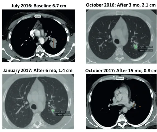
report referred as melanoma with a Breslow of 0.87 mm with peritumoral signs of regression (Stage I). The patient underwent clinical fol-low-up until June 2016, when he was admitted to the emergency room due to ataxia and headache. In the local hospital, the patient performed a cerebral MRI, which showed two lesions in the cerebellar hemisphere zone, with signs of recent bleeding and edema with mass effect. Immediate-ly, the patient underwent surgery to remove these lesions completely, which resulted as melanoma metastasis. A dermatological and an ophthalmo-logical examination, a gastroscopy, and colonos-copy were performed to exclude the presence of
de novo melanoma. In July 2016, total body CT
scan revealed only the presence of one metastatic lesion (6.7 cm) in left lung near to the ileus where it contacts the peri-bronchus structure. Although the patient had a unique metastasis in the lung and a good clinical condition, with an ECOG-PS=0, surgical resection was not feasible due to the position of lesion that would have made the procedure as difficult and would have drastical-ly reduced the patient’s quality of life. There-fore, the only therapeutic option was a systemic treatment, considering that BRAF was mutated (V600E) and the LDH level was normal. In July
2016, the patient started nivolumab treatment (3 mg/kg, every two weeks). After six cycles, the total body CT scan revealed the reduction of lung lesion (2.1 cm), considered as an unconfirmed partial response according to iRECIST. After three months of treatment, the patient performed a new radiological assessment that confirmed the partial response, according to iRECIST. The therapy with nivolumab is currently ongoing, and the patient performs a total body CT scan every three months as per guidelines in his setting. The last radiological assessment completed in Octo-ber 2017 confirmed the partial response of lung lesion (0.8 cm) (Figure 5). During the treatment, the only adverse event was diarrhea of grade 1.
Literature Review
Case reports from literature are summarized in Table I. In these reports, safety issues of nivolumab treatment have been reported more frequently than efficacy; however, when present, clinical outcomes are usually favourable16-24. Waiting for wide
re-al-world studies, these case reports describe im-mune-related adverse events that have not been re-corded in pivotal trials (i.e., intestinal perforation) for the limited number of patients enrolled that was not powered to detect so rare adverse events. Liver
immune-related injury was observed in a patient with malignant melanoma with multiple, after the first cycle of nivolumab16. He was initially treated
with interferon and, then, with nivolumab. Clinical response was relevant at the metastasis site, but grade 4 aminotransferase elevation was observed. Liver histology revealed drug-induced injury that was treated with steroid half-pulse therapy fol-lowed by oral methylprednisolone. However, even after five months ALT level did not completely re-cover to the normal range. Nivolumab was discon-tinued due to persistent hepatitis, but the patient showed remission of his metastatic lung lesion for further five months16. Seronegative rheumatoid
arthritis was reported in another patient with mel-anoma and metastatic lesions to multiple organs. After failure of vemurafenib and ipilimumab, the patient was treated with nivolumab for 20 months, with marked improvement. During treatment, the occurrence of polyarthritis and synovitis compro-mised her quality of life; she gained benefit and symptom control only from hydroxychloroquine 300 mg daily17. Cutaneous immune-related
reac-tions were described in two case reports of bullous pemphigoid-like lesions18,19. Bullous pemphigoid is
the most common blistering skin disorder; it nor-mally presents with an initial non-bullous phase of pruritus, followed by development of general-ized or localgeneral-ized tense blisters filled with serous or haemorrhagic fluid. In both cases, a treatment with corticoids was resolutive18,19. A case of
bilat-eral uveitis was reported after the third infusion of nivolumab in a patient with metastatic mela-noma, affecting the lymph nodes and duodenum and harbouring a BRAF V600E mutation20. The
patient complained of sudden bilateral visual acu-ity impairment, confirmed by the ophthalmologic evaluation. Nivolumab was stopped, and a local treatment with a topic corticosteroid eye drops (sodium phosphate dexamethasone 0.1%) was not enough to reduce the visual acuity decline, that was controlled only with oral corticosteroid treat-ment (1 mg/kg, prednisone); a complete recovery was obtained after one month of systemic treat-ment. Treatment with nivolumab was re-initiat-ed and corticosteroids were gradually decreasre-initiat-ed, without any further relapse of bilateral uveitis; corticosteroids were not completely stopped on nivolumab20. An immunologic reaction may have
a potential to influence intestinal perforation, but the mechanism of gastrointestinal perforation due to nivolumab is not understood. A patient with malignant melanoma in the anal canal and multi-ple metastases reported abdominal distension and
progressive diffuse abdominal pain after the third treatment with nivolumab: he had an intestinal perforation, requiring a surgical intervention. Af-ter surgery and medical treatment for sepsis, the patient completely recovered21. Nivolumab was
active on small intestine metastases, without in-ducing any side effect22. This patient, after 8
cycles of nivolumab, showed depigmentation on the melanoma macula, likely due to a reduction in epidermal melanocytes, following the successful treatment with immunotherapy22. Among
endo-crinology related-dysfunction, hypophysitis and thyroid impairment are frequent. Okano et al23
described a case of hypophysitis where a patient initially developed progressive fatigue and appe-tite loss, after sixth administration of nivolumab; laboratory data indicated eosinophilia and hypo-natremia, and ACTH and cortisol levels were low. The patient was treated with hydrocortisone (20 mg/d), and the 7th administration of nivolumab was
completed without exacerbating patient’s general condition. A case of sarcoid-like granulomatous reaction induced by nivolumab was reported in 201624. After 10 months, the patient achieved a
melanoma complete response, but he developed sarcoid-like granulomatous reaction in the medi-astinal lymph node and skin, which resumed after nivolumab arrest; melanoma did not relapse after 12 months of follow-up.
Discussion
Italian case reports confirm efficacy and safe-ty of nivolumab in patients who do not meet the inclusion criteria of a clinical trial, i.e., for brain metastasis, cardiovascular comorbidities, or elderly. In many cases, nivolumab has been successfully used as second-line therapy, after treatment with carbo-platin and taxanes, ipilim-umab, and BRAF inhibitors, confirming that the objective response is not affected by prior BRAF inhibitor therapy or prior ipilimumab therapy25.
The efficacy and safety of nivolumab is even independent of the mutational status of BRAF, which is, conversely, determinant in the choice of targeted therapy with RAF and MEK inhibi-tors25. According to international guidelines26,27,
patients with mutated BRAF can be treated with immunotherapy or targeted combined therapy. In long-term analysis, both therapeutic options have demonstrated a durable survival: the 3-year OS rate was 44% with BRAF and MEK inhibitors28,
ipilimum-ab and 52% with nivolumipilimum-ab alone13. However,
targeted therapies induced rapid responses in the majority of BRAF-mutant patients, but 50% of these responders developed resistance within approximately 13 months. In contrast, immuno-therapies, particularly inhibitors of PD-1, induced responses, which tended to be durable, in 40-55% of patients28.
Subgroup analyses of large clinical trials would help to identify which patient-centered factors are valuable in choice of first-line ther-apy: for instance, patients with low tumor bur-den could benefit of immunotherapies that can
continue for long time, with very long-lasting response and without side effects that compro-mise the quality of life or exacerbate existing comorbidities. In this series, two patients had pre-existing cardiovascular comorbidities and one of them had showed QT prolongation during treatment with vemurafenib. QT prolongation syndrome has been associated with the use of BRAF inhibitors in clinical studies, albeit with low frequency, and it should be early noticed and promptly managed29. Cardiotoxicity with
immunotherapy is rare, but multiple manifesta-tions of immune-related cardiac syndromes have
Table I. Literature review of case reports from 2016 to February 2018.
Patients Efficacy Safety
Matsubara 201816 42-year-old man with c Significant decrease in the ALT elevation grade 4 and
stage III malignant melanoma size of metastases immune-related liver injury treated
with multiple lung metastases with steroid half-pulse therapy
followed by oral methylprednisone
Haikal 201817 65-year-old Caucasian female Marked improvement in Symmetrical polyarthritis with
with Stage IV melanoma metastasis in multiple organs synovitis and swelling of both
(BRAF-positive) Metacarpophalangeal Joints (MCPs)
and (PIPs) Proximal Interphalangeal Joints bilaterally. Treatment with hydroxychloroquine with remarkable improvement
Anastasopoulou 48-year-old patient with Not reported Bullous pemphigoid-like
201818 melanoma skin lesions along with fever,
arthralgia and overt eosinophilia. Treatment with corticosteroids
Naidoo 201619 80-year-old male with Complete remission Bullous pemphigoid, treated with
metastatic melanoma systemic corticosteroids
Theillac 201820 55-year-old man with Not reported Bilateral granulomatous uveitis
metastatic melanoma, and unilateral Posterior retinal
affecting the lymph nodes serous detachment after the
and duodenum. BRAF V600E. third infusion. Treatment with
both local and systemic
corticosteroids
Yasuda 201721 73-year-old man with melanoma Not reported Intestinal perforation, successfully
in the anal canal with multiple resolved after surgical treatment
metastases in the lungs, liver, and bones, thyroid gland, and subcutaneous tissue.
Yamamura 201722 66-year-old woman with After two cycles, Not reported
metastatic melanoma, with reduction the lesion in the small intestine lesions small intestine; after 8 cycles,
dermoscopic changes
Okano 201623 50-year-old man with metastatic Effective for the primary Hypophysitis at the 7th cycle,
melanoma on the right side and mediastinal lesions treated with hydrocortisone
of the lingual root, with cervical and mediastinal metastases.
Danlos 201624 57-year-old man with At 10 months durable Sarcoid-like granulomatous
desmoplastic melanoma complete response reaction in the mediastinal
been observed; a case series based on the expe-rience of several institutions in the United States and Germany documented cases of autoimmune myocarditis, cardiomyopathy, heart failure, car-diac fibrosis and carcar-diac arrest. Therefore, the deterioration of heart function should be closely monitored, especially in patients with preexist-ing cardiac conditions30,31.
There are few evidence about the efficacy of nivolumab in patients with active brain metas-tasis. In this cases series, one patient with brain metastasis gained clinical benefits from nivolum-ab therapy, despite the previous progression after excision and gamma-knife radiosurgery (GKRS). Nordmann et al32 analyzed the experience of their
institution on concurrent treatment with PD-1 inhibitors and GKRS to enhance the treatment of metastatic melanoma: the combination showed some radiologic benefit in 13 out of 25 patients, and 2 radiologic pseudo-progressions, thus indi-cating that checkpoint inhibition may result in an accelerated response to GKRS. From a molecular point of view, the immune microenvironment in brain metastases may be an ideal target for im-munotherapy, since it is active with a high den-sity of tumor-infiltrating lymphocytes33. A recent
retrospective analysis34 suggested that anti-PD-1
antibodies (both nivolumab and pembrolizumab) obtained an intracranial overall response in 21% of patients and the disease control in 56%, with a median overall survival of 9.9 months (95% CI 6.93-17.74).
Nivolumab treatment resulted as generally well tolerated, with the most frequent treatment-relat-ed adverse events as dermatologic, gastrointes-tinal, endocrine, hepatic, renal, and pulmonary toxicities35. Most events have a low grade and are
successfully managed with supportive care, as per well-established safety guidelines; grade 3 to 4 adverse events are normally resolved with dose delay or permanent discontinuation, with or with-out administration of systemic corticosteroids or other suppressive immune-modulating agents35.
Treatment-related adverse events leading to dis-continuation were reported in 3% of patients (17 out 576) in pivotal trials, with the most common being colitis, increased alanine aminotransferase, increased lipase, and pneumonitis (two patients [0.3%] each); none instance of gastrointestinal perforation was reported35.
A case of intestinal perforation after nivolum-ab treatment has been described in real world practice22, thus confirming the importance of
post-marketing monitoring to identify rare
ad-verse events. The similar autoimmune response was associated with colitis that is more frequently observed and may lead to intestinal perforation. In phase I study, one serious adverse event of inflammatory colitis was observed in a patient36.
Other immune reactions may interest numer-ous organs, including liver, skin, joints, endo-crine system, as reported in case reports from literature16-24. Physicians should be aware of
po-tential immune-related risks and should promptly diagnose and treat these conditions. Immune-re-lated toxicities are rare, but often challenging to be managed. Monitoring for these adverse reactions is advisable to early diagnose and treat them and to avoid delay in nivolumab treatment that may compromise the clinical outcomes on metastatic melanoma.
Unfortunately, not all patients experience a favorable response to anti-PD1 treatment and a better selection of patients is mandatory. Several biomarkers have been investigated, but no con-sensus has been reached yet. The high PD-L1 expression on melanoma was found predomi-nantly in regions of abundant inflammation or TIL (tumor infiltrating lymphocytes) infiltrates, even in sanctuaries like brain metastases37,38,
but it failed to predict responses to nivolumab in metastatic melanoma12. Recently, a functional
method called “the ex vivo metastatic Lymph Node assay”, capable of assessing the reactivi-ty of infiltrating immune effectors (T and NK cells) during stimulation with various immune checkpoint blockers and their combinations, was coupled with a paired blood and tumor immune profiling to correlate immune fingerprints with clinical parameters38. Preliminary results
indi-cated that PD-L1 expression on circulating T cells was relevant in the prediction of resistance to ipilimumab, alone or combination with IL-2 or GM-CSF and that detectable levels of CD137 on circulating CD8+ T cells tended to pre-dict longer PFS for the anti-CTLA-4 + anti-PD-1 co-blockade38. On the other hand, it will be very
important to deeply investigate the mechanisms underlying the antitumor effect of nivolumab, from the in vitro to the clinical relevance, as reported in recent publication39.
Conclusions
These clinical cases demonstrate how nivolum-ab has changed the natural history of metastatic melanoma, leading it to become a disease
man-ageable for a long time. In the therapeutic deci-sion on metastatic melanoma, not only the BRAF status, but also disease characteristics, tumor bur-den, number of metastatic sites, LDH levels, and the performance status of the patient should be considered. Adverse events are often manageable and can be resolved within few weeks with delay or suspension of the therapy. Attention should be paid for rare immune-related toxicities that may be more challenging to both diagnose properly and treat efficiently.
Funding
Bristol-Myers Squibb S.r.l. funded editorial support, pro-vided by Content Ed Net, with the helpful contribution in drafting the test by Elisa Sala, Ph.D, Medical Writer.
Acknowledgements
Authors wish to thank Dr Pasquale Vitale for his precious contribution.
Conflict of Interest
The authors declare that the research was conducted in the absence of any commercial or financial relationships that could be construed as a potential conflict of interest.
References
1) Maverakis e, Cornelius la, Bowen GM, Phan T, PaTel
FB, FiTzMauriCe s, he Y, Burrall B, DuonG C, kloxin
aM, sulTani h, wilken r, MarTinez sr, PaTel F.
Met-astatic melanoma: a review of current and future treatment options. Acta Derm Venereol 2015; 95: 516-524.
2) roBerT C, ThoMas l, BonDarenko i, o’DaY s, weBer
J, GarBe C, leBBe C, Baurain JF, TesTori a, GroB JJ,
DaviDson n, riCharDs J, Maio M, hausChilD a, Miller
wh Jr, GasCon P, loTeM M, harMankaYa k, iBrahiM
r, FranCis s, Chen TT, huMPhreY r, hoos a, wolChok
JD. Ipilimumab plus dacarbazine for previously
untreated metastatic melanoma. N Engl J Med 2011; 364: 2517–2526.
3) weBer Js, D’anGelo sP, Minor D, hoDi Fs, GuTz -Mer r, neYns B, hoeller C, khushal ani ni, Miller
wh Jr, lao CD, lineTTe GP, ThoMas l, loriGan
P, GrossMann kF, hassel JC, Maio M, sznol M,
asCierTo Pa, Mohr P, ChMielowski B, BrYCe a,
svane iM, GroB JJ, kraCkharDT aM, horak C,
laMBerT a, YanG as, larkin J. Nivolumab
ver-sus chemotherapy in patients with advanced melanoma who progressed after anti-CTLA-4 treatment (CheckMate 037): a randomised, controlled, open-label, phase 3 trial. Lancet Oncol 2015; 16: 375-384.
4) Blank C, Brown i, PeTerson aC, sPioTTo M, iwai Y,
honJo T, GaJewski TF. PD-L1/B7H-1 inhibits the
effector phase of tumor rejection by T cell recep-tor (TCR) transgenic CD8+ T cells. Cancer Res 2004; 64: 1140-1145.
5) sinGh BP, salaMa ak. Updates in therapy for
ad-vanced melanoma. Cancers (Basel) 2016; 8: E17. doi: 10.3390/cancers8010017.
6) worlD healTh orGanizaTion. Skin cancers.
Avail-able from: http://www.who.int/uv/faq/skincancer/ en/index1.html. Accessed October 31, 2017. 7) BoloGnia Jl, Jorizzo Jl, raPini rP. Melanoma. In:
Dermatology. Amsterdam: Elsevier; 2003: 1789-1815.
8) BalCh CM, soonG sJ, GershenwalD Je, ThoMPson JF,
reinTGen Ds, CasCinelli n, urisT M, MCMasTers kM,
ross Mi, kirkwooD JM, aTkins MB, ThoMPson Ja, CoiT
DG, BYrD D, DesMonD r, zhanG Y, liu PY, lYMan Gh,
MoraBiTo a. Prognostic factors analysis of 17,600
melanoma patients: validation of the American Joint Committee on Cancer melanoma staging system. J Clin Oncol 2001; 19: 3622-3634 9) BarTh a, wanek la, MorTon Dl. Prognostic factors
in 1,521 melanoma patients with distant metasta-ses. J Am Coll Surg 1995; 181: 193-201.
10) karlsson ak, saleh sn. Checkpoint inhibitors for
malignant melanoma: a systematic review and meta-analysis. Clin Cosmet Investig Dermatol 2017; 10: 325-339.
11) roBerT C, lonG Gv, BraDY B, DuTriaux C, Maio M,
MorTier l, hassel JC, ruTkowski P, MCneil C, kalin -ka-warzoCha e, savaGe kJ, hernBerG MM, leBBé C,
Charles J, MihalCioiu C, Chiarion-sileni v, MauCh C,
CoGneTTi F, aranCe a, sChMiDT h, sChaDenDorF D,
GoGas h, lunDGren-eriksson l, horak C, sharkeY
B, waxMan iM, aTkinson v, asCierTo Pa. Nivolumab
in previously untreated melanoma without BRAF mutation. N Engl J Med 2015; 372: 320-330. 12) larkin J, Chiarion-sileni v, Gonzalez r, GroB JJ,
CoweY Cl, lao CD, sChaDenDorF D, DuMMer r,
sMYlie M, ruTkowski P, FerruCCi PF, hill a, waGsTaFF
J, Carlino Ms, haanen JB, Maio M, Marquez-roDas
i, MCarThur Ga, asCierTo Pa, lonG Gv, Callahan
Mk, PosTow Ma, GrossMann k, sznol M, Dreno B,
BasTholT l, YanG a, rollin lM, horak C, hoDi Fs,
wolChok JD. Combined nivolumab and
ipilimum-ab or monotherapy in untreated melanoma. N Engl J Med 2015; 373: 23-34.
13) wolChok JD, Chiarion-sileni v, Gonzalez r, ruT -kowski P, GroB JJ, CoweY Cl, lao CD, waGsTaFF J,
sChaDenDorF D, FerruCCi PF, sMYlie M, DuMMer r,
hill a, hoGG D, haanen J, Carlino Ms, BeChTer o,
Maio M, Marquez-roDas i, GuiDoBoni M, MCarThur
G, leBBé C, asCierTo Pa, lonG Gv, CeBon J, sosMan
J, PosTow Ma, Callahan Mk, walker D, rollin l,
Bhore r, hoDi Fs, larkin J. Overall survival with
combined nivolumab and ipilimumab in advanced melanoma. N Engl J Med 2017; 377: 1345-1356. 14) weBer J, ManDala M, Del veCChio M, GoGas hJ,
aranCe aM, CoweY Cl, Dalle s, sChenker M, Chiar -ion-sileni v, Marquez-roDas i, GroB JJ, BuTler Mo,
Gonzalez r, kuDChaDkar rr, sMYlie M, MeYer n,
MorTier l, aTkins MB, lonG Gv, BhaTia s, leBBé
C, ruTkowski P, YokoTa k, YaMazaki n, kiM TM, De
Pril v, saBaTer J, qureshi a, larkin J, asCierTo Pa;
CheckMate 238 Collaborators. Adjuvant nivolum-ab versus ipilimumnivolum-ab in resected stage III or IV melanoma. N Engl J Med 2017; 377: 1824-1835 15) Donia M, kiMPer-karl Ml, høYer kl, BasTholT l,
sChMiDT h, svane iM. The majority of patients with
metastatic melanoma are not represented in piv-otal phase III immunotherapy trials. Eur J Cancer 2017; 74: 89-95.
16) MaTsuBara T, nishiDa T, hiGaki Y, ToMiTa r, shiMakoshi
h, shiMoDa a, osuGi n, suGiMoTo a, Takahashi k,
nakaMaTsu D, Mukai k, YaMaMoTo M, Fukui k, aDaChi
s, inaDa M. Nivolumab induces sustained liver
in-jury in a patient with malignant melanoma: a case report. Intern Med 2018.
17) haikal a, BorBa e, khaJa T, DooliTTle G, sChMiDT
P. Nivolumab-induced new-onset seronegative
rheumatoid arthritis in a patient with advanced metastatic melanoma: a case report and literature review. Avicenna J Med 2018; 8: 34-36.
18) anasTasoPoulou a, PaPaxoinis G, DiaManToPoulos P,
ChrisToFiDou e, BenoPoulou o, sTraTiGos a, GoGas
h. Bullous pemphigoid-like skin lesions and overt eosinophilia in a patient with melanoma treated with nivolumab: case report and review of the literature. J Immunother. 2018.
19) naiDoo J, sChinDler k, querFelD C, BusaM k, Cun -ninGhaM J, PaGe DB, PosTow Ma, weinsTein a, luCas
as, CiCColini kT, quiGleY ea, lesokhin aM, Paik
Pk, ChaFT Je, seGal nh, D’anGelo sP, DiCkson M,
wolChok JD, laCouTure Me. Autoimmune bullous
skin disorders with immune checkpoint inhibitors targeting PD-1 and PD-L1. Cancer Immunol Res 2016; 4: 383-389.
20) TheillaC C, sTrauB M, BreTon al, ThoMas l, Dalle s.
Bilateral uveitis and macular edema induced by Nivolumab: a case report. BMC Ophthalmol 2017; 17: 227.
21) YasuDa k, Tanaka T, ishihara s, oTani k, nishikawa T,
kiYoMaTsu T, kawai k, haTa k, nozawa h, Masui Y,
shinTani Y, waTanaBe T. Intestinal perforation after
nivolumab immunotherapy for a malignant mela-noma: a case report. Surg Case Rep 2017; 3: 94. 22) YaMaMura k, oTsuka a, kaku Y, seiDel Ja, noMura M,
naGai h, MaTsuMoTo s, kaBashiMa k. Dermoscopic
changes in malignant melanoma after successful treatment with nivolumab: a case report. J Der-matol 2017; 44: 547-548.
23) okano Y, saToh T, horiGuChi k, ToYoDa M, osaki a,
MaTsuMoTo s, ToMaru T, nakaJiMa Y, ishii s, ozawa
a, shiBusawa n, shiMaDa T, hiGuChi T, ChikaMaTsu
k, YaMaDa M. Nivolumab-induced hypophysitis in
a patient with advanced malignant melanoma. Endocr J 2016; 63: 905-912.
24) Danlos Fx, PaGès C, BarouDJian B, verCellino l,
BaTTisTella M, MiMoun M, JeBali M, BaGoT M, Tazi
a, leBBé C. Nivolumab-induced sarcoid-like
gran-ulomatous reaction in a patient with advanced melanoma. Chest 2016; 149: e133-6.
25) larkin J, lao CD, urBa wJ, MCDerMoTT DF, horak
C, JianG J, wolChok JD. Efficacy and safety of
nivolumab in patients with BRAF V600 mutant and BRAF wild-type advanced melanoma: a pooled analysis of 4 clinical trials. JAMA Oncol 2015; 1: 433-440.
26) DuMMer, a. hausChilD, n. linDenBlaTT, G. PenTherou -Dakis, u. Keilholz. Cutaneous melanoma: ESMO
Clinical Practice Guidelines for diagnosis, treat-ment and follow-up. Ann Oncol 2015; 26: v126-132.
27) euPDaTe – Cutaneous Melanoma Treatment
Rec-ommendations Published: 19 September 2016. Authors: ESMO Guidelines Committee. Available from: www.esmo.org. Accessed 25 February, 2018.
28) lonG Gv, Fl aherTY kT, sTroYakovskiY D, GoGas
h, levChenko e, De BrauD F, larkin J, GarBe C,
JouarY T, hausChilD a, Chiarion-sileni v, leBBe C,
ManDal à M, MillwarD M, aranCe a, BonDarenko
i, ha anen JBaG, hansson J, uTik al J, Ferraresi
v, Mohr P, ProBaChai v, sChaDenDorF D, naThan
P, roBerT C, riBas a, Davies Ma, lane sr, leGos
JJ, MookerJee B, GroB JJ. Dabrafenib plus
trametinib versus dabrafenib monotherapy in patients with metastatic BRAF V600E/K-mu-tant melanoma: long-term survival and safety analysis of a phase 3 study. Ann Oncol 2017; 28: 1631-1639.
29) weBer Js, hoDi Fs, wolChok JD, ToPalian sl, sCha -DenDorF D, larkin J, sznol M, lonG Gv, li h,
waxMan iM, JianG J, roBerT C. Safety profile of
nivolumab monotherapy: a pooled analysis of patients with advanced melanoma. J Clin Oncol 2017; 35: 785-792.
30) BronTe e, BronTe G, novo G, BronTe F, BaveTTa MG,
lo re G, BranCaTelli G, Bazan v, naToli C, novo s,
russo a. What links BRAF to the heart function?
New insights from the cardiotoxicity of BRAF in-hibitors in cancer treatment. Oncotarget 2015; 6: 35589-35601.
31) heinzerlinG l, oTT Pa, hoDi Fs, husain an, Ta -JMir-riahi a, TawBi h, PausChinGer M, GaJewski TF,
liPson eJ, luke JJ. Cardiotoxicity associated with
CTLA4 and PD1 blocking immunotherapy. J Im-munother Cancer 2016; 4: 50.
32) norDMann n, huBBarD M, norDMann T, sPerDuTo
Pw, Clark hB, hunT Ma. Effect of gamma knife
radiosurgery and programmed cell death 1 recep-tor antagonists on metastatic melanoma. Cureus 2017; 9:e1943.
33) BerGhoFF as, venur va, Preusser M, ahluwalia Ms.
Immune checkpoint inhibitors in brain metasta-ses: from biology to treatment. Am Soc Clin Oncol Educ Book 2016; 35: e116-122.
34) Parakh s, Park JJ, MenDis s, rai r, xu w, lo s, DruM -MonD M, rowe C, wonG a, MCarThur G, haYDon a,
anDrews MC, CeBon J, GuMinski a, keFForD rF, lonG
Gv, Menzies aM, klein o, Carlino Ms. Efficacy
of anti-PD-1 therapy in patients with melanoma brain metastases. Br J Cancer 2017; 116: 1558-1563.
35) silva iP, lonG Gv. Systemic therapy in advanced
melanoma: integrating targeted therapy and im-munotherapy into clinical practice. Curr Opin On-col 2017; 29: 484-492.
36) BrahMer Jr, Drake CG, wollner i, PowDerlY JD, PiCus
J, sharFMan wh, sTankeviCh e, Pons a, salaY TM,
MCMiller Tl, Gilson MM, wanG C, selBY M, TauBe
JM, anDers r, Chen l, korMan aJ, ParDoll DM,
lowY i, ToPalian sl. Phase I study of single-agent
anti-programmed death-1 (MDX-1106) in refracto-ry solid tumors: safety, clinical activity, pharma-codynamics, and immunologic correlates. J Clin Oncol 2010; 28: 3167-3175.
37) BerGhoFF as, riCken G, wiDhalM G, raJkY o, DieCk -Mann k, Birner P, BarTsCh r, höller C, Preusser M.
Tumour-infiltrating lymphocytes and expression of programmed death ligand 1 (PD-L1) in melano-ma brain metastases. Histopathology 2015; 66: 289-299.
38) JaCqueloT n, roBerTi MP, enoT DP, rusakiewiCz s,
Ternès n, JeGou s, wooDs DM, soDré al, hansen
M, Meirow Y, saDe-FelDMan M, Burra a, kwek ss,
FlaMenT C, MessaouDene M, DuonG CPM, Chen l,
kwon Bs, anDerson aC, kuChroo vk, weiDe B, auBin
F, BorG C, Dalle s, BeaTrix o, aYYouB M, BalMe B,
ToMasiC G, Di GiaCoMo aM, Maio M, sChaDenDorF D,
Melero i, Dréno B, khaMMari a, DuMMer r, levesque
M, koGuChi Y, FonG l, loTeM M, BaniYash M, sChMiDT
h, svane iM, kroeMer G, MaraBelle a, MiChiels s,
CavalCanTi a, sMYTh MJ, weBer Js, eGGerMonT aM,
ziTvoGel l. Predictors of responses to immune
checkpoint blockade in advanced melanoma. Nat Commun 2017; 8: 592.
39) sun lM, liu YC, li w, liu s, liu hx, li lw, Ma r.
Nivolumab effectively inhibit platinum-resistant ovarian cancer cells via induction of cell apopto-sis and inhibition of ADAM17 expression. Eur Rev Med Pharmacol Sci 2017; 21: 1198-1205.