by
Matilde Marchi
for the award of the degree ofDOCTOR OF PHILOSOPHY
“Dynamic imaging of the intracellular trafficking of ERK suggests
a novel mechanism at the basis of the functional differences
between ERK1 and 2”
“As natural selection works solely by and for the good of each being, all corporeal and mental endowments will tend to progress towards perfection.”
INDEX
INDEX………... ABSTRACT………. WORKING HYPOTHESIS………. SPECIFIC AIMS………... SIGNIFICANCE OF THE WORK………. LIST OF ABBREVIATIONS………... LIST OF PAPERS DERIVED FROM THIS STUDY………...
INTRODUCTION ………
Signal transduction: the keystone of living matter ……… Why and how studying a molecular pathway? ... Studying cellular processes in living cells ………. INTRODUCTION: Part 1 ……….. The ERK1/2 cascade ………... Upstream of ERK ……… Tyrosine Kinase Receptors (TKRs) ………. Scaffold proteins ……….. Ras ………... The linear module Raf-MEK-ERK ……….
Raf ……….……….
MEK ……….…….
ERK ……….. The regulation of ERK localization by activation and inactivation mechanisms ……….…
ERK and its targets ………..
Cytoplasmic Targets ……….……… Ribosomal protein S6 Kinases (RSKs)……… ERK-mediated cell migration ………..………. Cytoskeleton regulation ………..………. Nuclear Targets………..………
The ETS transcription factor family ………. The AP-1 (activating protein-1) transcription factor family ……… Mitogen and Stress-activatedKinase (MSK) ……...
1 5 6 6 7 8 9 10 10 10 11 13 13 15 15 15 16 18 18 18 19 20 22 22 22 23 25 26 26 28 29
Duration, magnitude and compartmentalization of ERK response….
Duration ……… Magnitude and compartmentalization ……….
Modeling: a bridge between biochemistry and computation …..……. Nucleo-cytoplasmic shuttling of ERK ……….
Nuclear Pore Complexes: the gatekeepers of the nuclear entry
ERK1 and ERK2: the “different twins” ………..……….
INTRODUCTION: Part 2 ……….. Dissecting a pathway: techniques to investigate molecular mechanisms ………... Generation of fusion proteins and their validation ………...
Tracking protein movements ……… “Sliding molecules”: measuring protein movement with FRAP……..
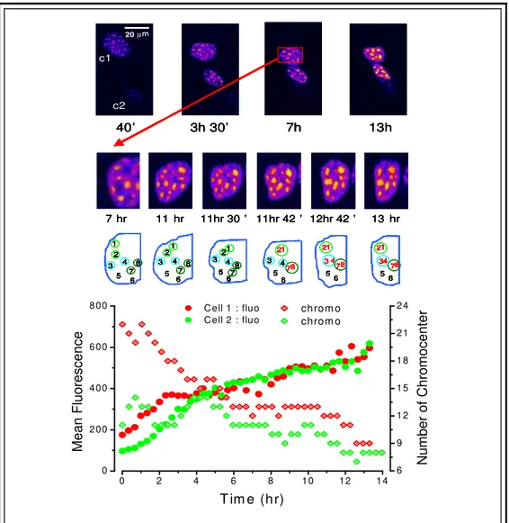
BOX 1: ERK variants ……….... BOX 2: MEK binding Partner 1 ……….. BOX 3: Long Term Time Lapse ……….
RESULTS ……… The ERK1/2 Cascade ……….. Fluorescent probe validation ………
Checking for correct post-translational modifications ……… Testing the catalytic activity ………...…... Chimera localization and expression level ………..…...
Resolving spatio-temporal dynamics of the ERK activation/inactivation ………
ERK2 in action: the nuclear translocation ………..………… MEK and Phosphatases: the hero and the villain? ………..………
ERK1 and ERK2: focus on nucleo-cytoplasmic shuttling properties….
Do ERK1 and ERK2 display different temporal patterns of localization?... Nucleo-cytoplasm exchange of ERK1 and ERK2 ………..………
29 30 32 32 34 34 37 39 39 40 43 44 50 52 53 55 55 55 55 56 57 60 61 64 70 70 72
ERK1 N-terminus: a key domain for understanding functional differences between ERK1 and ERK2? ………..
ERK1 ERK2 sequence comparison ……….…………...…. ERK1 N-terminus: specific functional domain or steric hindrance for nuclear access? ………..… Effects of shuttling rate on ERK phosphorylation: a quantitative model…. Toward a functional interpretation ……….………..
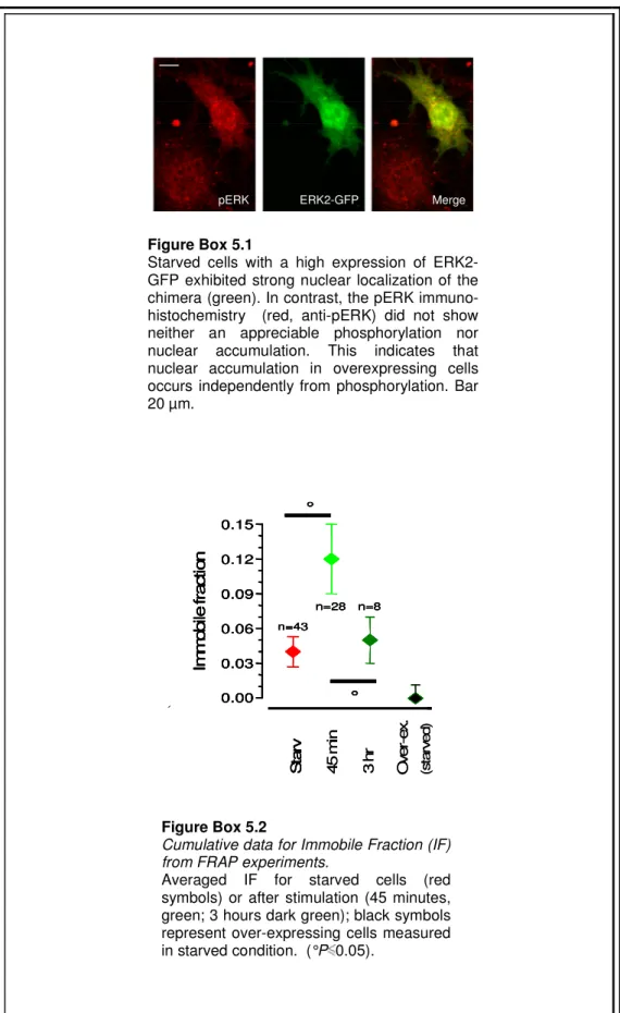
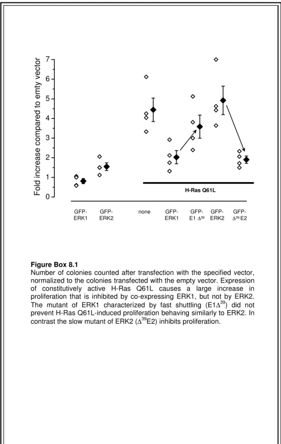
BOX 4: Correlation between the time constant and ERK2-GFP concentration ………. BOX 5: ERK2 immobile fraction in the nucleus……… BOX 6: Mechanism at the basis of ERK2-GFP nuclear accumulation BOX 7: Mapped important domains of ERK ………. BOX 8: Functional consequences of ERK trafficking speed ………...
DISCUSSION ………. Use of fluorescent probes to study cellular processes in living cells Dynamics of ERK activation/deactivation …...……….…. ERK2 nuclear entry/exit ……….. ERK1 ERK2 shuttle across the nuclear membrane with different rates ………...…….. ERK1 N-terminus is the domain responsible of slowing down ERK1. Phosphorylation levels of ERK1 and 2 in the nucleus and consequential functional outputs ………....
CONCLUSIONS and FUTURE DIRECTIONS ………. MATERIAL and METHODS ……… Plasmid preparation ………..
Swapped ERK clones ……… Deleted clones ……… Mutagenized clones ………..
Cell culture and transfection ……….. Immunoblotting ………. 76 76 81 88 92 93 95 97 102 103 105 107 111 117 118 121 123 124 124 125 126 126 127 127
Immunoprecipitation, pMBP reaction and immunoblotting ……... Immunofluorescence ……… Calibration of protein concentration ……… Fluorescence-based recordings ………
Acquisition of pERK immunohistochemistry ……….. FRAP experiments: nucleus-cytoplasm shuttling ……… FRAP experiments: spot photobleaching ……….. Strip-FRAP ……….. Modelling ……….. BIBLIOGRAPHY………. 128 129 129 130 131 131 132 133 133 135
ABSTRACT
In this thesis I studied the localization and trafficking in living cells of the Extracellular Regulated Kinase signaling by making visible ERK1 and ERK2 with a fluorescent tag. This approach allowed to identify different dynamical properties of the two kinases, posing the bases for the understanding of the functional differences between ERK1 and ERK2.
The nucleo-cytoplasmic trafficking of tagged ERK1/2 has been measured by means of FRAP experiments. Surprisingly, I found that ERK1 shuttles at a much slower rate than ERK2. Moreover, I demonstrated that this difference is caused by an unique domain of ERK1 located at its N-terminus, since the progressive deletion of these residues converts the shuttling features of ERK1 into those of ERK2. Conversely, the fusion of this ERK1 sequence at the N-terminus of ERK2 slows down its shuttling to a similar value found for ERK1 and, when fused to small cargos such as a GFP monomer, it is capable of hampering their shuttling too. In addition, I identified some crucial aminoacids at ERK1 N-terminus, responsible in large part of this phenotype. Finally, I have demonstrated that the speed of nucleo-cytoplasmic shuttling critically affects the ERK capability of activating downstream effectors.
In conclusion, I propose a novel biochemical model, in which the regulation of nucleo-cytoplasmic trafficking might provide a sensitive mechanisms through which cells modulate their response to extracellular stimulus. This mechanism significantly contributes to the differential ability of ERK1 and 2 to generate an overall signaling output.
WORKING HYPOTHESIS
This work focused on dissecting protein dynamics inside a molecular pathway of crucial importance: the Extracellular Regulated Kinase (ERK) signaling, which is activated by a wide plethora of stimuli and is involved in almost every cellular process.
Understanding the basic control mechanisms of a biological system like a molecular signaling cascade will help to develop new therapies, drugs and sensitive diagnostics tools.
This work is characterized by a multidisciplinary approach, indispensable to unveil differences between the two kinases ERK1 and 2 which share a high sequence homology and the majority of the regulatory partners. The proteins of interest have been made visible by fusing them with fluorescent proteins. The resulting chimerical proteins were expressed in mammalian cells and were tested by experiments of live imaging. Furthermore, the functional consequences of the slower trafficking of ERK1 have been evaluated by computational models and biochemical assays.
SPECIFIC AIMS
• Characterizing the biochemical-functional properties of GFP-ERK fusion proteins in order to validate them as investigation tools in living cells.
• Deciphering nuclear translocation dynamics of ERK2, investigating the interplay between the activation and deactivation systems.
• Measuring the nucleo-cytoplasmic shuttling of ERK1 and 2, by FRAP experiments.
• Evaluating the effects of ERK trafficking on the signaling to the nucleus. The regulation of nuclear access constitutes a fundamental check point to control downstream effects.
• Studying the capability of ERK1 N-terminus of influencing the trafficking across the nuclear barrier and the functional outputs.
• Identification of specific residues at ERK1 N-terminus, which are responsible of the slower phenotype of nuclear shuttling.
SIGNIFICANCE OF THE WORK
In this thesis the following results have been obtained:
The mechanisms at the basis of ERK regulation have been better elucidated focusing on the spatio-temporal patterns of ERK phosphorylation and trafficking in living cells. In particular, I have found that there is a continuous counterbalance between the activation drive (MEK mediated) and the deactivation reactions (by Phosphatases). This is true also in basal conditions, given that a small percentage of MEK activity is still present. This push-pull mechanism is highly modulated in strength and over time, and is responsible for the on/off switching of the pathway.
Furthermore, it has been measured the nucleo-cytoplasmic shuttling of ERK1 and 2, by FRAP experiments, finding out that ERK1 and 2 drastically differ in their capability of crossing the nuclear envelope. Computational, biochemical and functional evidences proved that this trafficking difference causes ERK1 and 2 to have different signaling capabilities to the nucleus. Indeed, I demonstrated that the rate of nucleo-cytoplasmic shuttling is a crucial regulator of the signaling to the nucleus, representing a novel possible target for the molecular control of this pathway.
Finally, it has been demonstrated that the difference in nuclear shuttling between ERK1 and 2 is caused by a short domain located at the N-terminus of ERK1. This region is necessary and sufficient to cause the differences of permeation and functional properties between ERK1 and 2. By directed mutagenesis some crucial residues have been identified to be responsible of the slow permeation of ERK1.
LIST OF ABBREVIATIONS
CLSM: Confocal Laser Scanning Microscopes cPLA2: cytosolic PhosphoLipase A2
CREB: Cyclic AMP Response Element-Binding CRM1: Chromosome Region Maintenance Protein 1 DSP: Dual Specificity Phosphatases
DsRFP: Discosoma Red Fluorescent Protein EGF R: Epidermal Growth Factor Receptor ERK: Extracellular Regulated Kinase
EYFP: Enhanced Yellow Fluorescent Protein FAK: Focal Adhesion Kinase
FGF: Growth Factor for Fibroblast
FRAP: Fluorescence Recovery After Photobleaching FRET: Fluorescence Resonance Energy Transfer GAP: GTPase-Activating Proteins
GEF: Guanine nucleotide Exchange Factors GFP: Green Fluorescent Protein
Grb: Growth factor Receptor-Bound protein GTP: Guanine Triphosphate
HePTP: Hematopoietic Protein Tyrosine Phosphatase IF: Immobile Fraction
JNK: c-Jun NH2-terminal kinase KD: Kinase Domain
KSR: Kinase Suppressor of Ras
MAP-2: Microtubule Associated Protein 2 MAPK: Mitogen Activated Protein Kinase MBP: Myelin Binding Protein
MeCP2: Methyl CpG–binding Protein 2 MKP: MAP Kinase Phosphatase MLCK: Myosin Light Chain Kinase MP1: MEK Partner 1
MSK: Mitogen- and Stress-activated ProteinKinase NES: Nuclear Export Signal
NGF: Nerve Growth Factor NGF: Nerve Growth Factor NLS: Nuclear Localization Signal NPC: Nuclear Pore Complex NUP: Nucleoporin
PDGF: Platelet-Derived Growth Factor
PEA-15: Phosphoprotein Enriched in Astrocytes, 15kDa PKC: Protein Kinase C
PP2A: Protein Phosphatase 2A
PPAR 1: Peroxisome Proliferator Activated Receptor 1 PTB: Phospho-Tyrosine Binding
PTK: Protein Tyrosine Kinase Rap1: Repressor activator protein 1 RSK: Ribosomal S6 Kinase
SAPK: Stress-Activated Protein Kinase SH: Src Homology
SMAD: this name is the combination between the C. elegans protein SMA and the drosophila protein Mothers Against Decapentaplegic
Sos: Son of Sevenless
SRF: Serum Response Factor TAD:Transactivation Domain TCF: Ternary Complex Factor TKR: Tyrosine Kinase Receptors TMP: Thrombin-Mimicking Peptide TRD: Transcription Repression Domain
LIST OF PAPERS DERIVED FROM THIS STUDY
Marchi M, D'Antoni A, Formentini I, Parra R, Brambilla R, Ratto GM, Costa M. The N-terminal domain of ERK1 accounts for the functional differences with ERK2.
PLoS ONE. 2008;3(12):e3873. Epub 2008 Dec 4.
Marchi M, Guarda A, Bergo A, Landsberger N, Kilstrup-Nielsen C, Ratto GM, Costa M.
Spatio-temporal dynamics and localization of MeCP2 and pathological mutants in living cells.
Epigenetics. 2007 Sep;2(3):187-97. Epub 2007 Sep 18.
Costa M, Marchi M, Cardarelli F, Roy A, Beltram F, Maffei L, Ratto GM. Dynamic regulation of ERK2 nuclear translocation and mobility in living cells.
INTRODUCTION
Signal transduction: the keystone of living matter Why and how studying a molecular pathway?
In biology, signal transduction refers to any process by which a cell converts an extra-cellular signal in a series of intracellular events leading to adaptive responses of the cell to the changing environment. This involves ordered sequences of biochemical reactions, carried out by enzymes and linked through second messengers or protein-protein interactions. Such processes are usually rapid, lasting on the order of milliseconds, as in the case of ion fluxes, to minutes for the activation of proteins and lipid-mediated kinase cascades. The main feature of most pathways is the capability of generating a “signal cascade”: indeed the number of proteins and other molecules participating in these events increases as the process goes on from the initial stimulus, providing amplification and the possibility of integrating different signals on common effectors. Each protein is committed to act in a well defined range of activities and it behaves like a fine sensor of the surrounding environment. All these components form highly interactive networks responsible of the first level of complexity in a living organism. The stunning perfection of the molecular orchestration inside a pathway is not only fascinating per se, but hides essential clues necessary to understand how a biological system works. Furthermore, understanding basic control mechanisms will help to develop new therapies, drugs and sensitive diagnostics tools.
In this thesis I focused on dissecting protein dynamics inside a molecular pathway of crucial importance: the Extracellular Regulated Kinase (ERK) signaling. ERK pathway is activated by a wide plethora of stimuli, including growth factors, cytokines, integrins and hormones. It is involved in almost every cellular process like cell cycle, motility, differentiation, apoptosis and synaptic plasticity. Indeed, the ERK pathway represents a hot spot of investigation because of its large impact on widespread and serious diseases: for example, it has been reported that ERK1/2 activity is massively upregulated in several human cancers (Hoshino et al., 1999).
The basic assembly of the ERK pathway is a three-component module conserved in all eukaryotes, from yeast to humans. This module includes three serially linked kinases (Raf/MEK/ERK) of which ERK is the last effector.
In mammals there are at least two “isoforms” for each level: Raf1, B-Raf and A-Raf for the upstream form, MEK1 and MEK2 for the intermediate and ERK1 and ERK2 at the lower level. In this work I will investigate the differences between these two last proteins, that, although sharing many known features, they still hold some secrets.
ERK1 and ERK2 have been considered as redundant isoforms for a long time, since they share the same regulatory machinery and targets. In addition, the identification of their individual role is complicated by technical difficulties in separating the action of each kinase. Given that ERK1 and 2 are characterized by a high degree of sequence homology (90% in humans), their existence raises the question of why they have been both conserved to date.
Studying cellular processes in living cells
Although biological processes and structures are dynamic in nature, most investigations into their mechanisms have been carried out in ex-vivo specimens using, for example, immunocytochemistry and biochemical assays. This incongruity has been imposed in large part by a lack of tool to analyze signal transduction in living cells. Recent developments in in vivo microscopy techniques and the discovery of the green fluorescent protein (GFP) and its spectral variants (Chalfie et al., 1994) have allowed the non destructive investigation of a wide range of dynamic processes in living cells [reviewed in (Lippincott-Schwartz et al., 2001; Misteli and Spector, 1997)]. In this work I took advantage of these new tools: ERK1 and 2 were made visible by fusion with fluorescent proteins. Then, the resulting chimeras were expressed in living cells to follow the dynamics of their movements between different compartments and their localization after specific stimuli. These experiments offered a different perspective on ERK function compared to the classic biochemical approach, and, as I will show, allowed to evidence differences between the two kinases ERK1 and 2 that were, upon now, unsuspected.
In the following of this introductory chapter two main sections will be developed:
° in the first part I will discuss the biological background of the proteins investigated; I will review the state of art of ERK biology specifically focusing on the molecular aspects concerning ERK targets, the regulatory mechanisms of the pathway and the communication between nucleus and cytoplasm.
° in the second part I will introduce the multidisciplinary approach that characterizes this work, indispensable to unveil differences between the two kinases. I will illustrate potentialities and advantages of the imaging techniques used in tight conjunction with more classical molecular and biochemical methods. This kind of approach has made possible to unveil different kinetic and functional properties of ERK1 and 2.
INTRODUCTION: Part 1 The ERK1/2 cascade
This pathway is one of the primordial signaling systems; it exists in all eukaryotes and controls fundamental cellular processes such as proliferation, differentiation, survival and apoptosis. The ERK cascade was originally discovered as a critical regulator of cell division and differentiation and this is the reason why it was firstly named Mitogen-Activated Protein Kinase(MAPK) cascade. As further details of this molecular signaling were worked out, it became clear that the ERK cascade is in fact a prototype for a family of signaling cascades that shares the motif of three serially linked kinases, which regulate each other by sequential phosphorylation. Thus, a revised nomenclature uses the term MAPK to refer to the entire superfamily of signaling cascades, comprising also SAPK/JNK (Stress-Activated Protein Kinase/c-Jun NH2-terminal kinase) and p38, and it specifies the prototype MAPK as ERK (Extracellular-Regulated Kinase). SAP/JNK and p38 are principally activated by cellular stresses including osmotic shock, inflammatory cytokines, lipopolysaccharides (LPS) and ultraviolet light. Conversely ERK1/2 are primarily recruited by mitogens (such as polypeptide growth factors as well as insulin) and neurotrophins, but also by cytokines and hormones through Tyrosine Kinase Receptors (TKRs). Here I will focus on the dynamic regulation of the ERK1/2 pathway, activated by growth factors. The basic arrangement in the ERK pathway includes membrane receptors (principally TKRs) and a G-protein (Ras) which sits upstream of a core module consisting of a MAPK Kinase Kinase (Raf), that phosphorylates and activates a MAPK Kinase (MEK), which finally activates ERK1/2 (Fig. 1).
Figure 1
Schematic representation of the major components involved in ERK pathway (activated by trophic factors). Cytoplasm P P P P P P P P Kinase Domain Heparan Sulfate Proteoglycan Monomer (e.g. EGF) Dimer
(e.g. PDGF) Monomers bind toproteoglycans (e.g. FGFs) P P P P P P P P SH2 Cytoplasm P P P P P P P P Kinase Domain Heparan Sulfate Proteoglycan Monomer (e.g. EGF) Dimer
(e.g. PDGF) Monomers bind toproteoglycans (e.g. FGFs) P P P P P P P P SH2 Figure 2
Tyrosine Kinase Receptors and their ligands
The binding of specific extracellular signals with the extracellular domains of Tyrosine Kinase Receptors (TKR) activate the intracellular Tyrosine Kinase Domain (TKD). Before or after ligand binding, the dimerization of TKRs occurs. To activate a TKR the ligand usually has to bind simultaneously to two adjacent receptor chains. PDGF is a dimer and crosslinks two receptors together; EGF is monomeric and FGFs, also monomers, form multimers by binding to heparan sulfate proteoglycans. Once activated, the TKD transfers a phosphate group (autophosphorylation) from ATP to selected tyrosine side chains, both on the receptor protein itself and on intracellular signaling proteins, that subsequently binds to the phosphorylated receptor. Phosphorylation of Tyr within the Kinase Domain (KD) increases the kinase activity of the enzyme. Phosphorylation of Tyr residues outside the KD creates high affinity docking sites for the binding of scaffold proteins (with SH2, PTB and SH3 domains).
Upstream of ERK
Tyrosine Kinase Receptors (TKRs)
Membrane spanning cell surface receptors of the TKR family are endowed with intrinsic tyrosine kinase activity, catalyzing the transfer of the -phosphate of ATP to the hydroxyl groups of tyrosines on target proteins. All TKRs frequently contain a glycosylated extracellular ligand binding domain, connected to the cytoplasmic domain, which has a conserved Protein Tyrosine Kinase (PTK) core and regulatory regions that are subjected to autophosphorylation and phosphorylation by other kinases (Schlessinger, 2000a). Nearly all TKRs are monomers at the cell membrane, with ligand binding or ectopic overexpression resulting in receptor dimerization and tyrosine autophosphorylation in trans (Fig. 2). As the first TKR to be discovered (Downward et al., 1984), the epidermal growth factor receptor (EGF R, also known as ErbB1 from the v-erb-B transforming protein of an avian retrovirus) has helped to establish many of the principles of TKR functions (Schlessinger, 2002).
Activating mutations and transforming overexpression, mimicking receptor oligomerization of EGFR and its fellow family members, have been implicated in numerous cancers, including mammary carcinomas, squamous carcinomas and glioblastomas (Blume-Jensen and Hunter, 2001).
Scaffold proteins
The EGF receptor contains at least nine tyrosine residues in its cytoplasmic domain capable of being phosphorylated, and seven of these are autophosphorylation sites (Levkowitz et al., 1999). The autophosphorylation of Tyr sites on EGFR and other TKRs provides a mechanism for the recognition of specific scaffold proteins and it represents a platform for the assembly of signaling complexes. Phosphorylated sites of EGFR are recognized and bound by Src Homology 2 (SH2) (Pelicci et al., 1992) and Phospho-Tyrosine Binding (PTB) domains (Schlessinger, 2000b). Shc can assist the binding of an other protein, the Growth factor receptor bound protein 2, Grb2 (Rojas et al., 1996), a cytosolic adaptor, containing a central SH2 domain flanked by two Src Homology 3 (SH3) domains, that allows it to associate constitutively with the proline-rich regions of the nucleotide exchange factor Son of Sevenless (Sos) (Li et al., 1993). The recruitment of Grb2 from the cytoplasm to the plasma membrane brings Sos near the membrane-bound Ras. Through guanine exchange, Sos
enhances GDP release and GTP binding to Ras, converting this GTPase into its active conformation.
In a general view, it can be considered that the mammalian ERK pathway contains a central fifth-tiered module, which is strongly conserved and includes:
• the GTPase protein Ras (H-Ras, K-Ras and N-Ras) and other still unknown kinases;
• Raf (Raf-1, Raf A and Raf B);
• MEK1 and 2;
• ERK1 and 2;
• RSK, MSK and MNK.
In the following, I will describe features and functions of each components, particularly focusing on the isoforms which are principally involved in ERK1 and 2 signaling (Raf1 and Raf-B, MEK1 and MEK2).
Ras
Ras is a notable member of the large family of GTPases, proteins that bind and hydrolyze GTP. First discovered as transforming oncogenes of murine sarcoma viruses (v-ras), three highly related 21 kDa mammalian proteins, Harvey-Ras (H-Ras), Kirsten-Ras (K-Ras) and Neuroblastoma-Ras (N-Ras) have been identified (Bos, 1989). Ras family members are anchored to the cytoplasmic side of the plasma membrane by carboxyl-terminal farnesylation (post-translational modification by the attachment of an isoprenoid to the C-terminal cysteine residue). This localization to the inner leaflet brings Ras into close proximity with Sos, stimulating the exchange of GDP bound to Ras with GTP from the cytosol (Fig. 3). This exchange conformationally activates Ras, allowing it to interact with a number of downstream effectors (Avruch et al., 1994). Within the ERK signaling cascade, active Ras functions as an adaptor that binds to the effectors Raf kinases with high affinity, causing their translocation to the cell membrane, where Raf activation takes place (Jelinek et al., 1996).
GEF
Ras
Ras
GDP GTP GDP GTP GAP Pi Inactive Active GEFRas
Ras
GDP GTP GDP GTP GAP Pi Inactive ActiveIt has been well established that specific alterations in members of the ras gene family can convert them into active oncogenes. These malignant transformations lead to a subversion of cellular pathways that regulate the proliferation, differentiation and survival of cells, resulting in altered cell growth (oncogenic transformation). In Ras family these alterations are either point mutations occurring in either codons 12, 13 or 61 or, alternatively, a 5- to 50-fold amplification of the wild-type gene resulting in Ras overexpression.
Activating mutations of these Ras isoforms, which impair GTPase activity and stabilize the GTP bound state, are found in nearly one-third of all human cancers, making these oncoproteins among the most potent transforming polypeptides known (Seger and Krebs, 1995).
The transformant properties of mutated Ras isoforms have been recently used to show that ERK1 and 2 differently transduce Ras-dependent cell signaling and proliferation (Vantaggiato et al., 2006). Ectopic expression of ERK1 but not of ERK2 in NIH 3T3 cells inhibits oncogenic Ras-mediated proliferation and colony formation.
Figure 3
Ras-GDP/GTP cycle
Ras functions as a switch, cycling in two distinctive conformational states: Active, when GTP is bound and Inactive when GDP is bound. Two classes of signaling proteins regulate Ras activity by influencing its transition: Guanine Nucleotide Exchange Factors (GEF) and
GTPase-Activating Proteins (GAP). GEF
stimulate the dissociation of GDP and the uptake of GTP, while GAP increase the rate of hydrolysis of bound GTP by Ras, inactivating Ras.
The linear module Raf-MEK-ERK
This module is actually the central core module. Once activated, it accomplishes for the convergence of different up-stream stimuli, providing for a high degree of signal integration.
Raf
Raf is a Ser/Thr protein kinase, catalyzing the phosphorylation of hydroxyl groups on specific Ser and Thr residues (Chong et al., 2003). Like Ras, Raf was first discovered in the form of a mutant retroviral transforming agent, v-raf (Rapp et al., 1983). Mammals have 3 Raf proteins, ranging from 70 to 100 kDa in size: Raf-1, Raf-A and Raf-B.
1 is ubiquitously expressed and studies on knockout mice indicated that Raf-1 may serve a general role in tissue formation (Mikula et al., 200Raf-1).
B-Raf is present in multiple isoforms and it is strongly expressed in fetal brain and adult cerebrum (Barnier et al., 1995) and it seems to fulfill more specialized duties (Wojnowski et al., 1997).
Recruitment to the plasma membrane by GTP-bound Ras is the initial event in Raf activation. Different Ras isoforms appear to activate Raf with varying ability, despite binding in vitro with comparable affinity. For example, K-Ras both recruits Raf-1 to the plasma membrane more efficiently, and activates the recruited Raf-1 more potently than H-Ras (Yan et al., 1998). It has also been suggested that Raf-B is the primary target of oncogenic Ras isoforms (Marais et al., 1997). Activating mutations of raf-B were reported in 66% of malignant melanomas (Davies et al., 2002).
MEK
Phosphorylated Raf activates MEK1 and MEK2 (Zheng and Guan, 1993). These kinases are about 45 kDa each and share 80% sequence identity. It is unclear why two MEKs exist, although conservation of both forms throughout eukaryotic species suggests non-redundant functions. Both MEKs are expressed ubiquitously in mammalian cells at micromolar levels, although some tissue-specific variation has been noted (Brott et al., 1993). Raf family activation of MEK1 and MEK2 occurs through phosphorylation of two Ser found in the activation loop (Alessi et al., 1994). While Raf isoforms are enzymes of relatively low abundance, the high concentration of MEKs allows for amplification of signaling (Huang and Ferrell, 1996). MEK1 or MEK2 may activate ERK1 or
ERK2. At endogenous levels of expression, there is evidence for preferential coupling, which may depend on the upstream kinase or adapter proteins in addition to differences in their direct interactions. Several studies showed that Raf-1 complexes preferentially with MEK1 and ERK2 (Huang et al., 1993; Jelinek et al., 1994).
MEK1 and MEK2 have distinct ways to contribute to the regulation of ERK activity and the mammalian cell cycle progression; MEK1 is required for Golgi fragmentation (Colanzi et al., 2000), whereas MEK2 is thought to be essential for progression through the G2/M checkpoint (Abbott and Holt, 1999). The phenotypes for loss of MEK1 versus loss of MEK2 have been studied in CT116 cells, a colon cancer line with WT p53 (Ussar and Voss, 2004). Depletion of either MEK subtypes by RNA interference generated a unique phenotype. The MEK1 knockdown led to the induction of a senescence-like phenotype and permanent ablation of MEK1 resulted in reduced colony formation potential, indicating the importance of MEK1 for long term proliferation and survival. In contrast, MEK2 deficiency was accompanied by a massive induction of cyclin D expression and the centrosome over-amplification, inducing a delay in mitosis. Knockout studies have demonstrated that theinactivationof MEK1 gene leads to
embryonic lethality, suggesting that MEK1 has a unique role during
embryogenesis; while MEK2is not necessary for the normal development of the embryo and its loss can be compensatedby MEK1 (Belanger et al., 2003). All these experiments demonstrated that the two isoforms of MEK are not interchangeable, since interfering with one or the other causes different phenotypes.
ERK
ERK was evidenced for the first time as a kinase protein phosphorylating the microtubule-associated-protein 2 (MAP2) in extracts of 3T3-L1 adipocytes (Sturgill and Ray, 1986). This polypeptide was identified to be the same found phosphorylated by many growth factors (Nakamura, 1983; Cooper, 1984; Cooper 1985; Khono, 1985) and phorbol esters (Gilmore, 1983); these fidings reinforced the possibility that it might be an ubiquitous effector of mitogenic stimuli. This realization prompted the ridesignation of acronym “MAP” from “microtubule-associated-protein” to “mitogen-activated-protein”. ERK genes were purified and cloned the beginning of the nineties’ (Boulton et al., 1991).
ERK1 and ERK2 are 44 and 42 kDa Ser/Thr kinases with 90% sequence identity in mammals. The two kinases are both expressed in most, if not all, mammalian tissues, with ERK2 levels generally higher than ERK1. Knockout studies in mice demonstrated that ERK2 may at least partially compensate for the other's loss, although ERK1 has been found to regulate specifically thymocyte maturation (Pages et al., 1999). Dual Thr and Tyr phosphorylations activate both ERK1 and 2 at Thr202/Tyr204 for human ERK1 and at Thr185/Tyr187 for human ERK2. Unlike MEK, significant ERK activation requires phosphorylation at both sites, with Tyr phosphorylation preceding that of Thr (Ferrell and Bhatt, 1997).
The regulation of ERK localization by activation and inactivation mechanisms In resting conditions ERK is mostly retained in the cytoplasm bound to MEK, which carries a Nuclear Export Signal, NES (Adachi et al., 1999; Rubinfeld et al., 1999). Indeed, ERK does not display any localization sequence, and theoretically could be homogeneously distributed. MEK prevents basal levels of ERK from entering the nucleus in unstimulated cells. Only upon stimulation, the cascade activation propagates through the different components till ERK phosphorylation by MEK and the subsequent detachment of the two proteins. This event determines the massive translocation of ERK in the nucleus (Chen et al., 1992), as exemplified in figure 4. Cytoplasm Nucleus
nes MEK1
ERK1/2
Cytoplasm Nucleusnes
P P P PMEK1
ERK1/2
Cytoplasm Nucleusnes MEK1
ERK1/2
Cytoplasm Nucleusnes
P P P PMEK1
ERK1/2
Figure 4In resting conditions ERK1/2 are mostly retained in the cytoplasm by MEK, which carries a NES (Nuclear Export Signal). Upon stimulation of the pathway MEK doubly phosphorylates ERK1 and 2, which detach from their cytosolic anchor and accumulate in the nucleus. Besides, also MEK has been demonstrated to be able to cross the nuclear barrier, being continuosly exchanged between the cytoplasm and the nucleus (Fukuda et al., 1997; Jaaro et al., 1997; Tolwinski et al., 1999).
Also MEK1 can be continuosly exchanged between the nucleus and the cytoplasm, as demonstrated by several researchers (Fukuda et al., 1997; Jaaro et al., 1997; Tolwinski et al., 1999). However, the presence of the NES and the mantainance of its localization in the cytoplasm also after stimulation suggest that MEK has not any nuclear target. In conclusion, it is plausible that MEK principally acts as ERK1 and 2 activator, retaining them in the cytoplasm ready for sequential cycles of burst activity.
Analyzing the control of ERK localization on multiple levels, it clearly emerges that there is a complex regulation operated by a network made of several components of the pathway. As already described, ERK activation is propelled by the Raf-MEK route and many feedbacks have been elucidated. For example, ERK has demonstrated to be able to phosphorylate Sos on multiple residues following growth factor stimulation (Waters et al., 1996). This phosphorylation destabilizes the Sos-Grb2 complex, eliminating Sos recruitment to the plasma membrane and interfering with Ras activation.
To counterbalance the activation process and to restore the basal conditions, there are at least two major effectors responsible of the switch down of the signaling: phosphatases (either in part up regulated by ERK itself) and sprouty.
Phosphatase action provides ERK dephosphorylation and makes possible to re-localize the kinase in the cytoplasm under MEK control. Because ERKs and other MAPKs require both Thr and Tyr phosphorylation for full activity, Dual Specificity Phosphatases (DSPs, more frequently called MKPs), that dephosphorylate both sites, are uniquely positioned to regulate MAPK signal transduction cascades. At least 9 MKPs have been identified in mammalian cells (Camps et al., 2000), but the MKPs more frequently associated with ERK inactivation include: MKP3, MKP4, and Phosphatase of Activated Cells 1 (PAC1). MKP3 is present in many tissues and is more specific for ERKs versus other MAPKs. MKP4, expressed in kidney, placenta and embryonic liver, strongly dephosphorylates ERKs, but it shows some reactivity with JNK and p38 as well. The hematopoietically expressed PAC1 also shows limited reactivity with JNK and p38 and it is transcriptionally upregulated by p53 (Yin et al., 2003). In addition to MKPs, the phosphatases PP2A and HePTP have been implicated in ERK2 dephosphorylation at Thr185 and Tyr187, respectively (Zhou et al., 2002).
ERKs are also capable to regulate negatively themselves by phosphorylating MKPs, reducing the degradation of these phosphatases through the ubiquitin-directed proteasome complex (Brondello et al., 1999).
Sprouty is an inhibitor of the ERK pathway that is phosphorylated on a tyrosine residue in response to growth factor stimulation and it acts as an inhibitor of ERK activation.
Recently, it has been demonstrated that human Sprouty2 coimmunoprecipitates with protein phosphatase 2A (PP2A) in cells upon FGF receptor activation (Lao et al., 2007). c-Cbl and PP2A compete for binding on Sprouty2 and it can find at least two distinct pools of Sprouty2, one that binds PP2A and another that binds c-Cbl. c-Cbl binding likely targets Spry2 for ubiquitin-linked destruction, whereas the phosphatase binding and activity are necessary to dephosphorylate specific Ser/Thr residues. The resulting change in tertiary structure, following dephosphorylation, enables the binding with Grb2, a necessary step for Sprouty2 to act as a Ras/ERK pathway inhibitor in FGF signaling.
ERK and its targets
ERK1 and ERK2 are proline-directed protein kinases which phosphorylate consensus P-X-S/T-P sequences in a large number of substrates throughout the cell, leading to diverse cellular outcomes. Docking sites present on physiological substrates confer additional specificity (Tanoue et al., 2000).
To date, about 160 ERK substrates have been identified, including several transcription factors and immediate early gene products that facilitate the dramatic effects of ERK activation on gene expression and cell functions.
The wide variety of incoming signals which conveys on the module Raf-MEK-ERK is converted to a variety of actions owing to the phosphorylation of downstream effectors both in the cytoplasm and in the nucleus. This spatial segregation provides for diverse temporal profile of the following downstream effects. Indeed, the activation of cytoplasmic targets is responsible of acute effects: the major targets are represented by cytoskeleton proteins and other cofactors, which cooperatively act in migratory processes and outgrowth. Conversely, the translocation of activated ERK1/2 in the nucleus (Lenormand et al., 1993) is a necessary step for the long-term actions of the pathway on gene expression (Brunet et al., 1999), for morphological transformation of fibroblasts (Cowley et al., 1994) and for neurite extension in PC12 (Robinson et al., 1998). In
the following, I will explore some of the most significant targets for each category, in more details.
Cytoplasmic Targets Ribosomal protein S6 Kinases (RSKs)
ERK1 and ERK2 indirectly regulate transcription by phosphorylating RSKs, a family of broadly expressed Ser/Thr kinases activated in response to mitogenic stimuli, including growth factors and tumor-promoting phorbol esters (Chen et al., 1991). RSK is phosphorylated in the cytoplasm and it shortly enters into the nucleus where it actually explicates its action on transcription factors. A highly conserved feature common to all RSK family members is the presence of tandem non-identical catalytic domains, involved in both exogenous phosphorylation and auto-activation (Dalby et al., 1998). These domains are activated in a sequential manner by a series of phosphorylation following the binding of active ERK1 or ERK2 to cytoplasmic RSK (Gavin and Nebreda, 1999). Active RSK plays a major role in transcriptional regulation, translocating to the nucleus and phosphorylating factors such as the product of proto-oncogene c-fos, serum response factor (SRF) and cyclic AMP response element-binding protein (CREB) (Chen et al., 1993b; Xing et al., 1996). Although RSK1 was initially purified and named on the base of its ability to phosphorylate the ribosomal protein S6 in vitro, this translational component is apparently the physiological substrate for the p70 S6 kinase, and not the RSKs (Chung et al., 1992).
ERK-mediated cell migration
I cannot get tired to outline the importance of spatial segregation; indeed, the intracellular and extracellular surface organization reflects a high degree of compartmentalization providing for precise and coordinated actions in response to exogenous signals. The targets herein discussed represent the major ERK-mediated effectors involved in cellular response to migratory signals. These are typical examples of asymmetric stimuli, given that they are often characterized by a spatial gradients in the extracellular environment. Cells are able to sense well defined spatial oriented stimuli, that can cause migration of the cell towards or away from the active substance. This local activation on restricted areas of the cell surface is transduced intracellularly with the recruitment of specific proteins, which coordinate opposite actions in different region of the cell (e.g., elongation of phylopodia towards the direction of the migratory stimulus versus retraction in
the diametrically opposite region). This fine regulation provides for an efficient machinery able to respond to external agents with great flexibility and a short time lag. Molecules involved in this process and that are regulated by ERK are the following:
Myosin light chain kinase (MLCK) phosphorylates myosin's regulatory light chain (and thus activates myosin) during nonmuscle cell contraction, cytokinesis, stress fiber formation and motility. Inhibition of the ERK pathway impairs MLCK and MLC phosphorylation and cell migration; expression of active MEK1 promotes phosphorylation of MLCK and MLC and enhanced cell migration in COS-7, MCF-7 human breast cancer and HT1080 fibrosarcoma cells (Klemke et al., 1997). Moreover, ERK phosphorylates MLCK and causes some increase in MLCK activity (Klemke et al., 1997).
Calpains are a family of Ca2+-activated proteolytic enzymes that are involved in cell migration (Dourdin et al., 2001; Huttenlocher et al., 1997). ERK phosphorylates m-calpain Ser50 both in vitro and in vivo (Glading et al., 2004) and this is required for adhesion turnover and cell migration because Ser50 mutation inhibits cell migration (Glading et al., 2004). m-calpain also associates with the N-terminus of FAK upon Src activation (Carragher et al., 2003); the FAK–m-calpain interaction is involved in targeting m-calpain to focal adhesions, where calpain degrades cytoskeletal proteins and causes adhesion disassembly (Cuevas et al., 2003).
Focal Adhesion Kinase (FAK) is a non-receptor protein tyrosine kinase that localizes at focal adhesions or focal contacts (Schaller, 2001). ERK phosphorylates FAK both in vitro and in vivo (Hunger-Glaser et al., 2003).
Paxillin is constitutively associated with MEK and extracellular stimuli induce the subsequent binding of active Raf and inactive ERK to paxillin, thus mediating ERK activation at focal complexes (Ishibe et al., 2003). The paxillin-FAK interaction is also involved in ERK activation (Subauste et al., 2004). Liu et al. have shown that ERK phosphorylates paxillin both in vitro and in hepatocyte-growth-factor-stimulated epithelial cells, and that paxillin phosphorylation in turn enhances paxillin-FAK association (Liu et al., 2002). However, Hunger-Glaser et al. have reported that ERK-mediated phosphorylation of FAK blocks the interaction of FAK with paxillin (Hunger-Glaser et al., 2003). These observations suggest that there might be a fine and complicated regulation of the FAK-paxillin complex, in which ERK might initially promote complex-assembly by
phosphorylation of paxillin and then promote disassembly by subsequent phosphorylation of FAK.
Integrins: ERK might also participate in cell migration by suppressing the ability of integrins to bind to their extracellular matrix ligands. It is well known that dynamic integrin activation is required for cell migration (Huttenlocher et al., 1996; Palecek et al., 1997) and that the Ras-Raf-MEK-ERK pathway regulates the affinity of integrins for their substrates (Chou et al., 2003; Hughes et al., 1997), although the molecular mechanism remains to be elucidated.
Cytoskeleton regulation
Microtubule associated protein 2 (MAP-2) was one of the first known substrates of ERK (Ray and Sturgill, 1987), this is the reason why it was also originally named Microtubule-Associated Protein-2 Kinase. MAPs are a group of proteins that stabilize microtubules, organize them into bundles, and connect them to membranes and intermediate filaments (Maccioni and Cambiazo, 1995). They are phosphorylated in response to cell stimulation and this inhibits their capacity to stabilize the microtubules (Jameson and Caplow, 1981).
In proliferating cells evidences suggests that MAPK is involved in cytoskeletal regulation (Reszka et al., 1995).
There is still no clear idea about the role of ERK in neurons; however several evidences pointed out to a regulational control of microtubule remodeling in axons. For example, Campenot chamber studies have shown that the ERK pathway is required for neurotrophin-induced axon assembly (Atwal et al., 2000), thus, application of pharmacological inhibitors of ERK to the side compartment blocks axon extension into the side chamber. Recent studies have also identified a potential downstream target of ERK, MAP-1b, reinforcing the idea of a link between ERK and axonal microtubule dynamics (Goold and Gordon-Weeks, 2005). Furthermore, ERK inhibition in growing axons has been shown to induce actin depolymerization and growth cone collapse (Atwal et al., 2000).
ERK signaling likely regulates actin filaments in the growth cone by using local protein translation, a mechanism that could ensure an efficient control of axon growth and guidance (Campbell and Holt, 2003). In support of this idea, increasing evidence shows that the synthesis of many cytoskeletal related proteins involved in axon growth and guidance is locally regulated in the axons;
e.g. mRNAs of b-actin (Bassell et al., 1998), the actin binding protein cofilin (Willis et al., 2005) and GAP43 (Smith et al., 2004).
ERK has been also demonstrated to be capable of phosphorylating Ser-Pro and Thr-Pro motifs in tau proteins, a specific class of microtubule-associated proteins that are abundant in neurons in the central nervous system (Weingarten et al., 1975), that have been found pathogenically hyperphosphorylated in Alzheimer disease (Anderton et al., 2001).
Nuclear Targets
ERK nuclear accumulation upon MEK-mediated phosphorylation is the crucial event to nuclear targeting: this provides a direct link between an extracellular signal, an internal signaling pathway and the genetic response. Even if ERK1/2 do not directly interact with the final effectors responsible of the transcriptional regulation, they control many nuclear targets, influencing and coordinating their activity. These effects are responsible of long term actions, leading to functional changes on wider time scales and inducing deep transformations of the cellular morphology. Herein, I will present the most important targets localized in the nuclear compartment.
The ETS transcription factor family
Cumulative data have revealed that this family of transcription factors are down-stream effectors of the Ras-MAPK signaling cascades (Wasylyk et al., 1998). The Ets family is defined by a conserved winged helix-turn-helix DNA binding domain (Papas et al., 1989; Wasylyk et al., 1998; Werner et al., 1995). Specific phosphorylation of Ets proteins greatly enhances their ability to activate transcription and regulate specific genes; this is achieved through interactions with other transcription factors on DNA.
Two major groups within this family have been extensively studied, the Ets group, including Ets1, Ets2 and Pointed, and the ternary complex factors (TCFs) which includes Elk1, Sap1a, Sap1b, Fli1 and Net (Fig. 5). The first group of Ets family members has a single MAPK phosphorylation site located near the pointed domain (Brunner et al., 1994; Wasylyk et al., 1997). TCFs, on the other hand, contain a transactivation domain that can be phosphorylated on multiple serine and threonine residues (Hipskind et al., 1994; Treisman, 1994). Phosphorylation generally enhances their ability to activate transcription by binding to specific sequences termed Ras-Responsive Elements (RREs) and Serum Response
Elements (SREs) present in the promoters of many immediate early response genes (Fig. 5).
Figure 5
The major members of ETS family.
These transcription factors are characterized by the presence of the ETS DNA-binding domain and sequence conservation, within this domain alone is sufficient to classify ETS-domain proteins into subfamilies. Elk-1 is recruited to the SRE (Serum Response Factor) by a combination of protein-DNA and protein-protein interactions.
Elk1 represents the founding member of the TCF subfamily; it is directly phosphorylated by ERK1 and ERK2 at multiple sites (Marais et al., 1993), presenting two different domains acting as MAP kinase docking sites, the D-domain (Jacobs et al., 1999; Yang et al., 1998) and the FxF motif (Jacobs et al., 1999). Elk1 forms a complex with the Serum-Response Factor (SRF) and it recognizes the regulatory sequence SREs. Elk1 phosphorylation both enhances Elk1 recruitment to DNA (either in ternary complexes or autonomously) and potentiates its transcriptional activation activity. ERK-mediated Elk1 phosphorylation is also thought to promote the formation of quaternary complexes containing two Elk1 molecules (Gille et al., 1996).
There are several indications that Elk1 might have a role in neurons, indeed, Elk1 is expressed in neuronal cell types in the rat brain, and becomes phosphorylated in response to the activation of glutamate receptors (Sgambato et al., 1998). Upon activation, Elk1 binds to the promoters of many immediate early genes, e.g. c-fos, egr-1, egr-2, nur77, pip92, b-actin, vinculin and jun-B (Wasylyk et al., 1997), however, the full spectrum of TCF target genes it is still not known. Elk1 is not the unique ERK target belonging to the TCF family, Sap1 and Sap2 have been shown to be phosphorylated by ERKs (Price et al., 1995).
The TCFs are direct targets also of other MAP kinases, like JNK (c-Jun N-terminal kinase) and p38 cascades (Cohen, 1997; Robinson and Cobb, 1997). In contrast with the ERK cascade, which is activated by growth factors and mitogens, the JNK and p38 cascades respond to cytokines and stress stimuli. The AP-1 (activating protein-1) transcription factor family
The activation of AP-1 family of transcription factors is one of the earliest nuclear event induced by growth factors that stimulates extracellular signal-regulated kinases (Karin, 1996). The AP-1 family consists of several bZIP (basic region leucine zipper) domain proteins, like Jun, Fos, ATF1 (Activating Transcription Factor 1), which all have to dimerize before they can bind to AP-1 DNA sequences, in order to regulate the gene expression of their DNA target sites (Fig. 6). Indeed, structural and functional analysis of c-Fos have revealed that it heterodimerizes with c-Jun and binds DNA through its bZIP DNA binding domain (Halazonetis et al., 1988; Kouzarides and Ziff, 1988; Nakabeppu et al., 1988; Sassone-Corsi et al., 1988).
Figure 6
Structure of the c-fos/c-jun/DNA complex
PONDR® predicted disorder and order are represented respectively by the red and the blue ribbons.
(Molecular Kinetics, Inc., Washington State University)
The levels of expression and activities of these proteins are regulated by a variety of extracellular stimuli. They are thought to function in nuclear signal transduction
processes in many different cell types. The role of Fos and Jun in gene transcription is complex and may be regulated in several ways including association with different dimerization partners, interactions with other transcription factors, effects on DNA topology, phosphorylation and finally, reduction/oxidation of a conserved cysteine residue in the DNA-binding domain. In the case of Fos, activation of ERKs leads to the coordinated stimulation of c-fos expression by acting on transcription factors bound at the c-fos promoter (Treisman, 1994; Whitmarsh et al., 1995); ERK also induces the post-translational modification of c-Fos by the direct phosphorylation of the c-Fos Carboxy-terminalTransactivation Domain (TAD) (Chen et al., 1993a; Murphy et al., 2002), thereby enhancing c-Fos transcriptional activity (Monje et al., 2003). However, the precise mechanism by which phosphorylation by ERKs alters the function of these transcription factors remains not fully understood. For example, reversible phosphorylation may result in changes in the stability, the nuclear localization, the rate of binding to target DNA sequences, and/or the positive or negative modulation of the transactivating activity of these transcription factors (Hill and Treisman, 1995). In the latter case, it is possible that the phosphorylation of specific residues may favor the interaction with other transcription factors or with the transcriptional initiation complex, either directly or through the recruitment of co-activators. In this regard, it has been shown that c-Fos interacts with the TATA box binding protein (Metz et al., 1994) and the transcriptional activator cAMP-responsive element binding protein (Bannister and Kouzarides, 1995).
Mitogen and Stress-activatedKinase (MSK)
MSK1 is localized in the nucleus and, as RSK, it catalyzes the phosphorylation of CREB at Ser133, indirectly linking ERK activity to the control of gene expression (Deak et al., 1998). MSK1 and the closely related MSK2 can mediate the stress-induced phosphorylation of CREB, because they are also activated in cells by SAPK2/p38 (Deak et al., 1998). In addition, the overexpression of MSK2 stimulates CREB-dependent reporter gene transcription in transfected cells (Pierrat et al., 1998). Finally, using knockout mice, MSKs were also found to phosphorylate histone H3 and the high-mobility-group protein HMG-14, facilitating the rapid induction of immediate early genes following mitogenic stimulation (Soloaga et al., 2003).
Duration, magnitude and compartmentalization of ERK response
The serially linked members of the ERK pathway provide not only for signal amplification, but, even more importantly, for additional regulatory interfaces that allow the specificity, duration and amplitude of activity to be precisely tuned. Despite having enjoyed a decade in the limelight of scientific investigation, which led to reveal a plethora of new insights into the circuitry of signaling pathways in general, this cascade still holds many secrets. These pertain mainly to how specific biological responses are encoded by spatial and temporal changes in the activity and sub-cellular distribution of the pathway components and how these fluctuations are orchestrated at the molecular level. The complexity of ERK signaling does not lie only in the enormous number of partners involved or in the variety of stimuli by which ERK can be activated; there are also other sophisticated levels of control represented by the duration, the magnitude and the subcellular compartmentalization of ERK activation/inactivation mechanisms. Accumulating evidences have demonstrated that differences in the duration, magnitude and subcellular compartmentalization of ERK activity determine signaling specificity. For example, ERK activation can elicit opposite outcomes depending on the situation: cell proliferation versus cell-cycle arrest, cell survival versus cell death, and so on. This cannot be fully explained by cell type specificity, because ERK activation has distinct outcomes even in the same cell type (Schaeffer and Weber, 1999; Tan and Kim, 1999).
In the following, I will separately present the most important determinants of the temporal pattern of ERK activation (paragraph “Duration”) and of the spatial segregation and amplification of ERK signaling (paragraph “Magnitude and compartmentalization”).
Duration
Treatment of PC12 cells with nerve growth factor (NGF) induces sustained activation of ERK and causes their differentiation into sympathetic-like neurons, which is characterized by neurite outgrowth. By contrast, epidermal growth factor (EGF) stimulates transient ERK activation and causes cell proliferation (Gotoh et al., 1990; Marshall, 1995); furthermore, when the EGF receptor (EGF-R) is overexpressed in PC12 cells, ERK activity becomes sustained and the cells undergo differentiation in response to EGF (Traverse et al., 1994). The duration of ERK activity therefore appears to determine PC12 cell fate.
The ERK-signal-duration affects different cellular responses also in other cell types. Sustained, but not transient, activation of ERK is required for quiescent fibroblasts to begin to proliferate (Balmanno and Cook, 1999; Dobrowolski et al., 1994). When quiescent fibroblasts are treated with thrombin or platelet-derived growth factor (PDGF), they display sustained ERK activation and enter S phase. By contrast, thrombin-mimicking peptide (TMP) or EGF stimulate transient ERK activation and cannot induce the onset of S phase (Murphy et al., 2002; Vouret-Craviari et al., 1993). Indeed, blocking ERK activity with a specific MEK inhibitor, even several hours after growth factor stimulation, effectively blocks S phase entry (Weber et al., 1997). Murphy et al. have provided clues for understanding how sustained ERK activation causes fibroblast proliferation (Murphy et al., 2004). Whereas both transient and sustained activation of ERK induce transcription of immediate early genes (e.i. Fos, Jun, Myc, Egr1), only sustained ERK activation causes phosphorylation and stabilization of the proteins they encode. Because most immediate early genes encode transcriptional factors, they should in turn change expression levels of other genes crucial for cell proliferation. In fact, the mRNA and protein levels of cyclin D1 (a transcriptional target of the Fos-Jun complex and important for S-phase entry) are both elevated and maintained by sustained ERK activation (Weber et al., 1997).
What causes the difference in the duration of the ERK signal? Several determinant have been evidenced, herein I will discuss the principal positive regulators.
Ras and Rap1. In PC12 both EGF and NGF induce transient Ras activation but NGF stimulation also leads to sustained Rap1 activation (York et al., 1998). Recently, a combination of computational simulations and experimental validation has provided a more detailed analysis of Ras and Rap1 dynamics (Sasagawa et al., 2005).
PKC. It stimulates ERK activation through activation of Raf. ERKs activate cytosolic phospholipase A2 (cPLA2), and the arachidonic acid produced by cPLA2 activates PKC. Inhibition of PKC signaling has no effect on initial ERK activation, but blocks sustained ERK activation.
Growth factor receptor. The dynamics of the activation of TRK receptors might contribute to the different effects on the duration of ERK activity (Di Fiore and Gill, 1999). Upon EGF stimulation, the EGF-R undergoes rapid internalization
followed by degradation, which terminates ERK activation and thus makes it transient.
I can conclude that the duration and extent of ERK activation is the results of the balance between activating processes, as described before, and inactivating reactions directly carried out by phosphatases (ERK dephosphorylation) or indirectly by sprouty, which sequesters Grb2, impairing full activation of Ras cascade and, consequentially, ERK recruitment (as described in the paragraph “The regulation of ERK intracellular localization by activation and inactivation mechanisms”).
Magnitude and compartmentalization
In addition to the simple linear Ras–Raf–MEK–ERK module, all of these core signaling proteins have additional means of regulation. The enormous potential for convergence and divergence of signals onto and from the Ras–Raf–MEK– ERK pathway raises the question of how specificity is achieved, and the answer lies partially in scaffold proteins, molecules that tether signaling proteins in the vicinity of one another.
Recent studies have revealed scaffolding as a mechanism that helps the ERK cascade to transduce signals with both high efficiency and specificity. Scaffold proteins could play an important role in the regulation of the magnitude of ERK activity and they physically segregate ERK in specific loci of the intra-cellular environment. Experiments in mammals have focused on two scaffolding proteins: Kinase Suppressor of Ras (KSR) and MEK Partner 1 (MP1). KSR1 translocates to the plasma membrane on receptor activation, localizing MEK and activated Raf at the plasma membrane and providing a docking platform for ERK. This facilitates the sequential phosphorylation events needed for ERK activation, after which ERK dissociates from the complex to bind substrates in the cytoplasm and/or nucleus (Kolch, 2005; Nguyen et al., 2002). Experiments with KSR-deficient mice indicate that KSR is not absolutely required, but it enhances signaling from Ras (Nguyen et al., 2002).
The specialized adapter protein MP1 tethers MEK with ERK and seems to favor the activation of ERK1 over ERK2, linking exclusively MEK1 with ERK1 (Schaeffer et al., 1998). The physiological significance of this differential activation is not understood, but reduction of MP1 using RNA interference results
in defective ERK signal transduction with a decreased ERK activation in response to growth factor stimulation (Teis et al., 2002).
Modeling: a bridge between biochemistry and computation
In silico modeling represents an important tool of investigation and I firmly believe that, in order to maximize its efficacy, there should be a tight integration between models and experimental data. This approach can provide not only the validation or refusal of the hypothesis formulated to explain the observed phenomena, but it also helps to guide the design of new experiments. In this perspective, data coming from experiments must be quantitatively analyzed, in order to get specific parameters that can be used as input data for the computational model. This method allows to reduce the number of freedom degrees assumed for a certain model and it might represent a far more powerful tool, compared to only computational simulations.
ERK signaling has been object of investigation of several scientists, particularly concerning the dynamics controlling the signaling network at the basis of ERK-dependent functional outputs.
Bhalla et al. have conducted computational simulations and they reported that sustained ERK activation in fibroblasts is brought by a positive feedback loop between ERK and protein kinase C (PKC) signaling (Bhalla and Iyengar, 1999; Bhalla et al., 2002).
Sasagawa and coworkers have developed a simulation model of ERK signaling by constraining in silico dynamics on experimental data from living cells. In particular, they correlated the temporal pattern of ERK activation of two stimuli (EGF and NGF) with different outputs, and they demonstrated the involvement of Ras and Rap1 dynamics in the specification of PC12 fate. In particular, Ras and Rap1 capture the temporal rate of the stimuli presentation and the concentration of growth factors and then, they encode these physical properties into transient and sustained ERK activation (Sasagawa et al., 2005).
In this work, I took advantage of this integrated approach by modeling our experimental data in order to quantitatively estimate the effects of ERK nuclear inactivation and trafficking speed on the nuclear ERK phosphorylation. I tested the hypothesis that the relevant differences observed between the two kinases could be explained with a basic assumption of an equilibrium between the different states in which ERKs can exist.
In conclusion, ERK activity is the result of the synergy of several control mechanisms and regulatory feedbacks that are continuously modulated and that can operate differently in different cellular compartments. The resulting “average” state of ERK activity, that is seen by the nuclear targets and that is crucial for determining cell fate, is due to the integration of these processes.
Nucleo-cytoplasmic shuttling of ERK
A hallmark of eukaryotic cells is their separation into compartments, which are surrounded by membranes, impermeable to macromolecules, so specific transport systems have evolved to allow protein to be exchanged between different compartments.
For specific molecular components it is essential to move towards a particular compartment (e.i., RNAs once translated in the nucleus must move to the cytoplasm, chromatin remodeling proteins have to localize in the nucleus, etc…). The directionality of this process is provided by the presence of specific tags, which specifies the compartment of destination. The transport of the cargo presenting such special signals is mediated by specific transporters energy-dependent, like the importins and karyopherins (see later).
Nevertheless, there are proteins that are able to continuously move back and forth between the nucleus and the cytoplasm without the help of any carrier and in an independent-energy manner; these proteins are key factors in conveying information on nuclear and cytoplasmic activities within the cell. One of these is ERK; it notifies the nucleus of biochemical events upstream its activation which occurs in the cytoplasm. Then, the biochemical information coded in the ERK activation state is transduced in the regulation of gene expression.
In this work I investigated the exchange of ERK1 and 2 across the nuclear membrane in different conditions of pathway activation. In the following chapter, I will present the main features of the trafficking through the nuclear barrier. In particular, I will describe the complex structure which characterizes nucleoporins and the main routes followed by most cargos, paying particular attention to the anomalous transport system (not completely elucidated yet) which is responsible of ERK delivery.
Nuclear Pore Complexes: the gatekeepers of the nuclear entry
The spatial separation of transcription and translation provides eukaryotes a powerful mechanisms for controlling gene expression, but also necessitates