Abstract. In oncology, liquid biopsy is used in the detection of next-generation analytes, such as tumor cells, cell-free nucleic acids and exosomes in peripheral blood and other body fluids from cancer patients. It is considered one of the most advanced non-invasive diagnostic systems to enable clinically relevant actions and implement precision medi-cine. Medical actions include, but are not limited to, early diagnosis, staging, prognosis, anticipation (lead time) and the prediction of therapy responses, as well as follow-up. Historically, the applications of liquid biopsy in cancer have focused on circulating tumor cells (CTCs). More recently, this analysis has been extended to circulating free DNA (cfDNA) and microRNAs (miRNAs or miRs) associated with cancer, with potential applications for development into multi-marker diagnostic, prognostic and therapeutic
signatures. Liquid biopsies avoid some key limitations of conventional tumor tissue biopsies, including invasive tumor sampling, under-representation of tumor heterogeneity and poor description of clonal evolution during metastatic dissemination, strongly reducing the need for multiple sampling. On the other hand, this approach suffers from important drawbacks, i.e., the fragmentation of cfDNA, the instability of RNA, the low concentrations of certain analytes in body fluids and the confounding presence of normal, as well as aberrant DNAs and RNAs. For these reasons, the analysis of cfDNA has been mostly focused on mutations arising in, and pathognomonicity of, tumor DNA, while the analysis of cfRNA has been mostly focused on miRNA patterns strongly associated with neoplastic transformation/ progression. This review lists some major applicative areas, briefly addresses how technology is bypassing liquid biopsy limitations, and places a particular emphasis on novel, PCR-free platforms. The ongoing collaborative efforts of major international consortia are reviewed. In addition to basic and applied research, we will consider technological transfer, including patents, patent applications and available information on clinical trials aimed at verifying the poten-tial of liquid biopsy in cancer.
Contents
1. The concept of liquid biopsy
2. Analytes in plasma: Examples of biomedical applications 3. Technologies
4. Experimental model systems for technological validation 5. Specific biomedical applications
6. PCR-free detection strategies
7. Patents and clinical trials on liquid biopsy and ultrasensitive detection systems
8. International networks focusing on liquid biopsy 9. Conclusions
Liquid biopsy and PCR-free ultrasensitive
detection systems in oncology (Review)
ALESSIA FINOTTI1, MATTEO ALLEGRETTI2, JESSICA GASPARELLO1,PATRIZIO GIACOMINI2, DEMETRIOS A. SPANDIDOS3, GIUSEPPE SPOTO4,5 and ROBERTO GAMBARI1,6
1Department of Life Sciences and Biotechnology, Ferrara University, 44121 Ferrara; 2Oncogenomics and Epigenetic Unit, IRCCS Regina Elena National Cancer Institute, 00144 Rome, Italy; 3Laboratory of Clinical Virology, Medical School, University of Crete, 71003 Heraklion, Crete, Greece; 4Department of Chemistry, Catania University, 95125 Catania;
5I.N.B.B. Consortium, c/o Department of Chemistry, Catania University, 95125 Catania; 6Interuniversity Consortium for Biotechnology (CIB), 34012 Trieste, Italy
Received May 17, 2018; Accepted July 31, 2018 DOI: 10.3892/ijo.2018.4516
Correspondence to: Professor Roberto Gambari, Department of Life Sciences and Biotechnology, Biochemistry and Molecular Biology Section, Ferrara University, Via Fossato di Mortara 74, 44121 Ferrara, Italy
E-mail: [email protected]
Abbreviations: CRC, colorectal cancer; HCC, hepatocellular carcinoma; CTCs, circulating tumor cells; cfDNA, circulating free DNA; cfRNA, circulating free RNA; miRNA or miR, microRNA; UTR, untranslated region; CDS, coding sequence; RISC, RNA-induced silencing complex; PCR, polymerase-chain reaction; RT, reverse transcription; qPCR, quantitative PCR; ddPCR, droplet digital PCR; WGS, whole genome sequencing; NGS, next generation sequencing; SPR-I, surface plasmon resonance imaging; SNPs, single nucleotide polymorphisms
Key words: liquid biopsy, circulating tumor cells, circulating free DNA, microRNA
1. The concept of liquid biopsy
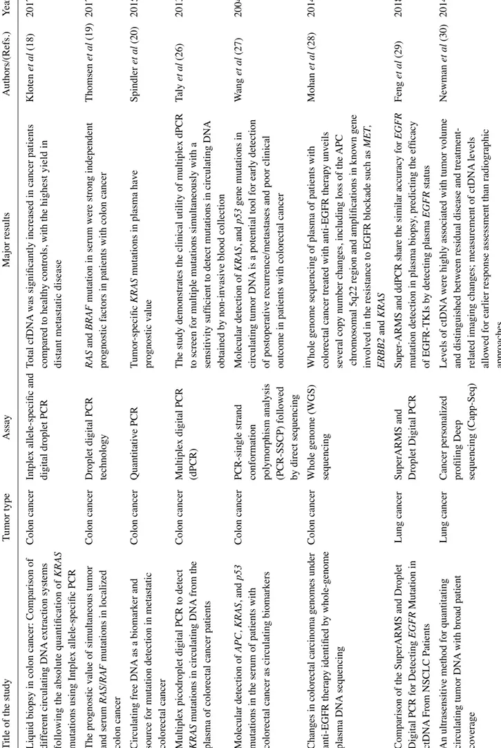
Liquid biopsy investigates circulating tumor cells (CTCs) and/ or cell-free nucleic acids in the peripheral blood of cancer patients (Fig. 1) and is considered one of the most advanced non-invasive diagnostic systems with which to obtain key molecular information relevant to clinical decisions and the practice of precision medicine (1-5). Diagnostic actions include, but are not limited to, early diagnosis, staging, prognosis, the prediction of therapeutic responses, and follow-up during therapeutic intervention (5-13). Historically, the applications of liquid biopsy for the characterization of cancer patients have been focused on CTCs (1). Looking for CTCs in peripheral blood has generated a very large number of reports focusing on diagnosis, prognosis and therapeutic management (6). The downstream characterization of CTCs, including the identifi-cation of possible therapeutic targets (e.g., mutations or other traits of aggressiveness) in this peculiar tumor cell subset not only has had a great impact on diagnosis and prognostication, but also has an impact on clinical protocols, charting the route to precision medicine (14-16). In this respect, an excellent example is colorectal cancer (CRC), one of the most frequent malignancies worldwide (17). As is known, the transformation of normal colonic epithelium into CRC is punctuated by the progressive accumulation of acquired genetic and epigenetic alterations deeply altering morphological parameters, cell growth potential and differentiation, and shutting down apop-tosis. Recent basic and clinical research on CTCs in patients with CRC has underlined that the molecular detection of CTCs in peripheral blood is feasible, and their phenotypic charac-terization drives therapeutic protocols for tailored clinical interventions (18). Moreover, the real-time monitoring of CTCs in patients with CRC has been extensively applied for
a better mechanistic understanding of the factors determining clinical outcome and the efficacy of therapeutic treatment, as well as the stability of therapeutic effects over time (18-21).
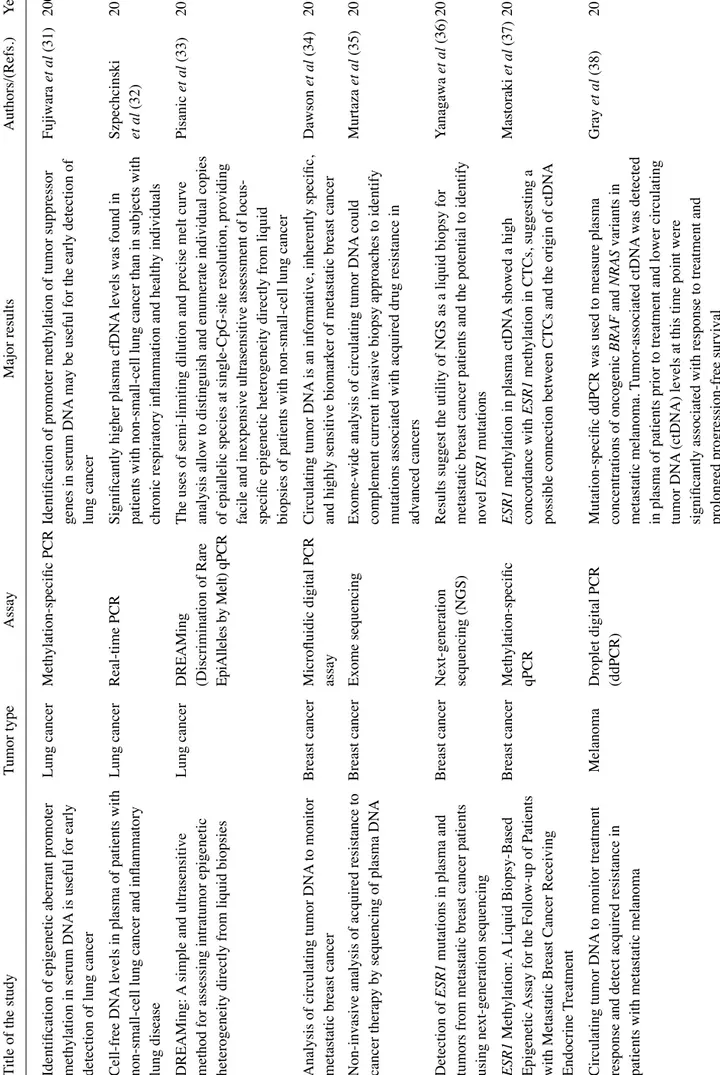
In addition to CTCs, the formal demonstration that free nucleic acids are present (although short-lived) in biological fluids (plasma being investigated by most authors), has led to the development of a large wealth of studies aimed at circulating DNA and RNA (22-24). This strategy, similar to CTC detection, allows for non-invasive diagnosis, and at the same time it represents a convenient method for directly inter-rogating tumor aberrations, addressing tumor heterogeneity and metastatic dissemination across multiple, longitudinally collected clinical specimens (6,7). On the other hand, this approach suffers from important drawbacks, i.e., the frag-mentation of circulating free DNA (cfDNA), the instability of RNA, low analyte concentrations, and the confounding, variable presence of DNA and RNA from normal tissues and mutated cells from the hematopoietic compartment (clonal hematopoiesis) (25). Limitations notwithstanding, the analysis of cfDNA has successfully identified mutations arising in, and the pathognomonicity of, tumor DNA, while the analysis of circulating free RNA (cfRNA) has been mostly focused on miRNA patterns strongly associated with neoplastic transfor-mation/progression (22). Examples of the detection of tumor cfDNA are presented in Table I (18-20,26-52), while examples of the detection of circulating miRNAs are presented in Table II (53-104).
2. Analytes in plasma: Examples of biomedical applications Molecular targets: Cancer genetic aberrations. One of the most robust evidence supporting the application value of liquid biopsies is the detection of circulating genomic aberrations, Figure 1. Applications of liquid biopsy in colorectal cancer (CRC).
Table I. Selected examples of liquid biopsy based on the analysis of circulating free DNA
(cfDNA).
Title of the study
Tumor type
Assay
Major results
Authors/(Refs.)
Year
Liquid biopsy in colon cancer: Comparison of
Colon cancer
Intplex allele-specific and
T
otal cfDNA
was significantly increased in cancer p
at ie nt s Kloten et al (18) 2017 dif
ferent circulating DNA
extraction systems
digital droplet PCR
compared to healthy controls, with the h
ig he st yi el d in
following the absolute quantification of
KRAS
distant metastatic disease
mutations using Intplex allele-specific PCR The prognostic value of simultaneous tumor
Colon cancer
Droplet digital PCR
RAS
and
BRAF
mutation in serum were strong independent
Thomsen et al (19) 2017 and serum RAS/RAF mutations in localized technology
prognostic factors in patients with colon cancer
colon cancer Circulating free DNA
as a biomarker and Colon cancer Quantitative PCR Tumor -specific KRAS
mutations in plasma have
Spindler
et al
(20)
2015
source for mutation detection in metastatic
prognostic value
colorectal cancer Multiplex picodroplet digital PCR to detect
Colon cancer
Multiplex digital PCR
The study demonstrates the clinical utility of multiplex dPCR
Taly
et al
(26)
2013
KRAS
mutations in circulating DNA
from the
(dPCR)
to screen for multiple mutations simultaneously with a
plasma of colorectal cancer patients
sensitivity sufficient to detect mutations in circulating DNA obtained by non
-invasive blood collection
Molecular detection of APC, KRAS , and p53 Colon cancer PCR-single strand Molecular detection of KRAS , and p53 gene mutations in W ang et al (27) 2004
mutations in the serum of patients with
conformation
circulating tumor DNA
is a potential tool for early detection
colorectal cancer as circulating biomarkers
polymorphism analysis
of postoperative recurrence/metastases and poor clinical
(PCR-SSCP) followed
outcome in patients with colorectal cancer
by direct sequencing Ch an ge s i n co lo re cta l c ar cin om a g en om es u nd er Colon cancer Whole genome (WGS)
Whole genome sequencing of plasma of patients with
Mohan
et al
(28)
2014
anti-EGFR therapy identified by whole-genome
sequencing
colorectal cancer treated with anti-EGFR therapy unveils
plasma DNA
sequencing
several copy number changes, including loss of the
APC
chromosomal 5q22 region and amplifications in known gene involved in the resistance to EGFR blockade such as
MET , ERBB2 and KRAS
Comparison of the SuperARMS and Droplet
Lung cancer SuperARMS and Su pe r-A RM S an d dd PC R sh ar e t he si m ila r a cc ur ac y fo r EGFR Feng et al (29) 2018
Digital PCR for Detecting
EGFR
Mutation in
Droplet Digital PCR
mutation detection in plasma biopsy
, predicting the efficacy
ctDNA
From NSCLC Patients
of EGFR-TKIs by detecting plasma
EGFR
status
An ultrasensitive method for quantitating
Lung cancer
Cancer personalized
Levels of ctDNA
were highly associated with tumor volume
Newman
et al
(30)
2014
circulating tumor DNA
with broad patient
profiling Deep
and distinguished between residual disease and
treatment-coverage
sequencing (Capp-Seq)
related imaging changes; measurement of ctDNA
levels
allowed for earlier response assessment than radiographic
Table I. Continued. Title of the study Tumor type Assay Major results Authors/(Refs.) Year
Identification of epigenetic aberrant promoter
Lung cancer
Methylation-specific PCR
Identification of promoter methylation of tumor suppressor
Fujiwara
et al
(31)
2005
methylation in serum DNA
is useful for early
genes in serum DNA
may be useful for the early detection of
detection of lung cancer
lung cancer
Cell-free DNA
levels in plasma of patients with
Lung cancer
Real-time PCR
Significantly higher plasma cfDNA
levels was found in
Szpechcinski
2015
non-small-cell lung cancer and inflammatory
patients with non-small-cell lung cancer than in subjects with
et al
(32)
lung disease
chronic respiratory inflammation and healthy individuals
DREAMing:
A simple and ultrasensitive
Lung cancer
DREAMing
The uses of semi-limiting dilution and precise melt curve
Pisanic
et
al
(33)
2015
method for assessing intratumor epigenetic
(Discrimination of Rare
analysis allow to distinguish and enumerate individual copies
heterogeneity directly from liquid biopsies
Ep iA lle les by M elt ) q PC R
of epiallelic species at single-CpG-site resolution, providing facile and inexpensive ultrasensitive assessment of locus- specific epigenetic heterogeneity directly from liquid biopsies of patients with non-small-cell lung cancer
Analysis of circulating tumor DNA
to monitor
Breast cancer
Microfluidic digital PCR
Circulating tumor DNA
is an informative, inherently specific,
Dawson
et
al
(34)
2013
metastatic breast cancer
assay
and highly sensitive biomarker of metastatic breast cancer
Non-invasive analysis of acquired resistance to
Breast cancer
Exome sequencing
Exome-wide analysis of circulating tumor DNA
could Murtaza et al (35) 2013
cancer therapy by sequencing of plasma DNA
complement current invasive biopsy approaches to identify mutations associated with acquired drug resistance in advanced cancers
Detection of
ESR1
mutations in plasma and
Breast cancer
Next-generation
Results suggest the utility of NGS as a liquid biopsy for
Yanagawa
et
al
(36)
2017
tumors from metastatic breast cancer patients
sequencing (NGS)
metastatic breast cancer patients and the potential to identify
using next-generation sequencing
novel ESR1 mutations ESR1 Methylation: A Liquid Biopsy-Based Breast cancer Methylation-specific ESR1
methylation in plasma ctDNA
showed a high M as to ra ki et al (37) 2 01 8 Epigenetic
Assay for the Follow-up of Patients
qPCR
concordance with
ESR1
methylation in CTCs, suggesting a
with Metastatic Breast Cancer Receiving
possible connection between CTCs and the origin of ctDNA
Endocrine T
reatment
Circulating tumor DNA
to monitor treatment
Melanoma
Droplet digital PCR
Mutation-specific ddPCR was used to measure plasma
Gray
et
al
(38)
2015
response and detect acquired resistance in
(ddPCR) concentrations of oncogenic BRAF and NRAS variants in
patients with metastatic melanoma
metastatic melanoma.
Tumor
-associated ctDNA
was detected
in plasma of patients prior to treatment and lower circulating tumor DNA
(ctDNA) levels at this time point were
Table I. Continued. Title of the study Tumor type Assay Major results Authors/(Refs.) Year Quantitative assessment of BRAF V600 Melanoma Allele-specific An increase of the BRAF V600mut ctDNA
copy number and
Schreuer
et
al
(39)
2016
mutant circulating cell-free tumor DNA
as a
quantitative PCR (qPCR)
fraction, identified disease progression with high sensitivity
tool for therapeutic monitoring in metastatic
and specificity
melanoma patients treated with BRAF/MEK inhibitors Pyrophosphorolysis-activated polymerization
Melanoma
Bidirectional
Bi-P
AP
assays detect and quantify ctDNA
in patients with Madic et al (40) 2012
detects circulating tumor DNA
in metastatic
pyrophosphorolysis-metastatic uveal melanoma
uveal melanoma
activated polymerization (bi-P
AP) real-time PCR
Personalized circulating tumor DNA
Ovarian
Droplet digital PCR
The use of personalized ctDNA
biomarkers in gynecologic Pereira et al (41) 2015
biomarkers dynamically predict treatmen
cancer
cancers can identify the presence of the residual tumor
response and survival in gynecologic cancers Non
-invasive identification and monitoring of
Ovarian
Tagged-amplicon deep
TAm-Seq is a flexible and cost-ef
fective platform for
Forshew
et
al
(42)
2012
cancer mutations by tar
geted deep sequencing
cancer sequencing (T Am-Seq) ap pl ica tio ns in n on -in va siv e c an ce r g en om ics an d di ag no sti cs . of plasma DNA
This method can be used for high-throughput sequencing of plasma samples to identify and monitor levels of multiple cancer mutations in circulating DNA
Cell-free DNA
level as a prognostic biomarker
Ovarian
Quantitative (real-time)
The pre-operative serum cfDNA
level of RAB25 may be a No et al (43) 2012
for epithelial ovarian cancer
cancer
PCR
useful biomarker predicting survival outcomes in patients with advanced ovarian cancer
RASSF1A
promoter methylation in high-grade
Ovarian
Real-time methylation
RASSF1A
promoter methylation provides significant
Giannopoulou 2017 se ro us ov ar ian ca nc er : A di re ct co m pa ris on st ud y cancer specific PCR (real-time
prognostic information in HGSC patients
et
al
(44)
in primary tumors, adjacent morphologically
MSP) and a
methylation-tumor cell-free tissues and paired circulating
sensitive high-resolution
tumor DNA
melting analysis (MS-HRMA)
Cancer genome scanning in plasma: Detection
He pa to ce llu lar Shotgun massively
Genome wide profiling of copy number aberrations and point
Chan et al (45) 2013 of tumor
-associated copy number aberrations,
carcinoma
parallel sequencing
mutations in the plasma of the cancer patients was found
single-nucleotide variants, and tumor
(MPS)
heterogeneity by massively parallel sequencing Methylation profiling of serum DNA
from He pa to ce llu lar Methylation, BeadChip,
The methylation status of circulating DNA
in hepatocellular Zhang et al (46) 2013
hepatocellular carcinoma patients using an
carcinoma
Hot-start PCR,
cancer (HCC) may serve as a potential biomarker
.
Infinium Human Methylation 450 BeadChip
Pyrosequencing
Table I. Continued. Title of the study Tumor type Assay Major results Authors/(Refs.) Year Th e p ro gn os tic v alu e o f c irc ul ati ng p las m a D NA H ep ato ce llu lar Quantitative (real-time)
Combination of circulating DNA
and allelic imbalance at
Ren et al (47) 2006 lev el an d i ts all eli c i m ba lan ce on ch ro m os om e 8 p carcinoma PCR
microsatellite D8S258 may predict the prognosis of patients
in patients with hepatocellular carcinoma
with hepatocellular carcinoma
Detecting circulating tumor DNA
in He pa to ce llu lar Droplet digital PCR
The DNAs from matched tumor and adjacent liver tissues or
Huang et al (48) 2016 he pa to ce llu lar ca rc in om a p ati en ts us in g ca rc in om a
peripheral blood mononuclear cells (PBMCs) were sequenced
droplet digital PCR is feasible and reflects
to identify the origin of circulating mutants. ctDNA
could be
intratumoral heterogeneity
readily detected in patients with hepatocellular carcinoma by targeting hotspot mutations using ddPCR and might reflect intratumoral heterogeneity
Tumor
-associated copy number changes in the
Prostate
Plasma-Seq
Shotgun DNA
sequencing of plasma ctDNA
is a potentially Heitzer et al (49) 2013
circulation of patients with prostate cancer
cancer
powerful tool for cancer detection, monitoring, and for
identified through whole-genome sequencing
studying tumor heterogeneity
Ci rc ul ati ng tu m or D NA g en om ics co rre lat e w ith Prostate
Whole-exome and deep
A lar
ge randomized phase II trial, based on liquid biopsies in
Annala et al (50) 2018 resistance to
Abiraterone and Enzalutamide in
cancer
tar
geted gene sequencing
a patient population representative of clinical practice,
prostate cancer
demonstrated the impact of common genomic alterations on patient response to the most widely used therapies for advanced prostate cancer
Characterization of cell-free circulating DNA
Prostate
Quantitative (real-time)
The study suggests that ccfDNA
integrity can be a useful
Delgado
et
al
(51)
2013
in plasma in patients with prostate cancer
cancer
PCR
biomarker to monitor prostate cancer progression, as the longer fragments are released of non-apoptotic cell death (for example necrosis) that is a frequent event in solid tumors
Prognostic and therapeutic implications of
Prostate
Droplet digital PCR
The study evaluates the circulating androgen receptor (AR)
Buelens
et
al
(52)
2017
circulating androgen receptor gene copy
cancer
gene copy number (CN) control and prostate cancer serum
number in prostate cancer patients using
samples. Poor prognosis in castration-resistant prostate
droplet digital polymerase chain reaction
Table II. Selected examples of liquid biopsy based on the analysis of circulating microRNAs (miRNAs or miRs). Title of the study Tumor type As sa y an d tar ge t m iR NA s Major results Authors/(Refs.) Year
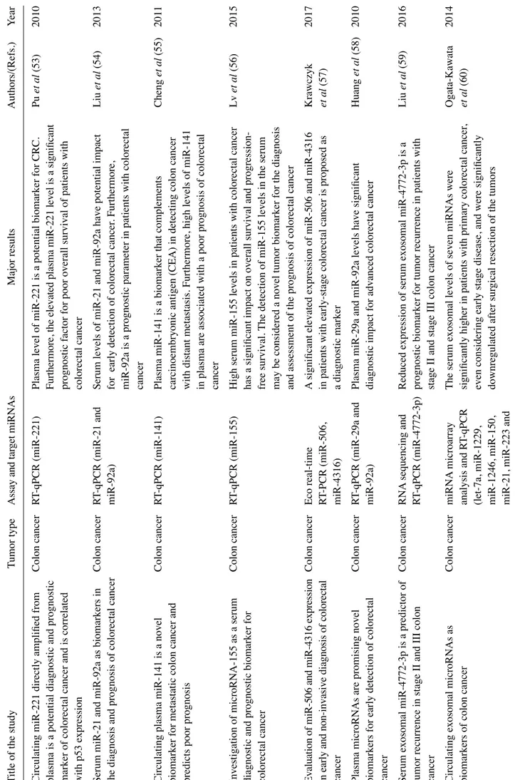
Circulating miR-221 directly amplified from
Colon cancer
RT
-qPCR (miR-221)
Plasma level of miR-221 is a potential biomarker for CRC.
Pu et al (53) 2010 pl as m a i s a p ot en tia l d ia gn os tic an d pr og no sti c
Furthermore, the elevated plasma miR-221 level is a significant
marker of colorectal cancer and is correlated
prognostic factor for poor overall survival of patients with
with p53 expression
colorectal cancer
Serum miR-21 and miR-92a as biomarkers in
Colon cancer
RT
-qPCR (miR-21 and
Serum levels of miR-21 and miR-92a have potential impact
Liu
et
al
(54)
2013
the diagnosis and prognosis of colorectal cancer
miR-92a)
for early detection of colorectal cancer
. Furthermore,
miR-92a is a prognostic parameter in patients with colorectal
cancer
Circulating plasma miR-141 is a novel
Colon cancer
RT
-qPCR (miR-141)
Plasma miR-141 is a biomarker that complements
Cheng et al (55) 201 1
biomarker for metastatic colon cancer and
carcinoembryonic antigen (CEA) in detecting colon cancer
predicts poor prognosis
with distant metastasis. Furthermore, high levels of miR-141 in plasma are associated with a poor prognosis of colorectal
cancer
Investigation of microRNA-155 as a serum
Colon cancer
RT
-qPCR (miR-155)
High serum miR-155 levels in patients with colorectal cancer
Lv
et
al
(56)
2015
diagnostic and prognostic biomarker for
has a significant impact on overall survival and
progression-colorectal cancer
free survival.
The detection of miR-155 levels in the serum
may be considered a novel tumor biomarker for the diagnosis and assessment of the prognosis of colorectal cancer
Ev alu ati on of m iR -5 06 an d m iR -4 31 6 e xp re ss io n Colon cancer Eco real-time
A significant elevated expression of miR-506 and miR-4316
Krawczyk 2017 in ea rly an d no n-in va siv e d iag no sis o f c ol or ec tal RT -PCR (miR-506,
in patients with early-stage colorectal cancer is proposed as
et al (57) cancer miR-4316) a diagnostic marker
Plasma microRNAs are promising novel
Colon cancer
RT
-qPCR (miR-29a and
Plasma miR-29a and miR-92a levels have significant
Huang
et
al
(58)
2010
biomarkers for early detection of colorectal
miR-92a)
diagnostic impact for advanced colorectal cancer
cancer Serum exosomal miR-4772-3p is a predictor of
Colon cancer
RNA
sequencing and
Reduced expression of serum exosomal miR-4772-3p is a
Liu
et
al
(59)
2016
tumor recurrence in stage II and III colon
RT
-qPCR (miR-4772-3p)
prognostic biomarker for tumor recurrence in patients with
cancer
stage II and stage III colon cancer
Circulating exosomal microRNAs as
Colon cancer
miRNA
microarray
The serum exosomal levels of seven miRNAs were
Ogata-Kawata
2014
biomarkers of colon cancer
analysis and R
T-qPCR
significantly higher in patients with primary colorectal cancer
,
et
al
(60)
(let-7a, miR-1229,
even considering early stage disease, and were significantly
miR-1246, miR-150,
downregulated after sur
gical resection of the tumors
Table II. Continued. Title of the study Tumor type As sa y an d tar ge t m iR NA s Major results Authors/(Refs.) Year Di ffe re nt ial ex pr es sio n of m icr oR NA s i n pl as m a Co lo n ca nc er RT -q PC R ar ra y In the study
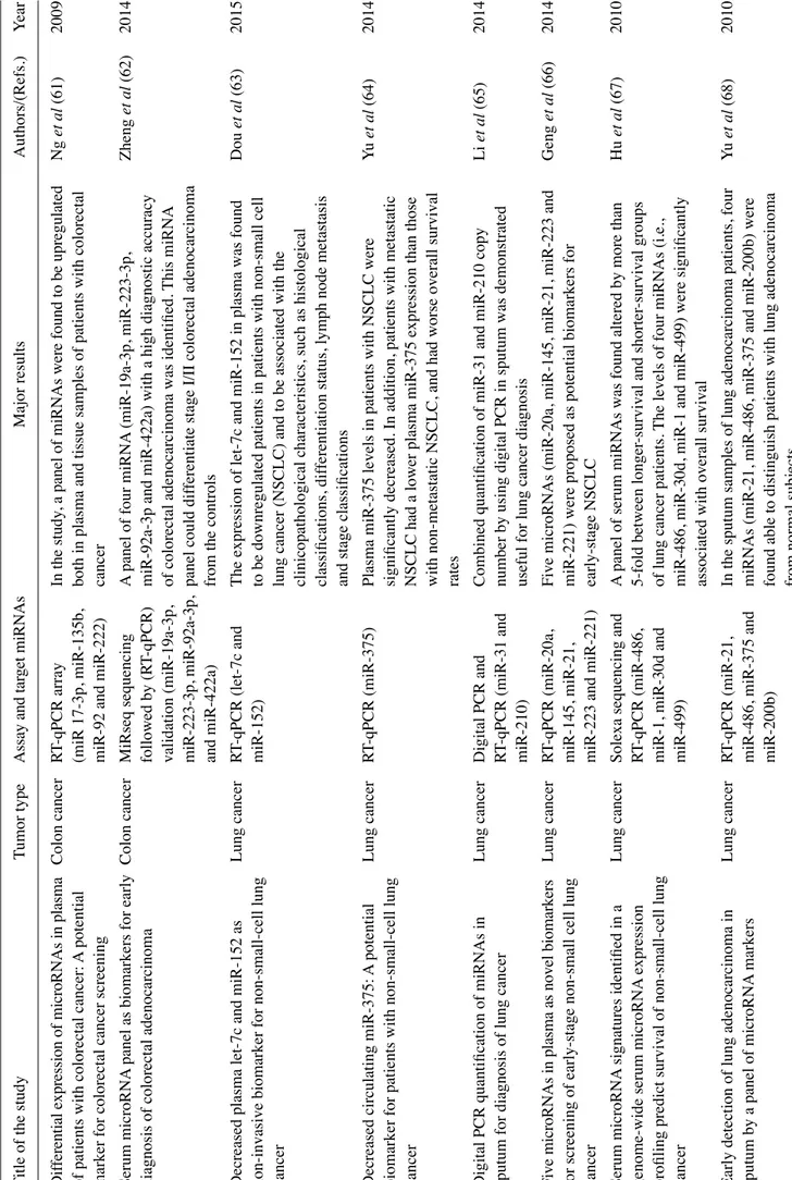
, a panel of miRNAs were found to be upregulated
Ng
et
al
(61)
2009
of patients with colorectal cancer:
A potential
(miR 17-3p, miR-135b,
both in plasma and tissue samples of patients with colorectal
marker for colorectal cancer screening
miR-92 and miR-222)
cancer
Serum microRNA
panel as biomarkers for early
Colon cancer
MiRseq sequencing
A panel of four miRNA
(miR-19a-3p, miR-223-3p, Zheng et al (62) 2014
diagnosis of colorectal adenocarcinoma
followed by (R
T-qPCR)
miR-92a-3p and miR-422a) with a high diagnostic accuracy
validation (miR-19a-3p,
of colorectal adenocarcinoma was identified.
This miRNA m iR -2 23 -3 p, m iR -9 2a -3 p,
panel could dif
ferentiate stage I/II co
lorectal adenocarcinoma
and miR-422a)
from the controls
Decreased plasma let-7c and miR-152 as
Lung cancer
RT
-qPCR (let-7c and
The expression of let-7c and miR-152 in plasma was found
Dou et al (63) 2015 non
-invasive biomarker for non-small-cell lung
miR-152)
to be downregulated patients in patients with non-small cell
cancer
lung cancer (NSCLC) and to be associated with the clinicopathological characteristics, such as histological classifications, dif
ferentiation status, lymph node metastasis
and stage classifications
Decreased circulating miR-375:
A potential
Lung cancer
RT
-qPCR (miR-375)
Plasma miR-375 levels in patients with NSCLC were
Yu
et
al
(64)
2014
biomarker for patients with non-small-cell lung
significantly decreased. In addition, patients with metastatic
cancer
NSCLC had a lower plasma miR-375 expression than those with non-metastatic NSCLC, and had worse overall survival
rates
Digital PCR quantification of miRNAs in
Lung cancer
Digital PCR and
Combined quantification of miR-31 and miR-210 copy
Li
et
al
(65)
2014
sputum for diagnosis of lung cancer
RT
-qPCR (miR-31 and
number by using digital PCR in sputum was demonstrated
miR-210)
useful for lung cancer diagnosis
Fi ve m icr oR NA s i n pl as m a a s n ov el bi om ar ke rs Lung cancer RT -qPCR (miR-20a,
Five microRNAs (miR-20a, miR-145, miR-21, miR-223 and
Geng
et
al
(66)
2014
for screening of early-stage non-small cell lung
miR-145, miR-21,
miR-221) were proposed as potential biomarkers for
cancer
miR-223 and miR-221)
early-stage NSCLC
Serum microRNA
signatures identified in a
Lung cancer
Solexa sequencing and
A panel of serum miRNAs was found altered by more than
Hu
et
al
(67)
2010
genome-wide serum microRNA
expression
RT
-qPCR (miR-486,
5-fold between longer
-survival and shorter
-survival groups
profiling predict survival of non-small-cell lung
miR-1, miR-30d and
of lung cancer patients.
The levels of four miRNAs (i.e.,
cancer
miR-499)
miR-486, miR-30d, miR-1 and miR-499) were significantly associated with overall survival
Early detection of lung adenocarcinoma in
Lung cancer
RT
-qPCR (miR-21,
In the sputum samples of lung adenocarcinoma patients, four
Yu
et
al
(68)
2010
sputum by a panel of microRNA
markers
miR-486, miR-375 and
miRNAs (miR-21, miR-486, miR-375 and miR-200b) were
miR-200b)
Table II. Continued. Title of the study Tumor type As sa y an d tar ge t m iR NA s Major results Authors/(Refs.) Year
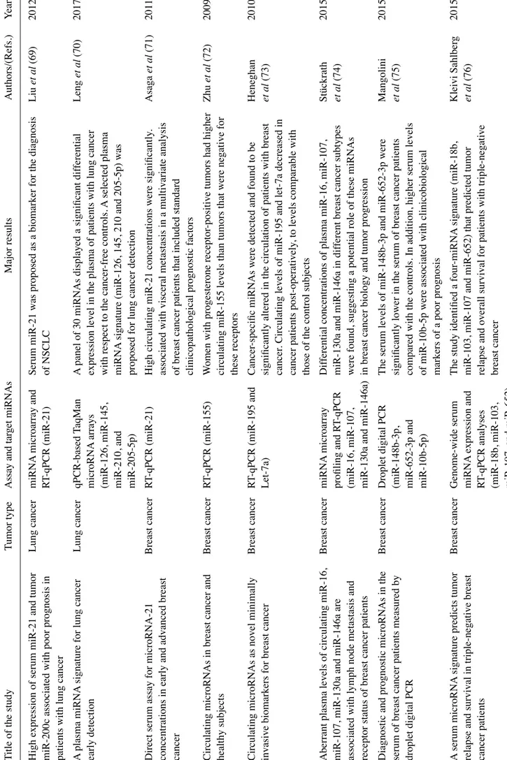
High expression of serum miR-21 and tumor
Lung cancer
miRNA
microarray and
Serum miR-21 was proposed as a biomarker for the diagnosis
Liu
et
al
(69)
2012
miR-200c associated with poor prognosis in
RT
-qPCR (miR-21)
of NSCLC
patients with lung cancer A plasma miRNA
signature for lung cancer
Lung cancer
qPCR-based
TaqMan
A panel of 30 miRNAs displayed a significant dif
ferential Leng et al (70) 2017 early detection microRNA arrays
expression level in the plasma of patients with lung cancer
(miR-126, miR-145,
with respect to the cancer
-free controls.
A selected plasma
miR-210, and
miRNA
signature (miR-126, 145, 210 and 205-5p) was
miR-205-5p)
proposed for lung cancer detection
Direct serum assay for microRNA-21
Breast cancer
RT
-qPCR (miR-21)
High circulating miR-21 concentrations were significantly
. Asaga et al (71) 201 1
concentrations in early and advanced breast
associated with visceral metastasis in a multivariate analysis
cancer
of breast cancer patients that included standard clinicopathological prognostic factors
Circulating microRNAs in breast cancer and
Breast cancer
RT
-qPCR (miR-155)
W
omen with progesterone receptor
-positive tumors had higher
Zhu et al (72) 2009 healthy subjects
circulating miR-155 levels than tumors that were negative for these receptors
Circulating microRNAs as novel minimally
Breast cancer
RT
-qPCR (miR-195 and
Cancer
-specific miRNAs were detected and found to be
Heneghan
2010
invasive biomarkers for breast cancer
Let-7a)
significantly altered in the circulation of patients with breast
et
al
(73)
cancer
. Circulating levels of miR-195 and let-7a decreased in
cancer patients post-operatively
, to levels comparable with
those of the control subjects
Aberrant plasma levels of circulating miR-16,
Breast cancer
miRNA
microarray
Dif
ferential concentrations of plasma miR-16, miR-107,
Stückrath
2015
miR-107, miR-130a and miR-146a are
profiling and R
T-qPCR
miR-130a and miR-146a in dif
ferent breast cancer subtypes
et
al
(74)
associated with lymph node metastasis and
(miR-16, miR-107,
were found, suggesting a potential role of these miRNAs
receptor status of breast cancer patients
miR-130a and miR-146a)
in breast cancer biology and tumor progression
Diagnostic and prognostic microRNAs in the
Breast cancer
Droplet digital PCR
The serum levels of miR-148b-3p and miR-652-3p were
Mangolini
2015
serum of breast cancer patients measured by
(miR-148b-3p,
significantly lower in the serum of breast cancer patients
et
al
(75)
droplet digital PCR
miR-652-3p and
compared with the controls. In addition, higher serum levels
miR-10b-5p)
of miR-10b-5p were associated with clinicobiological markers of a poor prognosis
A serum microRNA
signature predicts tumor
Breast cancer
Genome-wide serum
The study identified a four
-miRNA
signature (miR-18b,
Kleivi Sahlber
g
2015
relapse and survival in triple-negative breast
miRNA
expression and
miR-103, miR-107 and miR-652) that predicted tumor
et al (76) cancer patients RT -qPCR analyses
relapse and overall survival for patients with triple-negative
(miR-18b, miR-103,
breast cancer
Table II. Continued. Title of the study Tumor type As sa y an d tar ge t m iR NA s Major results Authors/(Refs.) Year
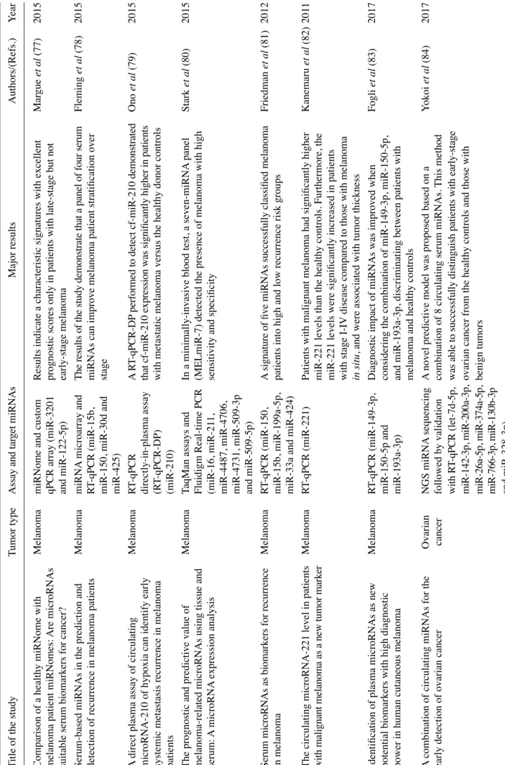
Comparison of a healthy miRNome with
Melanoma
miRNome and custom
Results indicate a characteristic signatures with excellent
Mar gue et al (77) 2015
melanoma patient miRNomes:
Are microRNAs
qPCR array (miR-3201
prognostic scores only in patients with late-stage but not
suitable serum biomarkers for cancer?
and miR-122-5p)
early-stage melanoma
Serum-based miRNAs in the prediction and
Melanoma miRNA microarray and Th e r es ul ts of th e s tu dy de m on str ate th at a p an el of fo ur se ru m Fleming et al (78) 2015
detection of recurrence in melanoma patients
RT
-qPCR (miR-15b,
miRNAs can improve melanoma patient stratification over
miR-150, miR-30d and
stage miR-425) A di re ct pl as m a a ss ay o f c irc ul ati ng M ela no m a RT -q PC R A R T-qPCR-DP
performed to detect cf-miR-210 demonstrated
On o et al (79) 2015
microRNA-210 of hypoxia can identify early
directly-in-plasma assay th at cf -m iR -2 10 ex pr es sio n w as si gn ifi ca nt ly hi gh er in pa tie nt s
systemic metastasis recurrence in melanoma
(R
T-qPCR-DP)
with metastatic melanoma versus the healthy donor controls
patients
(miR-210)
The prognostic and predictive value of
Melanoma
TaqMan assays and
In a minimally-invasive blood test, a seven-miRNA
panel Stark et al (80) 2015
melanoma-related microRNAs using tissue and
Fluidigm Real-time PCR
(MELmiR-7) detected the presence of melanoma with high
serum:
A microRNA
expression analysis
(miR-16, miR-21
1,
sensitivity and specificity
miR-4487, miR-4706, miR-4731, miR-509-3p and miR-509-5p)
Serum microRNAs as biomarkers for recurrence
M ela no m a RT -q PC R (m iR -1 50 ,
A signature of five miRNAs successfully classified melanoma
Fr ied m an et al (81) 2012 in melanoma miR-15b, miR-199a-5p,
patients into high and low recurrence risk groups
miR-33a and miR-424)
The circulating microRNA-221 level in patients
Melanoma
RT
-qPCR (miR-221)
Patients with malignant melanoma had significantly higher
Kanemaru et al (82) 201 1 wi th m ali gn an t m ela no m a a s a ne w tu m or m ar ke r
miR-221 levels than the healthy controls. Furthermore, the miR-221 levels were significantly increased in patients with stage I-IV
disease compared to those with melanoma
in situ
, and were associated with tumor thickness
Identification of plasma microRNAs as new
Melanoma
RT
-qPCR (miR-149-3p,
Diagnostic impact of miRNAs was improved when
Fogli
et
al
(83)
2017
potential biomarkers with high diagnostic
miR-150-5p and
considering the combination of miR-149-3p, miR-150-5p,
power in human cutaneous melanoma
miR-193a-3p)
and miR-193a-3p, discriminating between patients with melanoma and healthy controls
A combination of circulating miRNAs for the
Ovarian
NGS miRNA
sequencing
A
novel predictive model was proposed based on a
Yokoi
et
al
(84)
2017
early detection of ovarian cancer
cancer
followed by validation
combination of 8 circulating serum miRNAs.
This method
with R
T-qPCR
(let-7d-5p,
was able to successfully distinguish patients with early-stage
miR-142-3p, miR-200a-3p,
ovarian cancer from the healthy controls and those with
miR-26a-5p, miR-374a-5p, b en ig n t um or s
Table II. Continued. Title of the study Tumor type As sa y an d tar ge t m iR NA s Major results Authors/(Refs.) Year Circulating miRNA landscape identifies Ovarian miRNA microarray and
This study allowed the identification of circulating miRNAs
Todeschini
2017
miR-1246 as promising diagnostic biomarker
cancer
droplet digital PCR
with diagnostic relevance for high-grade serous ovarian
et
al
(85)
in high-grade serous ovarian carcinoma:
A
(miR-1246, miR-595 and
carcinoma (HGSOC)
validation across two independent cohorts
miR-2278)
Expression of serum miR-200a, miR-200b, and
Ovarian
RT
-qPCR (miR-200a and
The expression levels of miR-200a and miR-200c were found
Zuberi
et
al
(86)
2015
miR-200c as candidate biomarkers in epithelial
cancer
miR-200b, miR-200c)
to be significantly associated with disease progression, while
ovarian cancer and their association with
miR-200a overexpression was found be associated with tumor
clinicopathological features
histology and the stage of epithelial ovarian cancer
Serum microRNA-145 as a novel biomarker in
Ovarian
RT
-qPCR (miR-145)
Serum miR-145 levels could discriminate patients with
Liang
et
al
(87)
2015
human ovarian cancer
cancer
malignant ovarian cancer from the healthy controls
MicroRNA-200c and microRNA-141 as
Ovarian
RT
-qPCR (miR-200c and
The results of the study suggested that serum miR-200c and
Gao and W u (88) 2015 potential diagnostic and prognostic biomarkers cancer miR-141) m iR -1 41 w er e a bl e t o d isc rim in ate p ati en ts w ith o va ria n c an ce r fo r o va ria n ca nc er
from healthy controls. In addition, miR-200c and miR-141 may be predictive biomarkers for the prognosis of ovarian cancer
Urinary microRNA-30a-5p is a potential
Ovarian
miRNA
microarray and
Results indicated an increase in miR-30a-5p levels in the
Zhou
et
al
(89)
2015
biomarker for ovarian serous adenocarcinoma
cancer
RT
-qPCR (miR-30a-5p)
urine of patients with ovarian serous adenocarcinoma. In parallel, the inhibition of miR-30a-5p suppressed the malignant phenotypes of ovarian cancer
in vitr
o
Combining serum microRNA
and CA-125 as
Ovarian
RT
-qPCR (miR-375,
The combination of serum miR-375, miR-210 and CA-125
Shah
et
al
(90)
2018
prognostic indicators of preoperative sur
gical
cancer
miR-34a-5p and
can discriminate healthy versus patients with high-grade
outcome in women with high-grade serous
miR-210)
serous ovarian cancer
. The combination of miR-34a-5p
ovarian cancer
and CA-125 was the strongest predictor of completeness of surgical resection
Serum microRNA characterization identifies He pa toc ell ula r qPCR-based TaqMan
miR-885-5p is significantly elevated in the sera of patients
Gui et al (91) 201 1
miR-885-5p as a potential marker for detecting
carcinoma
microRNA
arrays
with liver pathologies, including hepatocellular carcinoma
liver pathologies
(miR-885-5p)
MicroRNA-500 as a potential diagnostic marker
H ep ato ce llu lar RT -qPCR (miR-500)
An increased amount of miR-500 was found in the sera of
Yamamoto
2009
for hepatocellular carcinoma
carcinoma
patients with hepatocellular carcinoma. In fact, miR-500
et
al
(92)
levels in the sera of patients with hepatocellular carcinoma returned to normal following sur
gical treatment
Circulating microRNAs, miR-21, miR-122, and
H ep ato ce llu lar RT -qPCR (miR-21,
Results indicated that serum miR-21, miR-122 and miR-223
Xu et al (93) 201 1
miR-223, in patients with hepatocellular
carcinoma
miR-122 and miR-223)
were elevated in patients with hepatocellular carcinoma or
carcinoma or chronic hepatitis
chronic hepatitis and these miRNAs have strong potential to serve as novel biomarkers for liver injury
, but not specifically
Table II. Continued. Title of the study Tumor type As sa y an d tar ge t m iR NA s Major results Authors/(Refs.) Year Pl as m a m icr oR NA p an el to d iag no se h ep ati tis B H ep ato ce llu lar miRNA microarray A microRNA
panel that provides a high diagnostic accuracy
Zhou et al (94) 201 1
virus-related hepatocellular carcinoma
carcinoma
analysis and R
T-qPCR
of hepatocellular carcinoma was described
(miR-122, miR-192, miR-21, miR-223, miR-26a, miR-27a and miR-801)
Se ru m m icr oR NA p ro fil es se rv e a s n ov el He pa to ce llu lar NG S m icr oR NA
The study demonstrates that serum miRNA
profiles can serve
Li et al (95) 2010 biomarkers for HBV
infection and diagnosis of
carcinoma
sequencing followed by
as non-invasive biomarkers for hepatitis B virus (HBV)
HBV
-positive hepatocarcinoma
validation with
TaqMan
infection and HBV
-positive hepatocellular carcinoma
probe-based R
T-qPCR
diagnosis
(miR-23b, miR-423, miR-375, miR-23a and miR-342-3p)
Circulating miR-106b-3p, miR-101-3p and
He pa to ce llu lar
RNAseq and droplet
Ci rc ul ati ng m iR -1 01 -3 p, m iR -1 06 b-3p an d m iR -1 24 6, ei th er Moshiri et al (96) 2018
miR-1246 as diagnostic biomarkers of
carcinoma
digital PCR (ddPCR)
individually or in combination, exhibit a considerable
hepatocellular carcinoma
(miR-106b-3p,
potential value as diagnostic biomarkers of hepatocellular
miR-101-3p and
carcinoma
miR-1246)
Combinations of serum prostate-specific
Prostate
Quantitative PCR (let-7c,
Combinations of let-7c, miR-30c, miR-141, miR-375 and
Kachakova
et
al
(97)
2015
antigen and plasma expression levels of let-7c,
cancer
miR-30c, miR-141 and
PSA
obtained even better discrimination and could be more
miR-30c, miR-141, and miR-375 as potential
miR-375)
useful that prostate-specific antigen (PSA) alone as
better diagnostic biomarkers for prostate cancer
non-invasive diagnostic biomarkers for the screening of prostate cancer
Changes in circulating microRNA
levels
Prostate
miRNA
microarray and
miR-200b and miR-375 levels are increased in the serum of
Bryant
et
al
(98)
2012
associated with prostate cancer
cancer
RT
-qPCR (miR-200b and
patients with metastatic prostate cancer compared with
miR-375)
patients with localized disease
Circulating microRNAs are associated with
Prostate RT -qPCR microRNA Th e s tu dy ha s i de nt ifi ed se lec ted ci rc ul ati ng m iR NA s, no tab ly Lin et al (99) 2014
docetaxel chemotherapy outcome in
castration-cancer
array cards and R
T-qPCR
those of the miR-200 and miR-17 families, associated with
resistant prostate cancer
(miR-200 family and
PSA
response and/or overall survival in patients with
miR-17 family)
castration-resistant prostate cancer
Circulating miRNAs 21 and 221 as biomarkers
Prostate
RT
-qPCR (miR-21 and
The study showed that serum miR-21 and miR-221 levels
Kotb
et
al
(100)
2014
for early diagnosis of prostate cancer
cancer
miR-221)
Table II. Continued. Title of the study Tumor type As sa y an d tar ge t m iR NA s Major results Authors/(Refs.) Year MicroRNA
profiling in prostate cancer - the
Prostate
RT
-qPCR microRNA
miR-205 and miR-214 levels are downregulated in prostate
Srivastava
2013
diagnostic potential of urinary miR-205 and
cancer
array cards and R
T-qPCR
cancer and may serve as a potential non-invasive molecular
et
al
(101)
miR-214
(miR-205 and miR-214)
biomarker for prostate cancer
Serum microRNA
expression patterns that
Prostate
miRNA
microarray and
Altered content of miR-103, miR-125b and miR-222 in the
Singh
et
al
(102)
2014
predict early treatment failure in prostate
cancer
RT
-qPCR (miR-103,
serum of patients with prostate cancer was found to be
cancer patients
miR-125b and miR-222)
associated with the outcome of clinical treatment
A study on circulating microRNAs identifies a
Prostate
RT
-qPCR (miR-17,
The study demonstrates that a novel previously unreported
Farran
et
al
(103)
2018
new potential biomarker panel to distinguish
cancer
miR-192 and
circulating miRNA
signature consisting of a combination of
aggressive prostate cancer
miR-181a)
interacting miRNAs (miR-17/miR-192) and an independent miRNA
(miR-181a) are capable of dif
ferentiating between
aggressive and non-aggressive prostate cancer
Di ffe re nt le ve ls of se ru m m icr oR NA s i n p ro sta te Prostate RT -qPCR (let-7c, let-7e,
let-7c, let-7e, let-7i, miR-26a-5p, miR-26b-5p, miR-18b-5p
Cochetti
et
al
(104)
2016
cancer and benign prostatic hyperplasia:
cancer
let-7i, miR-26a-5p,
and miR-25-3p were able to discriminate between patients
Evaluation of potential diagnostic and
miR-26b-5p,
with prostate cancer from those harboring benign prostatic
prognostic role
miR-18b-5p and
hyperplasia, both presenting altered PSA
levels
mainly mutations. The topic is extensive, and is the subject of a number of excellent reviews. Therefore, in this review, we focus on very specific examples, particularly in early-stage tumors. KRAS mutations are a case in point, since they serve as an actionable marker for EGFR blockade therapy, are highly prevalent, and have been thoroughly investigated. For instance, Brychta et al compared plasma and paired tumor samples from early-stage pancreatic cancer patients (105) by chip-based digital PCR. Their major aim was to identify selected KRAS codon 12 mutations (G12D, G12V and G12C) in circulating tumor DNA (ctDNA). Remarkably, circulating KRAS mutations were demonstrated in 72% of the patients, were associated with tumor burden, and were undetectable in the healthy controls. This study supports the use of liquid biopsy for early cancer diagnosis. Other studies focusing on KRAS mutations in ctDNA were reported by Kinusaga et al (pancreatic cancer) (106), Couraud et al (lung cancer) (107), Perez-Carbonell et al (CRC) (108) and Case et al (lympho-blastic leukemia) (109). Table I summarizes the applicative examples of liquid biopsy for the identification of oncogene mutations, including the detection of EGFR mutations in the blood of lung cancer patients, now approved by regulatory bodies. These assays are of outmost interest and exemplify the profound difference between non-invasive liquid biopsy and invasive tumor tissue biopsy. Tissue biopsy may not reflect the genomic profile of the tumor in its entirety due to intra-tumor heterogeneity, multiple foci poorly accessible to sampling, and/or changes occurring during tumor develop-ment and/or therapy. On the contrary, the non-invasive liquid biopsy of plasma, urine or saliva samples may more effec-tively recapitulate the mutational complexity of the many populations (cryptic and clinically evident) accounting for tumor burden in a given patient. This makes liquid biopsy particularly suitable to identify truncal aberrations that, when targeted, may result in a considerably greater systemic clinical benefit, as compared to targeting site-specific aberra-tions (Lin et al, 2015) (110).
Molecular targets: Gene methylation. A variation on the theme is to look at non-mutational events marking the cancer genome. Particularly relevant in this context is DNA meth-ylation. It has been known for quite some time that tumor progression is associated with the abnormal methylation of cancer genes. Both hypomethylation and hypermethylation have been reported. Most often, the specific DNA hypermeth-ylation of tumor suppressor genes is observed in the context of widespread DNA hypomethylation. Since aberrant DNA methylation at specific promoter regions is a defined molecular feature of cancer, ctDNA methylation has been developed into a very promising molecular marker. DNA methylation is to date one of the preferred consensus circulating biomarkers in pre-symptomatic and symptomatic patients with CRC. This has been discussed by Warton et al (111) and by Mitchell et al (112), who have considered methylation-specific PCR assays as a novel approach for the assessment of low levels of DNA methylation in 29 regions of 17 genes. Eight differ-entially-methylated regions (DMRs) residing in the BCAT1, GRASP, IKZF1 and IRF4 genes, exhibited low positivity in the plasma of healthy subjects and high positivity (>59%) in ctDNA from colonoscopy-confirmed patients with CRC.
Molecular targets: Circulating microRNAs. MicroRNAs (miRNAs or miRs) are a family of small (19 to 25 nucleotides in length) non-coding RNAs which play important roles in controlling post-transcriptional gene expression. Regulatory miRNAs reduce protein synthesis through selective interac-tions with complementary sequences of target messenger RNAs (mRNAs) (113,114). Single or multiple mRNAs can be targeted at their 3'untranslated region (UTR), coding sequence (CDS), 5'UTR sequences, and it is calculated that >60% of human mRNAs are miRNA targets (114). The miRNA/mRNA interaction occurs at the level of RNA-induced silencing complex (RISC) and is associated with the repression of translation or mRNA degradation, depending on the levels of complementarity with nucleotide sequences on the target mRNAs (115-118). Since their discovery and first characteriza-tion, the number of human miRNAs identified and deposited in the miRBase databases (miRBase v.22, www.mirbase.org) has been steadily increasing and is now >2,500 (119,120). Research on miRNAs has confirmed the complexity of this expanding miRNA/RNA network (117-122).
Alterations in miRNA expression have been associated with different human diseases. The guided alteration of specific miRNAs may potentially lead to innovative thera-peutic protocols (123,124). miRNAs function both as tumor promoters (oncomiRNAs and metastamiRNAs) and tumor suppressors (125,126), depending on their regulatory prefer-ence for oncoproteins with opposing influprefer-ences on cancer cells. Based on this, it is unsurprising that circulating cell-free miRNAs have been actively investigated as liquid biopsy analytes. OncomiRNAs are abundant in several extracellular body fluids (127-132), where they are protected and stabilized by exosome-like structures and small intraluminal vesicles produced by a variety of cells (including cancer cells) (127). Hence, elevated levels of several miRNAs (including miR-221, miR-222, miR-141, miR-92a, miR-21, miR-155, miR-506 and miR4316, miR-4772 and miR-29a) are present in the blood from patients with CRC (53-62,133,134) and may contribute to the diagnosis and prognosis of patients with CRC (134). Furthermore, miRNAs may aid in the monitoring of therapeutic approaches. For instance, Ogata-Kawata et al reported that serum exosomal miRNA levels (let-7a, miR-1229, miR-1246, miR-150, miR-21, miR-223 and miR-23a) were higher in patients with CRC than in the controls, that this abnormally high levels were already detectable at early disease stages, and that they were significantly downregulated following surgical resection (60).
3. Technologies
In order to identify specific DNA mutations and quantify miRNA levels in plasma and other body fluids of cancer patients, several types of technologies for DNA/RNA analysis have been proposed. For cfDNA analysis, the golden standards are possibly quantitative PCR (qPCR) and digital PCR; however, several additional technologies have been proposed (Table I), such as polymerase chain reaction-single strand conformation polymorphism (PCR-SSCP) analysis (27), multiplex digital PCR (dPCR), allele-specific qPCR (18,39), whole genome sequencing (WGS) (28), cancer personalized profiling deep sequencing (Capp-Seq) (30),
methylation-specific PCR (31,37,44), the Discrimination of Rare EpiAlleles by Melt qPCR (DREAMing) (33), bidirec-tional pyrophosphorolysis-activated polymerization (bi-PAP) real-time PCR (40) and tagged-amplicon deep sequencing (TAm-Seq) (42). For miRNA analysis, qPCR and reverse transcription (RT)-PCR (53-58), NGS RNA sequencing (63), miRNA microarray analysis (60) and digital PCR (65) are the most commonly used technologies (Table II).
A common step, and under many respects a complication of all the above-mentioned technologies, is the need to amplify the minute amounts of target analytes by an enzymatic reaction with DNA modifying enzymes, most often Taq polymerase and its derivatives. Biosensing platforms hold great promise for the simple and rapid detection of cfDNA and cfRNA (135), since they skip this time-consuming, analyte-dependent, PCR amplification step. Novel PCR-free biosensing approaches are able to detect KRAS and BRAF mutations in the serum of patients with lung cancer and melanoma (136).
Digital PCR (137) is based on the limiting dilution of DNA, and single molecule detection to identify and quantify the target mutated DNA in a given sample (138,139). This experimental approach is very useful for the identification of rare variants and in non-invasive diagnosis on peripheral blood, since only a small concentration of template is required for the analysis. Next generation sequencing (NGS) is a high throughput DNA sequencing technology which allows for the analysis, in a single reaction, a large variety of different DNA aberrations across multigene panels (140,141), although comprehen-siveness may somewhat detract from sensitivity. Different commercial NGS platforms are available, such as Genome Analyzer and HiSeq 2000 (Illumina), HeliScope (Helicos BioSciences), SOLiD and Ion Torrent (Life Technologies), Roche/454 (Roche). In these instruments, templates, primers or polymerase enzymes are immobilized on a solid support or on microbeads before sequencing, allowing the process of millions of microreactions carried out in parallel on each spatially distinct template.
However, as already pointed out, several challenges are related to liquid biopsy, the most important of which is the amount of target molecules to be detected and quantified. As far as cfDNA detection is concerned, these target molecules are so diluted by normal DNA that existing sequencing methods, such as Sanger sequencing, were not considered sufficiently sensitive to detect tumor-associated DNA mutation. As shown in Table I, the most commonly used approach was based on mutation-specific PCR, a technology proven to exhibit suffi-cient specificity and sensitivity allowing for the detection of the weak tumor signal present in the patient's circulation. This technology may be associated with important drawbacks when the quantification of miRNAs is considered, suffering from biases in the template-to-product ratios of the amplified target sequences (141). In addition, differential RT efficiency on different miRNA targets may also introduce variability when miRNA patterning is considered. Once again, PCR-free detection strategies are of great interest (142,143).
4. Experimental model systems for technological validation Liquid biopsy is a complex strategy requiring pre-analytical steps, post-analytical optimization, and the careful selection
of optimal analytes for specific biological queries. In vivo model systems may be very useful in addressing and isolating these numerous individual variables (that are both technical and biological), and validate complex multi-step approaches. It is surprising, in this respect, that only few reports are available focusing on the use of animal models. For example, Garcia-Olmo et al directly compared the tumor ctDNA concentration and the number of circulating cancer cells in rats with xenograft tumors during the spread of CRC (144). Of note, they found that high ctDNA levels preceded the presence of CTCs. Rago et al (145) developed an elegant and highly sensitive qPCR test to quantify ctDNA by targeting LINE-1 in mouse xenografts, demonstrating that this experimental system enables the monitoring of systemic tumor burden and close examination of the therapeutic management on a variety of animal tumor models. These studies demonstrate the impor-tance of ctDNA and how it intertwines with CTCs. In a more recent study, Thierry et al (146) evaluated the relative quantita-tive contributions of non-tumor, tumor and mutated ctDNA, as well as ctDNA integrity, in an animal model. In this case, they found differences between patients with CRC and nude mice xenografted with human colon cell lines, suggesting that further research is necessary to validate in vivo model systems based on mice xenografted with tumor cell lines.
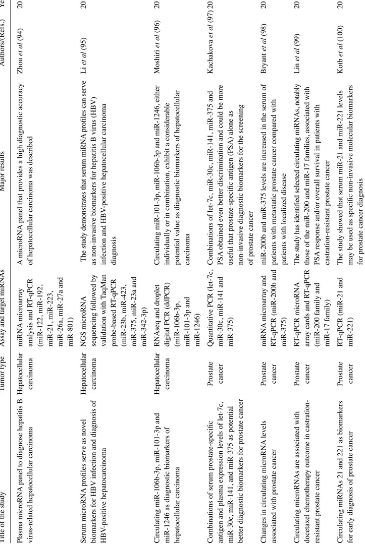
As for miRNAs, different independent studies have firmly demonstrated that miRNAs released into the circulation by tumor xenografts are distinct from ‘background’ mouse miRNAs. This is a key point, since pre-existing miRNAs present in mouse body fluids may be a powerful confounding parameter, possibly altering conclusions and implications of any circulating miRNA signature. In this respect, the use of laboratory mouse strains has the advantage that it sets a ‘background’ mouse miRNA pattern that is stable and easily quantifiable. Mitchell et al demonstrated that several miRNAs originating from xenografted human prostate cancer cells are present in the circulation (one of the most interesting being miR-141), and are readily measured in plasma, allowing a clear distinction between tumor-xenografted mice and controls (147). Selth et al (148) performed global miRNA profiling and iden-tified a set of miRNAs exhibiting significantly altered serum levels in transgenic mice bearing prostate adenocarcinoma tumors. Among the most interesting miRNAs, they focused their attention on miR-141 and miR-375. Waters et al observed a complex miRNA dysregulation in the circulation of athymic nude mice subcutaneously injected with MDA-MB-231 cells. Some miRNAs (such as miR-10b) were undetectable in the circulation, some others (miR-195 and miR-497) were significantly decreased, the miR-221 content was not altered, and a positive correlation was observed between miR-497 and miR-195. That study highlighted the distinct roles of miRNAs in the circulation and in disease dissemination and progres-sion, all of which may be candidates as molecular targets for diagnosis, as well as for systemic therapy (149). More recently, Greystoke et al developed a robust protocol that allowed for the specific profiling of human tumor miRNAs in microliters of tail vein plasma (150). In a recent study, Gasparello et al presented the analysis of KRAS variants and the content of miR-141, miR-221 and miR-222 in mice xenografted with colon cancer cell lines (151). These results support the exis-tence of multiple, finely tuned (non-housekeeping) control
gateways that selectively regulate the release/accumulation of distinct ctDNA and miRNA species in culture and tumor xenograft models (Fig. 2).
5. Specific biomedical applications
ctDNA and miRNAs find application in a variety of clinical cancer settings.
Early diagnosis. Liquid biopsy for early lung cancer detection has been reviewed by Hofman (152) and by Pérez-Ramírez et al (153). Coupled with thoracic imaging, liquid biopsy is a powerful diagnostic tool, and potentially provides surveillance recommendations for high-risk populations without a detect-able nodule. In a study on patients with CRC, Bedin et al (154) examined a large cohort of patients with CRC in comparison to healthy subjects and patients with adenomatous lesions. In Figure 2. Study workflow on an in vivo model system to validate liquid biopsy protocols. Three human colorectal cancer cell lines can be selected as proxies of clinical cancers and cultured in vitro (top left panel) or used to establish tumor xenografts (top right panel). DNA and RNA can be isolated from cells, supernatants and tumor xenografts. Circulating tumor DNA (ctDNA) and microRNAs (miRNAs) can be isolated from blood plasma and droplet digital PCR (ddPCR), reverse transcription (RT)-ddPCR and RT-quantitative PCR (qPCR) can be performed to detect KRAS mutations and miRNA analysis. Examples of published studies are reported within the boxes. Blue and orange arrows indicate positive events associated with miR-221 and mutG13D content.
their study, the presence and integrity of plasma cfDNA and the methylation profile of two gene promoters were evaluated. The cfDNA concentration and cfDNA integrity were found to be increased in patients with CRC, and were associated with a poor prognosis. A lower extent of DNA methylation was observed in cfDNA as compared to tissue DNA.
With respect to alterations affecting cancer drivers, a high prevalence was previously described by Allenson et al of mutant KRAS in circulating exosome-derived DNA from patients with early-stage pancreatic cancer (155). One very interesting observation of their study was that exosomes from viable cancer cells may reflect a different biology than cfDNA shed from dying tissues, including normal tissues. The information that the analyzed ctDNA is derived from actively metabolizing cancer cells with invasive potential, and not from normal cells, is certainly a crucial issue for early diagnosis. In this respect, Allenson et al (155) observed that the size of ctDNA differs depending on whether it is released from necrotic and dying cells or from live cells. The former is contained in cfDNA, the latter is included in the exosome-DNA fraction. The conclu-sion of their study was that exosomes should be considered as distinct sources of tumor DNA that may be complementary to other liquid biopsy DNA sources. In addition, circulating mutant KRAS was found in a minority of healthy samples, suggesting that care should be exercised when proposing liquid biopsy as a broad cancer-screening method.
As far as miRNA-based early diagnosis, an interesting study by Shimomura et al employed a highly sensitive micro-array assay for the evaluation of serum miRNA expression profiles (156). In this large study, a total of 1,280 serum samples from patients with breast cancer were tested. In addition, 2836 serum samples were obtained from non-cancer controls, 451 from patients with other types of cancers, and 63 from patients with non-breast benign diseases. The expression of miRNAs was compared between breast cancer and non-breast cancer patients. The conclusion was that a set of five miRNAs (1246, 1307-3p, 4634, 6861-5p and miR-6875-5p) discriminated breast cancer from healthy control and non-breast cancer patients.
Staging and prognosis. Schröck et al (157) presented a study on free-circulating methylated DNA for the diagnosis, staging and prognosis of head and neck squamous cell carcinoma patients. In their study, the DNA methylation of two genes [short stature homeobox 2 (SHOX2) and septin (SepT9)] was quantified in plasma before treatment, and thereafter longitudinally during follow-up. The methylation levels were associated with the tumor and nodal category, and increased DNA methylation levels were associated with a shorter survival. On the whole, the data independently obtained in different laboratories support the hypothesis that the testing of DNA methylation in plasma is a powerful diagnostic tool for staging, risk stratification and disease monitoring. Patients with initially high biomarker levels may benefit from intensi-fied treatment and surveillance. The marker-driven, timely detection of recurrent/metastatic disease may guide successive lines of treatment, thereby improving patient outcomes. Therapeutic outcome. One example demonstrating the possible role of liquid biopsy in predicting therapeutic outcome has
been reported by Quandt et al (158) who discussed how infor-mation obtained from liquid biopsies may contribute to the clinical decision-making process for cancer immunotherapy. This issue is of great interest since the application of immune checkpoint blockade over the past decade has revolutionized the treatment of a number of malignancies, leading to signifi-cantly improved survival. In this context, liquid biopsies are proposed to monitor treatment efficacy, acquired resistance to therapy and assign prognosis. A second example was published by Goodall et al (159) on cfDNA to guide prostate cancer treatment with poly(ADP)-ribose polymerase (PARP) inhibition. They reported whole exome sequencing of serial cfDNA samples collected during the treatment of patients with prostate cancer with the PARP inhibitor, olaparib. Decreases in the cfDNA concentration were found to be associated with a favorable outcome. All tumor tissue somatic DNA repair mutations were detectable in cfDNA, and allelic frequencies of somatic mutations decreased selectively in responding patients. At disease progression, following response to olaparib, multiple sub-clonal aberrations and somatic mutations in DNA repair genes (BRCA2 and PALB2) emerged as mechanisms of resistance. These data support the role of liquid biopsies as predictive, prognostic, response and resistance biomarkers in prostate cancer.
Final considerations on the management of cancer patients, follow-up and treatment monitoring. The results obtained thus far have indicated that liquid biopsy considerably affects systemic cancer therapy in metastatic cancer, due to the relevant information it provides to the medical oncologist. This is expected to improve key clinical parameters, such as patients overall survival and quality of life. This should be considered a major advantage of liquid biopsy (as outlined in Fig. 3), since the tissue biopsy of metastatic foci, is not only invasive, but is limited to certain locations, does not reflect clonal heterogeneity and multiple biopsies (even assuming they are feasible) may not be easily accepted, and may ingenerate doubts and contradictory diagnostic reports. Along this line, a droplet digital PCR (ddPCR) study by He et al (160) on 120 patients with a diverse EGFR mutational status supported an association between liquid biopsy and outcome. Of note, it was found that the mutant signature was stable and marked dynamic changes during the treatment allowing efficient and continuous disease profiling, which is expected to greatly facilitate the clinical decision-making process.
6. PCR-free detection strategies
Despite the fact that the majority of the analytical technologies are based on PCR and RT-PCR (see the Technologies section above and Tables I and II), PCR-free methods have attracted great interest in biomedicine. In fact, several articles have been published dealing with PCR-free methods for the detection of point mutations. In addition to the already cited limitation of PCR-based approaches, the need for repeated steps involving heating and cooling is an important limitation of all the PCR-based technologies, particularly when the PCR steps for the amplification of nucleic acids are associated with proce-dures performed in microfluidic-based devices (143,161-164). Several alternative isothermal-amplification methods (which