Index ________________________________________________________________________________
II
"La presente tesi è cofinanziata con il sostegno della Commissione Europea, Fondo Sociale Europeo e della Regione Calabria. L’autore è il solo responsabile di questa tesi e la
Commissione Europea e la Regione Calabria declinano ogni responsabilità sull’uso che potrà essere fatto delle informazioni in essa contenute"
Index
Abstract
I° Chapter : General introduction
Ia – The process of transcription in eukaryotic organism
Ia1.- Transcription iniziation, elongation and termination Ib1.- Role of chromatin state in the transcription
Ib1a- ATP-dependent chromatin-remodelling Ib1b- Covalent histone modifications
Ib – The Elongator Complex
Ib1.-Identification of Elongator Complex in yeast, Drosophila and Human Ib1a Functions of Elongator Complex
Ib2.- Identification of Elongator Complex in plant Ib2a Functions of Elongator Complex in plant
Ic - Objective of the work
II° Chapter – Section A: Study of Sec31 gene encoding a putative
Elongator’s interactor
IIa.- Introduction
IIb- Materials and Methods IIc.- Results
IIc1.- Phenotypical analysis of sec31 mutants
IIc2.- Multitprobe in situ hybridization of ELO3 and Sec31 in wild type Arabidopsis seedlings
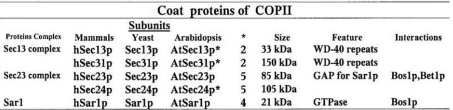
IIc3.- In silico analysis of protein-protein interactions between ELO3 and secretory pathway proteins
IIc4.- qRT-PCR expression analysis of Elongator-related genes in sec31 mutants
IId- Discussion
Pag.
4
6
7 8 12 13 14 18 18 23 26 29 3234
34 42 52 53 57 58 59 60Index ________________________________________________________________________________
III° Chapter- Section B: Study of Elongator-mediated gene
expression under darkness and light qualities
IIIa.- Introduction
IIIa1.- Skoto and Photomorphogenesis pathways
IIIa2. - The whole Elongator Complex takes part in regulation of Skoto/Photomorphogenesis
IIIb.- Materials and Methods IIIc.- Results
IIIc1.- Hypocotyl assay on Elongator subunit mutants and double mutants IIIc2.- Chromatin Immunoprecipitation assay (ChIP)
IIIc3.- qRT-PCR analysis
IIId.- Discussion
Acknowledgements
References
Appendix
A. Supplemental Material for Chapter 2
A.1 Table S1. List of interactors in between ELO3 and Sec31
B. Supplemental Material for Chapter 3
B.1. Figure of qRT-PCR results (first biological replicate) B.2. Figure of qRT-PCR results (second biological replicate)
website:
http://arabidopsis.info/NASC http://bar.utoronto.ca/efp https://bioinformatics.psb.ugent.be/cornetPag.
62
62 64 68 71 80 84 88 90 9497
98
112
112
1112114
114 118Abstract ________________________________________________________________________________
4
Abstract
The Elongator complex is a histone acetyltransferase complex associated with RNAPII to facilitate transcript elongation. It’s composed of six proteins (ELP1-6). ELP1-3 form the Elongator core subcomplex, while ELP4-6 form the accessory subcomplex. Elongator complex, firstly identified in yeast, was later isolated from animals and plants and all its six subunits are evolutionarily conserved. The Elongator activity is conferred by ELP3 that targets specifically histons H3 (lysine-14) and H4 (lysine-12) by acetylating histone in order to facilitate the RNAPII progresses through the nucleosome. In yeast, mutations in Elongator subunits induce delay in growth due to a slowly adaptation to changing environmental conditions. In human, mutations in Elongator components affect neuronal development and this leads to neuronal disease. Whereas, in plant Elongator stimulates plant growth acting a positive regulator of cell proliferation. At the phenotypic level, Elongator mutants, called elongata, are known for narrow leaves and short root.
In the present work, by using the model plant Arabidopsis thaliana, we investigated some aspects of molecular networks underlying Elongator activity and its interaction with environmental factors, mainly focusing on light conditions. Based on previous unpublished data obtained through TAP analysis, in the first period of PhD project we focused the attention on the functional study of
Sec31 gene encoding a protein involved in cell secretory pathway, identified as a putative direct
interactor of Elongator complex. To add information on this interaction we analyzed phenotypic and developmental characteristics of sec31 mutants to compare with elo3-6 mutant. The histological expression pattern of Sec31 and ELO3 transcripts in wild type seedlings was also investigated, through multiprobe in situ hybridization, to compare organ/tissue specific expression domains. The obtained results showed that expression pattern of the two genes is quite similar while sec31 mutants do not resemble elo3-6 phenotypes. Moreover further TAP experiments and in silico analysis of protein/protein interaction did not confirm previous data, thus excluding a direct interaction between ELO3 and Sec31.
However, expression analysis in sec31 mutants of some Elongator-related genes, performed by qRT-PCR, showed that Sec31 and ELO3 share common downstream target genes and both seem play a role in auxin pathway. Future trascriptomic analyses on auxin mutants on one side, and the identification of possible interactors/players of both genes on the other side, could be useful to deepen if the molecular circuits, by which Elongator complex and the secretory machinery act on auxin pathway, show some cross-talk or they work in an independent manner.
A further aspect of Elongantor molecular network that we investigated deals with role of Elongator in the skoto/photomorphogenesis pathways. In particular we investigated the elo3-6 mutant in darkness and under light condition (red, far-red and blue light) through microarray and
Abstract ________________________________________________________________________________ RNA-seq approaches. Gene ontology categories over representative in elo3-6 seedlins, identified by BINGO analysis, allowed us to discover the putative targets of Elongator both in darkness and in light, and to understand the position of whole Elongator complex along either pathways. The results suggested that Elongator complex takes part in the skotomorphogenesis and photomorphogenesis and is dependent on photoreceptors PHYA and PHYB. Microarray, RNA-seq, qRT-PCR and ChIP- qPCR analyses displayed that Elongator regulates transcription of some genes both in light and in darkness. In the specific, results displayed that Elongator complex participates in the skoto/photomorphogenic pathways by binding target genes such as HYH and LHY in light and darkness condition, respectively. Whereas it can regulate the activity of other putative targets such as Pifs gene (PIF4) in darkness and HY5 under light condition.
Chapter I ________________________________________________________________________________
6
I° Chapter: General introduction
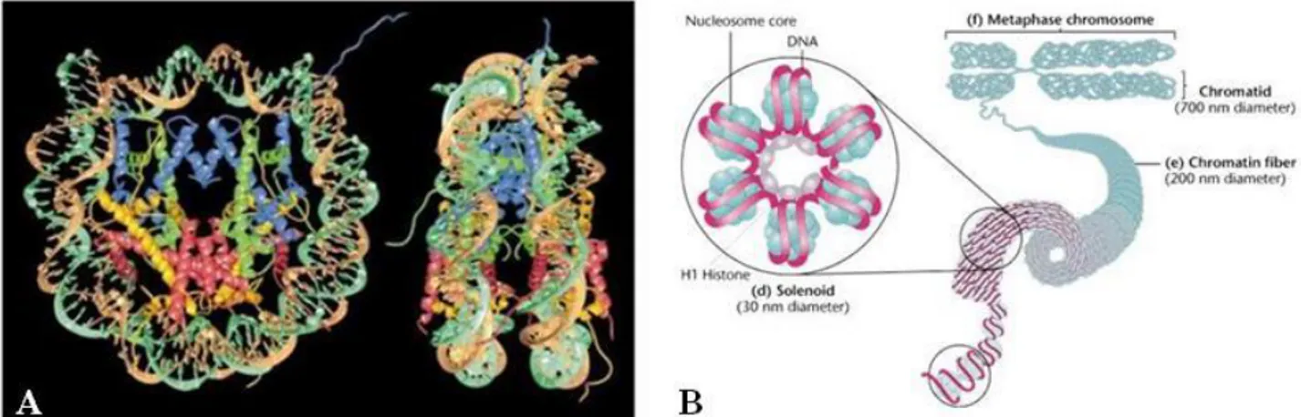
In all organisms the genetic information, that regulates their growth and development is stored in the DNA molecule. The eukaryotic cell developed a strategy to compact the long DNA molecule within the nucleus by wrapping it into a nucleoprotein complex which is referred as chromatin. Proteins that mediate the successive orders of DNA folding are represented by different histone classes: two copies each H2A, H2B, H3 and H4 histones form a protein core that wraps 146 base pairs of DNA tightly on its surface to form the core nucleosome (Kornberg & Lorch, 1999) (Figure 1). The linker histone H1 is not a part of core nucleosome but binds the DNA between two core particles and connects one nucleosome to the next one leading to the final chromatin structure. At the cytological level, in a non-dividing cell two different types of chromatin organization (i.e. decondensed or tightly dense) can be observed which correspond to different functional states (i.e. transcriptionally active or inactive DNA) an relate to a different DNA packaging and nucleosome arrangment: these two organization/functional states are indicated as euchromatin or heterochromatin (Jenuwein and Allis, 2001; Kouzarides, 2007; The ENCODE Project Consortium, 2007). Euchromatin is the region where DNA is accessible, representing an open conformation due to the relaxed state of nucleosome arrangement. The genomic regions of euchromatin are more flexible, and contain genes in active and inactive transcriptional states (Jenuwein and Allis, 2001; Kouzarides, 2007; The ENCODE Project Consortium, 2007; Koch, et al, 2007). Conversely, heterochromatin are areas where DNA is packaged into highly condensed forms that are inaccessible to transcription factors or chromatin-associated proteins (Jenuwein and Allis, 2001; Talbert and Henikoff, 2006; Huang, et al, 2004). The genomic regions within heterochromatin primarily consist of repetitive sequences and repressed genes associated with morphogenesis or differentiation (imprinting or X chromosome inactivation) (Reik, 2007; Feinberg and Tycko, 2004).
Chapter I ________________________________________________________________________________
Figure 1. (A). Nucleosome core particle: ribbon traces for the 146-bp DNA phosphodiester backbones (brown and turquoise) and eight histone protein main chains (blue: H3; green: H4; yellow: H2A; red: H2B. The views are down the DNA superhelix axis for the left particle and perpendicular to it for the right particle. For both particles, the pseudo-twofold axis is aligned vertically with the DNA center at the top. (B) Stabilization of the 30 nm chromatin by binding of the histone H1
First picture from (Luger, et al 1997). Second picture from Klug & Cummings (1997 p542)
Thus beside packaging DNA into a smaller volume to fit into the cell, relevant functions of chromatin are to control DNA replication, chromosomal stability and gene expression. The dynamics of chromatin structure is tightly regulated through multiple mechanisms including histone modification, chromatin remodeling, histone variant incorporation, and histone eviction. In this chapter we want focus our attention on mechanisms of chromatin regulation and how such regulation controls gene expression during transcription.
Ia - The process of transcription in eukaryotic organism
Transcription is the process by which the information stored in the DNA double strand is copied into a new molecule of messenger RNA (mRNA). RNA synthesis involves separation of the DNA strands and synthesis of an RNA molecule from 5' to 3' direction by RNA polymerase, using one of the DNA strands as a template. Transcriptional regulatory mechanisms modulate the accurate recruitment and activation of RNA polymerase at different locations in the genome, such as gene promoters. Transcriptional regulators can be grouped into three types. First, the preinitiation complex (PIC) which binds at the core promoter and recruits RNAPII. Second, DNA-binding transcription factors which bind to sites such as proximal promoter elements and enhancers. Third, enzymes that modify the higher-order chromatin structure by promoting the physical movement of nucleosomes relative to the genome and post-translationally modify histone molecules to alter the stability and accessibility of chromatin RNA polymerases are large multi-subunit enzymes that
Chapter I ________________________________________________________________________________
8 perform transcription and thus produce all the RNAs in the cell from a DNA template. These complexes are assembled and tightly bound at the promoter of genes before initiating transcription. Different RNA polymerases identified in eukaryotic organisms are responsible for the synthesis of ribosomal RNA (RNAPI), pre-messenger RNA (RNAPII) and small RNAs including transfer RNA (RNAPIII) respectively (Cramer, 2002). Plants are unique in having evolved multisubunit RNA polymerases IV and V in addition to RNAPI, II, and III, the three ubiquitous nuclear DNA-dependent RNA polymerases of eukaryotes. RNAPIV and V synthesize noncoding RNAs for transcriptional silencing of transposons, repetitive elements, and a subset of genes (Haag and Pikaard; 2011; Herr et al., 2005; Kanno et al., 2005; Onodera et al., 2005; Pontier et al., 2005; Ream et al., 2014). The RNA polymerase structure is broadly conserved from prokaryotes to all eukaryotes (Saltzman and Weinmann, 1989). All RNA polymerase types are believed to have diverged from a common ancestral protein, as some subunits are shared between them. RNAPII uniquely possesses an extra C-terminal domain (CTD) on its largest subunit, it acts both in the recruitment of RNAPII to active promoters and as a binding platform for other proteins involved in transcription. Here, we will focus on RNAPII and its regulation, as it is at the origin of the production of all proteins in the cell. The transcription is divided into an initiation stage, during which transcription factors and RNAPII bind to promoter sites and RNA synthesis starts, followed by an elongation stage, during which RNAPII traverses along the DNA assembling an RNA transcript (Ponting, 2002).
Ia1. - Transcription initiation, elongation and termination
Transcription iniziation
The RNAPII arranges on the promotor region a complex formed by several proteins including factors. These proteins form the basis of the pre-initiation complex and they are responsible for the position of polymerase at the core promoter region as well as induce the separation of DNA helix to facilitate polymerase movement along DNA and allow transcription elongation. The promoter region includes a sequence called TATA-box (about 30 bp upstream from the transcription start site) that is recognized by a multisubunits protein complex called TFIID (Transcription Factor for polymerase II). One subunit of TFIID that binds TATA-box is called TBP (TATA binding protein), whereas the other subunits of this complex are called TAF (TBP associated
Chapter I ________________________________________________________________________________ dynamic of TBP connection at DNA. TBPs are associated with TAF and these last show a structural homology with histone proteins. Accordingly it was proposed that TAF proteins could link the DNA in the same manner as histones, although experimental evidences for that have not yet been provided. So far, it has been only shown that in Drosophila TAF42 and TAF6 form a similar structure as the H3·H4 tetramer. These histone-like TAF proteins are localized in the TFIID complex and associated to enzymes involved in histones modification such as the SAGA complex. Currently, the role of the various transcription factors in directing transcription initiation is the subject of intense study. The TFIIB interacts directly with TBP, to form TFIIB-TBP-DNA complex, and with polymerase II. Therefore TFIIB, makes a bridge between TBP and polymerase II. While TBP and TFIIB play central roles in the nucleation of the transcription initiation complex, TFIIF, TFIIE, and TFIIH play roles at later steps. TFIIF interacts directly with RNA polymerase II and TFIIB and is required for stable assembly of RNAPII with the TATA-TBP-TFIIB complex. The factor TFIIE joins the complex TATA-TBP-TFIIB-RNA and this promotes the link with TFIIH. TFIIE and TFIIH promote melting and clearance through ATP hydrolysis. TFIID regulates the activities of TFIIH which possesses ATP-dependent helicase activity and also kinase activity which allows it to phosphorylate the C-terminal domain of RNAPII. Indeed the bigger subunit of RNAPII has a C-terminal domain (called CTD) which exhibits a tail rich in phosphorylation sites. In addition, TFIIE plays a direct role in promoter melting, perhaps by binding to the single -stranded region and thereby stabilizing the melted region of the promoter moreover it participates in the recruitment of TBP and TFIIA to the TATA box. The different GTFs together with the RNAPII complex form the “pre-initiation complex” (PIC) (Figure. 2).
In summary the activation of transcription by Polymerase requires DNA-binding by general transcription factors that form the PIC, so that transcriptional activator and co-activator then interact with the complex to initiate transcription.
Figure 2. Transcription initiation in eukaryotes. To start transcription, eukaryotic RNA polymerase II requires the general transcription factors. These transcription factors are assembled around TATA-box sequence. TFIIH uses ATP to pry apart the double helix at the transcription start point, allowing transcription to begin. TFIIH also phosphorylates RNA polymerase II, releasing it from the general factors so it can enter the elongation phase of transcription. (Alberini, 2009, Physiol Rev).
Chapter I ________________________________________________________________________________
10 Transcription elongation
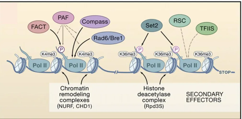
Following the transcription activation, elongation phase takes place when RNAPII get away from transcription initiation factors and moves into the (towards) coding region. This event triggers the recruitment of the elongation machinery, which includes the factors involved in polymerization, mRNA processing, mRNA export, and chromatin function (Hahn, 2004). Cells exploit a very sophisticated array of factors to control chromatin architecture during elongation, and the events and factors required at the beginning of the gene differ significantly from those required at the end (Li., et al, 2007). This is done not only to promote efficient RNA synthesis but also to ensure the integrity of the chromatin structure while RNAPII travels through the body of the gene. The transcription elongation requires new factors able to stimulate the elongation of RNAPII through phosphorylation of its CTD. The RNAPII CTD undergoes two major phosphorylation changes during elongation: Ser5 is phosphorylated by TFIIH at the 5’ end of the ORF, and Ser2 is phosphorylated by the Ctk kinase as RNAPII transits toward the 3’end. These phosphorylation events appear to control the elongation processes and couple them with alterations in chromatin structure. One of proteins involved in the elongation of RNAPII is a kinase P-TEFb which phosphorylate the CTD of RNAPII on Ser5 and Ser7. Important components which bind RNAPII during elongation are SAGA, FACT, PAF, TFIIS, RAD6/Bre1, COMPASS, and many others
(Figure 3). Most of these complexes play a function in relaxing the chromatin state to allow RNAPII passage during transcription but recently another level of transcriptional regulation has been identified during elongation.
Figure 3. Regulation of Nucleosome Dynamics during Transcription Elongation.
The chromatin landscape during elongation is determined by the factors associated with different forms of RNAPII. PAF facilitates the binding of FACT, COMPASS, and Rad6/Bre1 to the Ser5-phosphorylated CTD. (Li., et al, 2007).
Chapter I ________________________________________________________________________________
A series of discoveries suggest that many developmental and inducible Drosophila and mammalian genes, prior to their expression, contain RNAPII bound predominantly in their promoter proximal regions in a “stalled” state (Saunders, et al., 2006; Sims, et al., 2004) Genome-wide surveys of the phenomenon suggest that RNAPII stalling is likely to be a rate-limiting control on gene activation that poises developmental and stimulus-responsive genes for prompt expression when inducing signals are received. In addition, stalled RNAPII signals are associated with active histone modification marks, including trimethylation of lysine 4 on histone H3 (H3K4me3) and acetylation of H3 lysine 9 and 14 (H3K9ac and H3K14ac) (Guenther, et al., 2007). Thus, RNAPII promoter-proximal stalling could help to provide an active chromatin environment and prepare developmental and stimulus-responsive genes for timely expression (Saunders, et al., 2006; Lorincz and Schubeler, 2007). RNAPII promoter-proximal stalling was first described in Drosophila heat-shock-inducible
Hsp70 genes by using ultraviolet-crosslinking and chromatin immunoprecipitation (UV ChIP).
RNAPII was found to be recruited to the promoter of the un-induced Hsp70 gene, where initiates RNA synthesis but stalls after synthesis of 20-50 nucleotides of RNA (Saunders, et al., 2006; Rasmussen and Lis, 1993). Heat-shock stimulation enabled RNAPII to escape from the Hsp70 promoter-proximal region and transcribe the full-length RNA. This suggests that regulation through pausing is a fundamental step for controlling developmental programs and enabling rapid reaction to environmental stimuli (Zeitlinger, et al., 2007) Studies have revealed that TEFs regulate plant growth and development by modulating diverse processes including hormone signaling, circadian clock, pathogen defense, responses to light, and developmental transitions (Van Lijsebettens and Grasser, 2014). Indeed in plant the transcription of genes that are affected by the depletion of different TEFs can cause different types of growth and/or developmental defects.
Transcription termination
Termination occurs when RNAPII ceases RNA synthesis and both RNAPII and the nascent RNA are released from the DNA template. Transcription termination is important because it prevents RNAPII from interfering with downstream DNA elements, such as promoters, and promotes polymerase recycling. RNAPII termination can be elicited through different pathways, depending on the RNA 3′-end processing signals and termination factors that are present at the end of a gene (Richard & Manley, 2009; Lykke-Andersen & Jensen, 2007; Rondon, et al, 2008). Two models for transcription termination were proposed to explain the role of 3′-end processing. The first model, known as the allosteric or anti-terminator model, proposes that transcription through the
Chapter I ________________________________________________________________________________
12 poly(A) site leads to conformational changes of the elongation complex (EC) by dissociation of elongation factors and/or association of termination factors (Logan, et al., 1987). The second model, the torpedo model, is based on the observed rapid degradation of the 3′ RNA after cleavage at the poly(A) site. Cleavage creates an entry site for a 5′–3′ exonuclease at the uncapped 5′-monophosphate, which degrades the RNA and, according to this model, in some way promotes RNAPII release upon “catching up” with RNAPII (Connelly and Manley, 1988). In summary, transcription takes place through the recruitment and action of multiple regulatory factors.
Ib1. - Role of chromatin state in the transcription
In eukaryotic cells the genomic DNA is packaged with histone proteins and this organization has double function, one is the need to compact 2 m of DNA into the small space of the nucleus and the other one plays a role in the gene transcription regulation. Decompaction of chromatin to facilitate access to DNA has been most widely studied for RNAPII-mediated transcription of protein-coding genes, a process that requires rapid access to genes for the response to environmental signals and programmed cellular events, but the underlying principles are equally applicable to any process requiring interaction with DNA (George Orphanides and Danny Reinberg, 2000). Recent reports confirm that eukaryotic cells are equipped with specialized proteins that help RNAPII pass through chromatin during transcription elongation. In order to facilitate the transcription of specific genes the chromatin must be available and this chromatin state is called "open chromatin" since the structure of chromatin is disrupted from the promoter region of the gene to the entire transcribed domain. During transcription the chromatin transcribed region reveals alteration at the level of histone proteins themselves. Indeed chromatin state undergoes many modifications through the action of many different proteins. by using a chromatin remodelling proteins complex such as ATP-dependent chromatin-remodeling complexes and covalent histone modifications. The ATP-dependent chromatin-remodeling complexes manipulate chromatin structure by using energy from ATP hydrolysis to disrupt chromatin and make DNA more accessible to DNA-binding proteins. The other class of histone modifications, covalent histone modifications, act on histone tails by modification of one or more amino acids that results in activation or repression of transcription. The activity carried out from the complexes involved in chromatin modification plays an important role during the life of an organism, because in that way they can regulate gene activity and expression during the phase of organism development and differentiation, or in response to environmental stimuli.
Chapter I ________________________________________________________________________________
Ib1a - ATP-dependent chromatin-remodeling
The ATP-dependent chromatin-remodeling complexes. are conserved in evolution, and there is more than one type of complex in each cell All of the ATP-dependent chromatin-remodeling complexes contain an ATPase subunit that belongs to the SNF2 superfamily of proteins. Based on the identity of this subunit, they have been classified into two main groups, the SWI2/SNF2 group and the imitation SWI (ISWI) group (Eisen, et al., 1995). Their mechanism of action is similar but the SWI/ SNF and NURF complexes utilize the energy of ATP hydrolysis to alter nucleosome structure, however, while the Swi2/ Snf2 ATPase of SWI/SNF is induced by either DNA or nucleosomes (Coˆte´, et al., 1994). The SWI2/SNF2 group includes RSC that requires nucleosomes with intact histone amino-terminal tails for maximum stimulation. The results of Kasten et al, 2004, showed that Rsc4 is an integral and essential member of RSC preferential binding to the histone H3 peptide acetylated at Lys14 This means that both RSC and ATP-dependent chromatin-remodeling protein are involve the dynamic modification of chromatin architecture. In particular Rsc4 bromodomains recognize acetylated H3 Lys14 and cooperate with H3 Lys14 acetylation for their function. These results highlight the important role bromodomains play in the coordination of chromatin remodeling with histone acetylation in transcriptional regulation, and reveal new properties of tandem bromodomain function (Kasten, et al., 2004) The Arabidopsis genome encodes more than 40 different proteins belonging to ATP-dependent chromatin remodeling complexes, which can be grouped into three major types, namely SWI2/SNF2, ISWI and CHD based on the presence of other protein motifs in addition to the ATPase domain. Among them, only AtBRM, SPLAYED (SYD), PICKLE (PKL), DECREASE IN DNA METHYLATION (DDM1), MORPEUS MOLECULE (MOM), DRD1 and PHOTOPERIOD INDEPENDENT EARLY FLOWERING1 (PIE1) have been functionally characterized (Hsieh and Fischer, 2005).
In Arabidopsis SWI/SNF ATPases: SPLAYED (SYD) plays a specific role in maintenance of the stem cell pool in the shoot apical meristem, it’s involved in floral transitino and ovule development. Whereas BRM is involved in the control of expression of two regulators of cotyledon separation: the CUP-SHAPED COTYLEDON genes CUC1 and CUC3 (Kwon, et al., 2006). The SPLAYED (SYD) gene product acts as a LFY-dependent repressor of the meristem identity switch in the floral transition. SYD regulates flowering in response to environmental stimuli which in plant may be achieved in part by regulating transcription factor activity via alteration of the chromatin state. In addition SYD encodes a presumptive Arabidopsis homolog of the yeast Snf2p ATPase, which is implicated in transcriptional control via chromatin remodeling (Wagner and Meyerowitz, 2002).
Chapter I ________________________________________________________________________________
14 In yeast and mammals, ATP-dependent chromatin remodelling complexes of the SWI/SNF family play critical roles in the regulation of transcription, cell proliferation, differentiation and development. Homologues of conserved subunits of SWI/SNF-type complexes, including Snf2-type ATPases and SWI3-type proteins, participate in analogous processes in Arabidopsis (Archacki, et
al, 2009). This means that ATP-dependent chromatin remodelling complexes of the SWI/SNF
family have a functional role in plant, yeast and mammals.
Ib1b- Covalent histone modifications
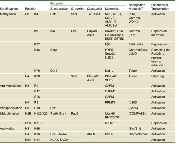
The chromatin modifications may affect higher-order chromatin structure by disrupting the contact between different histones in adjacent nucleosomes or the interaction of histones with DNA. Numerous studies have been performed to discover proteins catalysing histone modifications and understand their mode of action. Simplistically, the function of histone modifications can be divided into two categories: the establishment of global chromatin environments and the orchestration of DNA-based biological tasks. The histone tails are subject to a vast array of posttranslational modifications that include: methylation of arginine (R) residues; methylation, acetylation, ubiquitination, ADP-ribosylation, and sumolation of lysines (K); and phosphorylation of serines and threonines (Table 1). Modifications that are associated with active transcription, such as acetylation of histone 3 and histone 4 (H3 and H4) or di- or trimethylation (me) of H3K4, are commonly referred to as euchromatin modifications. Modifications that are localized to inactive genes or regions, such as H3 K9me and H3 K27me, are often termed heterochromatin modifications (Li.,et al, 2007). In the following section we discuss only about the function and role of “Histone
acetylation” on transcription regulation because it is the histone modification around which this thesis has developed.
Chapter I ________________________________________________________________________________
Table 1. Overview of different classes of modification identified on histones. The function have been associated with each modification are shown. (Li and Workman, 2007)
Histone acetylation
The acetylation and deacetylation are the most studied histone modifications, that play a role in gene activation and repression, respectively. The modification is reversible and is defined by a fine-tuned balance between the activity of histone acetyltransferases (HATs) and histone deacetylases (HDACs) to regulate gene expression. Histones are acetylated at N-terminal lysine residues. The source of the acetyl group in histone acetylation is acetyl-Coenzyme A and in histone deacetylation the acetyl group can be transferred back to Coenzyme A or to ADP-ribose by the NAD-dependent deacetylases (Denu, 2003). Histone acetylation neutralizes the positive charge of the target lysine residue, thereby changing the overall charge distribution of the histone tails and decreasing its affinity for DNA. The resulting change of nucleosomal conformation increases the accessibility of transcriptional regulatory proteins to the chromatin template, suggesting that histone acetylation is correlated with increased transcriptional activity. HAT activity is important for transcription initiation, in addition to a number of transcriptional coactivators. Several HAT
Chapter I ________________________________________________________________________________
16 complexes can be recruited to promoter regions by direct interactions with DNA-binding activators, resulting in increased DNA accessibility and stimulation of transcription initiation (Utley, et al, 1998; Chan & La Thangue, 2001). Whereas the histone acetylation induces transcriptional activation, the Histone deacetylases (HDACs) in turn interact with transcriptional repressors, suggesting that deacetylation is involved in repression and silencing (Courey and Jia, 2001).
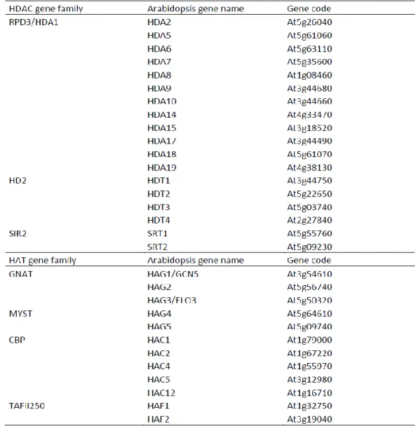
Based on the sequences outside the acetyltransferase domain, the HATs can be divided into four subfamilies: the Gcn5-related N-terminal acetyltransferases (GNAT), the MYST family, the CBP/p300 family, the family related to mammalian TAF250. Sequence and domain analyses of the
Arabidopsis genome have revealed four families of HATs, and three families of HDAC, consisting
of 12 genes and 18 genes respectively (Pandey, et al., 2002). The identification of plant-specific HD2 family of HDACs (Lusser, et al., 1997) provides an indication for functional diversification of histone-modifying proteins in plants. The N-terminal lysine residues of histone H3 (K9, K14, K18, K23, and K27) and H4 (K5, K8, K12, K16, and K20) are found to be acetylation/deacetylation targets in Arabidopsis (Zhang, et al., 2007; Earley, et al., 2007). This thesis focuses on the role of Elongator complex and their targets in plant development and their influence on the regulation on gene expression. Elongator complex is composed of six subunits (ELP1-ELP6), in particular Elp3 is a conserved member of the GNAT. Gcn5-related N-acetyltransferase) protein family, and recombinant Elp3 possesses acetyltransferase activity directed toward all four core histones (Wittschieben, et al, 1999). According to Winkler, 2002, both histones H3 and H4 are acetylated by holo-Elongator, the predominant site of acetylation in vitro is lysine-14 of histone H3 and lysine-8 of histone H4. Elp4, Elp5, and Elp6 subunits are required for Elongator HAT activity. As explained above the HAT group is divided into four types based on primary homology with yeast and mammalian. The GNAT (GCN5-related N-acetyltransferase) family is generally considered to comprise three subfamilies, designated GCN5 (General Control Nonderepressible protein5), ELP3 (a transcriptional Elongator complex Protein), and HAT1 in higher eukaryotes. In the Arabidopsis genome, a single homolog of each of the GCN5, ELP3, and HAT1 subfamilies (HAG1/AtGCN5, HAG3, and HAG2, respectively) has been identified (Pandey, et al., 2002) (Table 2) The
Arabidopsis genome contains two MYST family genes (HAG4 and HAG5), five p300/CBP genes
Chapter I ________________________________________________________________________________
Table 2. Genes encoding HAT and HDAC homologs in Arabidopsis (modified from Pandey, et al., 2002).
The mechanism involved in histone modification influence many processes of plant development. Two HATs in plants, namely AtGCN5 and ELO3, have been analyzed functionally and have a significant effect on plant growth. They are both part of conserved complexes that have been studied primarily in yeast. The yeast GCN5 is part of the SAGA and ADA regulatory complex, where it serves as a nucleosomal HAT, capable of acetylating histone H3 (Bertrand, et al., 2003). The Arabidopsis AtGCN5 has conserved HAT catalytic and bromodomains (Stockinger, et al., 2001). The role of histone acetylation has been assigned to cell cycle, cell division, response to abiotic stimuli such as light and temperature and biotic signals linked to growth or to stress.
Removal of the acetyl group from the histones is mediated by HDACs, they consist of 3 families in plants: RPD3/HDA1 (histone deacetylase 1 protein), SIR2 (silent information regulator protein 2) and a plant-specific family of HDACs, HD2 which was discovered in maize. Exactly Four HD2 proteins are identified in Arabidopsis and they all contain a conserved N-terminus with the HD2-
Chapter I ________________________________________________________________________________
18 type HDAC domain, a central acidic domain and variant C-terminal domain. By analyzing the sequence a single homologue is found in in the animal, fungi and Arabidopsis for all subfamilies (GCN5, ELP3, HAT1, HAG4 and HAG5) of the GNAT/MYST superfamily, suggesting that the plant proteins may form complexes similar to those formed in yeast and animals. In contrast the CREB-binding protein (CBP) and TAFII250 family of HAT proteins shows differences in domain architecture between plants and animals such as the absence of a bromodomain in the plant CBP-type HATs. The manner in which the chromatin organization influences the gene expression among different organisms depends on existing variety/diversity of enzyme involved in covalent modifications, suggesting that plants have developed mechanisms of global gene regulation related to their unique developmental pathways and environmental response.
Ib - The Elongator Complex
Ib1. - Identification of Elongator Complex in yeast, Drosophila and Human
In yeast
Studies suggested that in order to transcribe genes, the transcription machinery can recruit chromatin remodeling complex to facilitate the activation process. Among the chromatin remodeling complexes involved in post-transcriptional modification, the most prominent is histone acetyltransferase activity (HAT). It has been suggested that HATs might also assist RNAPII during transcript elongation through chromatin (Travers, 1992, Brownell and Allis, 1996; Cho, et al., 1998). The RNAPII isolated from native RNAPII/DNA/RNA ternary complexes in yeast chromatin is found tightly complexed with a novel multisubunit complex called Elongator. Elongator complex that was associated with the hyperphosphorylated form of RNAPII (Otero, et al., 1999). Holo-Elongator is an unstable six-subunit complex composed of two subcomplexes: core-Holo-Elongator, comprised of Elp1, Elp2, and Elp3 (Wittschieben, et al, 1999; Otero, et al, 1999; Fellows, et al, 2000), and a smaller three-subunit module composed of Elp4, Elp5, and Elp6 (Winkler, et al, 2001, Li, et al, 2001). The yeast Elongator complex consists of two WD40 proteins, Elongator factor protein 1 (ELP1) is involved in maintaining Elongator’s structural integrity and ELP2 that mediate protein-protein interaction. The ELP3 subunit possesses a C-terminal histone acetyl transferase (HAT) and radical S-adenosylmethionine domains activity, whereas the three RecA-like proteins, ELP4, ELP5, and ELP6, which form a hexameric RecA-like ATPase, all of which are conserved.
Chapter I ________________________________________________________________________________
The yeast ELP3 has motifs characteristic for the GCN5 family of HATs and acetylated in vitro histone H3 and H4 and all the core Elongator components are essential for in vivo histone acetylation (Winkler, et al, 2002). In nature the competition between organisms triggers different modes of action such as the secretion of toxic compounds that results in the killing or growth arrest of other species or genera. The yeast Kluyveromyces lactis secretes a toxin, called zymocin, which inhibits the growth of various sensitive yeast genera, including Saccharomyces cerevisiae. The native toxin is a heterotrimeric (αβγ) structure composed of three subunits, two of which are involved in facilitating toxin entry. Cytotoxicity resides solely within the γ subunit, and intracellular expression of this subunit alone in S. cerevisiae abrogates growth (Butler, et al, 1991). In particular the function of zymocin on S. cerevisiae is to inhibit the adenylate cyclase by the role of cAMP necessary for mitotic growth and cell division. Genetic screening for mutations that confer resistance toward the intracellular expression of the zymocin γ subunit identified genes that were named TOT1–7 (toxin target) (Jablonowski, et al, 2001, Frohloff, et al, 2001). Interestingly, the initially isolated genes TOT1-7 shown to be allelic to the genes encoding the subunits of each yeast Elongator (Elp1/Tot1/Iki3, Elp2/Tot2, Elp3/Tot3, Elp4/Tot7, Elp5/ Tot5/Iki1, and Elp6/Tot6). In addition, other genes in S. cerevisiae were identified which confer resistance to zymocin upon mutation, KTI11, KTI12, KTI13 and KTI14 (Fichtner, et al., 2002). The experimental results show that cells lacking KTI12 or Elongator (ELP) genes are insensitive to the toxin zymocin. The deletion of KTI12 doesn’t affect the integrity of Elongator complex, whereas it strongly supported the idea that Kti12 is required for the normal (HAT) activity of Elongator complex in vivo. Based on the latter finding, Kitamoto, et al., 2002 suggested that situations favoring histone hyperacetylation might reduce the cellular requirement for the HAT activity of Elongator and thereby reduce zymocin toxicity.The S. cerevisiae genome encodes other histone acetyltransferase (HAT) activities than the one associated with Elp3 (Tot3p) (Wittschieben, et al, 1999). Other HAT gene knockout, GNC5, HAT1, SAS3, HAPA3 were tested using zymocin eclipse assays, and only HAPA1 (isoallelic of TOT3/ELP3) remained zymocin sensitive. This lead to suppose that HAT activity appears to be required for zymocin action and in fact Elp3/Tot3p possess that activity (Figure 4). The Elongator mutants showed a resistance to zymocin but also a delay in growth. The slower adaptation is a consequence of decreased/delayed transcription of the GAL1-10 (galactose-inducible genes) transcripts.
Chapter I ________________________________________________________________________________
20 Figure 4. Effect of HAT gene deletions on zymocin sensitivity. Strains lacking non-Elongator HATs (sas3∆, hpa3∆, hat1∆, , hpa1∆, gcn5∆ , WT, tot3∆) are subjecte to a killer eclipse assay. Deletion of TOT3/ELP3 confers zymocin resistance, whereas the other HAT gene deletions tested arrre zymocine sensitive.
In Human and Drosophila
Subsequently to yeast, the Elongator complex has been purified in human as a six-subunit complex and it is composed of the IKAP homologue of Elp1, hELP3, StIP1 homologue to yeast Elp2, hELP4 and two additional unidentified proteins. In human IKAP was located in the nucleus, the nucleoli and cytoplasm by immunostaining suggesting multiple roles in the cell.
In 2001, Slaugenhaupt, 2002 and Anderson et al, 2001 reported that mutation in IKBKAP (IKAP is the protein encoded by IKBKAP) are responsible for a syndrome called Familial dysautonomia (FD). This disease is an autosomal recessive disease resulting from poor development and progressive degeneration of the sensory and autonomic nervous system. The individuals with this disease have a low numbers of neurons in peripheral nervous ganglions. To better understand the role played by IKAP and Elongator complex during transcription and in the same time learn about the molecular defects underlying the FD, an RNA interference (RNAi) was used to deplete the IKAP protein in the human cells. In yeast the silencing of ELP1 (homologue of IKAP in human) destabilizes the ELP3 catalytic subunit and of course induces a change in the Elongator complex integrity. In order to identify genes that require IKAP for their transcription, a microarray analysis was performed that identified several genes involved in cell motility or actin cytoskeleton remodelling. The cell motility has an important role in the developing nervous system, therefore motility/migration assay demonstrated that the IKAP depletion has functional consequence, in fact IKAP-depleted cells showed defect in migration which may be linked to the neurodevelopmental disorder that affects FD patients. Chromatin immunoprecipitation analysis was performed to
Chapter I ________________________________________________________________________________
investigate if or not the defects in cell migration resulted of impaired transcriptional elongation of IKAP-dependent genes. The results showed a decrease in histone H3 acetylation in the transcribed region of IKAP target genes and no defect in the recruitment of RNA polymerase II on the promoter region was observed. This indicates that IKAP/hELP1 has a specific effect on histone acetylation and on RNA polymerase across the target gene which is consistent with a direct effect of Elongator on transcriptional elongation in vivo. In human, the Amyotrophic lateral sclerosis (ALS) is a spontaneous, relentlessly progressive motor neuron disease, usually resulting in death from respiratory failure within 3 years. Mutation on SOD1 gene remains the most common genetic cause of ALS (Simpson, et al, 2009). In the study of 1483 individuals, ELP3 was associated with human motor neuron degeneration in the form of ALS (Amyotrophic lateral sclerosis, characterized by progressive motor disease, usually resulting in death from respiratory failure) in three different populations. In mammalian the regulation of cytoskeletal components such as microtubules assumes an important role in neuronal cell shape that are necessary in many aspects of cortical development. Indeed, mutations in α-tubulin or in the microtubule regulating proteins LISSENCEPHALY-1 (LIS1) and DOUBLECORTIN (DCX) result in neuronal migration defects in humans, thus pointing to a central role for microtubule regulation in neuronal migration (Wynshaw- Boris, 2009).
Post translational modification such as acetylation on microtubules and tubulin molecules influence their stability and interactions with other proteins. Creppe and colleagues supposed that depletion of ELP1 results in destabilization of ELP3 subunit of Elongator complex by short hairpin RNAs (shRNAs) reducing the migration speed of bipolar and multipolar cortical neurons in the mouse brain (Figure 5)Elongator acetylates histones in the nucleus but it also targets cytoplasmic proteins such as α-tubulin. In support of this hypothesis, SIRT2, a mainly cytoplasmic deacetylase, targets a-tubulin in this cell compartment but also moves into the nucleus once phosphorylated by cyclin-dependent kinases in the G2/M phase of the cell cycle in order to target histone H4 (North and Verdin, 2007; Vaquero, et al., 2006). The result obtained by Creppe, et al, 2009, showed that Elp3 in vitro promote the acetylation of α-tubulin, but not a HAT-defective Elp3 mutant, in contrast to the HDAC6-mediated a-tubulin deacetylation in HEK293 cells. Hence, Elongator in human binds to microtubules and promotes a-tubulin acetylation, a way in which it may regulate migration and maturation of cortical projection neurons.
Chapter I ________________________________________________________________________________
22 Figure 5. Modification of Elongator Complex subunit leads a reduction in migration and differentiation of projection neurons. (Left) Elongator may function in the cytoplasm of mouse neuronal cells to acetylate α-tubulin after microtubule polymerization, a modification that is associated with stable microtubules and is necessary for neuronal migration and differentiation. (Right) The depletion of ELP1 results in destabilization of ELP3, decreased tubulin acetylation in cultured cells, and defects in neuronal migration and branch formation in the developing mouse brain (Wynshaw-Boris, 2009).
The Elongator complex is conserved from yeast to humans and its biological activity was investigated also in Drosophila melanogaster. Deletion of the Elongator subunit Elp3 induced larval lethality at the pupa stage. Moreover, larval growth is dramatically impaired during early development, with progression to the third instar significantly delayed and pupariation occurring only at day 14 after egg laying. Interestingly, melanotic nodules appeared in larvae after 4 days (Walker, et al, 2011). Microarray analysis of late-stage Elp3 mutant larvae shows that many stress-induced genes like some involved in oxidative stress response are upregulated. As explained above Elp3 mutation is associated with amylotrophic lateral sclerosis (Simpson, et al., 2009), a neurodegenerative disorder, which may be associated with oxidative damage induced by free radicals. In addition during larval stage of elp3 mutant melanotic nodules appear that indicates dysregulation of the innate immune system (Figure 6). The ability of cells to modulate their transcriptional program in response to physiological stimuli is vital for the proper development of eukaryotic organisms.
Figure 6. Phenotype of Elp3 mutant larvae. (A) Larvae at 5 days. Heterozygotes are at the wandering stage, but Elp3 larvae remain in the food. Note at this stage the presence of melanotic nodules (blackspots). (B and C) Wandering stage Elp3 larvae show poorly developed leg and wing discs. (Karam, et al, 2010).
Chapter I ________________________________________________________________________________
During the transcription process the serine residues on C-terminal end of RNA polymerase are differentially phosphorylated. In Drosophila, the phosphorylation of histone H3 at Ser10 (H3S10ph) was recently shown to be accompanied by the recruitment of members of the 14-3-3 protein family to specific genes upon induction (Macdonald, et al, 2005; Zippo, et al, 2009). Several members of the 14-3-3 phospho-binding protein family interact with H3 only when phosphorylated at Ser10 by the action of the JIL-1 kinase. JIL-1 and 14-3-3 are required for Elp3 binding to chromatin, and this was confirmed by a reduction of H3K9 acetylation level when both JIL-1 and 14-3-3 were absence. The 14-3-3 physically interact with the Elp3 subunit of Elongator and the data of this study suggested that 14-3-3 proteins mediate cross-talk between histone phosphorylation and acetylation during transcription elongation by creating a bridge between JIL-1 and Elp3 (Karam, et al, 2010). This general overview reveals important functions of Elongator complex both in human and Drosophila during embryogenesis where loss of ELP3 in the nervous system resulted in expansion of synaptic boutons in the larval fly neuromuscular junction and during development of the mammalian cerebral cortex in which misregulation of ELP3 can induce neuronal degenerative disease.
Ib1a. - Function of Elongator Complex
Function of Elongator complex in transcription
Eukaryotic gene expression is regulated at many consecutive stages and the phosphorylated state of RNA polymerase II change during transcription is hypo-phosphorylated during initiation and hyperphosphorylated during elongation. Thus the C-terminal domain of RNA polymerase (CTD) is differentially phosphorylated during the transcription cycle, but the interaction between Elongator complex and RNA polymerase is stabilized on the hyperphosphorylated CTD- RNAPII. This indicated that Elongator complex plays a role in transcription elongation. To investigate if Elongator is involved during transcription elongation, the resistance to the drug 6-azauracil (6-AU) was tested that inhibit enzymes implicated in the nucleotide metabolism pathway with the depletion of UTP and GTP in yeast cell. This compromises the activity of RNA polymerase during elongation. For example, the only phenotype of yeast strains lacking the gene PPR2/SII, which encodes RNAPII elongation factor TFIIS, is sensitivity to 6-AU (Hubert, et al., 1983). In contrast the elp1∆ cells were less sensitive to 50 mg/ml 6-AU, the sii∆ showed a moderate sensitivity at the same concentration of 6-AU. The phenotypic analysis of double mutant elp1∆sii∆ suggested a role
Chapter I ________________________________________________________________________________
24 for Elongator in transcription elongator in vivo because the double mutant with their very small colonies confirmed the hypersensitivity to 6-AU. In order to detect if only the core-Elongator complex composed by ELP1, ELP2 and ELP3, were involved in facilitate with HAT activity the expression of some genes, were thus analyzed also the role of the other subunits ELP4, ELP5 and ELP6. At this aim by microarray on Elongator mutants were analyzed the expression level of a set of genes involved in different pathways and at least the expression level of 52 genes were reduced around 1,5 fold. The results clarified that the HAT activity resides in the catalytic subunit ELP3,indicating that core-Elongator and not the subcomplex is involved in histone acetylation but is required to form an active HAT. In the same microarray there were also genes that were up-regulated about 1,5 fold, so this means that in the Elongator mutant their expression increased. Most of these gene are involved in amino acid metabolism, and a similar subset of genes has previously been shown to be induced when S. cerevisiae cells were grown in the presence of carcinogenic alkylating agents, oxidizing agents, and ionizing radiation (Jelinsky, et al., 2000). In the beginning, the Elongator complex was considered to be involved in transcription because it co-immunoprecipitated with the elongating form of RNAPII. Moreover, in situ immunofluorescence of ELP1-ELP3 showed that Elongator is predominantly located in the cytoplasm and the authors suggest that Elongator may belong to a class of cytoplasmic, B-type histone acetyltransferases that are thought to catalyze acetylation events linked to transport of newly synthesized histone from the cytoplasm to the nucleus (Roth, et al., 2001). In conclusion it know the biological and molecular role of Elongator complex in the cell, though many are the questions that remains unclear such as the presence of this complex not only in the nucleo but also in the cytoplasm. Please phrase better To determine the nucleo-cytoplasmic distribution of Elongator was found that the nuclear localization sequence (NLS) in ELP1 subunit, may be essential for tRNA wobble uridine modification by acting as tRNA binding motif (Di Santo, et al, 2014).
Elongator function in tRNA modification
Most cellular non-coding RNAs require the addition of post-transcriptional nucleoside modifications to be fully functional. The tRNA modifications in translation efficiency and fidelity assume importance as in the case of human where some human diseases are associated with malfunctioning of this process. Recently, it has become increasingly apparent that tRNA wobble modifications also provide cells with a powerful tool to regulate gene expression at the translational level (Gustilo, et al., 2008). In the cytoplasm of eukaryotic cells, amethoxycarbonylmethyl (mcm)
Chapter I ________________________________________________________________________________
or carbamoylmethyl (ncm) group are added on side chains at position C5 of tRNA wobble uridine position. These modified nucleosides are important for efficient decoding during translation. Genes encoding the Elongator complex are required for an early step in the synthesis of mcm5U and ncm5U in the yeast Saccharomyces cerevisiae (Huang, et al., 2005). In Saccharomyces cerevisiae, mutations in any of the six Elongator protein subunit (ELP1–ELP6) genes or the three killer toxin insensitivity (KTI11–KTI13) genes cause similar pleiotropic phenotypes (Frohloff, et al, 2001). During tRNA modification all Elongator complex and also Kti11-Kti13 are required for the biosynthesis of modified uridine nucleosides present at the wobble position in tRNA. The new tRNA undergoes a set of post-transcriptional modification to generate a mature tRNA on four nucleoside such as: adenosine (A), guanosine (G), cytidine (C), and uridine (U). The group of Huang et al, 2005 have found that ELP1–ELP6 and KTI11–KTI13 genes are required for the formation of the modified nucleosides mcm 5U, mcm 5s 2U, and ncm 5U. Coimmunoprecipitation of Elp1 and Elp3 in vitro transcibed tRNAGlu UUC transcription indicated a direct involvement of Elongator in wobble uridine modification. The involvment of Elongator complex in tRNA modification was discovered by analysis of Elongator mutant elp1-elp6. In the beginning the study was focus on elp3 -null allele where it suppressed the formation of mcm 5s 2U, mcm 5U and ncm
5
U. All the elp deletion strains showed the same phenotype as elp3-null mutant. This demonstrated that all six subunits of the Elongator complex are required for the synthesis of mcm 5s 2U, mcm 5U, and ncm 5U. A mcm5 side-chain can be found in tRNAGlumcm5s2UUC, tRNALysmcm5s2UUU,
tRNAGlnmcm5s2UUG, tRNAGly mcm5UCC, and tRNAArg mcm5UCU (Johansson and Byström, 2002) but
only a reduction of tRNAGlumcm5s2UUC was found upon treatment with zymocin. These findings led
to the hypothesis that zymocin may be an RNAse that specifically cleaves certain modified, but not unmodified, tRNAs (Lu, et al., 2005). It is known that elp1-elp6 and kti11-kti13 mutants show a resistance to K.lactis killer toxin. The linkage between transfer RNA and γ-toxin toxicity was first suggested based on the finding that elevated level suppresses the zymocicity (Butler, et al., 1994). The tRNAGlumcm5s2UUC has an anticodon sequence U 34 U 35 C 36 in which U 34 is modified to
5-methoxycarbonylmethyl-2-thiouridine (mcm5s2U). Interestingly, the Elongator mutants, which are class II zymocin resistant, are all defective in the formation of mcm5s2U in tRNA (Huang, et al., 2005). Furthermore, phenotypes in transcription and exocytosis observed in the Elongator mutants can be suppressed by overexpression of tRNAs that normally contain mcm5s2U. This suggests that these phenotypes are secondary and are caused by inefficient translation due to the tRNA modification defect (Esberg, et al., 2006). Taken together, class II zymocin resistance is linked to a
Chapter I ________________________________________________________________________________
26 defect in mcm5s2U formation, suggesting that tRNAs containing this modified nucleoside could be the target of γ-toxin.
Ib2. - Identification of Elongator complex in plant
The existence of Elongator complex was investigated in Arabidopsis thaliana. The plant homologs of the yeast Elongator were identified through mutational analysis and BLAST. Tandem affinitty purification (TAP) with ELO1, ELO3 and AtELP5 as bait in cell suspension purified all the Elongator subunits. Hence, the plant Elongator complex consists of ELP1/ELONGATA2 (ELO2), ELP2, ELP3/ELO3, ELP4/ELO1, ELP5, and ELP6. In addition the in silico analysis showed that the functional domains of ELO2, ELO3, and ELO1 proteins were conserved between yeast and
Arabidopsis (Nelissen, et al, 2003) (Table 3).
Table 3. Characterization of Elongator subunits in yeast, Arabidopsis and human.
Nelissen, et al, 2003 isolated a mutant with altered leaf size and shape, upon screening a Dissociation (Ds)-mutagenized Arabidopsis population for leaf mutants, which identified the DEFORMED ROOT AND LEAVES1 (DRL1) gene, a homolog of the yeast TOT4/KTI12 gene (Butler, et al., 1994; Frohloff, et al., 2001; Fichtner, et al., 2002). The drl1-2 was isolated as a mutant with narrow leaves (figure 7 (A) and 7 (B)) where the palisade cells were larger and more irregularly shaped than in the wild typ. In this mutant also lateral growth was reduced severely, the lamina was thicker, and the midvein was less pronounced (Figure 7 (C)) (Nelissen, et al, 2003). The DRL1 protein (AtDRL1) shares a high level of homology with the TOT4/KTI12 protein of yeast
Human Yeast Arabidopsis AGI
AAH12173 TOT4/KTI12 DRL1/ELO4 At1g13870
IKAP TOT1/ELP1 ELO2 At5g13680
StIP1 TOT2/ELP2 ELP2 At1g49540
hELP3 TOT3/ELP3 ELO3 At5g50320
hELP4 TOT7/ELP4 ELO1 At3g11220
hELP5 TOT5/ELP5 ELP5 At2g18410
Chapter I ________________________________________________________________________________
(Saccharomyces cerevisiae) which is not a Elongator component but is associated with Elongator and regulated its function.An alignment presented by Fichtner, et al., 2002 showed the homology between the S. cerevisiae, S. pombe, C. elegans, D. melanogaster, M. musculus, Arabidopsis, and human sequences. Thus, DRL1 is not only conserved among eukaryotes: homologs also are found in archaea, suggesting that DRL1 is a universal and ancient protein (Nelissen, et al, 2003).
The drl1-2 mutant show meristematic defects such as significant reduction of leaf length, alteration in phyllotaxis and severel reduction of primary root growth. These results together with the finding of DRL1, and the putative homologs of the Elongator components ELP1-ELP6 in Arabidopsis, suggested that also in plant DRL1 can function as a putative regulator of Elongator.
Figure 7. Leaf phenotype in drl1-2 mutant. (A) and (B) Fully rosette of wild type and drl1-2 mutant. (C) Transverse sections of wild-type (top) and drl1-2 (middle and bottom) plants. mv, midvein; pc, palisade cells (Nelissen H., et al, 2003).
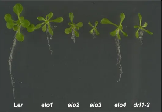
The group of Bernà et al, 1999 were interested to study genes involved in the control of leaf morphogenesis in a model system such as Arabidopsis thaliana. The mutants were divided into different classes according to the shape and size of the leaves and divided into 94 complementation groups. The elongata class contains 4 members, elo1-4, with a reduced leaf lamina width. The
elongata mutants exhibit a narrow leaf phenotype similar to the null mutant drl1-2. All elo mutants,
elo1, elo2, elo3 and elo4, have a delay in growth after germination, a reduction in leaf and root
Chapter I ________________________________________________________________________________
28 Figure 8. Cell division activity is also decreased in the yeast Elongator mutants; the cells undergo a G1 delay and are often not able to continue the cell cycle (Fichtner, L. and Schaffrath, R., 2002).
Figure 8. Comparison of the elo and drl1-2 mutant seedlings with Ler (22 DAG). (Nelissen H., et al, 2005).
The mutant phenotypes suggested a positive role for Elongator in lateral leaf and primary root growth, which coincides with the expression of ELO genes in these organs. In addition the pleiotropic phenotypes in elo mutants suggested that Elongator regulates several, but distinct, developmental pathways (Van Lijsebettens et al., 2014). To know the existing genetic interaction between the different Elongator genes the elo and drl homozygotes were crossed. The elo2elo3,
elo4elo1, and elo2 drl1-2 DMs had phenotypes similar to those of elo2, elo4drl1-4, and drl1-2,
respectively. Hence, DRL1 is epistatic to ELO2 and ELO1 in accordance with the proposed role for DRL1 as a regulator of the holocomplex (Fichtner et al.,2002). Furthermore, ELO1 was epistatic over ELO2 and ELO3, indicating the importance of the accessory subcomplex for the function of the core subcomplex. Finally, ELO2 was epistatic to ELO3, suggesting that the ELO2 scaffold protein is important for the maintenance of the integrity of Elongator as a HAT complex (Nelissen,
et al, 2005). Elongator Complex and SUPPRESSOR OF Ty4 (SPT4)/SPT5 are RNAPII-associated
transcript elongation factors that are implicated in chromatin dependent gene activation (Verséees, W.,et al. 2010; Hartzo, et al, 2013). The SPT4 and SPT5 genes are well studied in yeast and metazoa, indeed SPT5 is an essential gene in various organisms, whereas yeast cells lacking SPT4 are viable. In plants, the SPT4/SPT5 complex has been identified only recently, it pulled down the ELP3 subunit and subunit of RNAPII (Dürr, et al., 2014; Figure 9). The SPT5 is a nuclear protein
Chapter I ________________________________________________________________________________ that occurs in the transcriptionally active euchromatin and colocalizes with transcribing RNAPII (Dürr, ,et al, 2014). In Arabidopsis the immunoblot and mass spectrometry analysis demonstrated that SPT4/SPT5 complex exists and is conserved (Figure 9). Leaves of SPT4-RNAi plants are clearly smaller compared with wild type, and also the rosette leaf venation pattern is altered. In addition, the transcriptomes of the SPT4-RNAi line and elo3-1 mutants had common downregulated genes, including the auxin response and transport-related SHY2/IAA3 and LAX2, which are targeted by the Elongator complex for histone acetylation, transcription factors involved in cell elongation or organ morphogenesis, metabolic enzymes, membrane-located channels, and transporters (Van Lijsebettens, et al., 2014). In conclusion, the data suggest that Elongator and SUPPRESSOR OF Ty4 (SPT4)/SPT5 are transcript elongation factors may regulate the transcription of genes involved in the control of plant growth and development.
Figure 9. The structure of Elongator complex in Arabidopsis.
RNAPII subunits RPB1 and RPB2, all Elongator complex and the transcript elongation factor SPT4/SPT5 copurified with Elongator complex. Double-pointed arrows mark experimentally supported interactions between different protein (complexes), while interactions within complexes are not. (Van Lijsebettens, et al., 2014).
Ib2a. - Function of Elongator complex in plant
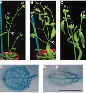
The phenotype of Elongator mutants are characterized by narrow and elongated leaves and petioles, short primary root, reduction in apical dominance defective venation patterning and altered phyllotaxis (Figure 10). In order to explain the phenotypes of the elo mutant microarray analysis identified a restricted number of significant GO categories (P < 0.001): chromatin assembly, pattern
Chapter I ________________________________________________________________________________
30 specification, vascular tissue development, and response to auxin stimulus (Nelissen, et al., 2010). Most of the down-regulated genes are involved in the auxin pathway such as: two SAUR, IAA14/SLR, IAA13, IAA11, ATHB8, IAA3/SHY2, IAA12/BDL, ARF10, ARF11, ARF18, ACL5, in addition to auxin biosynthesis (TAR2), and auxin transport (LAX2 and PIN4). All defects observed in elo mutants are connected to a defective auxin biology, indicating that auxin maxima are not properly established in elo mutants and suggesting a role for Elongator in auxin distribution or signaling. This suggested that Elongator likely controls plant development through its regulation of auxin-responsive genes. Indeed, the elo transcriptome identified a group of genes with a reduced expression level such as SHY2/IAA3 and BDL/IAA12 repressors, the ATHB8 auxin response gene the LAX2 influx carrier, the PIN4 efflux carrier, and the AP2/ERF ethylene response gene. Quantitative qPCR verified the down-regulation of these genes in the elo mutants. Chromatin immunoprecipitation (ChIP) were performed to investigate if the reduction in gene expression of some genes was related to reduced acetylation levels of histone H3 lysine-14 in their promoter and/or coding regions. Antibody against acetylated H3K14 or histone H3 and primers corresponding to the coding and 3′-untranslated regions showed reduced acetylation at the coding regions of the SHY2/IAA3 and LAX3 genes and confirmed that Elongator facilitates the expression of auxin-responsive genes by acetylation their coding region and thus facilitating the progress of RNA polymerase through the nucleosomes.
Figure 10. Auxin pathway is defective in elo mutant. (A)Wild-type inflorescence. (B) elo3-6 showing altered phyllotaxis of the secondary inflorescence branches
(arrowheads). (C) elo1-1 showing reduced apical dominance. (D) Defective venation pattern visualized by X-Gluc histochemical assay of wild type. (E) elo3-1 mutant transformed with pATHB8-GUS showing open venation (arrowhead). (Nelissen, et al., 2010).
Elongator complex plays many roles in plant in fact it is involved in abiotic stress responses, immune responses and in tRNA wobble uridine modification. Mutations in each subunit induced resistance to oxidative stress that is accompanied by increased expression of FSD1 (FE