I
University of Calabria-Italy
Inserm U1121University of Strasbourg-France
Thesis
Co-tutorship between University of Calabria, Doctorate in
“Life Sciences” and University of Strasbourg, Doctorate in
“Physics and Physical Chemistry”
Discipline: Physiology (BIO/09) and Biochemistry (BIO/10)
Title
Cateslytin and Chromofungin, two CgA derived peptides:
actors of the immune and cardiac systems
Dr. Francesco Scavello
Coordinator of the Doctorate in Life Sciences
Prof. Marcello Canonaco
Tutor
co-Tutor
Prof. Tommaso Angelone
Dr. Marie-Hélène Metz-Boutigue
Prof. Francis Schneider
University of Calabria
This study was supported by "Vinci Project 2014"- Università Italo-Francese
(project number C2_72)
I
INDEX
Index………I Abbreviations……….IV Resumé de la thèse………...1 Summary………...9 Introduction ... 121. Cardiac physiology and pathophysiology ... 13
2. Chromogranin A ... 15
2.1 Prohormone Chromogranin A and its derived peptides ... 16
3. Modulation of cardiac function and cardioprotection of Chromogranin A-derived peptides, Vasostatin and Catestatin ... 17
3.1 Vasostatin I and cardiovascular role ... 18
3.2 Catestatin and cardiovascular role ... 21
4. Chromofungin, the antifungal Chromogranin A (47–66)-derived peptide ... 23
5. Involvement of CgA and its derived peptides in immune system ... 25
5.1 Staphylococcus aureus and nosocomial infections ...... 32
5.2 Staphylococcus aureus and prosthetic valve endocarditis ...... 33
6. Antibacterial action of Cateslytin against Staphylococcus aureus ... 34
7. Aims of this thesis ... 38
Materials and Methods ... 40
1. Isolated and perfused rat Langendorff heart ... 41
1.1 Animals ... 41
1.2 Isolated heart preparation ... 41
2. Experimental protocols ... 42
2.1. Basal conditions ... 42
2.2. Drugs and chemicals ... 42
2.2.1. Chr stimulated preparations ... 43
2.2.2. Chr-dependent mechanism of action ... 43
2.3. Myocardial protective effects... 43
2.3.1. Ischemia/reperfusion ... 44
2.3.2. Cardiac function and infarct size ... 44
2.4. Cyclic guanosine monophosphate (cGMP)... 45
2.5. RNA preparation and quantitative real-time polymerase chain reaction for miRNA expression ... 45
II
3. Antibacterial characterization of Cateslytin derived-peptide ... 46
3.1. Preparation and characterization of the Cateslytin derived-peptides (DOPA*T*bCtl and T*bCtl) ... 46
3.2. Antibacterial assays against S. aureus ... 47
3.3. Purification of the fragments resulting from Ctl derived-peptides (T*bCtl and D*T*bCtl) digestion by the endoprotease Glu-C ... 48
3.4. Antibacterial assays of D*T*bCtl against S. aureus after the digestion by the endoprotease Glu-C ... 49
3.5. Prediction of secondary structure of Ctl-derived-peptides ... 49
3.6. Oxidation of D*T*bCtl: structural analysis and antibacterial activity ... 50
3.7. Stability analysis of D*T*bCtl in S. aureus supernatant and MHB medium ... 50
4. In vivo treatment with CgA derived-peptides in infected rat model ... 51
4.1 Animals ... 51
4.2 S. aureus infected rat models ... 51
4.3 Microbiological analysis ... 52
4.4 Plasma analysis of proinflammatory cytokines and damage marker ... 53
4.4.1 Enzyme-linked immunosorbent assay (ELISA) ... 53
4.4.2 Lactate dehydrogenase (LDH) determinations... 54
4.5 Western blotting analysis ... 54
5. Statistical analysis ... 55
Results ... 57
1. Basal cardiac effects and Postconditioning cardioprotective action of Chromofungin ... 58
1.1.Basal cardiac parameters ... 58
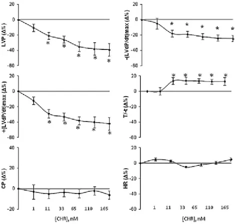
1.2.Chr effects on myocardial contractility and relaxation ... 58
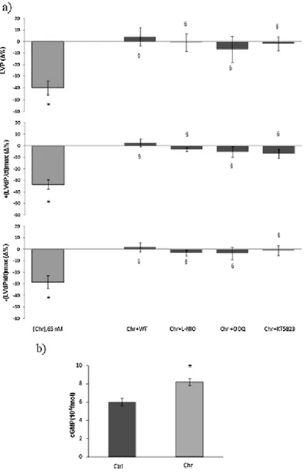
1.3.Mechanisms of action elicited by Chr ... 59
1.4.Chr effects on post-ischemic cardiac function ... 60
1.5.Chr influence on cardioprotective pathways ... 62
2. Antimicrobial action of Cateslytin-derived peptides: coating of prosthetic heart valves to prevent infection by S. aureus ... 64
2.1.Antibacterial activity of D*T*bCtl against S. aureus .... 65
2.2.Digestion of T*bCtl (L, D) by Glu-C protease from S. aureus V8 ... 66
2.3.Digestion of D*T*bCtl by Glu-C protease from S. aureus V8 ...... 67
2.4.Released Ctl from Glu-C cleavage and antibacterial effect against S. aureus ....... 68
2.5.Different secondary structure/different cleavage of Ctl-derived peptides ... 69
2.6.The oxidation of D*T*Ctl induces the formation of inactive aggregates ... 70
III 3.1.In vivo antibacterial action of Ctl against S. aureus in infected rat model ... 73
3.2.In vivo anti-inflammatory action of Ctl in S. aureus infected rat model ... 76
3.3.Mechanisms of action elicited by Ctl against S. aureus-induced cardiac inflammation .79 Discussion ... 80 1. Cardioinhibitory and cardioprotective effects of Chr ... 81 2. Antibacterial effect of D*T*bCtl against S. aureus ... 86
3. In vivo antibacterial and anti-inflammatory effect of Ctl in S. aureus infected rat model .. 89
Conclusions ... 92 References ... 95
IV
Abbreviations
5HD: 5-hydroxydecanoate
A. brassicola: Alternaria brassicola A. fumigates: Aspergillus fumigatus
A: Alanine aa: Amino acids
AKT: Protein-chinasi B AMP: Antimicrobial peptide AP: aortic pressure
AR: Adrenergic receptors
ARC: Activity-regulated cytoskeleton-associated protein b: Bovine
BNP: B-type natriuretic peptide
C. tropicalis: Candida tropicalis C. albicans: Candida albicans C. neoformans: Candida neoformans
CA: Catecholamine CaM: Calmodulin
CFU: Colony forming unit CgA: Chromogranin A
cGMP: Cyclic guanosine monophosphate Chr: Chromofungin
CNS: Central nervous system COX-2: Cyclooxygenase-2 CP: Coronary Pressure CPE: Carboxypeptidase E Ctl: Cateslytin
Cts: Catestatin
CTSL: Cysteine protease cathepsin L D*: DOPA-K-DOPA-K-DOPA
D*T*Ctl: DOPA-K-DOPA-K-DOPA-TLRGGE-RSMRLSFRARGYGFR D: Dextrorotation
DOPA: Levo-3,4-dihydroxyphenylalanine DPC: Dodecylphophatidylcholine
E. coli: Escherichia coli
E/S: Enzyme/Substrate E: Glutamic acid
EIA: Enzyme Immunoassay
ELISA: Enzyme-linked immunosorbent assay eNOS: Endothelial nitric oxide synthase ERK: Extracellular signal regulated kinase ET-1: Endothelin-1
F. culmorum: Fusarium culmorum F. oxysporum: Fusarium oxysporum
G: Glycine
GSK-3β: Glycogen synthase kinase-3β h: Human
V HPLC: High-performance liquid chromatography
HR: Heart Rate
HTR: Half Time Relaxation I/R: Ischemia/Reperfusion IL-1β: Interleukin-1β IL-6: Interleukin-6
iNOS: Inducible nitric oxide synthase
iPLA2: Calcium-independent phospholipase A2
ISO: Isoproterenol K: Lysine KH: Krebs-Henseleit solution L: Leucine L: Levorotation LDH: Lactic-dehydrogenase L-NIO: N(5)-(1-imino-3-butenyl)-l-ornithine LV: Left ventricle
LVEDP: Left ventricular end-diastolic pressure LVP: Left Ventricular Pressure
M. luteus: Micrococcus luteus
MALDI-TOF: Matrix Assisted Laser Desorption-Time Of Flight MH: Mueller-Hinton
MHB: Mueller-Hinton Broth
MIC: Minimal inhibitory concentration miRNA-21: mircoRNA-21
MitoKATP channel: Mitochondrial adenosine triphosphate-dependent potassium MRSA: Methicillin-resistant Staphylococcus aureus
MSSA: Methicillin sensitive Staphylococcus aureus
N. crassa: Neurospora crassa
N. haematococca: Nectria haematococca
NaIO4: Sodium Periodate
NO: nitric oxide OD: Optical density
ODQ: [1H-[1,2,4]oxadiazolo[4,3- a]quinoxalin-1-one] P: Proline
PBS: Phosphate-buffered saline PC: Prohormone convertases PCR: Polymerase chain reaction PD: PD98059
PI3K: Phosphoinositide 3-kinase PKC: Protein-chinasi C
PKG: Protein-chinasi G
PMN: Polymorphonuclear neutrophil PostC: Postconditioning
PTP: Prohormone thiol protease PVE: Prosthetic valve endocarditis PVL: Panton-Valentine leucocidin R: Arginine
VI ROS: Reactive oxygen species
RPP: Rate Pressure Product
S. aureus: Staphylococcus aureus S. cerevisiae: Saccharomyces cerevisiae
S: Serine
SAFE: Survivor Activating Factor Enhancement SDS: Sodium dodecyl sulfate
SDS-Page: Sodium dodecyl sulfate polyacrylamide gel electrophoresis sGC: Soluble guanylate cyclase
STA: Serine-Threonine-Alanine T*: TLRGGE
T*Ctl: TLRGGE-RSMRLSFRARGYGFR TFA: Trifluoroacetic acid
TFE: Trifuoroethanol
TMB: 3,3′,5,5′-tetramethylbenzidine TNF-α: Tumor necrosis factor α Vs-I: Vasostatin I
1
Université de Calabre-Italie
Inserm U1121Université de Strasbourg-France
Titre de la thèse
Cateslytine et Chromofungine, deux peptides dérivés de
la Chromogranine A qui sont de nouveaux acteurs des
systèmes immunitaire et cardiaque
Auteur
Dr. Francesco Scavello
2 La Chromogranine A (CgA) est une des protéines de la famille des granines. Elle est présente dans un très grand nombre de cellules nerveuses, neuro-endocrines, immunitaires et des cellules de la peau. La chromogranine A est une pro-hormone stockée dans les granules de sécrétion et elle subit une maturation protéolytique conduisant à la formation d’un très grand nombre de peptides dérivés. De plus, la CgA possède de nombreuses modifications post-traductionnelles (phosphorylation, O-glycosylation) qui confèrent une très grande variabilité aux fragments dérivés et aux activités biologiques correspondantes. La CgA est un marqueur plasmatique de désordres physiologiques (cancers, maladies cardio-vasculaires et maladies neurodégénératives).
Plusieurs peptides naturellement générés par clivage protéolytique possèdent des activités biologiques qui participent au retour à l’homéostasie après la réaction au stress. Ces peptides agissent au niveau du système cardio-vasculaire (inhibition de la vasoconstriction, effet inotropique), de la régulation hormonale (inhibition de la libération d’insuline), et possèdent des propriétés anti-microbiennes. Ainsi, la Chromofungine (Chr: CgA47-66) et la Cateslytine (Ctl: CgA344–358) possèdent des propriétés antimicrobiennes
(bactéries, champignons et levures). La Chr est un fragment du peptide naturel Vasostatine-I (1-76) et la Ctl correspond au domaine actif de la Catestatine (CgA344–364).
En plus de leur action directe sur les microorganismes ces peptides activent les neutrophiles et Ctl possède des propriétés anti-inflammatoires.
Staphylococcus aureus est un pathogène très virulent qui provoque un très grand nombre
de graves infections cliniques et il représente une des causes principales des infections nosocomiales. Cette bactérie a un fort impact de morbidité et mortalité en milieu communautaire et hospitalier. En effet S. aureus est la première cause d’infections en
3 milieu chirurgical et représente le pathogène Gram-positif le plus fréquent dans les cas de sepsis.
Dans le domaine des pathologies cardio-vasculaires, S. aureus provoque la destruction du tissu endocardiaque après implantation de valve cardiaque. Ce pathogène est remarquable par sa capacité à résister aux antibiotiques administrés et à répandre des clones résistants ce qui aggrave les cas d’infection liés au S. aureus. En 2013, Aslam et al., (Aslam et al., 2013) ont montré que Ctl est actif contre S. aureus et résistant à la dégradation par les protéases de S. aureus.
La présente thèse s’articule autour de 3 axes:
1- L’analyse dans le système de Langendorff des effets de Chr sur des coeurs de rats dans les conditions basales et pathologiques
2- L’analyse des propriétés antimicrobiennes d’un peptide synthétique dérivé de Ctl qui sera utilisé pour recouvrir des valves cardiaques pour combattre les infections contre S. aureus
3- L’étude in vivo sur des rats infectés par S. aureus de l’activité antibactérienne et cardio-protective de Ctl.
La première partie de l’étude a été réalisée en utilisant la technique de Langendorff sur coeurs isolés et perfusés, un test ELISA et la PCR en temps réel. Nous avons montré que’en conditions basales des doses croissantes de Chr (11–165 nM) induisent un effet inotropique négatif sans changement de la pression coronarienne. L’activation de la voie de signalisation AKT/eNOS/cGMP/PKG est responsable de cet effet de Chr. Nous avons aussi montré que Chr agit comme un agent de post-conditionnement contre les effets négatifs de l’ischémie/reperfusion en réduisant la taille de l’infarctus et le niveau de LDH.
4 a responsables de cette cardio-protection impliquent les cascades de signalisation PI3K, RISK, MitoKATP et miRNA-21.
L’ensemble de ces résultats suggère que Chr affecte directement la performance du travail du coeur, protège contre les blessures du myocarde par ischémie/reperfusion, par l’activation de kinases (Filice et al., 2015).
En conclusion, Chr peut être proposé comme un nouveau modulateur physiologique neuroendocrine capable de prévenir des dysfonctionnements cardiaques. Son potentiel clinique méritera d’être approfondi par de nouvelles recherches.
Dans la seconde partie de la thèse, 2 nouveaux peptides synthétiques contenant Ctl (RSMRLSFRARGYGFR) ont été designés:
D*T*Ctl (DOPA-K-DOPA-K-DOPA-TLRGGE-RSMRLSFRARGYGFR),
T*Ctl (TLRGGE-RSMRLSFRARGYGFR) with D*: DOPA-K-DOPA-K-DOPA and T*: TLRGGE.
Cette étude comprend une première partie d’expériences réalisées en solution. Elle est basée sur les propriétés adhésives de la séquence DOPA-K-DOPA-K-DOPA et sur l’aptitude de l’endoprotéase Glu-C pour cliver aorès la séquence TLRGGE.
En utilisant des techniques de biochimie, protéomique (séquençsage et spectrométrie de T*Ctl S. aureus de masse) et microbiologie, nous avons montré que la dégradation par la protése Glu-C de T*Ctl et D*T*Ctl génère la Ctl active. Puis l’analyse prédictive de la structure secondaire suggère la présence d’un domaine en hélice pour D*T*Ctl. Le
5 groupement D* semble stabiliser la structure secondaire et faciliter le clivage par la protése Glu-C pour libérer le peptide Ctl actif.
Le groupement D* stabiliserait la structure secondaire du peptide D*T*Ctl et faciliterait le clivage exclusif qui produit le peptide Ctl actif.
Mécanisme d’action de D*T*Ctl contre S. aureus
Pour pouvoire intégrer le peptide dans un revêtement de matériau il est nécessaire de procéder à une oxydation par NaIO4. Après cette étape on a recherché la production du
peptide actif Ctl et analysé l’activité antibactérienne.
L’étude protéomique a clairement montré par spectrométrie de masse la formation de polymères qui gênent le clivage par la protéase Glu-C et la formation de Ctl. En préambule nous avons montré une activité antibactérienne contre S. aureus pour le peptide D*T*Ctl avec une CMI de 75 µM. Cependant après oxydation on observe la formation d’aggrégats qui gênent le clivage nécessiare à la formation de Ctl.
L’ensemble de ce travail fait l’objet d’un manuscrit en cours de rédaction.
S. aureus
TLRGGE
RSMRLSFRARGYGFR
(DOPA)-K-(DOPA)-K-(DOPA)
Glu-CRSMRLSFRARGYGFR
Cleavage Direct Action Solution a b c Indirect Action d6 Dans la troisième partie de cette thèse nous avons évalué l’activité antibactérienne in vivo de Ctl ainsi que la protection cardiaque qui serait apportée par Ctl sur un modèle de rats infectés par S. aureus. Au niveau systémique et cardiaque nous avons cherché à identifier les cibles moléculaires de l’inflammation qui seraient visées par l’administration de Ctl. Ces expériences ont été réalisées par l’utilisation des techniques de Western blot, ELISA et analyses microbiologiques au niveau du coeur et du plasma.
Nous observons dans le plasma une forte diminution de la croissance bactérienne, de TNF-α, IL-1β et LDH. Les expériences de Western blot réalisées sur les extraits cardiaques montrent que le traitement par Ctl induit une diminution des marqueurs pro-inflammatoires tels que iNOS et COX-2.
Ces premiers résultats réalisés in vivo montrent que le traitement par Ctl de rats infectés par S. aureus combat l’infection et assure une protection du myocarde.
Dans le but de réaliser des études cliniques, nous rechercherons le plus peptide dérivé de Ctl assurant l’ensemble des propriétés recherchées.
7
Croissance bactérienne de S. aureus mesurée au niveau (a) plasmatique et (b) cardiaque sur des rats infectés par S. aureus et traités par Ctl.
Concentration de IL-1 dans le plasma de rats infectés par S. aureus
En conclusion, en utilisant 2 peptides de la CgA Chr et Ctl le travail de thèse présenté montre que:
1- Ctl assure une protection cardiaque dans un modèle de coeur isolé utilisant l’ischémie/reperfusion
2- Dans le but d’élaborer un nouveau revêtement de valves cardiaques le peptide D*T*Ctl se révèle intéressant dans des conditions non oxydantes car (1) il présente
0 10 20 30 40 50 60 70 80 90 100 S. aureus S. aureus+CTL (1.5 mg/Kg) CTL (1.5 mg/Kg) Heart % of B ac te ri al G ro w th * 0 10 20 30 40 50 60 70 80 90 100 S. aureus S. aureus+CTL (1.5 mg/Kg) CTL (1.5 mg/Kg) Plasma % of B ac te ri al G ro w th * (a) (b) 0 20 40 60 80 100 120 140 160 180
Control S. aureus S. aureus+CTL (1.5 mg/Kg) CTL (1.5 mg/Kg)
IL -1 β (pg /mL )
8 une activité antimicrobienne contre S. aureus; (2) en présence de S. aureus il permet par clivage protéolytique de libérer le peptide Ctl actif. Afin de finaliser cette étude des expériences sont actuellement en cours à l’Inserm U1121 pour réussir le recouvrement de matériau sans l’étape d’oxydation.
3- Une première expérience réalisée in vivo a montré le rôle de Ctl pour combattre l’infection à S. aureus au niveau systémique et au niveau cardiaque, mais aussi assurer la protection du myocarde.
9
Summary
Chromogranin A (CgA) belongs to the granin family of uniquely acidic secretory that are ubiquitous in secretory cells of the nervous, endocrine, immune system. Numerous cleavage products of the granins have been identified, some of these peptides showed biological activities and are costored in secretory granules of different cells. Chromofungin (Chr: CgA47-66) and Cateslytin (Ctl: CgA344–358) are peptides that display
antimicrobial activities and activate neutrophils, with important implications in inflammation and innate immunity. Staphylococcus aureus is an opportunistic pathogen and the leading cause of a wide range of severe clinical infections and one of the most important cause of hospital-acquired infections, in fact infections caused by this bacterium have classically an important impact in morbidity and mortality in the nosocomial and community scene. Furthermore, this pathogen is the primary cause of surgical site infections and the most frequently isolated pathogen in Gram-positive sepsis. In the specific field of cardiovascular disease S. aureus leading infective cause of destruction of endocardial tissue after implantation of prosthetic heart valve. This pathogen is also notorious for its ability to resist the available antibiotics and dissemination of various multidrug-resistant S. aureus clones that limit therapeutic options for a S. aureus infection. Aslam et al. in 2013 shown that Ctl is resistant to the degradation of S. aureus protease and is the most antibacterial CgA derived peptide against this bacterium. The aim of study was to evaluate the: 1) Effects of Chr on isolated and Langendorff perfused rat hearts in basal and pathological conditions; 2) In vitro antibacterial activity of a synthetic Cateslytin-derived peptide to cover artificial heart valves and prevent infection by S.
10 The first part of the study was performed by using the isolated and Langendorff perfused rat hearts, Elisa assay and real-time PCR. We found that, under basal conditions, increasing doses (11–165 nM) of Chr induced negative inotropic effects without changing coronary pressure. The AKT/eNOS/cGMP/PKG pathway mediated this action. We also found that Chr acted as a postconditioning (PostC) agent against ischemia/reperfusion (I/R) damages, reducing infarct size and LDH level. Cardioprotection involved PI3K, RISK pathway, MitoKATPand miRNA-21. Therefore, we suggest that Chr directly affects heart performance, protects against I/R myocardial injuries through the activation of prosurvival kinases. Results may propose Chr as a new physiological neuroendocrinemodulator able to prevent heart dysfunctions, also encouraging the clarification of its clinical potential.
In the second part of the study, two new synthetic peptides containing Ctl (RSMRLSFRARGYGFR) were designed: D*T*Ctl (DOPA-K-DOPA-K-DOPA-TLRGGE-RSMRLSFRARGYGFR), T*Ctl (TLRGGE-RSMRLSFRARGYGFR) with D*: DOPA-K-DOPA-K-DOPA and T*: TLRGGE. This study is based on the observation of the adhesive properties of the DOPA-K-DOPA-K-DOPA sequence and on the ability of S. aureus endoprotease Glu-C to cleave the TLRGGE sequence. Firstly, using techniques of biochemistry, proteomics (sequencing, mass spectrometry) and microbiology we shown that the digestion by the Glu-C protease of T*Ctl and D*T*Ctl is able to release active Ctl. The prediction analisys of the secondary structure suggested the presence of an alpha helix domain in the case of D*T*Ctl with respect to T*Ctl. The D* group stabilized the secondary structure and facilitated the cleavage by Glu-C to the release of the active peptide Ctl. Subsequently, the effect of the oxidation by NaIO4 of
11 analysis showed the formation of polymers inhibiting the action of Glu-C and the release of Ctl. We also shown that D*T*Ctl had a MIC value around 75 µM against different strains of S. aureus. This data shown that D*T*Ctl had a direct action against the bacteria without Glu-C cleavage. However, in oxidizing conditions the formation of aggregates of D*T*Ctl reduced the antibacterial action of this synthetic peptide.
In the last part of this thesis, we evaluated the in vivo antibacterial activity of Ctl and whether and to which extent Ctl elicit cardioprotection in rat infected with S. aureus, as a model of infection with this bacterium. Identification of specific molecular targets of tissue and systemic inflammation and damage were analysed by Western blotting, ELISA and microbiological analysis in cardiac homogenates and plasma. A strong reduction of plasma bacterial growth, TNF-α, IL-1β and LDH plasma levels was observed in infected rat treated with Ctl. Western blotting analysis of cardiac extracts showed that Ctl treatment is accompanied by reduction of expression of pro-inflammatory markers, such as iNOS and COX-2. These preliminary data suggest that in vivo Ctl treatment is able to counteract the deleterious effects of S. aureus, and elicits myocardial protection.
12
13
1. Cardiac physiology and pathophysiology
The heart, during its role of pump, is subjected to interaction with various molecules such as the endogenous hormones (Burley et al., 2007). Many of these factors, such as atrial natriuretic peptide (ANP) or brain natriuretic peptide (BNP), are able to (1) reduce contractility or relaxation, (2) improve the pump property, such as Urocortin, Serpinin (Sarzani et al., 2017; Díaz and Smani, 2013, Tota et al., 2014) and (3) induce vasoactive effects on coronary vessels (Nichols and Epstein, 2009).
In pathophysiological conditions such as heart failure, damage consequent to sepsis and myocardial infarction the pump function of the heart can be altered (Minucci et al., 2011; Suzuki et al., 2017) and several hormones may protect the heart against these alterations (Hausenloy et al., 2004 and 2016). In particular, several evidences reported that many hormones are able to protect the heart against the ischemia/reperfusion (I/R) damage (Burley et al., 2007). The most critical point in I/R damage is the time of reperfusion, because in this period the reoxygenation alters redox states that are implicated as the primary causes of oxidative stress and tissue damage in ischemic heart disease (Jeroudi et al., 1994). This deleterious phenomenon induces mitochondrial dysfunction, ROS generation, additional Ca2+ influx, lower concentration of ATP and apoptosis activation (Hausenloy et al., 2016). The powerful mechanisms for protecting the myocardium against this damage are ischemic preconditioning and postconditioning, in which transient non-lethal phases of myocardial ischemia (repeated respectively before and after the global ischemia) confer protection against the myocardial infarction (Hausenloy et al., 2004, 2005 and 2016). Recently several data showed that pharmacological administration before or after ischemia can also induce cardioprotection, and this approach represents a new therapeutic strategy against myocardial infarction (Hausenloy et al., 2004 and 2016).
14 In pre- and post-conditioning process the activation of several pro-survival kinases, during the time of reperfusion, confer powerful cardioprotection against myocardial I/R injury (Hausenloy et al., 2004 and 2005).
Sepsis is a systemic state of infection that induces severe organ dysfunctions related to a massive infection of host tissues. Several clinical studies estimated that patients associated with sepsis often die from cardiovascular complications (Suzuki et al., 2017). In fact, it has been reported that during sepsis, the consequences of the heart ischemia and reperfusion injury are more evident (Bar-Or et al., 2015); in fact, the infarct size is exacerbated by the local inflammation with a production of pro-inflammatory cytokines (Suzuki et al., 2017). The septic shock can induce destruction of myofilaments and degradation of contractile apparatus mediated by increased matrix metalloproteinase activity (Rudiger and Singer, 2007). These structural alterations of cardiac tissue, during a septic event, can induce hemodynamic disorders characterized by a reduced ejection fraction and enlarged end-diastolic volume index that lead to cardiac dysfunction during the early stages of sepsis (Calvin et al., 1981; Packer et al., 1984). The heart valves are most commonly affected by pathogen, but the infection may also occur in other structural areas of the heart including septal defects, patent ductus arteriosus or on the mural endocardium (Yusuf et al., 2012). The infection of cardiac valves occurs in four steps. In a first part, the microorganisms induce a valvular endothelial damage, after this process the pathogens form a platelet-fibrin thrombus. This platelet-thrombus plaque develops a surface that allows a major adherence of bacteria. Only then, the local bacterial proliferation with hematogenous seeding and consequent biofilm formation can occur (Frontera and Gradon, 2000). The most common microorganism associated with infective endocarditis is the Gram-positive Staphylococcus aureus (Yusuf et al., 2012). The mortality rate associated with S. aureus prosthetic valve endocarditis is high, and patients
15 with this complication are haemodynamically unstable and develop heart failure or valvular dysfunction (Attaran et al., 2012).
2. Chromogranin A
Chromogranin A (CgA) belongs to the granin family of uniquely acidic glyco-phospho proteins that are ubiquitous in secretory cells of the nervous, endocrine, immune system (Helle, 2004). This protein was the first granin to be characterized as stored and co-released with the catecholamine hormones from the bovine adrenal medulla (Banks and Helle, 1965). After this identification, CgA was also observed in the granules of many other cells such as cardiomyocytes (Glattard et al., 2006; Pieroni et al., 2007), keratinocytes (Corti and Ferrero, 2012), and neutrophils (Lugardon et al., 2000; Briolat et al., 2005). This protein displays important intracellular functions such as granule maturation, trafficking and exocytotic mechanisms (Helle and Aunis, 2000). Human and bovine CgA contain 439 and 431 amino acid residues, respectively. Mature human and bovine CgA have approximately 49 kDa molecular weight and contain post-translational modifications sites such as phosphorylations and glycosylations sites (Zhang et al., 1997; Strub et al., 1997; Gadroy et al., 1998; Bauer et al., 1999) and a large number of acidic residues (Metz-Boutigue et al., 1993a; Gadroy et al., 1998).
Bovine protein (bCgA1–431) is shorter than human (hCgA1–439) and rat (rCgA1–448) proteins
(Iacangelo et al.,1988). However, the N- and C-terminal domains, i.e. CgA1–76 and
CgA316–431, are highly conserved sequences in mammals (Yanaihara et al., 1998; Benedum
et al., 1986). The hypothesis that CgA may serve as a prohormone for shorter fragments with regulatory properties has been reported (Benedum et al., 1986; Eiden, 1987).
16
In fact, through proteolytic processing, often in a tissue-specific manner, CgA generates various smaller biologically active fragments that participate in peptide hormone regulation of physiological systems in health and disease conditions (Angelone and Tota, 2012).
2.1
Prohormone Chromogranin A and its derived peptides
Numerous pairs of basic amino acids, present in the sequence of CgA, indicate potential sites for cleavage by the prohormone convertases (PC1/3 and PC2), prohormone thiol proteases (PTP), and carboxypeptidase E (CPE) that occur as co-stored components of neurosecretory granules (Seidah and Chretien, 1999; Metz-Boutigue et al., 1993a). Numerous cleavage products of the granins have been identified, some of which show biological activities and are co-stored in secretory granules of different cells (Helle, 2004). The intracellular processing of CgA was shown for the first time in chromaffin cells whose secretory vesicles contain proteolytic enzymes. Among them, the neuroendocrine-specific carboxypeptidase E/H that removes basic residues from neuropeptides intermediates, the prohormone convertases (PC1/2; Seidah and Chretien, 1999), and the Lys/Arg aminopeptidases (Seidah and Chretien, 1999).
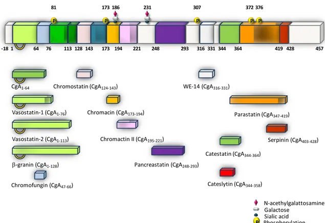
In the bovine chromaffin granules the proteolytic processing of CgA occurs at 13 sites, 5 of them at the N-terminal side, 2 in the middle and the rest at the C-terminal end. These sites are located at residues 3–4, 64–65, 76–77, 78–79, 115–116, 247–248, 291–292, 315– 316, 331–332, 350–351, 353–354, 358–359, 386–387, and 403–429 (Wohlfarter et al., 1989; Metz-Boutigue et al., 1993b; Koshimizu et al., 2011) (Figure 1).
17
Figure 1: Schematic illustration of biologically active Chromogranin A (CgA)-derived peptides originating from the stress-activated, diffuse neuroendocrine system (from Angelone and Tota, 2012).
3. Modulation of cardiac function and cardioprotection of
Chromogranin A-derived peptides, Vasostatin and
Catestatin
Several studies have shown the presence of CgA in secretory granules of rat atrial myoendocrine cells and in human ventricular myocardium (Steiner et al., 1990; Pieroni et al., 2007). Colocalization of CgA and atrial natriuretic peptides in rat heart cardiomyocytes (Steiner et al., 1990) and in human ventricular cardiomyocytes has been
shown and circulating levels of CgA are correlated with BNP (Pieroni et al., 2007). CgA has also been detected in rat Purkinje fiber cells of the conducting system, and in both rat atrium and ventricle (Weiergraber et al., 2000). Pasqua and co-workers (2013) have shown N-acethylgalattosamine Galactose Sialic acid P Catestatin (CgA344-364) Parastatin (CgA347-419) Chromostatin (CgA124-143) Pancreastatin (CgA248-293) WE-14 (CgA316-331) Vasostatin-2 (CgA1-113) Vasostatin-1 (CgA1-76) CgA1-64 -granin (CgA1-128) Chromacin (CgA173-194) Chromofungin (CgA47-66)
Chromactin II (CgA195-221) Serpinin (CgA403-428)
457 -18 1 64 76 113 128 143 173 194 221 248 293 316 331344 364 419 428 P 81 P 173 P 307 P 372 P 376 Phosphorylation 186 231 Cateslytin (CgA344-358)
18
a direct demonstration of the intracardiac processing of CgA in response to hemodynamic and excitatory challenges with a clear evidence that the heart produces and processes vasostatin-containing peptides (Glattard et al., 2006; Pasqua et al., 2013). In the rat heart, the detected PC1/3, PC2 and carboxypeptidase H/E might be involved in the intracellular CgA maturation process (Zheng et al., 1994; Muth et al., 2004). Some of these fragments may also result from extracellular processing, as reported for CgA in the adrenal gland (Metz-Boutigue et al., 1993). The presence of extracellular proteases both on
cardiomyocyte cell membranes and in the extracellular matrix may indicate an
extracellular processing (Glattard et al., 2006), as proposed for Angiotensin II (Jan Danser and Saris, 2002).
3.1
Vasostatin I and cardiovascular role
Vasostatin I (Vs-I) corresponds to the polypeptide CgA1-76, derived from the cleavage at
the first pair of basic amino acid residues of the N-terminal domain of CgA (Metz-Boutigue et al., 1993a). The name of this peptidederives from its ability to maintain blood flow by reducing vascular contraction evoked in arterial and venous preparations by both high potassium and agonists such noradrenalin and endothelin (Aardal and Helle, 1992; Angeletti et al., 1994).
Several studies in literature have shown that Vs-I acts as as inotropic modulator of cardiac performance in mammals with an “anti-adrenergic” potential (Cerra et al., 2006; Cerra et al, 2008; Gallo et al., 2007). Also the human recombinant CgA1-78 exerts a negative
inotropism and lusitropism on the rat heart (Cerra et al., 2006). This recombinant CgA-derived N-terminal peptide corresponds to the sequence of human Vs-I but also containing
19 the sequence of two residues (KK) at the C-terminus and three additional residues (STA) at the N-terminus, due to the molecular biology procedures (Corti et al., 1997).
On the Langendorff-perfused rat heart under basal conditions CgA1-78 causes a
dose-dependent reduction of Left Ventricular Pressure (LVP) and Rate Pressure Product (RPP), obtained by LVPx Heart Rate (HR), thus, inducing a negative inotropism (Cerra et al., 2006). Since the negative inotropy is obtained without affecting neither HR nor Coronary Pressure (CP), the reduction of contractile performance, and consequently of cardiac work, is consistent with a direct myocardial influence of the peptide which is independent from coronary reactivity (Cerra et al., 2006).
Also the native rat CgA1-64 (from 33 nM) induces negative inotropism and lusitropism,
and coronary dilation, counteracting the Isoproterenol (ISO) and Endothelin-1 (ET-1)-induced positive inotropic effects and ET-1-dependent coronary constriction in Langendorff perfused rat heart (Cerra et al., 2008). The CgA1-64 also depresses basal and
ISO-induced contractility on rat papillary muscles, without affecting calcium transients on isolated ventricular cells (Cerra et al., 2008). In addition, structure-function analysis, using three modified peptides (i.e., rCgA1–64 with S-S bridge, rCgA1–64 without S-S bridge,
and rCgA1–64 oxidized), has revealed the key role of disulfide bridge in the cardiotropic
action (Cerra et al., 2008).
Based on the ability of the domains CgA1-40 and CgA47-66to interact and pass through the
biological membrane (Maget-Dana et al., 2002; Zhang et al., 2009), the cardiac effects of human and rat Vs-I may be exerted by interacting with, or penetrating into, the cell membrane in a receptor independent-manner (Cerra et al. 2006). In line with this hypothesis, the inhibition of the ISO-mediated positive inotropism of both CgA1-64 and
CgA1-78 in the rat heart may be explained via an allosteric modulation of the β-adrenergic
20 intracellular signaling triggered by the activation of the β-adrenoceptor itself (Cerra et al., 2006, 2008).
The involvement of NO-cGMP-PKG pathway in the negative inotropic effect of CgA1-78
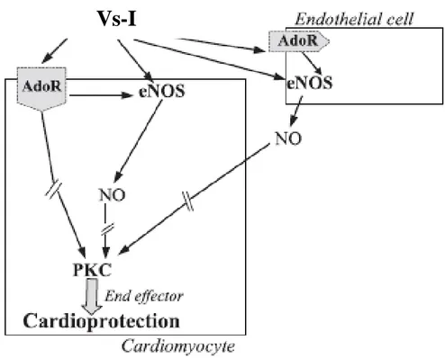
(Cerra et al., 2008; Tota et al., 2014) suggested the possibility that it exerts a protective effect against the extension of myocardial infarctions. Indeed, the administration of a low dose of CgA1-78 before Ischemia/Reperfusion (I/R) reduced the infarct size inducing a
preconditioning-like effect (Cappello et al., 2007). This protective effect is suppressed by eNOS inhibition and 50% reduced by the blockade of adenosine A1 receptors, indicating
that at least two different pathways may be hypothesized as possible mediators of cardioprotection (Figure 2) (Cappello et al., 2007).
Figure 2: Diagram of mechanisms of Vs-I (CgA1-78)-induced cardioprotection (from Cappello et al., 2007).
21
3.2
Catestatin and cardiovascular role
Catestatin (Cts) (CgA344–364) is a 21-residue long, cationic and hydrophobic peptide
(Mahata et al., 1997) located at the N terminal end of parastatin (Fasciotto et al., 1993) and derives from intragranular and extragranular processing of the prohormone (Mahata et al., 1997; Preece et al., 2004; Parmer et al., 2000). Cts is a strong non-competitive inhibitor of nicotinic receptor-mediated catecholamine (CA) release (Mahata et al., 1997). Cts inhibits the calcium-dependent CA release as well as the acetylcholine-induced desensitization of the nicotinic receptor itself (Mahata et al., 1997). Furthermore, through interaction with histamine receptors, Cts exhibits vasorelaxant and antihypertensive characteristics (Kennedy et al., 1998). This antihypertensive profile is documented by a decrease of its plasma levels in patients with essential hypertension (O’Connor et al., 2002). Several studies have documented the direct myocardial and coronary effects of Cts and its mechanisms of action. The cardiosuppressive influence of Cts was evaluated on the isolated Langendorff-perfused rat heart under both basal and chemically stimulated conditions (Angelone et al., 2008). Cts induces negative inotropism and lusitropism also involving β3-adrenergic receptors (β3-AR), but showing a higher affinity for β2-AR
(Angelone et al., 2012). These effects were mediated by β2
-ARs-Gi/oProtein-eNOS-NO-cGMP-PKG mechanisms (Angelone et al., 2012). Furthermore, Cts-induced activation of phosphodiesterases type 2 and the increased S-nitrosylation of both phospholamban and arrestin, suggest an additional mechanism for intracellular calcium modulation and β-adrenergic responsiveness (Angelone et al., 2012). Several studies have also shown that Cts acts as potent inhibitors of ISO exerting different coronary vasoactivity (Angelone et al., 2008, 2012). In isolated rat hearts Cts, given at reperfusion (Cts-Post), decreases the infarct size, limits contracture and improves the post-ischemic systolic function (Penna et
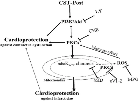
22 al., 2010). Cts also reduces infarct size and post-ischemic contracture given as a pre-conditioning agent, but less than Cts-Post (Penna et al., 2010). Moreover, only Cts-Post significantly improves post-ischemic recovery of developed LVP. Therefore, Cts seems more protective as PostC agent than as a pre-conditioning agent (Penna et al., 2010). Cts is also able to induce cardioprotection during the early reperfusion phase in isolated hearts, or if added during a challenging ischemia in isolated cells by pro-survival intrinsic signaling cascades, which include the Reperfusion Injury Salvage Kinases (RISK) and Survivor Activating Factor Enhancement (SAFE) pathways (Penna et al., 2010, 2014; Perrelli et al., 2013). Several studies focused on cardioprotective pathways activated by Cts. Perrelli et al., (2013) in isolated Langendorff rat hearts have shown that inhibition of PI3K or a large spectrum of PKC, PKCε and mitoKATP channels blockade or ROS
scavengering abolished the infarct sparing effect of Cts (75nM, Cst-Post) (Perrelli et al., 2013) (Figure 3).
23 Notably, Cts can reverse the S-nitrosylation downregulation induced by I/R in spontaneously hypertensive rats (Penna et al., 2014). In fact, the S-nitrosylation of L-type calcium channel may limit calcium overload and may allow a better functional recovery of surviving cardiomyocytes (Sun et al., 2006; Murphy et al., 2012). Furthermore, Cts increases expression of pro-angiogenetic factors such as HIF-1α and eNOS after two-hour reperfusion (Penna et al., 2014). Cts triggers anti-apoptotic factors, such as ARC, and induces reduction of pro-apoptotic factors such as active Caspase 3 (Penna et al., 2014).
4. Chromofungin, the antifungal Chromogranin A
(47–66)-derived peptide
In 2001, Lugardon et al. identified a natural CgA-derived peptide corresponding to the N-terminal Vs-I-derived sequence 47–66, and named Chromofungin (Chr) because of its antifungal activity (Lugardon et al., 2001). It is generated during infections after cleavage by Staphylococcus aureus endoprotease Glu-C (Aslam et al., 2012). Chr acts as an immediate protective shield against pathogens, in fact it displays antifungal activity at 2– 15 µM against filamentous fungi (N. crassa, A. fumigatus, A. brassicola, N.
haematococca, F. culmorum, F. oxysporum) and yeast cells (C. albicans, C. tropicalis, C. neoformans) (Lugardon et al., 2001). Furthermore, Chr is able to inhibit microbial cell
metabolism (Lugardon et al., 2001; Aslam et al., 2012), and to penetrate the cell membrane, thus inducing extracellular calcium entry by a CaM-regulated iPLA2 pathway
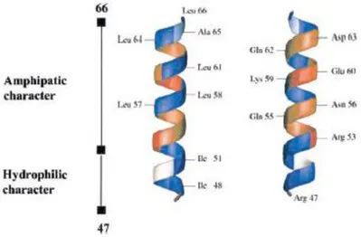
(Zhang et al., 2009). The 3-D structure of Chr has revealed the amphipathic helical character of the sequence 53–56, whereas the segment 48–52 confers hydrophobic character (Lugardon 2001) (Figure 4).
24
Figure 4:Three-dimensional representation of chromofungin (CgA47–66) (from Lugardon et al., 2001).
Among all synthetic Vs-I derived peptides, Chr actually displays the highest antifungal activity on both fungi and yeast. The increase of the surface pressure in presence of ergosterol indicated a specificity for the fungal membranes (Lugardon et al., 2001). In addition, Chr induces membrane destabilization with the possible formation of holes (Lugardon et al., 2001). Interestingly, a cardioactive role is also suggested for Chr. As shown on the frog heart bioassay, a fragment corresponding to the Chr sequence, is found to depress myocardial contractility by eliciting a direct negative inotropic effect (Tota et al., 2003). These observations represent an intriguing starting point for investigating the heart sensitivity to Chr.
25
5. Involvement of CgA and its derived peptides in immune
system
The innate immune system represents the primary defense in most living organisms. This system evolved as a set of very well organized events that protect the host from the invading pathogens such as bacteria, viruses, fungi, parasites and cancerous cells. Antimicrobial peptides (AMPs) are the unique component of the innate immune response that are highly conserved through evolutionary process (Ganz, 2003). AMPs are also called host defense peptides (Auvynet and Rosenstein, 2009). These are remarkably appreciable in their selection of target cells. These peptides are highly effective broad-spectrum antibiotics, which demonstrate their potential use as novel therapeutic agents. Antimicrobial peptides have been demonstrated to kill bacteria (including resistant strains), mycobacteria, enveloped viruses, fungi and even transformed or cancerous cells (Auvynet and Rosenstein, 2009). In addition, they also have the ability to enhance immunity by functioning as immunomodulators (Auvynet and Rosenstein, 2009). Antimicrobial peptides have two modes of action on microorganisms (Brogden, 2005). They can target cell membranes and permeabilize them according to several proposed mechanisms (Jenssen et al., 2006) or, alternatively, they act on intracellular targets to induce DNA binding, enzyme inhibition and cell wall synthesis inhibition. For numerous AMPs, the folded peptide adopts an amphipathic profile important for their microbial killing mechanism: the cationic character of AMPs induces an electrostatic attraction to the negatively-charged phospholipids of microbial membranes and their hydrophobicity aids the integration into the microbial cell membrane, leading to membrane disruption. Furthermore, the amphipathic structure also allows the peptides to be soluble both in aqueous environments and in lipid membranes (Yeaman and Yount, 2003).
26 Several Chromogranins-derived peptides have been characterized as new cationic antimicrobial peptides (Strub et al., 1996a, b; Metz-Boutigue et al., 1998; Lugardon et al., 2000; Briolat et al., 2005; Helle et al., 2007) and host defense agents derived from CgA during infections, were identified (Radek et al., 2008). The AMPs derived from CgA act at micromolar range against bacteria, fungi, yeasts and are non-toxic for mammalian cells. They are recovered in biological fluids involved in defense mechanisms (serum, saliva) and in secretion of stimulated human neutrophils (Lugardon et al., 2000; Briolat et al., 2005).
The CgA-derived peptides production and the molecular mechanism of the antimicrobial activities were characterized in several studies. In addition, when polymorphonuclear neutrophils (PMNs), known to accumulate at sites of infection, are stimulated by bacterial agents such as Panton-Valentine leucocidin (PVL), they produce and secrete complete and processed forms of CgA, such as Vs-I and Vs-II (Lugardon et al., 2000) and Cts (Briolat et al., 2005).
Others experiments have demonstrated the presence of Cts in infected skin (Radek et al., 2008). The potential relevance of Cts against skin pathogens was supported by the observation that CgA was expressed in keratinocytes. In human skin, CgA was found to be proteolytically processed to produce Cts (Radek et al., 2008). Interestingly, Cts expression in murine skin increases in response to injury and infection, providing potential for increased protection against infection (Radek et al., 2008). In fact, this peptide possesses antimicrobial activities at micromolar concentration against Gram-negative and Gram-positive bacteria, yeast, and fungi. (Briolat et al., 2005; Radek et al., 2008). The two human variants P370L and G364S display antibacterial activity against M. luteus with a minimal inhibitory concentration (MIC) of 2 and 1 µM, respectively, and against E. coli with a MIC of 20 and 10 µM, respectively (Briolat et al., 2005).
27 Cts acts against bacteria with a mechanism similar to that reported for Chr (Briolat et al., 2005). In fact, Cts is a cationic peptide with amphipathic structure such as Chr. This property allows to the hydrophobic domain to interact with the membrane while the cationic domain destabilizes and induces the pores formation (Figure 5).
Figure 5:Model for interaction of Cateslytin (antimicrobial domain of Cts) with bacterial-like membrane domains (from Jean-François et al., 2008a).
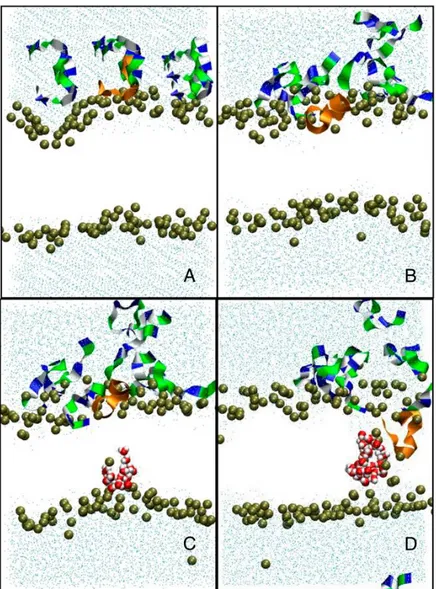
Moreover, the outer leaflet of external bacterial membranes comprises a large amount of negatively charged peptides (whereas that of animals or plants is globally neutral), so the susceptibility of a microorganism to a particular AMP is mainly linked to its charge, hydrophobicity, and amphipathicity. Indeed, Cts adopts the α-helical structure when it interacts with negatively charged membranes and this interaction involves positively charged residues such as arginine. After this interaction the peptide induces a rigidity increase of the membrane regions where it interacts, as well as a thickening of these regions and formation of pores (Sugawara et al., 2010) (Figure 6).
28
Figure 6: Molecular dynamics of pore formation induced in a 1,2-dimyristoyl-sn-glycero-3-phosphocholine bilayer (128 lipids, 6754 water molecules) by the presence of eight Cateslytins. (A) Starting conditions with the initial distribution of the peptides above the outer membrane leaflet; (B) the equilibrated bilayer and peptides on the outer membrane leaflet; and (C) the formation of a transient water defect. (D) Formation of a stable pore. The bilayer is only represented by the position of the interfacial phosphate groups (brown beads). Outside water molecules are in cyan, water molecules in the pore are in red. The peptides that do not contribute to the pore are represented as ribbons (residues: dark blue, basic; green, polar; and white, nonpolar). Peptide that inserts into the pore is in orange. (from Jean-François et al., 2008b).
However, the most active Cts peptide corresponds to the bovine sequence with higher number of residues with positive charges.
Literature data reported that bovine Vs-I, human recombinant Vs-I, and rat synthetic CgA7–57 possess antimicrobial activities at micromolar concentration (Lugardon et al.,
2000). Of note, bovine Vs-I displays antimicrobial activity against Gram-positive bacteria (Micrococcus luteus and Bacillus megaterium) with a MIC in the range 0.1-1 µM,
29 filamentous fungi (Neurospora crassa, Aspergillus fumigatus, Alternaria brassicola,
Nectria haematococca, Fusarium culmorum, Fusarium oxysporum) with a MIC of 0.5-3
µM and yeast cells (Saccharomyces cerevisiae, Candida albicans) with a MIC of 2 µM. However, Vs-I is unable to inhibit the growth of Escherichia coli and Staphylococcus
aureus (Lugardon et al., 2000).
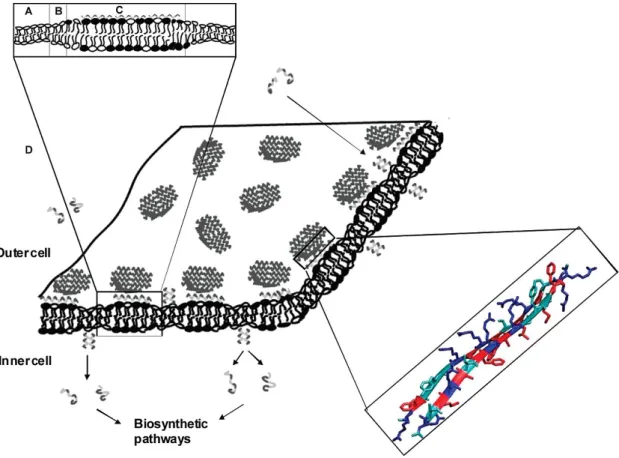
Biochemical techniques, confocal microscopy, flow cytometry, calcium imaging, surface plasmon resonance, and proteomic analysis have shown the ability of Chr and Cts to stimulate exocytosis from PMNs by provoking a transient Ca2+ influx, independent of
release from intracellular stores (Zhang et al., 2009).
For both peptides the mechanisms involve the calmodulin binding, the subsequent activation of the calcium-independent phospholipase A2 for the opening of the
store-operated channels to induce the secretion of numerous factors involved in innate immunity (Zhang et al., 2009; Aslam et al., 2012).
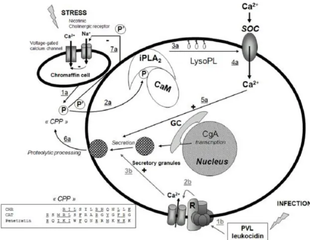
Stress and infection lead to two different pathways for stimulation of PMNs secretion, by release of CgA and CgA-derived peptides from the adrenal medulla, by PVL leucocidin stimulation and by Staphylococcus aureus infections (Aslam et al., 2012) (Figure7). The negative feedback is induced by Cts on nicotinic cholinergic receptor of chromaffin. The infective route leads to activation of the putative Panton-Valentine leucocidin receptor coupled to opening of Ca2+ channels and a rise in intracellular Ca2+ that converges on docking of secretory granules and subsequent secretion of proteins of relevance for innate immunity (Zhang et al., 2009; Aslam et al., 2012) (Figure7).
30
Figure 7:Model for the action of Chr and Cts on PMNs activation. Stress and infection lead to two different pathways for stimulation of PMN secretion, by release of CgA and CgA-derived peptides from the adrenal medulla, as indicated by 1a–6a (black), and by PVL leucocidin stimulation by Staphylococcus aureus infections, as indicated by 1b–3b (grey), respectively. Abbreviated symbols: P (Chr, Cts), P’ (Cts), CPP (Cell Penetrating Peptide), CaM (calmodulin), iPLA2 (calcium independent phospholipase A2) LysoPL (lysophospholipids); GC (Golgi complex); PVL (Panton-Valentine leucocidin), R (receptor). The stress-stimulated pathway leads to penetration of P into the cytoplasm (2a), resulting in removal of inhibitory CaM, activation of iPLA2 to produce LysoPL (3a) and activation of Ca2+ influx through SOC (4a), converging on activated docking of secretory granules (5a) and subsequent release of proteins of relevance for innate immunity (6a). The negative feedback induced by P’ on nicotinic cholinergic receptor of chromaffin cell is also indicated (7a). The infective route leads to activation of the putative PVL receptor coupled to opening of Ca2+-channels and a rise in intracellular Ca2+ (3b) that converges on docking of secretory granules and subsequent secretion of proteins of relevance for innate immunity (6a). Transcription of CgA in response to stress and infection is also indicated (from Zhang et al., 2009).
A well studied immunomodulatory activity of CgA-derived peptides is their ability to mediate monocyte chemotaxis (Shooshtarizadeh et al., 2010). It has been shown that human Cts is a potent chemotactic substance for monocytes, exhibiting its maximal effect at a concentration of 1 nM, comparable to the concentration of chemokines (Egger et al., 2008). Since the circulating concentration of Cts is around 2–3 nM (O’Connor et al.,
31 2002), the chemotactic effect of Cts is considered of physiological relevance. Additionally, lack of a chemotactic effect on neutrophils argues in favor of a specific chemotactic effect on monocytes (Egger et al., 2008).
In mouse model of experimental colitis, Cts has induced a reduction on colonic cytokine levels (Rabbi et al., 2014). Cts (1.5 mg/kg/day, 6 days, i.r.) has reduced IL-1β, IL-6 and TNF-α levels in colitic mice. This role was further studied by examining the ability of Cts to inhibit macrophages to produce pro-inflammatory cytokines by isolating macrophages from the peritoneal cavity of non-colitic or colitic mice treated or not in vivoand in vitro with the different peptides. These immune cells play an important role in the development of colitis, and recent data have demonstrated that these cells are the main producer of IL-1β, IL-6 and TNF-α (Rabbi et al., 2014). This study has shown that Cts regulates the macrophage pro-inflammatory cytokines release of monocytes and macrophages reducing inflammatory mechanism (Rabbi et al., 2014). These results can be explained by the fact the CgA or its derived peptides may have anti-inflammatory proprieties that need to be expressed not only during the acute phase but also during the remission phase to keep the inflammation under control via some anti-inflammatory mechanisms (Rabbi et al., 2014). These findings suggest that CgA or its derived peptides have an important role in the immune response to pathogens and in the development of inflammation, by regulating the infiltration of inflammatory cells and production of pro-inflammatory mediators.
32
5.1
Staphylococcus aureus and nosocomial infections
Staphylococcus aureus (S. aureus) is a dangerous gram positive bacterium which
represents one of the most important cause of hospital-acquired infections. In particular, methicillin-resistant Staphylococcus aureus (MRSA) is the major nosocomial factor that induces illness and death linked to infections (Klein et al., 2007). During the last years, the number of isolated MSRA strains is increased in intensive care units (Klevens et al., 2006) as well as in outpatients (Sieradzki e al., 1999; Johnson et al., 2007), thus producing serious economic costs for public health (Klein et al., 2007).
Nosocomial infections are often caused by biofilm colonization of medical implants (Riberio et al., 2016) and several studies in hospitals have identified S. aureus colonization of intravascular catheters as a strong predictor of subsequent S. aureus bacteremia, even in the absence of clinical signs of systemic infection (Hetem et al., 2011). In this case, bacteremia is prevented by antibiotic therapy (within 24 hours after catheter removal) (Hetem et al., 2011). Untreated bacteremia can induce complications, such as endocarditis, septic thrombosis, tunnel infection, or metastatic seeding (Mermel et al., 2001).
It is clear that, possible solutions for this health problem include greater attention in hospital surveillance and identification of reporting requirements for S. aureus and MRSA infections (Klein et al., 2007). On the other hand, the development of agents or functionalized biomaterials against S. aureus could represent an alternative strategy (Riberio et al., 2016).
33
5.2
Staphylococcus aureus and prosthetic valve endocarditis
In the specific field of cardiovascular research several scientific data suggest that the mortality rate associated with S. aureus prosthetic valve endocarditis (PVE) is high. The patients with PVE induced by S. aureus show cardiac and central nervous system (CNS) complications, and in severe cases, death within 90 days of the diagnosis (John et al., 1998). Cardiac complications, but not CNS complications, are associated with increased mortality and valve replacement surgery during antibiotic therapy is associated with decreased mortality (John et al., 1998). (John et al., 1998).
For PVE, the most important point for surgical guidelines is to understand which patients with this post-operative pathology need valve replacement surgery. Attaran and collegues (2012) in their study compared the outcome and survival between surgically and non-surgically treated patients with prosthetic valve endocarditis. This study showed that in patients haemodynamically unstable and with cardiac complications, such as heart failure or valvular dysfunction, the valve replacement surgery should be performed as soon as possible (Attaran et al., 2012), and this approach is also in accordance with the guidelines of the American College of Cardiology and American Heart association. Furthermore, in PVE cases the presence of S. aureus is considered an urgent surgical indication in the treatment of prosthetic valve endocarditis (Attaran et al., 2012). In fact, S. aureus PVE is often induced by more resistent strains to antibiotic treatments compared to the other micro-organisms that can induce PVE (Attaran et al., 2012).
Even if in hospital practice the surgery intervention is appropriately performed in the majority of patients, more recently, Chu et al., 2015 reported that one quarter of patients with surgical indications do not undergo surgery intervention but they are treated with medical therapy alone during the initial hospitalization (Chu et al., 2015). However,
34 mortality risk in S. aureus PVE remains high even with antibiotics therapy or surgical interventions (John et al., 1998; Attaran et al., 2012; Chu et al., 2015).
These same complications are also present in pacemaker patients with Gram-positive occult bacteremia. Also in these cases, S. aureus represents one of the main pathogens associated with this clinical problem (Golzio et al., 2010).
6. Antibacterial action of Cateslytin against Staphylococcus
aureus
Cateslytin (Ctl, CgA344-358) is the biologically active fragment of Cts (CgA344-364) which
displays antimicrobial activity (Briolat et al., 2005). It is a positively charged (5+), arginine-rich peptide (bCgA, RSMRLSFRARGYGFR). The biosynthesis of Ctl results by the action of prohormone convertases PC1/2 present in the granular matrix of chromaffin cells (Aslam et al., 2013). The prohormone thiol protease (PTP) was also reported to be essential for Cts production (bCgA344–364) by cleaving D-R and L-R (Lee
et al., 2003). In addition, in chromaffin secretory vesicles, the cysteine protease cathepsin L (CTSL) (Biswas et al., 2009) generates Ctl by the additional cleavage of the R-G linkage of Cts (Aslam et a., 2013).
Of note, the sequences of Cts and Ctl are highly conserved during evolution and their arginine ratio is in modulating the interaction with negatively charges of the membranes of microorganism. For hCts, bCts, and bCtl the arginine ratios are 15%, 23%, and 33% respectively. High arginine ratio of bCtl supports a strong interaction with the negatively charged lipid bilayer as compared to the both Cts and Chr (Lugardon et al. 2001; Aslam et al., 2013).
35 Several studies have analyzed the structure/function relationship of Ctl. It possesses a random structure in powder, water, or Dodecylphophatidylcholine (DPC) micelles, but adopts a helical structure in Trifuoroethanol (TFE), and is structured in β-sheets to mimic a membrane environment (Jean-François et al., 2007, 2008a). In particular, Ctl is converted into antiparallel β-sheets that aggregate mainly flat at the surface of negatively charged bacterial membranes (Jean-François et al., 2008a). This structured interaction peptides-membrane confers major rigidy and thick to membrane domains enriched in negatively charged lipids (Jean-François et al., 2008a). The rigidity and thickness bring about phase boundary defects that ultimately lead to permeability induction and peptide crossing through bacterial membranes (Jean-François et al., 2008a). In fact, after the first binding Ctl is able to form pores with 1 nm diameter and 0.25 nS conductance. The structure in pore formation is close to a simulation of α-helical peptides. Indeed, the electrostatic forces play a crucial role in the process of pore formation (Jean-François et al., 2008b). Much less interaction is detected with neutral mammalian model membranes, as reflected by only minor percentages of β-sheets or helices in the peptide secondary structure. In fact, no membrane destruction was detected for mammalian model membranes.
Even if several CgA derived peptides display antimicrobial properties (Metz-Boutigue et al., 1998), data concerning killing of S. aureus is reported only for Ctl (Aslam et al., 2013). In fact, it is known that Vs-I (Lugardon et al., 2000), Chr (Lugardon et al., 2001), and Cts (Briolat et al., 2005; Aslam et al, 2013) are active against different bacterial strains at micromolar range, but not against S. aureus.
Many bacterial strains express a variety of proteases able to degrade, in specific and non-specific manner, many proteins involved in innate immunity (Potempa and Pike, 2009).
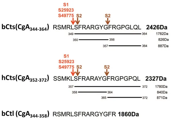
36 It has been suggested that several S. aureus proteases may degrade Cts to generate inactive peptides. For instance, several Metalloproteases (Sep A) (Lai et al., 2007) or leucine Aminopeptidase (LAP, pepZ) (Carroll et al., 2012), could cleave Cts in the sites S-F, G-F and L-S, respectively, that correspond to the proteolytic cleavage sites obtained by the incubation of Cts with different S. aureus strains (Figure 8). This proteolytic degradation could explain the observed low antibacterial activity of Cts (Aslam et al., 2013). Incubation with S. aureus supernatants has demonstrated that bovine and human Cts are similarly degraded to generate inactive fragments. For bovine and human Cts, the cleavage sites induced by the supernatants are different depending on bacterial strains.
On the contrary, Ctl resists to the degradation of S. aureus supernatant and maintains its activity against this gram-positive bacterium with a MIC value of 37–45 µg/mL (21 µM) (Aslam et al., 2013) (Figure 8).
Figure 8:The proteolytic cleavage sites induced after treatment of bCts, hCts and bCtl with different S.
aureus strains (ATCC25923, ATCC49775, S1 and S2) (from Aslam et al., 2013).
bCts(CgA
344-364)
bCtl (CgA
344-358)
37 Another property that may explain the best action of Ctl against S. aureus concerns its secondary structure.
Both hCts and bCts adopt helical structures (residue 7 to residue 11) in the presence of high concentrations of DPC (Sugawara et al., 2010). In a similar way, Ctl adopts a β-sheet structure only on negatively charged membranes, whereas it is essentially unstructured in water (Jean-François et al., 2008a). These β-sheet formations give more stability to peptide respect to the helical formation (Jean-François et al., 2008a) as previously reported for Cts (Sugawara et al., 2010). In addition, in Ctl structure, the arginine and hydrophobic residues are in close proximity, thus creating a deep penetration of charged residues into the membrane (Ziegler et a., 2008). In fact, as previously described, the electrostatic interaction between positively charged arginine residues and negatively charged lipids appears to be responsible for binding of Ctl to the lipid bilayer, and at the same time, the aromatic residues stabilize the lipid-peptide interaction (Jean-François et al., 2008a, b; Aslam et al., 2013).
Furthermore, it is known that Ctl has antimicrobial activity against Micrococcus luteus, few yeasts and fungal strains, and it is not hemolytic for human erythrocytes (Briolat et al., 2005). Recently, a mechanism by which the peptides favor destabilization of bacterial membrane, allowing the antibiotics to rapidly penetrate inside bacterial cells to reaching their site of action, has been postulated (Aslam et al., 2013). These Authors have demonstrated that Ctl shows synergistic effects with antibiotics, such as Minocyclin or Voriconazole, and this co-treatment induces a reduction of the antibiotics concentration used and can potentiate their activities.
38
7. Aims of this thesis
Antimicrobial peptides are now viewed as multifunctional factors. Indeed, in addition to their antimicrobial activity, they also mediate inflammatory and immune responses. Furthermore, several antimicrobial peptides have been shown to exert either detrimental or beneficial effects on the cardiovascular system (Li, 2009). For example, PR-39 and LL-37, both belonging to Cathelicidin family, showed opposite effects: the first one inducing cardioprotective effects against I/R damage (Ikeda et al., 2001) while, LL-37 promoting atherogenesis and cardiovascular disease (Edfeldt et al., 2006). Among CgA-derived peptides, Vs-I and Cts, also show both antimicrobial activity and benefical cardiovascular effects (Helle et al., 2007; Angelone et al., 2012a).
Thus, on the basis of these previous data, a link between immune-derived CgA peptides and cardiovascular pathophysiology may be hypothesized.
The aim of this thesis was to evaluate the possible contribution of antimicrobial peptides Chr (Vs-I-derived fragment) and Ctl (Cts-derived fragment) in the cardiovascular pathophysiology and immune response.
The first part of the study was focused to evaluate the action of Chr in maintaining basal cardiac function and its putative role in cardioprotection. Furthermore, Tota and co-workers (2003) have shown the cardiac effects of Chr in an avascular model of the vertebrate myocardium. This peptide induced a depression of myocardial contractility by eliciting a direct negative inotropic effect isolated frog heart (Rana esculenta). The antimicrobial activity of Chr against fungi and yeast (Lugardon et al., 2001) is well recognised. In this thesis, for the first time, it has been evaluated: