Cell Cycle 10:15, 2568-2573; August 1, 2011; © 2011 Landes Bioscience
RepoRt
*Correspondence to: Francesca Bosetti; Email: [email protected] Submitted: 04/04/11; Revised: 05/26/11; Accepted: 05/31/11 DOI: 10.4161/cc.10.15.15946
Introduction
Adult neurogenesis occurs in the dentate gyrus (DG) of the hippocampus1,2 and in the subventricular zone (SVZ) of the
lateral ventricle.3 Hippocampal neurogenesis plays a role in
the maintenance of normal hippocampal function, including learning and memory.4,5 Hippocampal stem cells (HSCs) lie
in the subgranular zone (SGZ), along the border between the hilus and the granule cell layer (GCL).6 Many of the HSCs die
shortly after their birth,7 whereas the surviving cells migrate
into the dentate GCL, differentiate into granule cells8 and
project their axons to the CA3 region resulting in their mor-phological and functional integration into the existing hip-pocampal circuitry.9 The formation of new granule cells in
the DG can be affected by a large number of physiological and pathological stimuli, such as aging and neurodegenerative diseases.10-13
Neuroinflammation is thought to be involved in the patho-physiology of several neurodegenerative diseases, including Alzheimer disease.14 Inflammation can affect the
microenviron-ment of the neurogenic niche15 and influence proliferation of
progenitor cells, survival and migration of new neuroblasts, dif-ferentiation of new neuroblasts to specific neuronal phenotypes and development of functional synaptic connectivity.16,17
Growing evidence indicates that neuroinflammation can alter adult neurogenesis by mechanisms as yet unclear. We have previously demonstrated that the neuroinflammatory response and neuronal damage after lipopolysaccharide (LpS) injection is reduced in cyclooxygenase-1 deficient (CoX-1-/-) mice. In this study, we investigated the role of
CoX-1 on hippocampal neurogenesis during LpS-induced neuroinflammation, using CoX-CoX-1-/- and wild-type (Wt) mice.
We found that LpS-induced neuroinflammation resulted in the decrease of proliferation, survival and differentiation of hippocampal progenitor cells in Wt but not in CoX-1-/- mice. thus, we demonstrate for the first time that CoX-1 is
involved in the inhibition of BrdU progenitor cells in proliferation and hippocampal neurogenesis after LpS. these results suggest that CoX-1 may represent a viable therapeutic target to reduce neuroinflammation and promote neurogenesis in neurodegenerative diseases with a strong inflammatory component.
Cyclooxygenase-1 is involved in the
inhibition of hippocampal neurogenesis after
lipopolysaccharide-induced neuroinflammation
Isabella Russo,1,2 panomwat Amornphimoltham,3 Roberto Weigert,3 Sergio Barlati2 and Francesca Bosetti1,†,*
1Molecular Neuroscience Unit; Brain physiology and Metabolism Section; National Institute on Aging; National Institutes of Health; Bethesda, MD USA; 2Division of Biology and Genetics; Department of Biomedical Sciences and Biotechnologies and National Institute of Neuroscience; University of Brescia; Brescia, Italy;
3Intracellular Membrane trafficking Unit; oral and pharyngeal Cancer Branch; National Institute of Dental and Craniofacial Research; National Institutes of Health;
Bethesda, MD USA
†Current address: National Institute of Neurological Disorders and Stroke; National Institutes of Health; Bethesda, MD USA
Key words: neurogenesis, cyclooxygenase-1, lipopolysaccharide, inflammation, brain
Neuroinflammation is mediated by microglial activation and subsequent release of pro-inflammatory mediators such as prosta-glandins, cytokines, chemokines and reactive oxygen or nitrogen species.18 Prostaglandin H synthases or cyclooxygenases (COX)-1
or -2 catalyze the conversion of arachidonic acid to prostaglan-dins.18 Because of its localization in microglia, and thus, being
able to immediately synthesize prostaglandins in response to microglial activation, COX-1 recently emerged as a major player in the acute inflammatory response.19-21 A recent study also
sug-gested a role for COX-1 expressed in brain perivascular cells in mediating the immune-to-brain signaling in response to a sys-temic lipopolysaccharide (LPS) challenge.22
In this study, we used mice with a null mutation of the COX-1 gene to investigate the role of COX-1 on hippocampal neuro-genesis during acute neuroinflammation induced by intracere-broventricular injection of LPS.18 LPS binds to a CD14 receptor
and together with the extracellular adaptor protein MD-2 binds to the toll-like receptor 4 (TLR4) expressed by microglia and astrocytes,23 causing a massive glial activation accompanied by a
robust and transient transcriptional activation of genes encoding for pro-inflammatory mediators.18
We have previously shown that the genetic deletion or phar-macological inhibition of COX-1 significantly attenuated glial activation, release of pro-inflammatory markers, as well as
RepoRt RepoRt
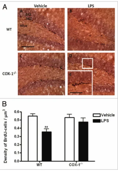
cells proliferation in the DG, BrdU-labeled cells were counted 24 h after LPS injection. The distribution of the BrdU-labeled pro-liferative cells was found mainly in clusters at the border between the granule cell layer and the hilus, regardless of treatment or gen-otype (Fig. 1A). Within the area encompassing the granule cell layer and subgranular zone (Fig. 1A), LPS treatment decreased by 35% the mean density of BrdU-positive cells in WT mice (0.36 ± 0.16 vs. 0.55 ± 0.027 in vehicle-treated WT mice, p < 0.01, Fig. 1B). In contrast, the number of BrdU-labeled proliferative cells was not significantly different in LPS-injected COX-1-/-
com-pared with vehicle-injected COX-1-/- mice (0.47 ± 0.05 vs. 0.53
± 0.04) (Fig. 1B).
LPS injection reduces hippocampal neurogenesis in WT but not in COX-1-/- mice. To determine the fate of new hippocampal
cells, we examined the phenotype of newly BrdU-positive cells generated 4 weeks after LPS injection by concomitant immu-nolabeling of BrdU with neuronal (NeuN) and glial (S100β) markers (Fig. 2A). S100β, a marker for astrocytes and oligo-dendrocytes, labels cell bodies with a better definition than GFAP. The percentage of BrdU-positive cells that co-localized with NeuN immunolabeling was significantly reduced by 35% in LPS-injected compared with vehicle-injected WT mice (p < 0.001). In contrast, the number of neurons did not differ significantly in LPS-injected COX-1-/- mice as compared with
vehicle-injected COX-1-/- mice (97.3% ± 0.6% vs. 100% ± 0%; Fig. 2B). Furthermore, LPS-injected WT mice showed a 35% increase (p < 0.001) in the number of BrdU-positive cells that differentiated into glial cells compared with vehicle-injected mice (p < 0.001; Fig. 2C). Only a small percentage of BrdU-positive cells differentiated into glia in LPS-injected COX-1-/- mice
com-pared with their vehicle-injected controls (2.7% ± 0.7% vs. 0%, p < 0.01; Fig. 2C).
COX-1 is involved in the LPS-induced survival effect of new hippocampal cells. To test whether COX-1 is involved in the post-mitotic BrdU-cell survival, we analyzed the density of BrdU-cells in the granule cell layer and subgranular zone of DG 4 weeks after LPS injection in COX-1-/- and WT mice. LPS
significantly increased the density of BrdU-survival cells com-pared with vehicle-injection in WT mice (0.29 ± 0.01 vs. 0.22 ± 0.006, 24%, p < 0.001), but did not affect survival of new cells in COX-1-/- mice compared with their respective vehicle-injected
controls (0.24 ± 0.005 vs. 0.22 ± 0.003) (Fig. 3).
When we compared the survival BrdU-cells at 4 weeks with the BrdU-cells present at 24 h in the DG, after LPS or vehicle injection in WT and COX-1-/- mice, the density of surviving
BrdU-cells was drastically decreased. In particularly, the surviving BrdU-cells in vehicle-injected WT and COX-1-/- mice was
drasti-cally reduced by 60% after 4 weeks (p < 0.001). LPS-injected COX-1-/- mice showed a minor reduction of surviving BrdU-cells
after 4 weeks (49%), whereas the number of BrdU-cells was not significantly affected in LPS-injected WT mice (Fig. 4).
Discussion
In this study we examined the role of COX-1 in the proliferation, survival and differentiation of new hippocampal progenitor cells blood-brain barrier disruption and recruitment of peripheral
leu-kocytes to the inflamed brain in response to LPS.18,20,24 Since LPS
has been shown to decrease hippocampal neurogenesis,12,25,26 in
this study we investigated whether COX-1 gene deletion posi-tively modulates hippocampal neurogenesis during LPS-induced neuroinflammation. We examined the proliferation, survival and differentiation of neuronal progenitor cells in the DG after LPS injection and demonstrated for the first time that COX-1 gene deletion resulted in an increased proliferation and differentiation of hippocampal progenitor cells in the adult mouse brain during neuroinflammation.
Results
LPS injection reduces proliferation of hippocampal progenitor cells in WT but not in COX-1-/- mice. To evaluate progenitor
Figure 1. (A) Hippocampal BrdU proliferative cells after LpS-induced
neuroinflammation in CoX-1-/- and Wt mice. Representative photomi-crographs of BrdU immunohistochemistry in the DG for Wt (A and B) and CoX-1-/- mice (C and D) euthanized 24 h after i.c.v. injection of LpS or
vehicle. High-magnification images of BrdU immunostaining is shown in (e). Scale bars = 100 μm (A–D); 50 μm (e). (B) Hippocampal BrdU-proliferative cells after LpS-induced neuroinflammation in CoX-1-/- and
Wt mice. Quantification of BrdU-labeled cells per μm3 of DG, in Wt and
CoX-1-/- mice euthanized 24 h after i.c.v. injection of LpS or vehicle. Data
Our results show that inhibition of the proliferation and dif-ferentiation of progenitor cells after LPS injection can be caused by the inflammatory environment, and particularly, through an indirect effect on hippocampal progenitor cells. Indeed, neuronal progenitor cells do not express COX-1 (data not shown) either under physiological conditions or after LPS. Thus, the positive modulation of neurogenesis by COX-1 deletion could be an indi-rect effect attributable to the attenuation of the neuroinflamma-tory response.18 In this regard, the transcription factors STAT3
and NFκB, which are upregulated after LPS-induced inflam-mation,18,31 induce the transcription of many pro-inflammatory
after LPS-induced neuroinflammation. We found that LPS-induced neuroinflammation altered the proliferation of progenitor cells, the production rate of new neurons, and the survival of new hippocampal progenitor cells in WT but not in COX-1-/- mice.
Previous data from our lab showed that COX-1 plays a critical role in the neuroinflammatory response to LPS18 or β-amyloid injection.27 Specifically, COX-1
gene deletion or treatment with the COX-1 specific inhibitor SC-560 resulted in decreased glial activa-tion, reduced expression of pro-inflammatory cyto-kines and chemocyto-kines, and limited neuronal damage after central injection of LPS.18 Taken together these
data suggest that during neuroinflammation, COX-1 plays a critical role in the propagation of the inflam-matory response and in the modulation of the regen-erative potency of hippocampal progenitor cells.
COX-1 is predominantly expressed by microglia and perivascular cells, and thus, can be activated, within seconds to minutes, following an acute chal-lenge, and produce prostaglandins that contribute to the inflammatory response.28,29 Activation of
COX-1-expressing microglia and subsequent release of pro-inflammatory mediators such as IL-1β, IL-6,
TNFα, NO and prostanoids, which have antineurogenic effects, could be responsible for the adverse effects of inflammation on neurogenesis.12,30
TLR4 is abundantly expressed by neural stem/progenitor cells and the absence of TLR4 results in enhanced proliferation and neuronal differentiation,31 suggesting that TLR4 directly
modu-lates self-renewal and the cell-fate decision of neuronal progenitor cells.31 Rolls et al. (2007) reported that LPS treatment decreases
the proliferation and differentiation of cultured neural stem/ progenitor cells via a nuclear factor kappaB (NFκB) signaling TLR-4 dependent.31
Figure 2. (A) Hippocampal BrdU-cells differentiation
after LpS-induced neuroinflammation in CoX-1-/- and Wt
mice. Confocal images of DG for Wt (A and B) and CoX-1
-/-mice (C and D) euthanized 4 weeks after i.c.v. injection of LpS or vehicle. Sections were triple-labeled for BrdU (red), NeuN (green) and S100β (blue). Scale bars = 20 μm. (B) Differentiation of new hippocampal cells 4 weeks after LpS-induced neuroinflammation in CoX-1-/- and Wt mice.
Quantification of percentage of BrdU-cells differentiated into neurons in Wt and CoX-1-/- mice after 4 weeks of i.c.v.
injection of LpS or vehicle. Data are means ± SeM (n = 4), ***p < 0.001, LpS-injected Wt mice vs. vehicle-injected Wt mice; ###p < 0.001, LpS-injected CoX-1-/- mice vs.
LpS-injected Wt mice. (C) Differentiation of new hippocampal cells 4 weeks after LpS-induced neuroinflammation in CoX-1-/- and Wt mice. Quantification of percentage of
BrdU-cells differentiated into glia in Wt and CoX-1-/- mice
4 weeks after i.c.v. injection of LpS or vehicle. Data are means ± SeM (n = 4), ***p < 0.001, LpS-injected Wt mice vs. vehicle-injected Wt mice; **p < 0.01, LpS-injected CoX-1-/- mice vs. vehicle-injected CoX-1-/- mice; ###p < 0.001, LpS-injected Wt mice vs. LpS-injected CoX-1
survival effect in the new hippocampal progenitor cells to replace damaged neurons in the inflamed brain, although many new progenitor cells will differentiate into glial cells.
When we compare the survival of BrdU positive cells 24 h and 4 weeks after LPS injection, COX-1-/- mice showed a strong
decrease in surviving cells that was not observed in WT mice (49% vs. 17%, respectively). This difference could be linked to the reduction of inflammatory response and neuronal damage in COX-1-/- mice after LPS, which would make unnecessary for
new hippocampal cells to survive as in vehicle-injected WT and COX-1-/- mice.
New hippocampal neurons that survive chronic inflammatory stress differentiate and integrate as granule neurons, although with a heightened degree of synaptic plasticity.41,42 Recent work
showed that new neurons exposed to the chronic pathological environment are severely controlled,41,43,44 and can exhibit subtle
changes in development of dendritic arborizations and spine den-sity,45 which are dependent on the characteristics of the
patho-logical environment.45 Apical dendrites reached the molecular
layer during the first 2 weeks after their birth, and the dendritic arborizations and the development of spines occurs during the first 3–4 weeks.41 Since pilot studies in our lab showed that
LPS-induced neuroinflammation peaks at 24 h and then resolves in few days (Choi et al. 2008), one could speculate that new hippo-campal cells develop normally and integrate into the hippocam-pal network, without undergoing morphological or functional changes. However, only electrophysiological studies can confirm a functional integration of new hippocampal cells into the hip-pocampal functional circuitry.
Neuroinflammation is a complex mechanism, in which dif-ferent pathways, intracellular and extracellular, can interact and both directly and indirectly modulate the neurogenic niche. In addition to a TLR4-dependent mechanism, we suggest that COX-1 plays a role in the inhibition of hippocampal neurogene-sis after LPS injection.32,34,46 COX-1-derived prostaglandins, such
as PGE2, could directly affect the neurogenic niche, although it genes18 and play a critical role in controlling the proliferation of
neuronal stem cells32 and neuronal differentiation.18 Supporting
a role for these transcription factors, our group has previously demonstrated that, in response to LPS, COX-1-/- mice or WT
treated with SC-560 have decreased activation and translocation of NFκB and STAT-3, and reduced levels of the pro-inflamma-tory cytokines IL-1β, IL-6 and TNFα.18
The differentiation of neuronal progenitor cells toward neuronal or glial lineage is determined by specific signaling cascades regulating the activation of different transcription fac-tors. Multiple processes directing cell fate determination can be modulated by different cytokines and transcription factors. IL-6, TNFα and IL-132,33 promote astrocytic differentiation by
activat-ing the transcription factors STAT3 and NFκB.34,35 Furthermore,
activation of cytokine receptors on neuronal progenitor cells stimulates the Notch1 pathway signaling, which inhibits neu-ronal differentiation through a hairy-enhancer-of split 1 (Hes-1)-mediated mechanism.15,36 Notch signaling increases the
expression of Hes-1,36 which antagonizes pro-neural basic
helix-loop-helix transcription factors and represses the commitment of neuronal progenitor cells to a neuronal fate.37 The cross-talk
of all these transcription factors and their co-activators could modulate astrocytic fate specification in hippocampal progenitor cells after LPS-induced neuroinflammation. Furthermore, LPS injection downregulates the expression of NeuroD2, a marker of neuronal progenitor cells, which is thought to play a role in the determination and maintenance of the neuronal cells fate.38
Functional stimuli, including learning, memory and environ-mental enrichment can increase the number of surviving BrdU-cells by a “survival promoting effect.”39,40 In our study, we showed
that neuroinflammation also has a survival promoting effect on new hippocampal progenitor cells. After 4 weeks of LPS injec-tion, WT mice showed a 24% increase in surviving BrdU-cells compared with vehicle-injected WT mice. This LPS-induced survival promoting effect was not observed in COX-1-/- mice.
This could be due to the reduced neuroinflammatory response observed in COX-1-/- mice, as neuroinflammation could trigger a
Figure 3. Survival of new hippocampal cells 4 weeks after LpS-induced
neuroinflammation in CoX-1-/- and Wt mice. Quantification of
BrdU-labeled cells per μm3 of DG in Wt and CoX-1-/- mice 4 weeks after i.c.v.
injection of LpS or vehicle. Data are means ± SeM (n = 4), ***p < 0.001, LpS-injected Wt mice vs. vehicle-injected Wt mice; ##p < 0.01,
LpS-injected CoX-1-/- vs. LpS-injected Wt mice.
Figure 4. Comparison between survival of BrdU-labeled cells at 24 h
and 4 weeks after LpS injection in Wt and CoX-1-/- mice. the surviving
BrdU-cells in LpS-injected CoX-1-/- mice was severely reduced by 49%
after 4 weeks, whereas the number of BrdU-cells was not significantly affected in LpS-injected Wt mice. Data are means ± SeM (n = 5, 24 h after LpS; n = 4, 4 weeks after LpS); **p < 0.01, LpS-injected CoX-1-/- vs.
triple labeling for BrdU were performed on free-floating 40 μm sagittal sections that were pretreated by denaturing DNA, as described previously in reference 48. The antibodies were mouse BrdU-antibody (1:100, DAKO, Denmark), rat anti-BrdU for triple labeling (1:200; Accurate Chemical and scientific, Westbury NY), rabbit anti-S-100β (1:200; Abcam, Cambridge, MA), mouse anti-NeuN (1:1,500; Chemicon, Billerica, MA). For immunohistochemistry the peroxidase method (ABC sys-tem) with biotinylated donkey anti-mouse IgG antibodies and diaminobenzidine as chromogen was used (Vector laborato-ries). Immunohistochemistry images were detected with a light microscope Olympus CKX41, using X40 and X60 objective (Olympus). The fluorescent antibodies 488-Alexa Flour anti-mouse IgG (1:500), 594-Alexa Fluor anti-rat IgG (1:500) and 405-Alexa Fluor anti-rabbit IgG (1:500) (Invitrogen, Carlsbad, CA) were used. Fluorescent signals were detected and processed using an inverted confocal microscope (model IX81, Olympus America Inc., Center Valley, PA) with Fluoview 1000 scanning head. Fluorescent images were acquired using a UPlanSApo X10 numerical aperture (NA) 0.4 dry objective and a UPlanSApo X60 NA 1.42 oil immersion objective (Olympus America Inc.), and were processed using Imaris 5.7 (Bitplane) and assembled using Adobe Photoshop CS.
Cell counting and volumetric analysis. BrdU-labeled cells were counted in the subgranular zone, which was defined as a 2-nucleus-wide band below the apparent border between the GCL and the hilus.8 We detected one every six sections and
cov-ered the entire area of dentate gyrus. To measure the DG area, slices were stained with hematoxylin (Vector, Burlingome, CA) and the area was traced using ImageJ Software. The mean den-sity of BrdU-labeled cells in each mouse was calculated as the total number of labeled nuclei divided by the volume of DG (cell density per μm3).49 To determine the percentage of neuronal
dif-ferentiation of newborn cells, one every six sections was analyzed for the entire area of dentate gyrus. For each animal 50 BrdU-positive cells were randomly selected and analyzed.49
Statistical analysis. All data are expressed as means ± SEM. Statistical significance was assessed with a two-way ANOVA followed by Bonferroni’s post hoc test using GraphPad Prism. Significance was taken at p < 0.05.
Acknowledgments
This research was supported by the Intramural Research Program of the NIH, NIA. We would like to thank Henriette Van Praag for useful discussion and technical advice, Yosuke Mukoyama for kindly providing microscopy and Erik Runko for critically read-ing and editread-ing this manuscript.
remains to be fully elucidated which type(s) of prostaglandins and downstream receptors play a role in the modulation of neuro-genesis.42 A recent study by Keen et al. (2009) tested the
hypoth-esis that activation of specific PGE2 receptors is responsible for suppression of hippocampal neurogenesis after LPS-induced neuroinflammation. They demonstrated that EP1, but not EP2, is expressed by progenitor cells. EP1 receptor gene deletion pro-tected hippocampal progenitor cells from PGE2-mediated toxic-ity directly and also indirectly by partially suppressing microglia activation and the propagation of inflammatory response. In con-trast, EP2 was not detectably expressed by progenitor cells, and its ablation did not protect from direct toxic effect of PGE2.
Overall, our past data suggest that the inhibition of hippo-campal neurogenesis by LPS in WT, but not COX-1-/- mice,
could be linked to the increase in NFκB activation, expression of pro-inflammatory cytokines, propagation of neuroinflamma-tion and PGE2 levels, all of which are attenuated by COX-1 gene deletion.18 We demonstrate for the first time that in LPS-induced
neuroinflammation, COX-1 activity is detrimental to the gen-eration of new hippocampal progenitor cells. Thus, COX-1 inhibition may represent a viable therapeutic target to promote neurogenesis in neurodegenerative diseases with a strong inflam-matory component, where neuronal loss and memory deficits are known to occur.
Materials and Methods
Animals and procedures. All animal procedures were performed under an animal study proposal approved by the National Institutes of Health (NIH) Animal Care and Use Committee, in accordance with NIH guidelines on the care and use of labo-ratory animals. Three-month-old male WT and homozygous (COX-1-/-) mice on a C57BL/6-129/Ola genetic background
were used.47 Vehicle (sterile saline, 5 μl) and LPS (Escherichia coli
serotype 0127:B8; 5 μg in 5 μl of sterile saline) were adminis-tered by intracerebroventricular (i.c.v.) injection into the lateral ventricle as previously described.18
5-bromo-2'-deoxyuridine (BrdU) was used to label the neu-ronal progenitor cell in proliferation. Mice were given 2 BrdU injections with an interval of 12 h, the first 30 min prior to LPS injection (dissolved in 0.9% NaCl; 50 mg/Kg; 10 mg/ml, i.p.; Sigma Aldrich, St. Louis, MO). To detect the proliferation of hippocampal progenitor cells, mice were euthanized 24 h after LPS injection. To detect the phenotype of new born progenitor cells, mice were euthanized 4 weeks after LPS injection.
DNA denaturation, immunohistochemistry and immuno-flourescence. Immunohistochemistry and immunofluorescent
References
1. Laplagne DA, Esposito MS, Piatti VC, Morgenstern NA, Zhao C, van Praag H, et al. Functional conver-gence of neurons generated in the developing and adult hippocampus. PLoS Biol 2006; 4:409; PMID: 17121455; DOI: 10.1371/journal.pbio.0040409. 2. Toni N, Teng EM, Bushong EA, Aimone JB, Zhao C,
Consiglio A, et al. Synapse formation on neurons born in the adult hippocampus. Nat Neurosci 2007; 10:727-34; PMID: 17486101; DOI: 10.1038/nn1908.
3. Doetsch F, Scharff C. Challenges for brain repair: insights from adult neurogenesis in birds and mam-mals. Brain Behav Evol 2001; 58:306-22; PMID: 11978948; DOI: 10.1159/000057572.
4. Jessberger S, Clark RE, Broadbent NJ, Clemenson GD Jr, Consiglio A, Lie DC, et al. Dentate gyrus-specific knockdown of adult neurogenesis impairs spatial and object recognition memory in adult rats. Learn Mem 2009; 16:147-54; PMID: 19181621; DOI: 10.1101/ lm.1172609.
5. Zhang CL, Zou Y, He W, Gage FH, Evans RM. A role for adult TLX-positive neural stem cells in learn-ing and behaviour. Nature 2008; 451:1004-7; PMID: 18235445; DOI: 10.1038/nature06562.
6. Cameron HA, McKay RD. Adult neurogenesis produc-es a large pool of new granule cells in the dentate gyrus. J Comp Neurol 2001; 435:406-17; PMID: 11406822; DOI: 10.1002/cne.1040.
7. Biebl M, Cooper CM, Winkler J, Kuhn HG. Analysis of neurogenesis and programmed cell death reveals a self-renewing capacity in the adult rat brain. Neurosci Lett 2000; 291:17-20; PMID: 10962143; DOI: 10.1016/S0304-3940(00)01368-9.
8. Kempermann G, Gast D, Kronenberg G, Yamaguchi M, Gage FH. Early determination and long-term persistence of adult-generated new neurons in the hippocampus of mice. Development 2003; 130:391-9; PMID: 12466205; DOI: 10.1242/dev.00203. 9. van Praag H, Schinder AF, Christie BR, Toni N,
Palmer TD, Gage FH. Functional neurogenesis in the adult hippocampus. Nature 2002; 415:1030-4; PMID: 11875571; DOI: 10.1038/4151030a.
10. Limke TL, Rao MS. Neural stem cells in aging and disease. J Cell Mol Med 2002; 6:475-96; PMID: 12611637; DOI: 10.1111/j.1582-4934.2002. tb00451.x.
11. Kuzumaki N, Ikegami D, Imai S, Narita M, Tamura R, Yajima M, et al. Enhanced IL-1beta production in response to the activation of hippocampal glial cells impairs neurogenesis in aged mice. Synapse 2010; 64:721-8; PMID: 20336624.
12. Russo I, Barlati S, Bosetti F. Effects of Neuroinflammation on the Regenerative Capacity of Brain Stem Cells. J Neurochem 2010; 116:947-56. 13. Martino G, Pluchino S. The therapeutic potential of
neural stem cells. Nat Rev Neurosci 2006; 7:395-406; PMID: 16760919; DOI: 10.1038/nrn1908. 14. Skaper SD. The brain as a target for inflammatory
pro-cesses and neuroprotective strategies. Ann NY Acad Sci 2007; 1122:23-34; PMID: 18077562; DOI: 10.1196/ annals.1403.002.
15. Keohane A, Ryan S, Maloney E, Sullivan AM, Nolan YM. Tumour necrosis factor alpha impairs neuronal differentiation but not proliferation of hippocampal neural precursor cells: Role of Hes1. Mol Cell Neurosci 2010; 43:127-35; PMID: 19840854; DOI: 10.1016/j. mcn.2009.10.003.
16. Ekdahl CT, Kokaia Z, Lindvall O. Brain inflammation and adult neurogenesis: the dual role of microglia. Neuroscience 2009; 158:1021-9; PMID: 18662748; DOI: 10.1016/j.neuroscience.2008.06.052.
17. Pluchino S, Muzio L, Imitola J, Deleidi M, Alfaro-Cervello C, Salani G, et al. Persistent inflammation alters the function of the endogenous brain stem cell compartment. Brain 2008; 131:2564-78; PMID: 18757884; DOI: 10.1093/brain/awn198.
18. Choi SH, Langenbach R, Bosetti F. Genetic deletion or pharmacological inhibition of cyclooxygenase-1 attenu-ate lipopolysaccharide-induced inflammatory response and brain injury. FASEB J 2008; 22:1491-501; PMID: 18162486; DOI: 10.1096/fj.07-9411com.
19. Choi SH, Aid S, Bosetti F. The distinct roles of cyclo-oxygenase-1 and -2 in neuroinflammation: implica-tions for translational research. Trends Pharmacol Sci 2009; 30:174-81; PMID: 19269697; DOI: 10.1016/j. tips.2009.01.002.
20. Aid S, Bosetti F. Targeting cyclooxygenases-1 and -2 in neuroinflammation: Therapeutic implications. Biochimie 2010; 93:46-5.
21. Bosetti F, Choi SH. Rethinking the role of cyclooxy-genase-1 in neuroinflammation: more than homeosta-sis. Cell Cycle 2010; 9:2919-20; PMID: 20714215; DOI: 10.4161/cc.9.15.12715.
22. García-Bueno B, Serrats J, Sawchenko PE. Cerebrovascular cyclooxygenase-1 expression, regula-tion and role in hypothalamic-pituitary-adrenal axis activation by inflammatory stimuli. J Neurosci 2009; 29:12970-81; PMID: 19828811; DOI: 10.1523/ JNEUROSCI.2373-09.2009.
23. Beutler B. Inferences, questions and possibilities in Toll-like receptor signalling. Nature 2004; 430:257-63; PMID: 15241424; DOI: 10.1038/nature02761. 24. Choi SH, Aid S, Choi U, Bosetti F. Cyclooxygenases-1
and -2 differentially modulate leukocyte recruit-ment into the inflamed brain. Pharmacogenomics J 2010; 10:448-57; PMID: 20038958; DOI: 10.1038/ tpj.2009.68.
25. Ekdahl CT, Claasen JH, Bonde S, Kokaia Z, Lindvall O. Inflammation is detrimental for neurogenesis in adult brain. Proc Natl Acad Sci USA 2003; 100:13632-7; PMID: 14581618; DOI: 10.1073/pnas.2234031100. 26. Monje ML, Toda H, Palmer TD. Inflammatory
block-ade restores adult hippocampal neurogenesis. Science 2003; 302:1760-5; PMID: 14615545; DOI: 10.1126/ science.1088417.
27. Choi SH, Bosetti F. Cyclooxygenase-1 null mice show reduced neuroinflammation in response to beta-amyloid. Aging (Albany NY) 2009; 1:234-44; PMID: 20157512.
28. Milatovic D, Zaja-Milatovic S, Montine KS, Shie FS, Montine TJ. Neuronal oxidative damage and dendritic degeneration following activation of CD14-dependent innate immune response in vivo. J Neuroinflammation 2004; 1:20; PMID: 15498098; DOI: 10.1186/1742-2094-1-20.
29. Aïd S, Bosetti F. Targeting cyclooxygenases-1 and -2 in neuroinflammation: Therapeutic implications. Biochimie 2011; 93:46-51; PMID: 20868723; DOI: 10.1016/j.biochi.2010.09.009.
30. Das S, Basu A. Inflammation: a new candidate in modulating adult neurogenesis. J Neurosci Res 2008; 86:1199-208; PMID: 18058947; DOI: 10.1002/ jnr.21585.
31. Rolls A, Shechter R, London A, Ziv Y, Ronen A, Levy R, et al. Toll-like receptors modulate adult hip-pocampal neurogenesis. Nat Cell Biol 2007; 9:1081-8; PMID: 17704767; DOI: 10.1038/ncb1629. 32. Widera D, Mikenberg I, Elvers M, Kaltschmidt C,
Kaltschmidt B. Tumor necrosis factor alpha trig-gers proliferation of adult neural stem cells via IKK/ NFkappaB signaling. BMC Neurosci 2006; 7:64; PMID: 16987412; DOI: 10.1186/1471-2202-7-64. 33. Bourke E, Kennedy EJ, Moynagh PN. Loss of Ikappa
B-beta is associated with prolonged NFkappaB activity in human glial cells. J Biol Chem 2000; 275:39996-40002; PMID: 10998424; DOI: 10.1074/jbc. M007693200.
34. Mondal D, Pradhan L, LaRussa VF. Signal transduc-tion pathways involved in the lineage-differentiatransduc-tion of NSCs: can the knowledge gained from blood be used in the brain? Cancer Invest 2004; 22:925-43; PMID: 15641490; DOI: 10.1081/CNV-200039679. 35. Cimini A, Ceru MP. Emerging roles of peroxisome
pro-liferator-activated receptors (PPARs) in the regulation of neural stem cells proliferation and differentiation. Stem Cell Rev 2008; 4:293-303; PMID: 18561036; DOI: 10.1007/s12015-008-9024-2.
36. Chojnacki A, Shimazaki T, Gregg C, Weinmaster G, Weiss S. Glycoprotein 130 signaling regulates Notch1 expression and activation in the self-renewal of mam-malian forebrain neural stem cells. J Neurosci 2003; 23:1730-41; PMID: 12629177.
37. Nakamura Y, Sakakibara S, Miyata T, Ogawa M, Shimazaki T, Weiss S, et al. The bHLH gene hes1 as a repressor of the neuronal commitment of CNS stem cells. J Neurosci 2000; 20:283-93; PMID: 10627606.
38. Bonow RH, Aid S, Zhang Y, Becker KG, Bosetti F. The brain expression of genes involved in inflamma-tory response, the ribosome and learning and memory is altered by centrally injected lipopolysaccharide in mice. Pharmacogenomics J 2009; 9:116-26; PMID: 18957951; DOI: 10.1038/tpj.2008.15.
39. Kempermann G, Kuhn HG, Gage FH. More hip-pocampal neurons in adult mice living in an enriched environment. Nature 1997; 386:493-5; PMID: 9087407; DOI: 10.1038/386493a0.
40. Leuner B, Mendolia-Loffredo S, Kozorovitskiy Y, Samburg D, Gould E, Shors TJ. Learning enhances the survival of new neurons beyond the time when the hippocampus is required for memory. J Neurosci 2004; 24:7477-81; PMID: 15329394; DOI: 10.1523/ JNEUROSCI.0204-04.2004.
41. Jakubs K, Bonde S, Iosif RE, Ekdahl CT, Kokaia Z, Kokaia M, et al. Inflammation regulates func-tional integration of neurons born in adult brain. J Neurosci 2008; 28:12477-88; PMID: 19020040; DOI: 10.1523/JNEUROSCI.3240-08.2008. 42. Keene CD, Chang R, Stephen C, Nivison M, Nutt
SE, Look A, et al. Protection of hippocampal neu-rogenesis from toll-like receptor 4-dependent innate immune activation by ablation of prostaglandin E2 receptor subtype EP1 or EP2. Am J Pathol 2009; 174:2300-9; PMID: 19389932; DOI: 10.2353/ ajpath.2009.081153.
43. Zhao C, Teng EM, Summers RG Jr, Ming GL, Gage FH. Distinct morphological stages of dentate granule neuron maturation in the adult mouse hippocampus. J Neurosci 2006; 26:3-11; PMID: 16399667; DOI: 10.1523/JNEUROSCI.3648-05.2006.
44. Jakubs K, Nanobashvili A, Bonde S, Ekdahl CT, Kokaia Z, Kokaia M, et al. Environment matters: syn-aptic properties of neurons born in the epileptic adult brain develop to reduce excitability. Neuron 2006; 52:1047-59; PMID: 17178407; DOI: 10.1016/j.neu-ron.2006.11.004.
45. Wood JC, Jackson JS, Jakubs K, Chapman KZ, Ekdahl CT, Kokaia Z, et al. Functional integration of new hippocampal neurons following insults to the adult brain is determined by characteristics of pathological environment. Exp Neurol 2011; 229:484-93. 46. Matousek SB, Hein AM, Shaftel SS, Olschowka
JA, Kyrkanides S, O’Banion MK. Cyclooxygenase-1 mediates prostaglandin E(2) elevation and contextual memory impairment in a model of sustained hippo-campal interleukin-1beta expression. J Neurochem 2010; 114:247-58; PMID: 20412387.
47. Langenbach R, Morham SG, Tiano HF, Loftin CD, Ghanayem BI, Chulada PC, et al. Prostaglandin syn-thase 1 gene disruption in mice reduces arachidonic acid-induced inflammation and indomethacin-induced gastric ulceration. Cell 1995; 83:483-92; PMID: 8521478; DOI: 10.1016/0092-8674(95)90126-4. 48. van Praag H, Kempermann G, Gage FH. Running
increases cell proliferation and neurogenesis in the adult mouse dentate gyrus. Nat Neurosci 1999; 2:266-70; PMID: 10195220; DOI: 10.1038/6368.
49. van Praag H, Christie BR, Sejnowski TJ, Gage FH. Running enhances neurogenesis, learning and long-term potentiation in mice. Proc Natl Acad Sci USA 1999; 96:13427-31; PMID: 10557337; DOI: 10.1073/pnas.96.23.13427.