Review Article
The Role of Oxidative Stress in the Pathomechanism of
Congenital Malformations
Nicola Laforgia
,
1Antonio Di Mauro
,
1Giovanna Favia Guarnieri,
1Dora Varvara,
2Lucrezia De Cosmo,
1Raffaella Panza,
1Manuela Capozza,
1Maria Elisabetta Baldassarre
,
1and Nicoletta Resta
21Neonatology and Neonatal Intensive Care Unit, Department of Biomedical Science and Human Oncology,“Aldo Moro” University
of Bari, Policlinico Hospital-Piazza Giulio Cesare n. 11, 70124 Bari, Italy
2Medical Genetics Unit, Department of Biomedical Sciences and Human Oncology,“Aldo Moro” University of Bari,
Policlinico Hospital-Piazza Giulio Cesare n. 11, 70124 Bari, Italy
Correspondence should be addressed to Nicola Laforgia; [email protected]
Received 8 February 2018; Revised 20 August 2018; Accepted 30 October 2018; Published 30 December 2018 Academic Editor: Carlo Dani
Copyright © 2018 Nicola Laforgia et al. This is an open access article distributed under the Creative Commons Attribution License, which permits unrestricted use, distribution, and reproduction in any medium, provided the original work is properly cited. Congenital anomalies are significant causes of mortality and morbidity in infancy and childhood. Embryogenesis requires specific signaling pathways to regulate cell proliferation and differentiation. These signaling pathways are sensitive to endogenous and exogenous agents able to produce several structural changes of the developing fetus. Oxidative stress, due to an imbalance between the production of reactive oxygen species and antioxidant defenses, disrupts signaling pathways with a causative role in birth defects. This review provides a basis for understanding the role of oxidative stress in the pathomechanism of congenital malformations, discussing the mechanisms related to some congenital malformations. New insights in the knowledge of pathomechanism of oxidative stress-related congenital malformations, according to experimental and human studies, represent the basis of possible clinical applications in screening, prevention, and therapies.
1. Introduction
Embryogenesis represents a complex process requiring temporal and spatial regulatory mechanisms [1]. These mechanisms have evolved to be particularly resilient to stressor factors, but experimental studies have shown that embryonic stages are very sensitive to internal or external stressors because of reduced protecting mechanisms [2].
Environmental drugs, chemicals, and physical agents can produce congenital malformations and reproductive effects. The most common known cause is genetic, but the largest group, unfortunately, is unknown.
It is important to remember that a teratogenic exposure includes not only the agent but also the dose and the time in pregnancy when the exposure has to occur. The dose is a crucial component in determining the risk, since those tera-togenic agents follow a toxicologic dose-response curve [3].
Known agents that have been demonstrated to result in malformations cannot produce every type of malformation. So, it is easier to exclude an agent as a cause of birth defects than to conclude definitively that it was responsible for birth defects [3].
Oxidation–reduction (redox) homeostasis, like pH con-trol, is central to life. Redox processes pervade practically all fundamental processes of life from bioenergetics to metabolism and life functions [4]. Biological redox reactions are manifold and organized according to the principles of the redox code [5].
Oxidative stress is an imbalance between oxidants and antioxidants in favour of the oxidants, leading to a disruption of redox signaling and control and/or molecular damage [6]. Oxidative stress is two sided; whereas excessive oxidant challenge causes damage to biomolecules, maintenance of a physiological level of oxidant challenge, termed oxidative
Volume 2018, Article ID 7404082, 12 pages https://doi.org/10.1155/2018/7404082
eustress, is essential for governing life processes through redox signaling [4].
Biological redox equilibria do not denote, as a matter of fact, true thermodynamic equilibria but instead are “none-quilibria” as defined by steady state [7]. Important deviations from the set point in metabolic steady states may ultimately cause damage to biomolecules and can modulate, and even disrupt, physiological redox signaling.
Embryonic development requires specific signaling events that regulate cell proliferation and differentiation to occur at the correct place and the correct time in order to build a healthy embryo. Signaling pathways are sensitive to perturbations of the endogenous redox state and are also susceptible to modulation by reactive species and antioxi-dant defenses, contributing to a spectrum of passive versus active effects that can affect redox signaling and redox stress [8].
Redox signaling plays a pivotal role in developmental processes, and it is largely regulated during embryogenesis. Disruption of redox signaling pathway alters the control of intracellular redox potential and causes redox stress through the generation of reactive oxygen species (ROS) [4]. These disruptions can include altered cell fate decisions that can lead to structural and functional changes in developing animals, including in specific tissues [9]. ROS and oxidative stress act as teratogenic agents, leading, during embryogene-sis, to several structural changes in the developing fetus [8].
In addition to ROS, further important reactive species have notable impacts on redox biology and, consequently, on oxidative stress: reactive nitrogen species (RNS) [10], reactive sulfur species (RSS) [11], reactive carbonyl species (RCS), and reactive selenium species (RSeS) [12].
Enzymes such as superoxide dismutase (SOD), catalase, and glutathione peroxidase (GPx) are important in scaveng-ing ROS and have been shown to increase 150% durscaveng-ing the last 15% of gestation. There are three forms of SOD that have been identified: copper-zinc superoxide dismutase (Cu/ZnSOD) that is present primarily in the cytoplasm, manganese superoxide dismutase (MnSOD) in the mito-chondria, and extracellular superoxide dismutase (EC-SOD) located in the extracellular spaces in adults but primarily intracellular in newborns. The only known function of SOD is to convert extremely reactive superoxide radicals to hydrogen peroxide and water. Catalase, GPx, and glutathione reductase then convert hydrogen peroxide to water [13].
Antioxidant enzyme expression generally increases in most fetal compartments throughout the progression of pregnancy. Qanungo and Mukherjea found that SOD, catalase, GPx, and glutathione reductase activities increased with gestational age, as evidence of lipid peroxidation decreased in human placental and fetal tissues [14].
Development of the antioxidant system during fetal life must also include redox signaling in the maintenance of pregnancy through uterine-placental-fetal interactions [15].
There is evidence of regulation of antioxidant enzymes in the context of local nitric oxide (NO) generation via nitric oxide syntheses and downstream NO-dependent signaling in the placenta, critically important to normal vascular development.
In this review, we present data concerning redox sig-naling in developmental processes and discuss the role of oxidative stress during pregnancy and embryogenesis. Models of congenital malformations in which redox mod-ulations affect the development and function of the system are also shown.
2. Redox Signaling and Oxidative Stress during
Embryonic Development
Intracellular ROS are important factors in signaling mecha-nism, as they modulate physiological processes during embryogenesis. Besides, cellular proliferation, differentiation, and apoptosis are often driven by specific redox signals.
Intracellular superoxide (O2-) is mainly produced by the oxidation of NADPH by NAPH oxidase enzymes (NOXs) or by electron leak from aerobic respiration in the mitochon-dria. Superoxide is then quickly converted into hydrogen peroxide (H2O2) by superoxide dismutases (SODs). H2O2 may either oxidize cysteine residues on proteins to initiate redox biology, or it may be converted to H2O by cellular anti-oxidant proteins, such as peroxiredoxins (PRx), glutathione peroxidase (GPx), and catalase (CAT). When H2O2 levels raise significantly, hydroxyl radicals (OH) form via reactions with metal cations (Fe2+) and irreversibly damage cellular macromolecules [5].
Hence, the homeostasis of intracellular oxidizing and reducing equivalents is modulated through a fine balance between antioxidant systems, enzymes, and metabolic pro-cesses to permit the normal cellular function that occurs when cell signaling is maintained and cellular viability is preserved [6].
Under normal physiological conditions, ROS are quickly destroyed by the antioxidant defense system. Free radical-mediated cellular damage may occur in case of genetic deficiency of free radical scavenging enzyme activity. An imbalance between diminished host antioxidant defenses and increased formation of free radicals (FRs) causes oxi-dative stress.
An increased production of ROS during organogenesis, period in which cells continue to differentiate, disrupts criti-cal signaling events causing structural abnormalities, loss of cellular function, or spontaneous abortion of the developing fetus [7].
Different conditions may produce abundant ROS in human tissues, resulting in a state of oxidative stress for both the mother and the developing fetus. Although the relation-ship between oxidative stress and congenital malformations is not clear and need further investigations, experimental studies in animal models have shown that oxidative stress might play a significant causal role in birth defects.
For instance, Long and colleagues investigated the relationship between the toxicological effects of bacterial component LPS via oxidative stress and pulmonary dysplasia in chick embryos. The FGF and Wnt signaling pathways are considered to control lung development. GATA binding protein 6 (GATA-6) is a member of the zinc finger GATA protein family that presumably plays a key role in maintain-ing the balance between the proliferation and differentiation
of pulmonary epithelial progenitor cells during lung develop-ment through modulating Wnt signaling. Long and col-leagues demonstrated that LPS could induce the oxidative stress, which subsequently led to altered embryonic lung development. Specifically, LPS induced an intracellular ROS production enhancement, which was partially blunted by the addition of vitamin C to the culture medium. LPS signif-icantly inhibited GATA-6 expression. However, GATA-6 was partially restored by vitamin C. Moreover, LPS induced downregulation of SP-C, ABCA3, and GATA-6 expressions, which again could be restored by vitamin C [16].
Over the course of development, the delicate balance between oxidants and antioxidants can be disrupted by various factors (e.g., thalidomide, phenytoin, ethanol, and maternal diabetes) that induce ROS production and lead to oxidative stress. Many investigators have evaluated the effects of antioxidants on embryonic development. In general, anti-oxidants reduce the abundance of highly reactive ROS by becoming radicalized themselves. The most important, gluta-thione, exists in a couple of its oxidized (GSSG) and reduced (GSH) forms. Another significant group of antioxidants are selenium and selenoproteins, such as thioredoxin, GSH per-oxidase (GPX4), and selenoprotein W. Lipoic acid is a potent natural antioxidant. Mice deficient in lipoic acid are retarded in their development and die early with a lack of organization and smaller size. The heterozygotes have significantly reduced erythrocyte GSH levels and lower antioxidant capacity.
The enzyme G6PD is also very important to oxidative stress. It is a developmentally critical enzyme that protects the embryo from endogenous and xenobiotic-initiated oxida-tive stress and DNA damage. In fact, G6PD-deficient dams have higher embryonic oxidation and more fetal death and birth defects than their wild-type counterparts.
Other antioxidants, including vitamin C, are vital to the fetoplacental unit, so that in cryopreserved embryos, addition of ascorbate reduced the levels of hydrogen peroxide, increased the rate of metabolism, and enhanced inner cell mass development [17].
A better understanding of the mechanisms behind the relation between oxidative stress and congenital malforma-tions may be important from both diagnostic and therapeutic perspective, providing both early prenatal diagnostic tools and new possible preventive treatments with antioxidant administration during pregnancy to reduce any oxidative damage of abundant ROS during organogenesis.
Oxidative stability can be assessed using different markers such as antioxidant enzymes: glutathione peroxidase (PxG), superoxide dismutase (SOD), catalase (CAT), gluta-thione S-transferase (GST), vitamin C, vitamin A, vitamin E, glutathione (GSH), or by determining the total antioxidant capacity (TAC) and nitrite levels [8].
Oxidative stress may also affect the levels of PAPP-A andβ-HCG, already used to assess the risk of chromosomal aberration in the first trimester [9].
3. Oxidative Stress and Down Syndrome
Plentiful evidence both from in vivo and in vitro studies and animal models have suggested that the pathogenesis of Down
syndrome (DS), due to complete or partial trisomy of chromosome 21, might be linked to the effects of oxidative stress on early embryonic development [18].
Trisomy of chromosome 21 causes deregulation of gene/protein expression either in dosage-sensitive genes (gene dosage hypothesis) or in several other genes (amplified developmental instability hypothesis) [19]. Furthermore, additional environmental factors—such as increased produc-tion of ROS—could play a central role in determining pheno-type severity and the wide clinical variability observed in DS patients [20].
Previous studies have been supposed that a chronic oxi-dative injury in the brain would act as a risk factor for abnor-mal brain development, the higher incidence of hyperactivity with attention deficits and Alzheimer disease clinical features of DS patients [21, 22].
Genes located on chromosome 21, overexpressed due to triplication of the chromosome, include copper-zinc super-oxide dismutase (SOD1), amyloid precursor protein (APP), transcription repressor BACH1 genes and other several genes with a role in ROS metabolism [23].
SOD1 gene encodes for the enzyme catalyzing the conversion of superoxide anion (O2⋅−) into the reactive oxy-gen species (ROS) hydrooxy-gen peroxide (H2O2) converted into water (H2O) and molecular oxygen (O2) by catalase (CAT) and glutathione peroxidase (GPX). The triplication of chro-mosome 21 causes an excessive activity of SOD1 and an altered ratio of SOD-1/CAT and GPX, resulting in the accumulation of endogenous H2O2 and/or its conversion products (hydroxyl radical) with cellular oxidative damage [24]. Interestingly, in several cells and tissues of DS patients, including erythrocytes, B- and T-lymphocytes, and fibro-blasts, SOD1 levels are 50% higher than normal, and in all DS tissues there was an altered SOD1/GPX activity ratio [25]. A decreased expression of peroxiredoxin 2, an antioxi-dant enzyme that detoxifies hydrogen peroxide, was also detected in DS fetal brain [26]. These conditions make neurons of DS patients more sensitive to ROS attack and prone to apoptosis and degeneration [27].
It has also been reported both an increase of intracellular ROS and elevated levels of lipid peroxidation in primary human DS cortical cultures established from cerebral cortex of 16–19 weeks gestation. These evidences suggest that increased generation of ROS in fetal DS neurons leading to neuronal apoptosis may contribute to abnormal brain devel-opment and mental retardation and predispose to the early onset of Alzheimer’s disease in DS as well [28].
Moreover, the analysis of oxidative stress biomarkers including enzymatic antioxidant defenses (CAT, SOD, and GPX) and oxidative damage antioxidants (protein carbonyls levels and lipoperoxidation), all measured spectrophotomet-rically in whole blood of 20 DS patients and 18 healthy controls, showed an increase in the SOD and CAT activities and a decrease in protein carbonyls levels in DS individuals, revealing a systemic prooxidant status in the blood of DS patients. Finally, in a cross-sectional study, total SOD activity in plasma from 36 DS children measured by spectrophoto-metric methods was found increased when compared with 40 healthy controls [29].
Trisomy 21 also causes overexpression of BACH1 gene, a basic leucine zipper protein belonging to the cap’n’collar (CNC) family. BACH1 is a transcription repressor that binds the antioxidant response elements of DNA (AREs) and suppresses the expression of specific genes/proteins con-trolled by ARE such as quinone oxidoreductase-1 (NQO1), glutathione S-transferase (GST), glutamate-cysteine ligase (GCL), and heme oxygenase-1 (HO-1). In oxidative stress conditions, the function of BACH1 is suppressed thus pro-moting the expression of these genes involved in the cell stress response. It was supposed that BACH1 overexpression might promote oxidative stress blocking the expression of oxidative stress-responsive and antioxidant genes. Increased total BACH1 protein levels were found into frontal cortex tis-sue from 16 DS individuals coupled with reduced induction of brain HO-1 compared to healthy individuals, suggesting that BACH1 overexpression in DS leads to the repression of HO-1 transcription and may contribute to the increased OS found in DS [30].
In addition, the overexpression of the amyloid precur-sor protein (APP) gene, located on chromosome 21, in DS patients causes an increased production of amyloid beta-peptide (Aβ) that is considered to be the most impor-tant pathogenic molecule in Alzheimer’s disease representing the core protein of neuritic plaques. Aβ accumulates in the brains of DS individuals as early as 8–12 years of age, and this accumulation increases during the lifespan resulting in Alzheimer’s disease-like neuropathology found in all DS individuals over 40 years of age. Soluble forms of Aβ gener-ated from APP commonly end at C-terminal residue 40 (Aβ40) or 42 (Aβ42). Plasma concentrations of both Aβ40 and Aβ42, quantitated by sandwich ELISA from 35 DS chil-dren and adolescents, were reported significantly higher in DS patients than in controls and the ratio of Aβ42/Aβ40 was lower in DS than in controls [31]. This Aβ-peptide overexpression leads to accumulation into neuritic plaque inducing neuronal loss and cognitive dysfunction and could be associated with ROS production and oxidative stress [32]. Moreover, the overexpression of APP may also induce mitochondrial dysfunction independently from aberrant Aβ deposition, thus aggravating oxidative stress conditions [33]. Furthermore, there is another candidate gene for oxida-tive stress in DS patients encoding for the enzyme carbonyl reductase (CBR) that normally detoxifies the cytotoxic metabolic intermediates carbonyls catalyzing the reduction of free carbonyls compounds to their corresponding alcohols. Carbonyls are cytotoxic metabolic intermediates that are detoxified by either oxidation catalyzed by aldehyde dehy-drogenase (ALDH) or by reduction to alcohols by CBR and/or alcohol dehydrogenase (ADH). Of note, increased levels of CBR protein have been shown in different brain regions of DS patients [34]. Thus, increased levels of CBR could be considered as a marker of oxidative stress, due to its role in detoxification of carbonyls produced by oxidative stress-dependent increases in SOD1 activity.
A systemic prooxidant status in DS individuals has been confirmed in various studies that demonstrated an increased activity of some important antioxidant enzymes (SOD1, CAT, and GR) together with decreased glutathione (GSH)
levels in DS whole blood and higher levels of biomarkers of oxidative damage, such as protein carbonyls, malondialde-hyde (MDA), allantoin, or 8-hydroxydeoxyguanosine than in controls [35].
Finally, the prooxidant condition in DS patients may be linked to reduced activity of complex I in the respiratory electron transport chain in the mitochondria associated with an increase in cellular ROS [36]. The oligonucleotide micro-arrays analysis of the expression profile of several genes located on chromosome 21 in 10 samples from cardiac tissue obtained from DS fetuses at 18–22 weeks of gestation after therapeutic abortion revealed a downregulation of genes encoding mitochondrial enzymes and upregulation of genes encoding extracellular matrix proteins. These results show that dosage-dependent upregulation of chromosome 21 genes alters the function of genes involved in mitochondrial function as well as the extracellular matrix organization of the fetal heart of DS patients [37].
In an effort to better understand the role of oxidative stress in DS, a set of oxidative biomarkers were evaluated in amniotic fluid collected from ten women undergoing amniocentesis and carrying confirmed DS fetuses compared with ten women carrying normal fetuses in a retrospective matched case control study [38]. Increased levels of circulat-ing oxidative stress biomarkers were found. Particularly, pro-tein carbonyls and HNE-propro-tein adducts, both evaluated by slot-blot analysis, were found significantly increased in AF from women carrying DS fetuses, suggesting an improving of protein oxidation and lipid peroxidation pathways even at the fetal stage in DS. Glutathione assay results showed a reduction of total glutathione and an increase of GSSG levels with lower Trx levels in DS AF with respect to controls, confirming a loss of thiol-disulfide reductive systems. Fur-thermore, three heat shock proteins (HSP 70, heat shock protein 70; Grp 78 glucose regulated protein 78; and HO-1, heme oxygenase 1), acting through a cytoprotective mechanism under oxidative stress conditions, evaluated by western blot experiments, were found to be upregulated in DS AF.
It is clear from these data that DS fetuses are exposed to oxidative stress early in pregnancy with consequent damage of many fetal organs and tissues [39].
In conclusion, it has been suggested that trisomy of chro-mosome 21 causes stress oxidative conditions and oxidative injury early in embryogenesis altering gene/protein expres-sion and particularly inducing overexpresexpres-sion of SOD1 and also reduction of antioxidant enzymes. Moreover, the over-production of Aβ also affects redox imbalance and could exacerbate oxidative damage into the brain.
Table 1 summarizes the evidence discussed above. Based on these findings, the administration of anti-oxidant nutrients could have a role in ameliorating the clinical pattern of DS patients. In previous studies on the effects of antioxidant elements, controversial results were obtained. In a 2-year randomized, double-blind, and placebo-controlled trial with daily oral antioxidant supple-mentation in DS patients and dementia (900 IU of alpha-to-copherol, 200 mg of ascorbic acid, and 600 mg of alpha-lipoic acid), it was demonstrated that the supplementation was safe
and well tolerated but not associated with any stabilization or improvement in the cognitive function [40].
Further studies are needed in order to elucidate the rela-tionship between oxidative stress and DS clinical expression to identify clinical biomarkers of early oxidative stress and damage and tofind any possible therapeutic agents.
4. Oxidative Stress and Heart Malformation
The incidence of congenital heart defects (CHDs) varies from 4/1000 to 50/1000 live births [41].The embryonal heart tube is composed of myocardium and an inner lining of endocardial cells separated by an extensive extracellular matrix the so-called cardiac jelly. The formation of cardiac cushions is a complex event under the direction of specific signaling pathways.
Nowadays, despite there are some progresses in understanding the genetics of heart defects, only 15% of CHDs can be attributed to a genetic cause. All other cases result from a complex interaction between genetic susceptibility and environmental factors (maternal cocaine and alcohol intake, cigarette smoking, exposure to indus-trial chemicals, viral infections, and so on) whose com-mon embryotoxic effect might be related to excessive
production of reactive oxygen species and to reduced antioxidant-defense mechanisms [42].
Despite the role of ROS in cardiovascular diseases (CVD) is well documented [43, 44], there are only few reports concerning the role of ROS in children with congenital heart defects (CHD) [45].
In a study by Ercan and colleagues, the relationship between congenital heart diseases and oxidative stress in children with cyanotic and acyanotic congenital heart diseases was investigated. The authors concluded that the oxidant and antioxidant values of the cyanotic patients were significantly higher than the acyanotic and control groups. So, they have speculated that due to the underlying anatomical defect, hyp-oxia develops and increases both the free oxygen radicals and the antioxidant substances for compensation afterwards [46]. Increased oxidative stress and reduced antioxidant capac-ity might lead to CHDs, through ROS production, which affect many intra- and intercellular signaling pathways [47, 48].
Furthermore, recent evidences in humans, i.e., mothers of offspring with congenital heart disease, have shown ele-vated homocysteine level related to low folate and/or vitamin B12 levels thus supporting that folic acid pathway alteration may exert an indirect embryotoxic effect by increasing oxida-tive stress. The metabolic pathway from homocysteine to glutathione is referred to as the transsulfuration pathway. Approximately 50% of homocysteine generated from methi-onine is metabolized to cystathimethi-onine. This is an irreversible reaction that permanently removes homocysteine from the methionine cycle for the synthesis of cysteine and glutathi-one. Elevated homocysteine is associated with alterations in the transsulfuration pathway that lead to greater oxidative stress [49]. In a previous publication, evidence of impair-ment in remethylation of homocysteine was shown by lower methionine and S-adenosylmethionine concentra-tions and higher S-adenosylhomocysteine concentraconcentra-tions among women with CHD-affected pregnancies [50]. Current findings indicate that the higher homocysteine observed among women with CHD affected pregnancies may extend beyond impairments in remethylation of homocysteine to impairments in the transsulfuration of homocysteine. Specif-ically, in comparison to controls, cases with CHD-affected pregnancies had significantly lower concentrations of vita-min B-6, GluCys, and GSH and significantly higher concen-trations of GSSG [50].
Experimental models have suggested that, in addition to evidence of a direct teratogenic effect, elevated homocysteine may have an indirect embryotoxic effect by increasing oxidative stress through excessive production of reactive oxy-gen species and by decreasing the glutathione-dependent antioxidant-defense mechanism. Hobbs and colleagues indi-cate that higher homocysteine observed among women with CHD-affected pregnancies may extend beyond impair-ments in remethylation of homocysteine to impairimpair-ments in the transsulfuration of homocysteine. Specifically, in com-parison to controls, cases with CHD-affected pregnancies had significantly lower concentrations of vitamin B-6, glu-tamylcysteine (GluCy), and reduced glutathione (GSH) and significantly higher concentrations of oxidized glutathi-one (GSSG).
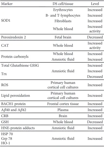
Table 1: Summary of the markers investigating in relation to oxidative damage in Down syndrome.
Marker DS cell/tissue Level
SOD1
Erythrocytes Increased B- and T-lymphocytes Increased Fibroblasts Increased Whole blood Increased activity
Peroxiredoxin 2 Fetal brain Decreased
CAT Whole blood Increased
activity
Protein carbonyls Whole blood Increased
Amnioticfluid Increased Total Glutathione GSSG Amnioticfluid Increased Increased Trx Decreased
ROS Primary human
cortical cell cultures Increased Lipid peroxidation cortical cell culturesPrimary human Increased BACH1 protein Frontal cortex tissue Increased
Aβ40 and Aβ42 Plasma Increased
CRB Brain Increased
GSH Whole blood Decreased
HNE-protein adducts Amnioticfluid Increased HSP 70
Grp 78 HO-1
Role of oxidative stress for congenital cardiovascular malformations is well studied in maternal diabetes [51]. Congenital heart disease occurs in 5% of infants of dia-betic mothers.
In fact, diabetic pregnancy is considered an independent risk factor for major embryonic malformations, and cardiac outflow tract defects are among the most frequent alterations observed in epidemiological studies [52]. The highest relative risk for major cardiovascular defects occurs if the mother has gestational diabetes and develops insulin resistance in the 3rd trimester.
Studies on rats have shown that hyperglycemia in the embryo induces production of reactive oxygen species that, together with a reduced ability of fetal cells to activate antiox-idant defense mechanisms, mediate adverse effects on cardiac neural crest migration and cardiac outflow tract septation through proapoptotic signaling [53, 54].
In humans, oxidative stress markers have been evaluated in the cord blood of newborns delivered by mothers with diabetes, suggesting that hyperglycemia induces oxidative stress [55]. However, glucose itself is not a mutagen; instead, it may exert a teratogenic effect via a signaling pathway regulating insulin sensitivity. Insulin sensitivity is thought to be involved in the pathophysiology of both type 1 and type 2 diabetes mellitus and insulin, and related signaling path-ways are also key mediators of embryogenesis and early development [56].
Glucose may also affect gene expression in embryonic development via epigenetic changes (histone acetylation and microRNA expression) [57].
The alternative that offspring CHD reflects maternally inherited genetic or epigenetic variations that confer risk of both diabetes mellitus and cardiac abnormalities is less likely, because the risk of maternal diabetes mellitus subsequent to birth of a child with CHD was only modestly increased. Conotruncal defect risk increased in the offspring of diabetic women consistent with experimental study findings that hyperglycemia in early pregnancy affects regulatory gene expression in the embryo, leading to cardiac neural crest cell death and increased CHD risk, particularly for conotruncal and outflow tract abnormalities.
Detailed mechanistic studies will be required to define the role of glucose sensitivity in cells from the neural crest and anterior second heart field during cardiac development. Maternal diabetes mellitus was also associated with the entire spectrum of CHD phenotypes. The nonspecific nature of the association suggests that hyperglycemia in early pregnancy may not only influence specific sequences in cardiac develop-ment but affects cardiac development in general or exerts its detrimental effect before formation of the primitive heart tube, with subsequent early and late consequences for fetal cardiac development.
Studies in rats have shown that oxidative stress during pregnancy can be reduced by using vitamin E and Vitamin C as antioxidants [58, 59], thereby supporting that ROS are involved in the embryonic dysmorphogenesis of dia-betic pregnancy.
Furthermore, recent evidences in humans, i.e., mothers of offspring with congenital heart disease, have shown
elevated homocysteine level related to low folate and/or vitamin B12 levels [50, 60], thus supporting that folic acid pathway alteration may exert an indirect embryotoxic effect by increasing oxidative stress [61].
Despite some human studies have shown that women using multivitamin supplements and folic acid during the periconceptional period had a lower risk of having babies with congenital heart defects, there are still concerns about the role of multivitamin supplements in reducing embryo-toxic effect and risk for CHD in humans [62, 63].
Recent studies have also shown that ROS overproduction and/or imbalance in the antioxidant system could lead to pulmonary hypertension in cases of CHD associated with increased pulmonary blood flow in lamb [64] and rodent experimental model of congenital diaphragmatic hernia [65] through a disordered process of vascular remodeling leading to smooth muscle cell hyperplasia, hypercontractility, and endothelial dysfunction [66].
Further studies are needed to identify the key factors in the development of CHDs in order to develop and imple-ment effective primary prevention program with preconcep-tional screening and development of nutripreconcep-tional intervention. Table 2 summarizes the factors that favour or prevent CDH via oxidative stress.
5. Role of Oxidative Stress in Neural
Tube Defect
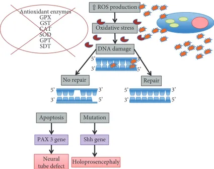
The human brain is particularly vulnerable to the damaging effects of reactive oxygen intermediates due to both its complexity and the long period of development (Figure 1).
Embryonic and fetal brain tissues are especially suscep-tible to peroxidative injury due to the fact that their membranes are rich in easily oxidable polyunsaturated fatty acid side chains [67].
Several studies indicate that antioxidant enzymes and molecules exhibit extremely low activities in fetal tissues, especially the brain [25, 68]. Cim and colleagues demon-strated higher levels of MDA, indicating an increased oxidant status in amniotic fluid of pregnant women with fetal congenital malformations of the central nervous system [69]. Although this antioxidant defense system is adequate to protect brain development under normal conditions, it is easily overcome by ROS, resulting in neurological and morphological abnormalities of SNC [70].
Holoprosencephaly (HPE) is one of the most common birth defects and is characterized by midline defects of the brain, facial, and oral structures. In humans, it has been estimated that HPE affects 1 in every 5000–10,000 live births and 1 in every 200–250 miscarried fetuses [71]. Many cases of Table 2: Factors that favour or prevent CHD via oxidative stress.
Favouring Preventing
(i) Maternal diabetes (i) Vitamin E
(ii) Hyperhomocysteine (ii) Vitamin C
(iii) Folic acid (iv) Vitamin B12
human HPE occur following fetal alcohol exposure or as a result of maternal diabetes both associated with elevated levels of reactive oxygen species. Studies on the teratogenic mechanism of ethanol-induced HPE in mice have showed that ethanol may impair Sonic hedgehog (Shh) gene expres-sion by activation of protein kinase A (PKA), a potent endogenous negative regulator of Shh signaling during the development of the neural tube [72, 73].
Shh, produced by the axial mesendoderm, prechordal mesoderm (PME), and notochordal plate, acts as a cru-cial signal in mammalian brain and facru-cial development, and Shh gene alterations are the most frequent causes of autosomal-dominant inherited HPE in humans and Shh2/2 mouse embryos exhibit severe HPE [74–76].
Ethanol-induced activation of PKA in the anterior PME results in a reduction of Shh expression and enhanced apoptosis of anterior PME cells causing the characteristic severe midline defects of HPE.
The inhibition of PME cells apoptosis by antioxidants, i.e., vitamin C and vitamin E, may protect from the terato-genic actions of ethanol.
Neural tube defects (NTDs) are a group of common and devastating congenital malformations that arise early in pregnancy due to the disturbance of normal neural tube closure. NTDs occur in about one in every 1000 established pregnancies worldwide [77], and it is estimated that over 323,000 births were affected with NTDs globally in 2001 [78]. The aetiology of NTD is thought to be heterogeneous, including genetic and environmental factors and their inter-actions [77, 79]. Factors that have been found to be associated with the risk of NTDs include insufficient folate [80] or multivitamin [81] intake, pregestational and gestational diabetes [82], pesticides [83], and antiepileptic drugs [84]. However, the proportion of NTD cases that can be attributed to known risk factors is lower than one-third [85]. Studies to delineate the mechanism underlying maternal diabetic
embryopathy have demonstrated that oxidative stress is a major contributor in NTD formation [86–89]. Excess apo-ptosis may be one of the mechanisms by which oxidative stress induces malformations. Apoptosis occurs at various developmental stages as a homeostatic mechanism to main-tain cell populations in tissues [90]. During the formation of the neural tube, apoptosis appears to be dispensable; how-ever, excessive apoptosis could potentially result in NTDs by causing insufficient cells to be present in the fusing neural folds or by disrupting the physical continuity of the dorsal midline [77, 90]. Growing evidence indicates that oxidative stress can stimulate apoptosis, which may lead to insufficient cell numbers to participate in folding and fusion of neural walls of the neural tube [90, 91].
Moreover, oxidative stress induces DNA damage and defects in DNA repair mechanisms. Single- and double-stranded fractures, base modifications (base partici-pation, rearrangement in some cases), and nucleoside dam-age may occur in DNA. There may also be crosslinking between DNA and protein depending on oxidative damage [92, 93]. Early embryonic development is vulnerable to oxidative stress because of the immaturity of free radical scavenging mechanisms [69]. The paired box 3 (Pax3) gene plays a major role in the development of neuroepithelium of embryos. In the absence of Pax3, neural tube defects occur [94]. Oxidative stress occurring before Pax3 expression leads to an increased risk of neural tube defects and diminished gene expression [95].
Myelomeningocele (MM) is a common congenital mal-formation that occurs when the embryonic neural tube fails to close properly during early embryogenesis. Common pathogenetic mechanisms for MM include folate deficiency, genetic susceptibility, environmental factors, in utero drug exposure, and biochemical factors [95–100].
Numerous reports have described free radical-mediated congenital defects [86–89]. Kao and colleagues supported
⇧ ROS production Oxidative stress DNA damage Repair No repair Antioxidant enzymes GPX GST CAT SOD GPT SDT Apoptosis Mutation Shh gene PAX 3 gene Neural
tube defect Holoprosencephaly 3’ 5’ 3’ 5’ 3’ 5’ 5’ 3’ 5’ 3’ 5’ 3’
the role of folate in modulating intracellular oxidative stress and suggest an additional mechanism for the etiology of folate deficit-associated MM [101]. Indeed, there is a direct relationship between antioxidant enzymes and the develop-ment of MM. GPX, GST, and SOD enzymes are the most important protective systems in humans for neural tube defects. An impaired responsiveness of the antioxidant enzymes, such as CAT, SOD, GPT, and SDT that play an active role in the detoxification of hydrogen peroxidase, has crucial effects in oxygen-induced embryopathy and might result in MM [102, 103].
In addition, SOD is involved in FR-mediated neurologi-cal diseases and acting a fundamental role in modulating reactive oxygen species toxicity [104]. In tissues lacking significant catalase activity, detoxification of hydrogen per-oxidase becomes critically dependent on GPX. In the study of Graf and colleagues, enzyme activity was abnormal in MM children compared to a control group underlying as deficiencies of enzyme are directly linked to neural tube defects [105].
Arslan and colleagues found that malondialdehyde (MDA), an oxidative damage marker, and a lower activity of erythrocyte carbonic anhydrase, an antioxidant enzyme which regulates the acid-base homeostasis, differ in new-borns with MM and in their mothers compared to healthy newborns and their mothers [106].
This study suggests that thefinding of low antioxidant enzyme activities in addition to ultrasound and maternal serum alpha fetoprotein may be an index of suspicion of neural tube defect.
In pregnant women with high risk, antioxidant enzymes administration together with folic acid may be an opportu-nity to reduce the incidence of neural tube defect.
6. Conclusion
Pregnancy is a state of oxidative stress as a consequence of high metabolic activity in the fetoplacental compartment. Fetal tissues are especially sensitive to oxidative damage because of the rapidly growing nature of their cells, which makes them vulnerable to the harmful effects of free radicals. Despite reactive oxygen species and free radicals, in the presence of a good antioxidant capacity, are important for developing embryos, promoting and controlling cellular fate, and playing a crucial role in normal development through cellular signaling, when overproduced, in the absence of a parallel increase in antioxidative activity, resulted in a wide range of biological toxic effects.
Due to the rapidly growing nature of their cells, fetal tis-sues are especially sensitive to oxidative damage that lead to lipid, protein, and polysaccharides oxidation and DNA dam-age with disruption of apoptosis processes that, during organogenesis, are highly needed in an appropriate location and temporal pattern.
Thus, the increase of oxidative stress, together with the impaired antioxidant activity, is clearly related to the induction of fetal malformations.
There are still gaps in our knowledge in the role of oxidative injury in the activation of complex array of genes
involved in different biological processes of fetal structure such as inflammation, coagulation, fibrinolysis, cell cycle, cell adhesion, and signal transduction. Future studies addressing the role of oxidative stress in thisfield are encouraged.
Moreover, there are few published studies evaluating oxidative stress biomarkers and management of oxidative stress with antioxidants therapeutic approaches.
Oxidative stress is widely implicated in failed repro-ductive performance, including infertility, miscarriage, diabetes-related congenital malformations, and preeclamp-sia. Poston et al. have focused on the role of free radicals and antioxidant capacity in preeclampsia. By measuring markers of lipid peroxidation and antioxidant capacity, they demonstrated the role of oxidative stress in this disorder [107].
Recent studies suggest that ischemia-reperfusion in the placenta as well as endoplasmic reticulum stress in the pla-centa may contribute to oxidative stress in trophoblasts. The recognition of oxidative stress in the placenta and the maternal circulation has led to evaluate the potential benefit of prophylactic antioxidant supplementation in women with a known risk of preeclampsia, particularly with an early supplementation with vitamins C and E [108]. However, until now, trials have shown no evidence that these supple-ments can prevent preeclampsia, but it is important to under-line that no RCT has yet addressed prophylaxis over the periconceptual period.
Other potential approaches include the use of supple-ments in the preconceptual period, selenium supplesupple-ments, antiperoxynitrite strategies, and statins [109, 110].
In clinical practice, early markers of oxidative stress might reveal that gravidic prophylactic use of antioxidants could help to prevent or at least reduce oxidative stress-related malformations in fetuses. Anyway, maternal antioxidant supplementation during pregnancy is important for protecting newborns against oxidative DNA damage.
Potential therapies for ROS-induced disease include both enzymatic and nonenzymatic antioxidant preparations. Supplementation with enzymatic and/or nonenzymatic anti-oxidants might have beneficial effects in decreasing injury from excess production of ROS, particularly in disorders such as bronchopulmonary dysplasia, retinopathy of prema-turity, periventricular leukomalacia, and necrotizing entero-colitis in preterm newborns who are especially susceptible to ROS-induced damage because of inadequate antioxidant stores at birth, as well as impaired upregulation in response to oxidant stress [13]. Nonenzymatic proteins (transferrin, ferritin, and ceruloplasmin), enzymes (superoxide dismu-tases, catalase, and glutathione peroxidase), oxidizable molecules (glutathione, vitamins E, A, C, carotenoids, and flavonoids), and trace elements (copper, zinc, and selenium) all play a role in maintaining a delicate balance between ROS production and oxidant damage to tissues and organs [111, 112].
More research is required to determine the beneficial effects of supplemental antioxidant therapy. There are multiple potential therapeutic antioxidants currently under investigation that could benefit newborns, particularly pre-mature infants. For example, one protein under investigation
is Pon3 that was shown in laboratory studies to have antiox-idant properties and to be upregulated in rat, sheep, and human cord blood late in gestation [113]. Other clinical trials include supplementation of preterm infants with lactoferrin and cysteine, examination of concentrations of beta-caro-tene, lutein, and lycopene in preterm infants fed formulas with mixed carotenoids and the effects on the developing eye, early administration of human erythropoietin in very preterm infants, NAC administration to women with intra-amniotic infection and/or inflammation, early enteral administration of vitamin E to extremely premature infants, and multiple trials involving inhaled nitric oxide. The results from these trials may change the way we treat many common neonatal conditions.
Caution must be taken since ROS are critical second messengers in various cell signaling pathways that control normal cellular functions, but strategies that maintain nor-mal antioxidant balance may be beneficial to the newborns. New studies should more extensively investigate the diag-nostic and therapeutic value of various oxidative stress bio-markers and antioxidants to reduce oxidative tissue injury to developing newborns.
Conflicts of Interest
The authors declare that there is no conflict of interests regarding the publication of this paper.
References
[1] T. W. Theunissen and R. Jaenisch, “Mechanisms of gene regulation in human embryos and pluripotent stem cells,” Development, vol. 144, no. 24, pp. 4496–4509, 2017. [2] R. L. Brent, S. Tanski, and M. Weitzman, “A pediatric
perspective on the unique vulnerability and resilience of the embryo and the child to environmental toxicants: the importance of rigorous research concerning age and agent,” Pediatrics, vol. 113, 4 Supplement, pp. 935–944, 2004. [3] R. L. Brent, “Environmental causes of human congenital
malformations: the pediatrician’s role in dealing with these complex clinical problems caused by a multiplicity of environmental and genetic factors,” Pediatrics, vol. 113, 4 Supplement, pp. 957–968, 2004.
[4] H. Sies, C. Berndt, and D. P. Jones,“Oxidative stress,” Annual Review of Biochemistry, vol. 86, no. 1, pp. 715–748, 2017. [5] D. P. Jones and H. Sies,“The redox code,” Antioxidants &
Redox Signaling, vol. 23, no. 9, pp. 734–746, 2015.
[6] H. Sies and D. P. Jones,“Oxidative stress,” in Encyclopedia of Stress, G. Fink, Ed., vol. 3, pp. 45–48, Elsevier, Amsterdam, 2nd edition, 2007.
[7] M. Kemp, Y.-M. Go, and D. P. Jones, “Nonequilibrium thermodynamics of thiol/disulfide redox systems: a perspec-tive on redox systems biology,” Free Radical Biology and Medicine, vol. 44, no. 6, pp. 921–937, 2008.
[8] A. R. Timme-Laragy, M. E. Hahn, J. M. Hansen, A. Rastogi, and M. A. Roy, “Redox stress and signaling during verte-brate embryonic development: regulation and responses,” Seminars in Cell & Developmental Biology, vol. 80, pp. 17–28, 2018.
[9] P. A. Dennery,“Oxidative stress in development: nature or nurture?,” Free Radical Biology and Medicine, vol. 49, no. 7, pp. 1147–1151, 2010.
[10] L. A. del Río, “ROS and RNS in plant physiology: an overview,” Journal of Experimental Botany, vol. 66, no. 10, pp. 2827–2837, 2015.
[11] G. I. Giles, K. M. Tasker, and C. Jacob,“Hypothesis: the role of reactive sulfur species in oxidative stress,” Free Radical Biology & Medicine, vol. 31, no. 10, pp. 1279–1283, 2001. [12] V. M. Labunskyy, D. L. Hatfield, and V. N. Gladyshev,
“Selenoproteins: molecular pathways and physiological roles,” Physiological Reviews, vol. 94, no. 3, pp. 739–777, 2014. [13] J. W. Lee and J. M. Davis,“Future applications of antioxi-dants in premature infants,” Current Opinion in Pediatrics, vol. 23, no. 2, pp. 161–166, 2011.
[14] S. Qanungo and M. Mukherjea,“Ontogenic profile of some antioxidants and lipid peroxidation in human placental and fetal tissues,” Molecular and Cellular Biochemistry, vol. 215, no. 1/2, pp. 11–19, 2000.
[15] S. C. Land,“Oxygen-sensing pathways and the development of mammalian gas exchange,” Redox Report, vol. 8, no. 6, pp. 325–340, 2003.
[16] Y. Long, G. Wang, K. Li et al.,“Oxidative stress and NF-κB signaling are involved in LPS induced pulmonary dysplasia in chick embryos,” Cell Cycle, vol. 17, no. 14, pp. 1757– 1771, 2018.
[17] P. A. Dennery, “Effects of oxidative stress on embryonic development,” Birth Defects Research Part C: Embryo Today: Reviews, vol. 81, no. 3, pp. 155–162, 2007.
[18] G. Pagano and G. Castello,“Oxidative stress and mitochon-drial dysfunction in Down syndrome,” in Neurodegenerative Diseases, S. I. Ahmad, Ed., Springer, New York, NY, USA, 2012.
[19] M. A. Pritchard and I. Kola, “The ‘gene dosage effect’ hypothesis versus the ‘amplified developmental instability’ hypothesis in Down syndrome,” Journal of Neural Trans-mission: Supplementum, vol. 57, pp. 293–303, 1999. [20] M. Perluigi and D. A. Butterfield, “Oxidative stress and
Down syndrome: a route toward Alzheimer-like dementia,” Current Gerontology and Geriatrics Research, vol. 2012, Article ID 724904, 10 pages, 2012.
[21] R. C. Iannello, P. J. Crack, J. B. de Haan, and I. Kola, “Oxidative stress and neural dysfunction in Down syn-drome,” Journal of Neural Transmission: Supplementum, vol. 57, pp. 257–267, 1999.
[22] M. Shichiri, Y. Yoshida, N. Ishida et al., “α-Tocopherol suppresses lipid peroxidation and behavioral and cognitive impairments in the Ts65Dn mouse model of Down syn-drome,” Free Radical Biology & Medicine, vol. 50, no. 12, pp. 1801–1811, 2011.
[23] F. V. Pallardó, A. Lloret, M. Lebel et al., “Mitochondrial dysfunction in some oxidative stress-related genetic diseases: ataxia-telangiectasia, Down syndrome, Fanconi anaemia and Werner syndrome,” Biogerontology, vol. 11, no. 4, pp. 401– 419, 2010.
[24] M. Zana, Z. Janka, and J. Kálmán,“Oxidative stress: a bridge between Down’s syndrome and Alzheimer’s disease,” Neuro-biology of Aging, vol. 28, no. 5, pp. 648–676, 2007.
[25] J. B. de Haan, M. J. Tymms, F. Cristiano, and I. Kola, “Expression of copper/zinc superoxide dismutase and glutathione peroxidase in organs of developing mouse
embryos, fetuses, and neonates,” Pediatric Research, vol. 35, no. 2, pp. 188–195, 1994.
[26] E. Engidawork and G. Lubec, “Molecular changes in fetal Down syndrome brain,” Journal of Neurochemistry, vol. 84, no. 5, pp. 895–904, 2003.
[27] M. F. Sánchez-Font, J. Sebastià, C. Sanfeliu, R. Cristòfol, G. Marfany, and R. Gonzàles-Duarte, “Peroxiredoxin 2 (PRDX2), an antioxidant enzyme, is underexpressed in Down syndrome fetal brains,” Cellular and Molecular Life Sciences CMLS, vol. 60, no. 7, pp. 1513–1523, 2003.
[28] J. Busciglio and B. A. Yankner,“Apoptosis and increased gen-eration of reactive oxygen species in Down’s syndrome neu-rons in vitro,” Nature, vol. 378, no. 6559, pp. 776–779, 1995. [29] J. He, T. Li, J. Chen et al.,“Plasma antioxidant enzymes and lipoperoxidation status in children with Down syndrome,” Clinical Biochemistry, vol. 49, no. 1-2, pp. 61–65, 2016. [30] F. di Domenico, G. Pupo, C. Mancuso et al.,“Bach1
overex-pression in Down syndrome correlates with the alteration of the HO-1/BVR-A system: insights for transition to Alzhei-mer’s disease,” Journal of Alzheimer’s Disease, vol. 44, no. 4, pp. 1107–1120, 2015.
[31] P. D. Mehta, G. Capone, A. Jewell, and R. L. Freedland, “Increased amyloid beta protein levels in children and adoles-cents with Down syndrome,” Journal of the Neurological Sciences, vol. 254, no. 1-2, pp. 22–27, 2007.
[32] D. Praticò, L. Iuliano, G. Amerio et al.,“Down’s syndrome is associated with increased 8,12-iso-iPF2α-VI levels:
evi-dence for enhanced lipid peroxidation in vivo,” Annals of Neurology, vol. 48, no. 5, pp. 795–798, 2000.
[33] M. Perluigi, F. Di Domenico, and D. A. Buttterfield, “Unraveling the complexity of neurodegeneration in brains of subjects with Down syndrome: insights from proteomics,” Proteomics - Clinical Applications, vol. 8, no. 1-2, pp. 73– 85, 2014.
[34] B. Balcz, L. Kirchner, N. Cairns, M. Fountoulakis, and G. Lubec,“Increased brain protein levels of carbonyl reduc-tase and alcohol dehydrogenase in Down syndrome and Alzheimer’s disease,” Journal of Neural Transmission: Supple-mentum, vol. 61, pp. 193–201, 2001.
[35] M. E. Garcez, W. Peres, and M. Salvador,“Oxidative stress and hematologic and biochemical parameters in individuals with Down syndrome,” Mayo Clinic Proceedings, vol. 80, no. 12, pp. 1607–1611, 2005.
[36] D. Valenti, G. A. Manente, L. Moro, E. Marra, and R. A. Vacca, “Deficit of complex I activity in human skin fibro-blasts with chromosome 21 trisomy and overproduction of reactive oxygen species by mitochondria: involvement of the cAMP/PKA signalling pathway,” Biochemical Journal, vol. 435, no. 3, pp. 679–688, 2011.
[37] A. Conti, F. Fabbrini, P. D'Agostino et al.,“Altered expression of mitochondrial and extracellular matrix genes in the heart of human fetuses with chromosome 21 trisomy,” BMC Genomics, vol. 8, no. 1, p. 268, 2007.
[38] M. Perluigi, F. di Domenico, A. Fiorini et al., “Oxidative stress occurs early in Down syndrome pregnancy: a redox proteomics analysis of amnioticfluid,” Proteomics - Clinical Applications, vol. 5, no. 3-4, pp. 167–178, 2011.
[39] S. Perrone, M. Longini, C. V. Bellieni et al.,“Early oxidative stress in amniotic fluid of pregnancies with Down syn-drome,” Clinical Biochemistry, vol. 40, no. 3-4, pp. 177– 180, 2007.
[40] I. T. Lott, E. Doran, V. Q. Nguyen, A. Tournay, E. Head, and D. L. Gillen,“Down syndrome and dementia: a randomized, controlled trial of antioxidant supplementation,” American Journal of Medical Genetics Part A, vol. 155A, no. 8, pp. 1939–1948, 2011.
[41] J. I. E. Hoffman and S. Kaplan, “The incidence of congenital heart disease,” Journal of the American College of Cardiology, vol. 39, no. 12, pp. 1890–1900, 2002.
[42] J. M. Matés, C. Pérez‐Gómez, L. Olalla, J. M. Segura, and M. Blanca,“Allergy to drugs: antioxidant enzymic activities, lipid peroxidation and protein oxidative damage in human blood,” Cell Biochemistry and Function, vol. 18, no. 2, pp. 77–84, 2000.
[43] I. Dalle-Donne, R. Rossi, R. Colombo, D. Giustarini, and A. Milzani, “Biomarkers of oxidative damage in human disease,” Clinical Chemistry, vol. 52, no. 4, pp. 601–623, 2006. [44] J. Redón, M. R. Oliva, C. Tormos et al.,“Antioxidant activities and oxidative stress byproducts in human hypertension,” Hypertension, vol. 41, no. 5, pp. 1096–1101, 2003.
[45] S. Nouri,“Congenital heart defects: cyanotic and acyanotic,” Pediatric Annals, vol. 26, no. 2, pp. 92–98, 1997.
[46] S. Ercan, A. Cakmak, M. Kösecik, and O. Erel,“The oxidative state of children with cyanotic and acyanotic congenital heart disease,” Anatolian Journal of Cardiology, vol. 9, no. 6, pp. 486–490, 2009.
[47] B. B. Warner and J. R. Wispé,“Free radical-mediated diseases in pediatrics,” Seminars in Perinatology, vol. 16, no. 1, pp. 47– 57, 1992.
[48] D. Bernstein,“Epidemiology and genetic basis of congenital heart disease,” in Nelson Textbook of Pediatrics, R. E. Behrman, R. M. Kliegman, and H. B. Jenson, Eds., pp. 1499–1502, Saunders, Philadelphia, PH, USA, 17th edition, 2004. [49] R.-F. S. Huang, Y.-C. Hsu, H.-L. Lin, and F. L. Yang,“Folate
depletion and elevated plasma homocysteine promote oxida-tive stress in rat livers,” The Journal of Nutrition, vol. 131, no. 1, pp. 33–38, 2001.
[50] C. A. Hobbs, M. A. Cleves, S. Melnyk, W. Zhao, and S. J. James, “Congenital heart defects and abnormal maternal biomarkers of methionine and homocysteine metabolism,” The American Journal of Clinical Nutrition, vol. 81, no. 1, pp. 147–153, 2005.
[51] S. C. Morgan, F. Relaix, L. L. Sandell, and M. R. Loeken, “Oxidative stress during diabetic pregnancy disrupts cardiac neural crest migration and causes outflow tract defects,” Birth Defects Research Part A, Clinical and Molecular Teratology, vol. 82, no. 6, pp. 453–463, 2008.
[52] R. M. Simeone, O. J. Devine, J. A. Marcinkevage et al., “Diabetes and congenital heart defects,” American Journal of Preventive Medicine, vol. 48, no. 2, pp. 195–204, 2015. [53] D. G. Molin, P. A. Roest, H. Nordstrand et al.,“Disturbed
morphogenesis of cardiac outflow tract and increased rate of aortic arch anomalies in the offspring of diabetic rats,” Birth Defects Research Part A, Clinical and Molecular Teratol-ogy, vol. 70, no. 12, pp. 927–938, 2004.
[54] P. Yang, E. A. Reece, F. Wang, and R. Gabbay-Benziv, “Decoding the oxidative stress hypothesis in diabetic embryopathy through proapoptotic kinase signaling,” Amer-ican Journal of Obstetrics and Gynecology, vol. 212, no. 5, pp. 569–579, 2015.
[55] M. Bis-Głuchowska, B. Marciniak, E. Szpringer-Boguń, R. Rola, B. Leszczyńska-Gorzelak, and J. Oleszczuk,
“Determination of antioxidative-peroxidative balance in the cord blood of newborns delivered to mothers with diabetes type G1,” Ginekologia Polska, vol. 72, no. 12A, pp. 1255– 1258, 2001.
[56] M. A. Basson,“Signaling in cell differentiation and morpho-genesis,” Cold Spring Harbor Perspectives in Biology, vol. 4, no. 6, 2012.
[57] D. J. Hoffman, R. M. Reynolds, and D. B. Hardy, “Develop-mental origins of health and disease: current knowledge and potential mechanisms,” Nutrition Reviews, vol. 75, no. 12, pp. 951–970, 2017.
[58] C. Martin Simán, A. C. Gittenberger-De Groot, B. Wisse, and U. J. Eriksson,“Malformations in offspring of diabetic rats: morphometric analysis of neural crest-derived organs and effects of maternal vitamin E treatment,” Teratology, vol. 61, no. 5, pp. 355–367, 2000.
[59] C. M. Simán and U. J. Eriksson,“Vitamin C supplementation of the maternal diet reduces the rate of malformation in the offspring of diabetic rats,” Diabetologia, vol. 40, no. 12, pp. 1416–1424, 1997.
[60] R. M. Shawky, A. R. M. Ramy, S. M. N. El-Din, S. M. Abd Elmonem, and M. A. Abd Elmonem, “Abnormal maternal biomarkers of homocysteine and methionine metabolism and the risk of congenital heart defects,” Egyptian Journal of Medical Human Genetics, vol. 19, no. 1, pp. 7–12, 2018. [61] S. E. Vollset, H. Refsum, L. M. Irgens et al.,“Plasma total
homocysteine, pregnancy complications, and adverse preg-nancy outcomes: the Hordaland Homocysteine study,” The American Journal of Clinical Nutrition, vol. 71, no. 4, pp. 962–968, 2000.
[62] L. D. Botto, J. Mulinare, and J. D. Erickson,“Occurrence of congenital heart defects in relation to maternal mulitivitamin use,” American Journal of Epidemiology, vol. 151, no. 9, pp. 878–884, 2000.
[63] L. D. Botto, J. Mulinare, and J. D. Erickson,“Do multivitamin or folic acid supplements reduce the risk for congenital heart defects? Evidence and gaps,” American Journal of Medical Genetics Part A, vol. 121A, no. 2, pp. 95–101, 2003. [64] S. Aggarwal, C. Gross, J. R. Fineman, and S. M. Black,
“Oxidative stress and the development of endothelial dysfunction in congenital heart disease with increased pulmonary blood flow: lessons from the neonatal lamb,” Trends in Cardiovascular Medicine, vol. 20, no. 7, pp. 238– 246, 2010.
[65] R. Aras-López, J. A. Tovar, and L. Martínez, “Possible role of increased oxidative stress in pulmonary hypertension in experimental diaphragmatic hernia,” Pediatric Surgery International, vol. 32, no. 2, pp. 141–145, 2016.
[66] V. G. DeMarco, J. Habibi, A. T. Whaley-Connell et al., “Oxidative stress contributes to pulmonary hypertension in the transgenic (mRen2)27 rat,” American Journal of Physiology-Heart and Circulatory Physiology, vol. 294, no. 6, pp. H2659–H2668, 2008.
[67] A. G. Fantel, “Reactive oxygen species in developmental toxicity: review and hypothesis,” Teratology, vol. 53, no. 3, pp. 196–217, 1996.
[68] A. G. Fantel, R. E. Person, R. W. Tumbic, T. D. Nguyen, and B. Mackler,“Studies of mitochondria in oxidative embryo-toxicity,” Teratology, vol. 52, no. 4, pp. 190–195, 1995. [69] N. Cim, H. E. Tolunay, E. Karaman et al.,“Amniotic fluid
oxidant–antioxidant status in foetal congenital nervous
system anomalies,” Journal of International Medical Research, vol. 46, no. 3, pp. 1146–1152, 2018.
[70] M. R. Brzezinski, H. Boutelet-Bochan, R. E. Person, A. G. Fantel, and M. R. Juchau,“Catalytic activity and quantitation of cytochrome P-450 2E1 in prenatal human brain,” The Journal of Pharmacology and Experimental Therapeutics, vol. 289, no. 3, pp. 1648–1653, 1999.
[71] M. M. Cohen and K. Shiota,“Teratogenesis of holoprosence-phaly,” American Journal of Medical Genetics, vol. 109, no. 1, pp. 1–15, 2002.
[72] K. Aoto, Y. Shikata, D. Higashiyama, K. Shiota, and J. Motoyama,“Fetal ethanol exposure activates protein kinase A and impairs Shh expression in prechordal mesendoderm cells in the pathogenesis of holoprosencephaly,” Birth Defects Research Part A: Clinical and Molecular Teratology, vol. 82, no. 4, pp. 224–231, 2008.
[73] M. Hammerschmidt, M. J. Bitgood, and A. P. McMahon, “Protein kinase A is a common negative regulator of hedge-hog signaling in the vertebrate embryo,” Genes & Develop-ment, vol. 10, no. 6, pp. 647–658, 1996.
[74] E. Belloni, M. Muenke, E. Roessler et al.,“Identification of sonic hedgehog as a candidate gene responsible for holo-prosencephaly,” Nature Genetics, vol. 14, no. 3, pp. 353– 356, 1996.
[75] C. Chiang, Y. Litingtung, E. Lee et al.,“Cyclopia and defective axial patterning in mice lacking sonic hedgehog gene func-tion,” Nature, vol. 383, no. 6599, pp. 407–413, 1996. [76] E. Roessler, E. Belloni, K. Gaudenz et al.,“Mutations in the
human sonic hedgehog gene cause holoprosencephaly,” Nature Genetics, vol. 14, no. 3, pp. 357–360, 1996.
[77] A. J. Copp, P. Stanier, and N. D. Greene,“Neural tube defects: recent advances, unsolved questions, and controversies,” The Lancet Neurology, vol. 12, no. 8, pp. 799–810, 2013. [78] A. Christianson, C. P. Howson, and B. Modell,“March of
dimes global report on birth defects,” in The Hidden Toll of Dying and Disabled Children, p. 30, March of Dimes Birth Defects Foundation White Plains, New York, 2006. [79] J. B. Wallingford, L. A. Niswander, G. M. Shaw, and R. H.
Finnell, “The continuing challenge of understanding, pre-venting, and treating neural tube defects,” Science, vol. 339, no. 6123, pp. 1222002–1222002, 2013.
[80] R. J. Berry, Z. Li, J. D. Erickson et al., “Prevention of neural-tube defects with folic acid in China,” The New England Journal of Medicine, vol. 341, no. 20, pp. 1485–1490, 1999. [81] A. E. Czeizel and I. Dudás,“Prevention of the first occurrence
of neural-tube defects by periconceptional vitamin supple-mentation,” The New England Journal of Medicine, vol. 327, no. 26, pp. 1832–1835, 1992.
[82] J. E. Becerra, M. J. Khoury, J. F. Cordero, and J. D. Erickson, “Diabetes mellitus during pregnancy and the risks for spe-cific birth defects: a population-based case-control study,” Pediatrics, vol. 85, no. 1, pp. 1–9, 1990.
[83] A. Ren, X. Qiu, L. Jin et al.,“Association of selected persistent organic pollutants in the placenta with the risk of neural tube defects,” Proceedings of the National Academy of Sciences, vol. 108, no. 31, pp. 12770–12775, 2011.
[84] H. Nau, R. S. Hauck, and K. Ehlers,“Valproic acid-induced neural tube defects in mouse and human: aspects of chirality, alternative drug development, pharmacokinetics and possible mechanisms,” Pharmacology & Toxicology, vol. 69, no. 5, pp. 310–321, 1991.
[85] A. J. Agopian, S. C. Tinker, P. J. Lupo, M. A. Canfield, L. E. Mitchell, and the National Birth Defects Prevention Study, “Proportion of neural tube defects attributable to known risk factors,” Birth Defects Research Part A: Clinical and Molecu-lar Teratology, vol. 97, no. 1, pp. 42–46, 2013.
[86] S. el-Hage and S. M. Singh,“Temporal expression of genes encoding free radical-metabolizing enzymes is associated with higher mRNA levels during in utero development in mice,” Developmental Genetics, vol. 11, no. 2, pp. 149– 159, 1990.
[87] M. Ishibashi, S. Akazawa, H. Sakamaki et al.,“Oxygen-induced embryopathy and the significance of glutathione-dependent antioxidant system in the rat embryo during early organogen-esis,” Free Radical Biology & Medicine, vol. 22, no. 3, pp. 447– 454, 1997.
[88] A. Ornoy, “Embryonic oxidative stress as a mechanism of teratogenesis with special emphasis on diabetic embryopa-thy,” Reproductive Toxicology, vol. 24, no. 1, pp. 31–41, 2007. [89] S. Weksler-Zangen, P. Yaffe, and A. Ornoy, “Reduced SOD activity and increased neural tube defects in embryos of the sensitive but not of the resistant Cohen diabetic rats cultured under diabetic conditions,” Birth Defects Research Part A: Clinical and Molecular Teratology., vol. 67, no. 6, pp. 429– 437, 2003.
[90] S. Lin, A. Ren, L. Wang et al.,“Oxidative stress and apoptosis in benzo[a]pyrene-induced neural tube defects,” Free Radical Biology and Medicine, vol. 116, pp. 149–158, 2018.
[91] L. Wang, S. Lin, D. Yi et al.,“Apoptosis, expression of PAX3 and P53, and caspase signal in fetuses with neural tube defects: apoptosis and fetal NTDs,” Birth Defects Research, vol. 109, no. 19, pp. 1596–1604, 2017.
[92] M. S. Cooke, M. D. Evans, M. Dizdaroglu, and J. Lunec, “Oxi-dative DNA damage: mechanisms, mutation, and disease,” The FASEB Journal, vol. 17, no. 10, pp. 1195–1214, 2003. [93] M. D. Evans and M. S. Cooke,“Factors contributing to the
outcome of oxidative damage to nucleic acids,” BioEssays, vol. 26, no. 5, pp. 533–542, 2004.
[94] S. A. Phelan, M. Ito, and M. R. Loeken,“Neural tube defects in embryos of diabetic mice: role of the Pax-3 gene and apoptosis,” Diabetes, vol. 46, no. 7, pp. 1189–1197, 1997. [95] T. I. Chang, M. Horal, S. K. Jain, F. Wang, R. Patel, and M. R.
Loeken,“Oxidant regulation of gene expression and neural tube development: insights gained from diabetic pregnancy on molecular causes of neural tube defects,” Diabetologia, vol. 46, no. 4, pp. 538–545, 2003.
[96] J. Gelineau-van Waes and R. H. Finnell,“Genetics of neural tube defects,” Seminars in Pediatric Neurology, vol. 8, no. 3, pp. 160–164, 2001.
[97] L. Pani, M. Horal, and M. R. Loeken,“Polymorphic suscepti-bility to the molecular causes of neural tube defects during diabetic embryopathy,” Diabetes, vol. 51, no. 9, pp. 2871– 2874, 2002.
[98] C. E. Pippenger, “Pharmacology of neural tube defects,” Epilepsia, vol. 44, Supplement 3, pp. 24–32, 2003.
[99] R. H. Finnell, A. Gould, and O. Spiegelstein,“Pathobiology and genetics of neural tube defects,” Epilepsia, vol. 44, Supplement 3, pp. 14–23, 2003.
[100] W. Zhao, B. S. Mosley, M. A. Cleves, S. Melnyk, S. J. James, and C. A. Hobbs, “Neural tube defects and mater-nal biomarkers of folate, homocysteine, and glutathione
metabolism,” Birth Defects Research Part A: Clinical and Molecular Teratology., vol. 76, no. 4, pp. 230–236, 2006. [101] T.-T. Kao, C.-Y. Chu, G.-H. Lee et al., “Folate
deficiency-induced oxidative stress contributes to neuropathy in young and aged zebrafish–implication in neural tube defects and Alzheimer’s diseases,” Neurobiology of Disease, vol. 71, pp. 234–244, 2014.
[102] W. D. Graf, O. E. Oleinik, T. A. Glauser, P. Maertens, D. N. Eder, and C. E. Pippenger, “Altered antioxidant enzyme activities in children with a serious adverse experience related to valproic acid therapy,” Neuropediatrics, vol. 29, no. 4, pp. 195–201, 1998.
[103] M. F. C. M. Knapen, P. L. M. Zusterzeel, W. H. M. Peters, and E. A. P. Steegers,“Glutathione and glutathione-related enzymes in reproduction,” European Journal of Obstetrics & Gynecology and Reproductive Biology, vol. 82, no. 2, pp. 171–184, 1999.
[104] L. M. Gaeta, G. Tozzi, A. Pastore, G. Federici, E. Bertini, and F. Piemonte,“Determination of superoxide dismutase and glutathione peroxidase activities in blood of healthy pediatric subjects,” Clinica Chimica Acta, vol. 322, no. 1-2, pp. 117– 120, 2002.
[105] W. D. Graf, C. E. Pippenger, and D. B. Shurtleff, “Erythrocyte antioxidant enzyme activities in children with myelomenin-gocele,” Developmental Medicine and Child Neurology, vol. 37, no. 10, pp. 900–905, 1995.
[106] M. Arslan, M. Melek, H. Demir et al.,“Relationship of antiox-idant enzyme activities with myelomeningocele,” Turkish Neurosurgery, vol. 22, no. 3, pp. 300–304, 2012.
[107] L. Poston, N. Igosheva, H. D. Mistry et al.,“Role of oxidative stress and antioxidant supplementation in pregnancy disor-ders,” The American Journal of Clinical Nutrition, vol. 94, suppl_6, pp. 1980S–1985S, 2011.
[108] L. C. Chappell, P. T. Seed, A. L. Briley et al.,“Effect of antiox-idants on the occurrence of pre-eclampsia in women at increased risk: a randomised trial,” The Lancet, vol. 354, no. 9181, pp. 810–816, 1999.
[109] M. P. Rayman, P. Bode, and C. W. G. Redman, “Low selenium status is associated with the occurrence of the pregnancy disease preeclampsia in women from the United Kingdom,” American Journal of Obstetrics and Gynecology, vol. 189, no. 5, pp. 1343–1349, 2003.
[110] F. Montecucco and F. Mach, “Update on statin-mediated anti-inflammatory activities in atherosclerosis,” Seminars in Immunopathology, vol. 31, no. 1, pp. 127–142, 2009. [111] C. Debier and Y. Larondelle,“Vitamins A and E: metabolism,
roles and transfer to offspring,” British Journal of Nutrition, vol. 93, no. 2, pp. 153–174, 2005.
[112] P. Giordano, P. Scicchitano, M. Locorotondo et al., “Carotenoids and cardiovascular risk,” Current Pharmaceuti-cal Design, vol. 18, no. 34, pp. 5577–5589, 2012.
[113] G. Belteki, S. L. Kempster, A. J. Forhead et al., “Paraoxo-nase-3, a putative circulating antioxidant, is systemically up-regulated in late gestation in the fetal rat, sheep, and human,” The Journal of Clinical Endocrinology & Metabo-lism, vol. 95, no. 8, pp. 3798–3805, 2010.