Advancing induced pluripotent stem cell (iPSC)
technology by assessing genetic instability
and immune response
Jordi Requena OseteAquesta tesi doctoral està subjecta a la llicència Reconeixement- NoComercial 3.0. Espanya de
Creative Commons.
Esta tesis doctoral está sujeta a la licencia Reconocimiento - NoComercial 3.0. España de
Creative Commons.
This doctoral thesis is licensed under the Creative Commons Attribution-NonCommercial 3.0.
- 1 -
UNIVERSITY OF BARCELONA
FACULTY OF FARMACY AND FOOD SCIENCES
CAMPUS DIAGONAL, AV. JOAN XXIII, 27-31, 08028
ADVANCING INDUCED PLURIPOTENT
STEM CELL (IPSC) TECHNOLOGY BY
ASSESSING GENETIC INSTABILITY AND
IMMUNE RESPONSE
BIOMEDICINE PHD PROGRAM
- 3 -
ADVANCING INDUCED PLURIPOTENT STEM CELL (IPSC)
TECHNOLOGY BY ASSESSING GENETIC INSTABILITY AND
IMMUNE RESPONSE
Memoria presentada por Jordi Requena Osete, Licenciado en Biología (2008-2012), para optar al Título de Doctor por la Universidad de Barcelona. Programa de Doctorado en Biomedicina (2013-2017).
Tesis Doctoral realizada en la Facultad de Medicina de la Universidad Barcelona bajo la dirección del Dr. Michael John Edel y bajo la tutoría del Dr. JosepMª Canals Coll.
TO OBTAIN
BIOMEDICINE PHD
PRESENTED BY
JORDI REQUENA OSETE
DIRECTOR
MICHAEL JOHN EDEL
TUTOR
JOSEP Mª CANALS COLL
- 5 -
“Que tot està per fer, i tot és possible...”
- 7 -
- 9 -
In the first place, I would like to acknowledge and to express my gratitude to my thesis director Dr. Michael John Edel, who gave me the opportunity to join his team to do this project, for his guidance and counsel. I am very thankful for his patience with my mistakes and for reminding me to stay focused at all time. Thank you for sharing your experience and knowledge as mentor.
Secondly, I would like to express special thanks to Dra. Belén Álvarez Palomo. Without her hard work it would not be possible to finish this research. I have learnt so much from her insightful observations. Thank you for assisting me and teaching me so many different research techniques.
I am really thankful and want to acknowledge as well Dra. Jovita Mezquita Pla, for her expert advice in molecular genetics techniques. Thank you for your encouragement and motivation during this intense period of research.
I have been able to count with you all three, Mike, Belén and Jovita, since the very first day and all along the 4 years of this PhD and previous master. Thank you to you all for leading me during these first steps in the scientific world while working and designing experiments together.
My sincere thanks also goes to my tutor Dr. JosepMª Canals Coll, for his assistance and help. I also would like to acknowledge to all my other fellow labmates from the Pluripotency laboratory in the Faculty of Medicine: Helena Sarret, Carme Grau, Adrià Martínez, Martí Sal, David, Isart Roca and Dra. Cristina Menchón. Thank you for the stimulating lab-meeting discussions and for all the fun we have had in the last four years.
This thesis would not have been possible without the excellent facilities the University of Barcelona provides to researchers and the people who run them such professionally, whose wise advice has been of huge value. In special to the Flow Cytometry unit: Isabel Crespo, Cristina Xufré, Cristina López and Sara Ozcoz; to Fluorescence Microscopy unit: Maria Calvo, Elisenda Coll and Anna Bosch and to the Animal House personal: JosepMª Marimón and Garikoitz Azkona.
I want to acknowledge all the collaborators that have contributed to this work in other research centers: to Dr. Manel Esteller, Dr. Raúl Delgado and Dr. Sebastian Moran from the Cancer Epigenetics group in IDIBELL; to Dr. Alejandro Vaquero and Dra. Irene Santos from the Chromatin Biology group in IDIBELL and to Dra. Antonella Consiglio from the Stem Cells and Neuroplasticity laboratory at IBUB.
I also would like to acknowledge all the scientists from other Groups and Departments of the Medicine Faculty and Hospital Clinic that have contributed and helped in the development of this thesis. In special to Dra. Carmen Barrot from the Forensic Anatomy Department for RT-PCR analysis; to Dr. Manel Joan and Dra. Anna Boronat from the Immunology Department; to Montse Pau and Dr. Xavier Gassull from Neurofisiology; Rafael Oliva and his group for the proteomic analysis from the Molecular Genetics group and to the Cytogenetic Unit: Dolors Costa, Joan and Candi.
During my short but intense stays in other laboratories I have been warmly received and treated with nice hospitality; I would like to acknowledge to Dra. Victoria Moreno from
- 10 -
the Research Center Principe Felipe in Valencia, as well as to Eric López Mocholi and Ana Alastrue Agudo for animal surgery and technical support; to Dra. María Blasco and Dra. Agueda Tejera from CNIO, in Madrid; to Dr. Rodney Dilley from the Harry Perkins Research Institute in Perth, Western Australia and to Dr. Jordi Calderó and Dra. Ana Casanovas from the Biomedical Research Center in Lleida.
Last, but not the least, I also want to acknowledge my family, to my father Antonio and my mother Ramona and to my brother JosepMª and sister MªJesús, for all their unforgettable support and for their continuous back-up. As well to my flatmate, uncle Fernando, for his company while I was living in Barcelona.
Finally, to the grants that have given financial support to enable this research project: FBG project 307900 and MINECO project BFU2011–26596 and BFU2014-54467-P.
- 11 -
- 13 - ACKNOWLEDGMENTS --- - 7 - INDEX --- 11 - GLOSSARY --- 17 - ABSTRACT --- 23 - INTRODUCTION --- 27 - 1. STEM CELLS--- 29 -
2. EMBRYONIC STEM CELLS (ESC) --- 31 -
3. INDUCED PLURIPOTENT STEM CELLS (IPSC)--- 33 -
3.1 REPROGRAMMING METHODS: --- 34 -
3.1.1 Transfection of synthetic modified mRNA: --- 34 -
3.1.2 Sendai virus: --- 35 -
3.1.3 Episomal vectors: --- 35 -
3.2 REPROGRAMMING FACTORS--- 36 -
3.3 REPROGRAMMING EFFICIENCY --- 38 -
3.4 THERAPEUTIC POTENTIAL OF IPSC --- 39 -
3.4.1 IPSC Cell Therapy --- 39 -
3.4.1.1 Spinal Cord Injury (SCI) --- 40 -
3.5 TUMORIGENIC THREAT OF IPSC --- 42 -
3.5.1 Ki67 --- 43 -
4. C-MYC AND CYCLIN D1 --- 45 -
4.1 C-MYC --- 45 -
4.1.1 C-Myc pleiotropic effects --- 45 -
4.1.2 C-Myc in cancer --- 46 -
4.2 CYCLIN D1 --- 47 -
4.2.1 CELL CYCLE --- 47 -
4.2.2 Cyclin D1 in cancer --- 48 -
5. IPSC GENETIC STABILITY --- 51 -
5.1 CELL STRESS --- 52 - 5.1.1 Sirtuin 1 --- 52 - 5.1.2 Apoptosis--- 53 - 5.2 EPIGENETIC STABILITY --- 53 - 5.3 GENETIC STABILITY --- 54 - 5.3.1 Aneuploidy --- 54 -
5.3.2 Single Nucleotide Polymorphism (SNP) --- 55 -
5.3.3 DNA double strand breaks (DSBs) --- 55 -
5.3.3.1 Histone H2AX --- 56 -
- 14 -
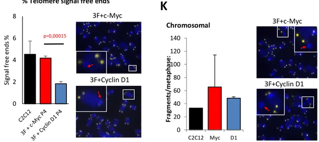
5.3.3.3 Myc and Cyclin D1 in DSB pathways --- 59 -
5.3.4 Copy Number Variation (CNV) --- 59 -
5.3.5 TELOMERE LENGTH IN IPSC --- 60 -
5.3.5.1 Multitelomeric Signal (MTS) --- 61 -
5.3.5.2 Signal-free ends --- 61 -
6. IMMUNE SYSTEM --- 63 -
6.2 INNATE IMMUNE RESPONSE --- 64 -
6.2.1 Toll like receptors (TLRs) --- 64 -
6.2.1.1 Toll-like receptor 3 (TLR3) --- 65 -
6.2 ADAPTIVE IMMUNE RESPONSE --- 66 -
6.2.1 CYTOTOXIC T LYMPHOCYTES (CTL) --- 66 -
6.3 IPSC IMMUNE RESPONSE --- 67 -
6.3.1 Allogeneic iPSC immune alterations --- 67 -
6.3.2 Syngeneic Models --- 67 -
OBJECTIVES AND HYPOTHESISE --- 71 -
MATERIALS AND METHODS--- 75 -
RESULTS --- - 119 - DISCUSSION --- - 151 - CONCLUSIONS--- - 165 - BIBLIOGRAPHY --- - 171 - APPENDICES --- - 210 - ANNEXES --- - 219 -
- 17 -
- 19 -
ATM: Ataxia Telangiectasia Mutated bFGF: Basic fibroblast growth factor BIN1: Bridging Integrator 1
BMP: Bone Morphogenetic Protein
BRCA2: Breast Cancer Type 2 susceptibility protein CCD1: Cyclin D1
CCL2: Chemokine (C-C motif) ligand 2 CDK: Cyclin Dependent Kinase
CM: Cardiomyocytes
CML: Chronic myelogenous leukemia CNS: Central Nervous System
CNV: Copy Number Variation CTLs: Cytotoxic T Lymphocytes DH iPSC: Cyclin D1 made human iPSC DHFR: Dihidroxifolate reductase
DNA-PKcs: DNA dependent protein kinases DSB: Double Strand Break
ENDO: Endoderm cells ESC: Embryonic Stem Cells FGF2: Fibroblast Growth Factor 2 GFP: Green Fluorescent Protein hESC: human embryonic Stem Cells HFFs: Human Foreskin Fibroblasts HLA: human leukocyte antigen HR: Homologous Recombination ICM: inner cell mass
IL-10: Interleukin 10 IL-15: Interleukin 15 IL-6: Interleukin 6
IPSC: induced Pluripotent Stem Cells KLF4: Krupple-Like Factor 4
LIF: Leukaemia inhibitory factor LIG3: Ligase 3
LPS: Lypopolysaccharides
MCP1: Monocyte Chemotactic Protein 1 MEFs: Mouse Embryonic Fibroblasts mESC: mouse Embryonic Stem Cells MH iPSC: C-Myc made human iPSC MHC: Major Histocompatibility Complex MN: Motor Neuron
MRN: Mre11-Rad50-NBS1 mRNA: messenger RNA
MSC: Mesenchymal Stem Cells
NBS1: Nijmegen Breakage Syndrome 1 NHEJ: Non-Homologous End Joining NSC: Neural Stem Cells
OCT4: Octamer-binding Transcription Factor 4 OSKM: Oct4, Sox2, Klf4 and c-Myc
- 20 -
PAMP: Pathogen Associated Molecular Pattern PARP1: Poly (ADP-ribose) polymerase-1 PI3K: Phosphoinositide 3-kinase
Pol µ: Polimerase µ Pol λ: Polimerase λ
POLQ or Pol θ: Polymerase θ
Poly(I:C): Polyinosinic:polycytidylic acid ROS: Reactive Oxigen Species
RPA: Replication Protein A RPE: Retinal Pigmented Epithelial SCI: Spinal Cord Injury
SNP: Single Nucleotide Polymorphism
SIRT1: Silent mating-type information regulation 2 homologue 1 SMA: Spinal Muscle Atrophy
SNP: Single Nucleotide Polymorphism
SOX2: SRY (sex determining region Y)-Box 2 TCR: T Cell Receptor
TGFβ: Transforming Growth Factor β TLRs: Toll-Like Receptors
TLR3: Toll-Like Receptor 3 TLR4: Toll-Like Receptor 4 TSS: Transcription Start Site VPA: Valproic Acid
- 23 -
- 25 -
Induced pluripotent stem cells (iPSC) can be made from adult somatic cells by reprogramming them with Oct4, Sox2, Klf4 and c-Myc. IPSC have given rise to a new technology to study and treat human disease (Takahashi et al., 2007). However, before iPSC clinical application, we need to step back and address two main challenges:
(i) Genetic stability of iPSC.
(ii) Immune response of iPSC-derived cells.
To address these key issues, the overall mission of this PhD thesis is to advance iPSC technology by addressing two objectives. First, is to replace c-Myc with Cyclin D1 in the reprogramming cocktail (Oct4, Sox2, Klf4 and c-Myc or Cyclin D1) and second, to study the immune response of iPSC-derived cells.
The quality of the starting iPSC determines the quality of the differentiated cells to be transplanted for clinical applications. In terms of genetic stability, aberrant cell reprogramming leads to genetic and epigenetic modifications that are the most significant barriers to clinical applications of patient iPSC derivatives (Gore et al., 2011). Such aberrations can result from the cellular stress that accompanies reprogramming or from the reprogramming factors themselves (Lee et al., 2012a). IPSC made with c-Myc are neoplastic in mouse models and have a higher tumorigenic potential than embryonic stem cells, prompting a search for new pluripotency factors that can replace the oncogenic factors Klf4 and c-Myc (Huangfu et al., 2008; Miura et
al., 2009; Okita et al., 2007). We chose Cyclin D1 to replace c-Myc because of previous
observation it can be used to reprogram cells to iPSC (Edel et al., 2010) and because of its DNA repair function (Chalermrujinanant et al., 2016). In this thesis we adopt a synthetic mRNA method to demonstrate that Cyclin D1 and c-Myc made iPSC have equal pluripotency using standard methods of characterisation. Moreover, no significant changes in copy number variation were found between starting skin cells and iPSC highlighting it is the method of choice for generating high quality iPSC. Further in-depth analysis revealed that Cyclin D1 made iPSC have reduced genetic instability assessed by: (i) reduced DNA double strand breaks (DSB), (ii) higher nuclear amount of the homologous recombination key protein Rad51, (iii) reduced multitelomeric signals (MTS) and (iv) reduced teratoma growth kinetics in vivo, compared to c-Myc made iPSC. Moreover, we demonstrate that Cyclin D1 iPSC derived neural stem cells engraft successfully, survive long term and differentiate into mature neuron cell types with high efficiency, with no evidence of pathology in a spinal cord injury rat model.
As we move towards the clinic with iPSC-derived cells for cell transplantation, the immunogenic response is thought to be one of the main advantages of iPSC technology for clinical application, because of its perceived lack of immune rejection of autologous cell therapy. We hypothesize that iPSC derived cells are unlikely to provoke an immune response. Here we have performed an analysis of the innate and adaptive immune response of human skin cells (termed F1) reprogramed to iPSC and then compared to iPSC-derived cells (termed F2) using proteomic and methylome arrays. We found little differences between MHCI expression and function; however, we discovered a short isoform of the Toll-like receptor 3 (TLR3), essential for viral dsRNA innate immune recognition, which is predominantly upregulated in all iPSC derived cells analysed and not seen in normal endogenous cells. High levels of the TLR3 isoform is associated with unresponsiveness to viral stimulation measured by lack of IL6 secretion in iPSC derived neural stem cells. We propose a new model that TLR3 short isoform competes with the full length wild type isoform destabilizing the essentially required TLR3
- 26 -
dimerization process. These differences could result in supressed inflammatory effects for transplanted human iPSC-derived cells in response to viral or bacterial insult. Further work to determine the in vivo effects is warranted and calls for screening of iPSC lines for TLR3 isoform expression levels before clinical use. In conclusion, this thesis has advanced iPSC technology by defining a new method that is a significant advance with novel insights that has immediate impact on current methods to generate iPSC for clinical application and more accurate disease modelling.
- 27 -
- 29 -
1. STEM CELLS
Stem cells are cells with self-renewing capability and the ability to differentiate into various cell types. They can be classified according to their capacity to differentiate and their origin.
Depending on their capacity to differentiate into other cell types stem cells are classified as:
Totipotent:
Stem cells with the ability to differentiate into all possible cell types. They have a self-organizing ability to generate a whole organism (Niwa et al., 2007). Only the zygote and early blastomere are totipotent in mammals.
Pluripotent:
Stem cells that can give rise to cell types from the three germ layers: ectoderm, mesoderm and endoderm. They differ from totipotent cells as pluripotent cells cannot give rise to the trophectoderm, which will form the placenta in a developing embryo. For instance, inner cell mass (ICM) cells that are specific to the early embryo are pluripotent (Pera et al, 2010).
Multipotent:
Stem cells that can give rise to all cell types within a particular lineage. For instance, mesenchymal stem cells can give rise to bone, muscle or adipose cells. Neural stem cells and Hematopoietic stem cells are other examples.
Unipotent:
Stem cells that can only generate one type of cell. For example spermatogonial stem cells can only form sperm (Jaenisch et al., 2008).
Depending on their origin, stem cells are classified into:
Embryonic:
Embryonic Stem Cells (ESC) are derived from the epiblast of the blastocyst and can be expanded indefinitely under proper culture conditions in vitro (Evans et al., 1981; Thomson et al., 1998). In embryos in vivo, pluripotent cells are transiently present before differentiating into somatic cells. ESC can give rise to all cell types of the three germ layers of the foetus: ectoderm, mesoderm and endoderm. When ESCs are injected into adult mice they produce teratomas, which contain tissues comprising the three germ layers. When taken from the blastocyst and cultured in vitro, cells keep their pluripotent
- 30 -
potential even after prolonged culture. It was first shown with mouse Embryonic Stem Cells (mESC) after being reintroduced into a blastocyst by their complete integration into the developing embryo (Beddington et al., 1989). Unlike mESC, human Embryonic Stem Cells (hESC) cannot contribute to the germ line after introduction into a host blastocyst (Yu et al., 2008).
Germinal:
Germinal stem cells or embryonic germ cells (EGC) are the cells that give rise to the gametes (sperm and eggs) in adults. They can be derived from primordial germ cells in vitro. Mouse EGS are also pluripotent and are undistinguishable from mESC morphologically, also expressing typical mESC markers. Furthermore, they can contribute to chimaeric mice upon blastocyst injection (Yu et al., 2008).
Somatic:
Somatic stem cells are undifferentiated cells found in adult or fetal somatic differentiated tissues. They have limited self-renewal capability and generally can differentiate only into cell types associated with the organ system in which they reside. Many tissues have niches of somatic stem cells, like: pancreas, brain, bone marrow, mammary gland, liver, skeletal muscle, the gastrointestinal tract, skin, dental pulp, blood and the eye.
- 31 -
2. EMBRYONIC STEM CELLS (ESC)
Mouse embryonic stem cells (mESC) were the first embryonic stem cells to be isolated from the inner cell mass (ICM) of mouse blastocysts and successfully cultured in vitro (Evans et al., 1981; Martin et al., 1981). They were initially cultured on top of a feeder layer of mitotically inactivated mouse embryonic fibroblasts (MEFs). Posterior analysis with filtered feeder layer produced conditioned medium led to the identification of leukaemia inhibitory factor (LIF). However, in serum-free medium, LIF alone was not able to prevent mESC differentiation, although with bone morphogenetic protein (BMP) pluripotency was sustained (Yu et al., 2008).
Later in 1998, human Embryonic Stem Cells (hESC) were also isolated from embryos (Thomson et al., 1998). Initially it was achieved with a suboptimal culture media conditions using a feeder layer and serum-containing medium. This time however, LIF and other related cytokines failed to support human and non-human primate ESC. Instead, Basic fibroblast growth factor (bFGF) and transforming growth factor β (TGFβ)/Activin/Nodal signalling were reported to be of high importance for the culture of undifferentiated hESC (Yu et al., 2008). Because of the critical requirement of hESC on bFGF, they are thought to be derived from a later stage of the inner cell mass development than mESC.
Introduction figure 1. Embryonic stem cells (ESC). ESC are isolated from the inner cell mass of the blastocyst. In vitro they can self-renew indefinitely and differentiate into cell types from the three germ layers: mesoderm, endoderm and ectoderm. Therefore ESC can be expanded in high numbers in vitro, differentiated into specific cell lineages such as: liver, pancreatic islet cells, intestinal cells, neurons, blood cells or muscle cells, and then transplanted to patients with damaged target tissues.
- 33 -
3. INDUCED PLURIPOTENT STEM CELLS (IPSC)
Due to the difficulty of finding a stable source of hESCs and the ethical issues associated to work with human embryos to isolate them, there was an urgency to find an alternative method to isolate pluripotent cells for the regenerative medicine field. Hence, in 2006 the discovery of induced pluripotent stem cells (iPSCs) signified a great leap in the field, which was awarded with the Nobel Prize later in 2012. IPSC avoid the ethical concerns of ESC, since they come from somatic cells of an adult organism. Induced pluripotent stem cells (iPSCs) are originated from adult somatic cells that have been reprogrammed, through ectopic expression of four defined factors: Oct4, Sox2, Klf4 and c-Myc (OSKM) (Takahashi et al., 2007) to cells with similar characteristics to embryonic stem cells (ESCs), meaning they can self-renew and differentiate to tissues of the three germ layers: mesoderm, endoderm and ectoderm. It was first discovered in 2006 by Dr. Shinya Yamanaka with mouse cells (Takahashi et al., 2006), and was confirmed later in human cells (Takahashi et al., 2007). Previously, reprogramming only had been possible by transfer of nuclear contents into oocytes (Gourdon et al, 1962, Wilmut et al, 1997) or by cell fusion with ES cells (Cowan et al, 2005).
Human iPSC can be established from various tissues: adult and embryonic fibroblasts (Takahashi et al., 2007; Yu et al., 2007), keratinocytes (Aasen et al., 2008), adipose tissue (Sun et al., 2009), peripheral blood (Loh et al., 2009), cord blood (Giorgetti et al., 2009), amniotic fluid-derived cells (Ye et al., 2009), neural precursor cells (Kim et al., 2009), among others.
Human iPSCs express marker genes, growth properties and morphology similar to human ESCs, however, they are not identical. Thus, it has been of interest the investigation to optimize the reprogramming method to yield iPSCs fully equivalent to Introduction figure 2. Induced pluripotent stem cells (iPSC) are reprogrammed from adult cells using defined factors: OCT4, SOX2, KLF4 and C-MYC (Takahashi et al., 2006). They can self-renew indefinitely and can differentiate into cell types from the three germ layers: mesoderm, endoderm and ectoderm.
- 34 -
ES cells. The assays used for establishing pluripotency equivalence between ESC and iPSC are four criteria (Lujan et al., 2010):
1) By subcutaneously injecting iPSC into mice it results in teratoma formation, containing tissues from the three germ layers: ectoderm, mesoderm and endoderm.
2) Injecting iPSC into blastocysts develops into contribution to chimera animal formation.
3) Germline transmission: progeny is able to display transgene expression.
4) Full embryo contribution (ability to generate more live-births “all-iPS cell embryos”) by tetraploid (4N) embryo complementation.
Human ESC and iPSC present differences in important aspects like global gene expression, epigenetic landscape and genomic imprinting. However, iPSC acquire ESC cell cycle properties during reprogramming (Ghule et al., 2010). But, although extended culture of iPSC makes them transcriptionally closer to ESC (Chin et al., 2009), it as well triggers chromosomal aberrations (Mayshar et al., 2010). It has also to be taken into account that iPSC derived from adult cells harbor residual DNA methylation signatures from their tissue of origin, retaining a specific epigenetic memory that influence differentiation propensity. Thus the forming potential of iPSCs depends on the differentiation status of the donor cell. For instance, blood-derived iPSC yielded more hematopoietic colonies than fibroblast-derived iPSC (Kim et al., 2010).
3.1 REPROGRAMMING METHODS:
Several methods have been established for reprogramming cells to a pluripotent state. The first method to make human iPSCs used a retroviral vector delivery system, carrying the risk of transgene reactivation and insertional mutagenesis (Takahashi et al., 2006; Takahashi et al., 2007). Since then many other groups have used the same methods to reprogram cells to pluripotency (Aasen et al., 2008; Stadtfeld et al., 2008; Okita et al., 2007; Yu et al., 2007; Woltjen et al., 2009Edel et al., 2010; McLenachan et
al., 2012). Research efforts thus focused on searching different ways to induce
pluripotency with non-viral methods to prevent transgene reactivation and avoid the risk of genomic recombination or insertional mutagenesis (Fusaki et al., 2009; Zhou et al., 2009; Warren et al., 2010; Masuda et al., 2013; Yoshioka et al., 2013). Hence, currently only three non-integrative methods appear to be appropriate for reprogramming patient cells for clinically safe cellular therapy: Sendai virus, mRNA transfections and episomal vectors (Introduction Fig. 3).
3.1.1 Transfection of synthetic modified mRNA:
Consists on the administration of messenger RNA (mRNA) modified to overcome innate antiviral immune responses. Although a daily transfection regime is required to maintain a sustained expression, mRNA reprogramming allows a higher reprogramming
- 35 -
efficiency (Warren et al., 2010). It offers the best option for future clinical applications as expression is transient over 48 hours and you can control the dose. In this thesis we have focused on reprogramming to iPSC by mRNA transfections as a substitute to retroviral transduction.
3.1.2 Sendai virus:
Non-integrative Sendai virus have the advantages of wide host specificity and low pathogenicity, and the disadvantage of strong immunogenic response (Fusaki et al., 2009), triggering the applicability of this method to firstly require the development of less antigenic vectors.
3.1.3 Episomal vectors:
Consist on introducing episomal genes that are ectopically expressed in the cells. Afterwards, the episome is naturally withdrawn by dilution while the iPSC divide (Yu
et al., 2009). Furthermore, in the case of episomal vectors and Sendai virus, final clones
have to be shown to be free of the original vector or virus.
However, regardless the method used, cell reprogramming always led to chromosome abnormalities and genomic instability (Gore et al., 2011), which is one of the main barriers to clinical application. Indeed, about half of the human adult derived iPSC clones exhibit genetic and epigenetic variations, thought to result from incomplete reprogramming, mutation in somatic cells and cellular stress during reprogramming (Koyanagi-Aoi et al., 2013).
Futhermore, an extensive comparison of all the non-integrating reprogramming methods has reported that, although having a lower successful rate and having a higher workload, mRNA transfections is more efficient, colonies emerge earlier and has a lower rate of aneurploidy than Sendai virus and episomal vectors (Schlaeger et al., 2015).
Introduction figure 3. Three main non-integrative methods for reprogramming adult cells to iPSCs in a clinical grade way in order to avoid insertional mutagenesis or transgene reactivation: a) Direct transfection of synthetic modified mRNA (Warren et al., 2010), b) Sendai virus (Fusaki et al., 2009) and c) Episomal vectors: (Yu et al., 2009). Diagram has been made by the author.
- 36 -
3.2 REPROGRAMMING FACTORS
In order to discover the cocktail combination of reprogramming factors, Dr. Yamanaka screened 24 candidate factors: Ecat1, Dppa5, Fbxo15, Nanog, ERas, Dnmt3l, Ecat8, Gdf3, Sox15, Dppa4, Dppa2, Fthl17, Sall4, Oct3/4, Sox2, Rex1, Utf1, Tcl1, Dppa3, Klf4, β-catenin, c-Myc, Stat3 and Grb2 (Takahashi et al., 2006). Finding the most suitable reprogramming factors cocktail combination is important to obtain high quality iPSCs carrying few or no genetic aberrations to become less tumorigenic. Accordingly, other transcription factors alternative to classic Oct4, Sox2, Klf4 and c-Myc (OSKM) have been evaluated as potential reprogramming factors, such as the orphan nuclear receptor Esrrb, which was used as a substitute of Klf4 together with Oct4 and Sox2 (Feng et al., 2009), or Glis1 that was able to substitute c-Myc reducing the number of partially reprogramed cells (Maekawa et al., 2011). Similarly, it was observed reprogramming by removing c-Myc and only using Oct4, Sox2 and Klf4 (Yamanaka et
al., 2006), although it was highly inefficient and most clones had issues to get
expanded. Even, it has been explored cell lines that can be reprogrammed with just Oct4 and Sox2 (Giorgetti et al., 2009; Giorgetti et al., 2010; Montserrat et al., 2013) and even only with Oct4 in adult neural stem cells (Kim et al., 2009). However, they were only successful starting from specific cell origins like cord blood (CB) cells or neural stem cells, which are not easily available, and using lentiviral vectors instead of clinical grade methods. Yet, the main transcription factors used for reprogramming are Oct4, Sox2, Klf4 and c-Myc, described below:
OCT4:
The main transcription factors to maintain pluripotency are Oct4, Sox2 and Nanog. Octamer-binding transcription factor 3/4, also known as Oct4 or Pou5f1, is a POU domain containing core transcription factor that binds specific target loci for maintaining pluripotency (Boyer et al., 2005). Oct4 expression is necessary to develop the inner cell mass in vivo. It is therefore highly expressed in ESC and iPSC and when its expression diminishes cells differentiate and lose pluripotency. Actually, the main classical four reprogramming transcriptional factors: Oct4, Sox2, Klf4 and c-Myc must have a specific stoichiometry: OSKM in a proportion of 3:1:1:1 (Kawamura et al, 2009). Hence, a critical amount of Oct4 is required to sustain pluripotency since only a 50% increase or decrease of Oct4 can lead to ESC differentiation (Yamanaka, 2008; Niwa et al., 2000).
SOX2:
Sex-determining region Y (SRY)-related HMG-box 2 (Sox2) transcription factor is part of a large family of 20 proteins sharing the DNA-binding motif HMG box. Sox2 is expressed in ESC, iPSC, extra-embryonic ectoderm, trophoblast stem cells and neural stem cells (Avilion et al., 2003). Similarly to Oct4, Sox2 dysregulation results in rapid differentiation (Fong et al., 2008; Yamanaka et al., 2008). Sox2 dimerizes with Oct4 (Yuan et al., 1995) being involved in the self-renewal of ESC and iPSC. Indeed, half of Oct4 bound genes are also bound by Sox2 and 87% of these are also co-occupied with Nanog (Boyer et al., 2005)
- 37 -
KLF4:
Kruppel-like factor 4 (Klf4) was first considered a candidate in the Reprogramming factors cocktail since it had been shown to contribute to the long-term maintenance of the ES cell phenotype and the rapid proliferation of ES cells in culture (Li et al., 2005a). Later, it has been shown that Klf4 induces epithelial properties by up-regulating E-cadherin directly (Li et al., 2010). Therefore, Klf4 mainly acts at the initial phase of reprogramming to initiate mesenchymal-to-epithelial transition (Chen et al., 2011). On the other hand, Klf4 had previously been reported to suppres proliferation as well through activating p21 (Zhang et al., 2000), to inhibit apoptosis induced by c-Myc (Zindy et al., 1998) and to repress p53 (Rowland et al., 2005).
C-MYC:
Myc genes are key regulators of cell proliferation, and their deregulation contributes to the origin of most tumours in humans. Specifically, c-Myc is a proto-oncogene that can give raise to tumor formation (Okita et al., 2007). It has many targets that enhance proliferation and transformation (Adhikary et al., 2005), many of which may have roles in the generation of iPSC. In ESC, c-Myc has been shown to maintain pluripotency and self-renewal (Varlakhanova et al., 2010). On the other hand, c-Myc role in iPSC reprogramming is not only in cell proliferation increase but also through the control of histone acetylation (Fernandez et al., 2003; Araki et al., 2011), since it associates with histone acetyltransferase (HAT) complexes and induces global histone acetylation allowing Oct3/4 and Sox2 to bind to their specific target loci. Therefore, in the absence of c-Myc, the overall efficiency of reprogramming is drastically reduced and the reprogramming time is increased (Habib et al., 2013).
The first part of the thesis is based on a previous observation that c-Myc oncogene, one of the four reprogramming factors, can be replaced by cell cycle gene Cyclin D1 in the reprogramming cocktail (Edel et al., 2010). With this replacement we pursue to reduce iPSCs cancer threat and genetic instability.
- 38 -
3.3 REPROGRAMMING EFFICIENCY
The issue of low efficiency in reprogramming still remains one of the main challenges to solve in the iPSC field. Reprogramming efficiency is determined by counting the number of iPSC colonies formed after reprogramming and dividing it by the total number of infected cells seeded. However, efficiency can be increased by modulating key components, like hypoxic condition (5% O2) (Yoshida et al., 2009) during reprogramming or by activation of enhancers or inhibition of barriers.
For example, a manner to increase efficiency is to modulate certain genes such as: downregulation of p53-p21 pathway (Kawamura et al., 2009), overexpression of Lin28 that inhibits Let7 induced differentiation (Heo et al., 2009), overexpression of Nanog (Han et al., 2010) or depletion of the structural component Mbd3, which promoted near 100% induction of somatic cells reprogrammed to pluripotency through suppressing the formation of the Nucleosome Remodelling and deacetylases (NuRD) complex (Rais et al., 2013), among others.
Other approaches to increase efficiency are based on chemicals that are added during the reprogramming process. For instance the addition of vitamin C (Esteban et al., 2009); cytokines like tamoxifen (Yang et al., 2010b); the histone deacetylase inhibitor Valproic acid (VPA1) (Huangfu D. et al., 2008); the inhibitor of DNA
methyltransferases 5-azacytidine (5-AZA) (Shi et al., 2008); inhibitor of MAPK (Yang.
et al., 2010) or the inhibitor of GSK3 to suppress differentiation (Ying et al., 2008).
Also, depending on the reprogramming method used efficiency is going to be different. It has been reported that viral reprogramming efficiency (0.04%) is lower than modified mRNA efficiency (1.4%) (Warren et al, 2010). This is another reason why in this thesis we wanted to reprogram cells using mRNA transfections.
1 The lack of chromatin silencing caused by VPA demethylation also renders the activation of the Dlk1-Dio3 gene
- 39 -
3.4 THERAPEUTIC POTENTIAL OF IPSC
Human iPSC technology holds great promise in the regenerative medicine field because of their capability to differentiate into any cell type of the three germ layers. Even elderly patient cells also have the ability to be reprogrammed into iPSCs (Dimos et al., 2008). Potential clinical applications range from drug screening, disease modelling and autologous cell therapies.
Drug screening in iPSC derived cells is advantageous because of the possibility to do the screening on specific cell types that without iPSC differentiation are difficult to find a source to obtain. Furthermore iPSC can be easily expanded to produce higher number of differentiated cells to work with.
Disease modelling using iPSC derived cells is advantageous to unveil the mechanism of specific diseases since scientists can work with iPSC containing the specific mutation that confers the illness. For example, iPSC technology has been used for setting cellular models for studding human diseases and performing drug screening tests. For instance, in 2010, Rett syndrome (RTT) patient-iPSC-derived neurons were produced as a model to study the disease. These cells were reported to exhibit fewer glutaminergic synapses formation, reduced neuritis spines density, smaller soma size, electrophysiological defects and altered calcium signalling; showing thus a neural network maturity deficiency (Marchetto et al., 2010). Thus, there is still a need to improve the differentiation methods employed not only for being more suitable for transplants but to reach an acceptable cellular platform for clinical drug screening research as well.
3.4.1 IPSC Cell Therapy
Regarding cell therapy, the potential application of iPSCs-derived cells offers a source for therapeutic treatment, either autologous or allogeneic, of a wide range of diseases, like neurodegenerative disorders, spinal cord injury, anaemia or diabetes among others. Correspondingly, numerous studies have been conducted since iPSC discovery to prove iPSCs derived cells therapeutic potential in animal models, aiming engraftment potential and illness recovery.
The first graft trial of iPSC-derived tissue in animal models was done in 2007 to treat sickle cell anaemic mice (Hanna et al., 2007). Hematopoietic progenitors (HP) were derived from iPSCs reprogrammed from mouse fibroblasts. In those iPSCs, genetic defects responsible for the anaemia were repaired through homologous recombination. The transplantation of those HPs reconstituted the haematopoietic system of sickle mice correcting the disease phenotype (Hanna et al., 2007).
Afterwards, in 2008 it was reported that loss of motor neurons from spinal cord and the motor cortex could be replaced with patient specific iPSCS-derived motor neurons in mice with amyotrophic lateral sclerosis (ALS) (Dimos et al., 2008). Even elderly patients cells were able to be reprogrammed into iPSCs.
Later on, engraftment of iPSC-derived myocites was successfully viable in dystrophic mice suffering from Duchenne muscular dystrophy (MD), in which regeneration and functional improvement was observed by transplanting satellite cells into
dystrophin-- 40 dystrophin--
deficient mice (Darabi et al., 2012). Moreover, the amount of stem or progenitor cells needed to be appropriate to succeed in graft transplantation (Darabi et al., 2012).
IPSC-transplantation therapy research has also focus on ischemic stroke of infarcted brains. Formerly sensorimotor function in rat brain was significantly improved after transplanted iPSCs migrated to injured areas, and differentiated into neuron-like cells successfully (Jiang et al., 2011). And most recently iPSC-derived neurons and astrocytes were transplanted into brain damaged areas after ischemic stroke injury in a rat model in which function recovery was significantly improved in comparison with control groups (Yuan et al., 2013).
In another disease model, human iPSC derived from fibroblasts of patient with spinal muscle atrophy, were genetically corrected. Afterwards, iPSC were derived into motor neurons and were transplanted into a spinal muscle atrophy mouse model. A significant recovery was shown in the cells injected group compared with the control sham group (Corti et al., 2012).
These previously described approaches in neurodegenerative disorders demonstrate the potential viability of transplantation therapy of human iPSCs-derived neuron. This new biomedical tool also offers the promises of potential treatment for Parkinson and Diabetes for example, as it has been reported the generation of functional human pancreatic β cells in vitro (Pagliuca et al., 2014), among other diseases.
Early this year, it has been published the first human clinical trial for iPSC transplantation therapies has been finished in the RIKEN institute, Japan (Mandai et al., 2017). It has consisted on iPSC derived retinal pigmented epithelial (RPE) cells transplantation in two age-related macular degeneration (AMD) patients. In the second patient however, the experiment was suspended since it were found genetic instabilities in that patient iPSC. The trial has demonstrated the viability and safety of the procedure and that although there has not been reported any improvement in patients sight, at least the degeneration did not worsen but was maintained. Authors insisted though, in the fact that only one patient was not enough to claim any conclusion. However, the first clinical trial of iPSC in human has been overall satisfactory.
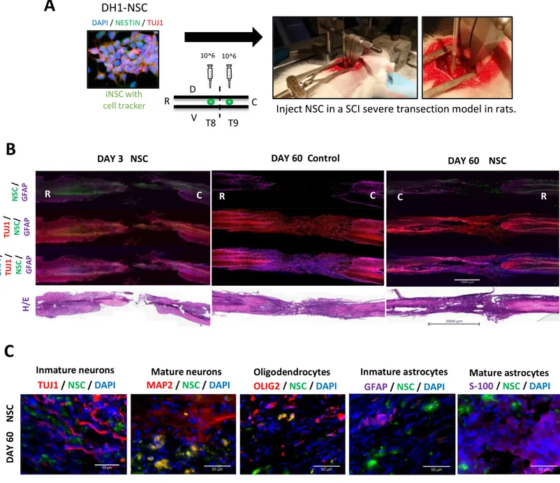
In this thesis, we have assessed iPSC derived Neural Stem Cells (NSC) survival, engraftment and differentiation potential in a spinal cord injury (SCI) rat model and iPSC derived Motor Neurons (MN) in a Spinal Muscle Atrophy (SMA) mouse model.
3.4.1.1 Spinal Cord Injury (SCI)
According to the world Health Organization, every year between 250.000 and 500.000 people worldwide suffer a spinal cord injury (SCI) (WHO webpage, 2017). In the last decades, progress had been made in surgical and rehabilitation treatments for SCI, however these approaches are only palliative. Transection of the spinal cord typically creates limited repair and poor functional recovery after the loss in motor and sensory function below the injury site. The accumulative death of neurons, astroglia, and oligodendroglia in and around the lesion site leads to neural circuitry disruption and dysfunction (Beattie et al., 2000). After the lesion, because of the scar formation,
- 41 -
injured axons are unable to grow, regenerate or reconnect, triggering a permanent interruption of the injured nervous route (Beattie et al., 2000; Blight et al., 1992; Grossman et al., 2001). Over the following hours after the traumatic insult, by-products of cell necrosis (DNA, ATP, glutamate) are released into the microenvironment leading to a rapid and progressive secondary injury cascade which generates further cell death and activation of pro-inflammatory microglia (Choo et al., 2007; LaPlaca et al., 2007). Macrophages and microglia infiltrate and generate ROS while phagocyting debris. Neutrophils and later lymphocytes also infiltrate the immune-privileged blood-spinal cord barrier and contribute to inflammatory response (Waxman, 1989; Ulndreaj et al., 2016).
During the following days, neurons are hampered to regenerate by an interlaced network of hyperproliferative astrocytes forming the glial scar around the lesion. The extracellular matrix also contains chondroitin sulfate proteoglycan (CSPGs) deposits which form a formidable barrier to neuronal growth. Therefore, inability to remyelinate spared axons and failure of axons to build again the signal conduction because of the astrocyte scar contribute to the incurability of SCI (Deumens et al., 2006; Dietz et al., 2006; Harel et al., 2006).
A number of interventions transplanting cell types derived from the adult Central Nervous System (CNS), have shown therapeutic efficacy in various animal models of SCI (Ogawa et al., 2002). However, there were still some barriers to clinical translation like the issues of isolation and expansion of large numbers of cells in a uniform manner and patient immunosuppression after transplant. In this sense, numerous pluripotent and multipotent cell types have been investigated for treating SCI (Tobias et al., 2003; Erceg et al., 2010; Tetzlaff et al., 2011; Vawda et al., 2012; Lopez-Serrano et al., 2016). Therefore, since the discovery of induced pluripotent stem cells (Takahashi et al., 2006), iPSC derived cells have held great promise for the regenerative medicine field. IPSC provide an autologous cell source and avoid the ethical and moral concerns of other stem cell sources. These pluripotent cells can be expanded indefinitely in vitro and can provide a large quantity of differentiated cells for transplantation, including specific cells of neuronal or glial fates (Benzing et al., 2006; Gerrard et al., 2005; Itsykson et al., 2005; Keirstead et al., 2005; Lee et al, 2007; Li et al., 2005b; Ludwig et al., 2006; Erceg et al., 2008).
IPSC technology tool offers the promises of potential treatment for a wide range of diseases described above. However, autologous iPSC-derived cellular transplantations still bargain the hurdle of tumorigenic threat.
- 42 -
3.5 TUMORIGENIC THREAT OF IPSC
One of the main concerns of iPSC is the potential tumorigenic threat of iPSC-derived cells after transplantation into patients, since stem cells and cancer cells share some characteristics. Some shared features are: rapid proliferation rate, lack of contact inhibition, propensity for genomic instability, high activity of telomerase, high expression of oncogenes, miRNA signatures and epigenetic status (Ben-David et al., 2011b). Self-renewal capacity and pluripotency are double-edged swords that prompt ESC and iPSC tumorigenic as well as cancer cells. Spontaneous transformation of ESC in culture increases the risk of formation of teratocarcinomas (Werbowetski-Ogilvi et al., 2009) on transplantation of differentiated cells derived from ESC. However, iPSC-derived teratomas develop faster, more efficiently and more aggressively than ESC-derived teratomas (Moriguchi et al., 2010). Even the formation of benign teratomas is also unacceptable regarding transplantation therapy. There is a need to understand the carcinogenic aspects of iPSC. Therefore, ESCs and iPSC tumorigenicity is a big hurdle hindering clinical application nowadays.
IPSC cancer risk has been studied widely in mouse models (Alvarez et al., 2013; Mc Lenachan et al., 2012; Kiuru et al., 2009; Miura et al., 2009; Okita et al., 2007). Interestingly, the tumorigenicity of virally derived iPSC and transgene-free iPSC showed no significant differences (Moriguchi et al., 2010). Thus, regardless the method used in reprogramming there is a genotoxic stress response that can lead to aneuploidy with aberrant iPSC karyotypes (Mayshar et al., 2010). Also iPSC derived from mature somatic cells that have lived longer enough to acquire mutation that randomly confer anti-apoptotic advantages, are more prone to be selected during culture (Mayshar et al., 2010). Regarding reprogramming factors, c-Myc is a well-established oncogene involved in tumor development (Albihn et al., 2010); however, Oct4, Sox2 and Klf4 are also known to be associated with cancer progression in specific tumours (Wang et al., 2010; Ji et al., 2010; Tian et al., 2010).
Tumorigenesis in ESC and iPSC has been proposed to be Nanog-related, because regardless of the reprogramming genes used, its pre-transduction treatment activated the carcinogenic program (Grad et al 2011). Also, during reprogramming, inactivation of p53-p21 antitumor system either yields iPSC generation or renders cancer (Hussein et al., 2011). Hence, downregulation of p53 pathway during reprogramming can lead to high levels of DNA damage, therefore compromising genomic integrity. Damage can be noted as a rise in the CNV levels, found higher in early-passaged iPSCs. However, most of these changes are lost by the selection pressure of the culture (Hussein et al., 2011). Several strategies have been proposed to cope with tumorigenicity and improve safety. As only few several hundreds of pluripotent cells are enough to generate tumours, it is required a 100% pure population of differentiated cells to safely apply ESC and iPSC derived treatments. Eliminating the remaining pluripotent cells by cytotoxic antibodies (Choo et al., 2008) or even separating ESC using MACS or FACS (Fong et al., 2009) don’t result in 100% pure cultures of only differentiated cells. Or even introduction of suicide genes to attack the tumour (Schuldiner et al., 2003).
- 43 -
3.5.1 Ki67
Once iPSC pluripotency capabilities are assessed in vivo with teratoma formation assays2 to corroborate differentiation into the three germ layer tissues in vivo,
assessment of proliferation rate of teratomas can be measured with Ki67 staining. Ki67 (or MIB1) is a nuclear protein that is a cellular marker for proliferation (Scholzen et al., 2000). Furthermore, it is associated with ribosomal RNA transcription (Bullwinkel et al., 2006). Through interphase, Ki67 antigen can be solely detected in the cell nucleus. However, in mitosis most of the protein is relocated to the surface of the chromosomes. During all active phases of cell cycle (G1, S, G2, and mitosis) Ki67 is
present, but is lacking in resting cells (G0) (Bruno et al., 1992).
Ki67 is an excellent marker to define the growth portion of a population of cells. Interestingly, the fraction of Ki67 positive tumour cells, also called Ki67 labelling index, is very often correlated with the clinical course of cancer. The main examples for this marker are carcinomas of the prostate, brain, breast and nephroblastoma. Prognostic values for survival and tumour reappearance has repeatedly been proven in uni- and multivariate analysis for these types of tumours.
- 45 -
4. C-MYC AND CYCLIN D1
Since the aim of this first part of the thesis is to remove the oncogenic threat of c-Myc from the reprogramming cocktail and replace it with Cyclin D1, here these two genes are introduced and an overview is given.
4.1 C-MYC
Identified three decades ago, c-Myc is a well-established oncogene, associated with many human cancers, involved in tumour development (Dang, 2010; Wasylishen and Penn, 2010; Albihn et al., 2010). All Myc family proteins (C-Myc, N-Myc and L-Myc) regulate processes involved in many if not all aspects of cell fate. Indeed, in vitro and in vivo DNA-binding studies, have designated an increasing number of genes as Myc targets (Ji et al., 2011; Margolin et al., 2009; Shaffer et al., 2006; Wasylishen and Penn, 2010). Likewise, numerous transcription factors and chromatin regulating factors interact with Myc (Cheng et al., 1999; Cowling and Cole, 2006; Eilers and Eisenma, 2008; Rahl et al., 2010; Wasylishen and Penn, 2010).
Regarding genetic aberrations, Myc dysregulation is directly linked with gene amplification. Myc overexpression elevated DHFR gene copy number within 3 weeks by 10 fold (Denis et al., 1991) or even just within 72h after overexpression (Mai et al., 1994; Mai et al., 1996). Indeed, every single cell expressing the conditional Myc gene showed DHFR3 amplification (Mai et al., 1994). Other genes found amplified after 72h
of c-myc overexpression are: ribonucleotide reductase R2 (R2) (Kuschak et al., 1999), the carbamyl-P synthetase, aspartate transcarbamylase, dihydro-orotase (CAD) enzyme conding gene (Miltenberger et al., 1995; Fukasawa et al., 1997; Chernova et al., 1998; Eberhardy and Farnham 2001), ornithine decarboxylase (George et al., 1996; Rounbehler et al., 2009), Cyclin B1 and Cyclin D2 (Mai et al., 1999, 2005).
4.1.1 C-Myc pleiotropic effects
Myc carry out important cellular functions by targeting regulator genes involved in proliferation, differentiation, metabolism, apoptosis, translation, stromal remodelling, inflammation, angiogenesis and invasion (reviewed in Sodir et al., 2009). Accordingly, Myc acts as a master regulator of tumor development by activating or repressing genes related with all this pathways. Therefore, Myc dysregulation initiates a dynamic process of genomic instability that is linked to tumor initiation.
- 46 -
4.1.2 C-Myc in cancer
MYC genes are dysregulated in numerous human neoplasias. Indeed, more than 70% of all tumours have some form of c-Myc gene dysregulation (Nesbit et al., 1999). Therefore, Myc pathology has been studied in neoplasms including Burkitt lymphoma (Lombardi et al., 1987), B and T cell lymphoma (Slack et al., 2011), multiple myeloma (Anguiano et al., 2009; Chng et al., 2011), plasmacytoma (Shen-Ong et al., 1982), hepatocarcinoma (Kawate et al., 1995), lung carcinoma (Little et al., 1983), breast carcinoma (Lavialle et al., 1989), pancreatic cancer (Hessmann et al., 2016) , among others.
Myc genes are induced as response upon almost every signal transduction pathways known to be altered in cancer, comprising, for instance, those ruled by tyrosine kinase growth factor receptors, NF-κB and β-catenin (Kelly et al., 1983; Renan, 1989; Duyao
et al., 1990; Marcu et al., 1997; Zou et al., 1997; He et al., 1998). In turn, Myc proteins
have a role as master transcriptional regulators of a wide range of target genes that execute a cellular response. As a matter of fact, 11% of all cellular loci are candidates to be bound by Myc (Fernandez et al., 2003; Orian et al.,2003; Hulf et al., 2005). Actually, MYC/MAX heterodimer are estimated to occupy more than 45% of all replication origins in human cells carrying Myc-binding E-box motifs (Swarnalatha et al., 2012). At the karyotype level, Myc overexpression cause chromosomal changes such as formation of extrachromosomal elements, centromere and telomere fusions, chromosome and chromatid breaks, ring chromosomes, translocations, deletions and inversions, aneuploidy, and the formation of Robertsonian chromosomes (Mai et al., 1996; Felsher and Bishop 1999; Rockwood et al., 2002; Guffei et al., 2007; Goncalves Dos Santos Silva et al., 2008; Silva et al., 2010; Chen et al., 2011).
Dysregulated Myc also modifies the nuclear architecture of cells, affecting therefore the positional organization of telomeres and chromosomes, which initiate a dynamic process of ongoing genomic instability (Chuang et al., 2004; Louis et al., 2005; Mai and Garini 2005; 2006; Vermolen et al., 2005). Myc dysregulation, even for as short as 2-12h, resulted in nuclear remodelling of the 3D organization and position of telomeres and chromosomes (Louis et al., 2005; Mai and Garini 2005). Therefore, by affecting nuclear organization, Myc drives dynamic remodelling of chromosomes, genes and their structural order. This is particularly relevant as gene activation, function and Introduction figure 4. C-Myc pleiotropic effects. C-Myc target genes involved in a great diversity of pathways, such as enhancement of proliferation, inflammation, angiogenesis, metabolism and invasion and inhibition of differentiation. Image from Sodir et al., 2009.
- 47 -
nuclear space are functionally linked (Solovei et al., 2009). Furthermore, nuclear remodelling occurs during early malignancy and set the stage for neoplastic transformation (Mai and Garini 2005, 2006; Gadji et al., 2011, 2012).
4.2 CYCLIN D1
Cyclin D1 is a well-known cell cycle gene responsible for enabling the progress of the cell cycle from G2 to S phase. Its classical role is to interact with CDK4 and form a complex that phosphorylates the retinoblastoma (Rb) (Connell-Crowley et al., 1997). Then, phosphorylated retinoblastoma derepresses E2F, which internalizes to the nucleus and acts as a transcription factor to activate the synthesis of genes involved in cell cycle progression.
On the other hand, Cyclin D1 also carries out important non-canonical roles apart from cell cycle progression, in other cellular processes recently reviewed (Hydbring et al., 2016), such as: DNA damage repair, control of cell death, differentiation, migration, immune response and metabolism.
Here we want to seize both the capability of Cyclin D1 to facilitate the cell cycle progression, bypassing the limiting step of speeding the cell cycle during early reprogramming stages, and its advantageous role in DNA damage repair to propose Cyclin D1 as a safer candidate to replace c-Myc in the reprogramming cocktail.
4.2.1 CELL CYCLE
Cell cycle is a sequence of events that eventually leads to division and duplication of a cell. Multiple checkpoints are present within the cell cycle to regulate progression through various stages. These mechanisms are controlled by a family of cyclins and cyclin-dependent kinases (CDKs), where CDK functionality is dependent on their association with an active cyclin. Eukaryotic cells have some CDK-cyclin complexes that play defined roles at different phases of the cell cycle. These complexes comprise ten cyclins belonging to four different types (A, B, D, and E type), three interphase CDKs (CDK2, CDK4, and CDK6) and a mitotic CDK1 (Schwartz et al., 2009) (introduction figure 5).
Somatic cells are generally dependent on receiving specific mitogen signals, such as growth factors, to divide and proliferate (Evan et al., 2001). Once cells have received enough mitogen exposure, DNA damage has been checked and cells have confirmed they have all required machinery proteins needed for successful division, then they enter the cell cycle. On the contrary, ESC and iPSC do not require mitogenic factors since they express Oct4, Sox2 and Nanog and are cultured in medium with FGF2 and TGFβ1 that maintain pluripotency and continuous self-renewal of the cells (reviewed in Huang et al., 2015).
- 48 -
These cyclins activate CDKs by binding to them depending on their presence and levels. For example, Cyclin D complexes with CDK4 and CDK6 to stimulate the initiation of G1 phase and the start of the cell cycle, when the cell prepares for DNA synthesis (Schwartz et al., 2009). In normal cells, CDK activity is regulated by two types of inhibitors: INK4 proteins (A-D) and Cip/Kip family proteins (p21, p27, and p57) (Schwartz et al., 2009).
In cancer, cell cycle defects are frequently generated by changes in CDK activity as a result of accumulated mutations (Evan et al., 2001; Schwartz et al., 2009). These mutations lead to hyperactive CDKs that trigger unscheduled proliferation, such as CDK4 in melanoma (Schwartz et al., 2009), CDK6 in pro-B acute lymphocytic leukemia (Kuo et al., 2011), CDK5 in pancreatic adenocarcinomas (Eggers et al., 2011) or CDK1 and CDK2 in colon adenomas (Vermeulen et al., 2003).
4.2.2 Cyclin D1 in cancer
Cyclin D is involved in various types of cancers, however, it is not the only cyclin dysregulated. Cyclin E has also been reported to be overexpressed in breast and colon cancer (Vermeulen et al., 2003) and cyclin A and E are amplified in certain lung carcinomas. D type Cyclins function as growth sensors, which connect mitogen stimuli with the cell cycle progression (Choi et al., 2014a). Therefore, Cyclin D translocations, amplifications, missense mutations, and elevated protein levels are potential causes of cancer. Mutations and abnormities may increase cyclin D activity, resulting in enhanced cell cycle progression to S-phase and cell proliferation (Ashgar et al., 2015). Elevated levels of cyclin D proteins in cancer have been attributed to defective mechanisms of Introduction figure 5. Cell Cycle phases and Cyclins. Overview of the cell cycle phases: G1, S, G2
and M (mitosis). Cell-cycle progress is controlled by cyclins and their CDKs. In the G1 phase, Cyclin D-CDK4 complex phosphorylates Rb protein, which derepresses E2F inducing Cyclin E transcription. On the other hand, p21 and p27 oppose these effects and can result in the exit of the cell-cycle finishing in G0. To ensure the integrity of the DNA there are several checkpoints in S phase after DNA replication.
For example, chromosome abnormalities and DNA damage are reported to cyclin B/CDK1 complex via diverse pathways to delay or stop progression into mitosis. Image from Niehrs et al., 2012.
- 49 -
degradation as well (Lahne et al., 2006). For example, overexpression of cyclin D1 linked to gene amplification has been studied in breast, esophageal, bladder, lung, and squamous cell carcinomas (Vermeulen et al., 2003). In B-cell lymphoma, overexpression of cyclin D1 has been observed due to gene translocation (Vermeulen et al., 2003). In prostate cancer however, low levels of cyclin D1 and D3 have been observed (Olshavsky et al., 2008).
In a ChiP promoter array assay, Cyclin D1 associated with approximately 900 genes in close proximity to the transcriptional start site (Fu et al., 2005). A large number of gene sets were associated with cell division (most involved in G2/M phase and cellular mitosis); other genes were found to regulate chromosomal stability. Hence, Cyclin D1 overexpression contributes to chromosomal instability by directly regulating a transcriptional program that governs it (Casimiro et al., 2012). Indeed, transient expression of Cyclin D1 over 7 days in the mammary gland induced chromosomal instability (Casimiro et al., 2012). Its overexpression also correlated with aneuploidy, supernumerary centrosomes, and spindle defects in mouse hepatocytes (Nelsen et al., 2005), and with aneuploidy and polyploidy in lymphoid tumors (Aggarwal et al., 2007). Still the number of genes targeted by Cyclin D1 is lower than c-Myc.
- 51 -
5. IPSC GENETIC STABILITY
Genetic instability refers to a series of chromosomal changes that enables cells to develop new and aggressive phenotypes to adapt to the changing selection pressures (Bayani et al., 2007). It is generally classified into two major types: DNA base changes that occur due to defects in the DNA repair processes including base excision repair, mismatch repair and nucleotide excision repair (Kolodner et al., 1995; Modrich et al., 1997; Rajagopalan et al., 2003) and abnormal karyotypes with structural and numerical chromosome alterations (Lengauer et al., 1997).
Human Pluripotent Stem Cells genomic instability was first documented in 2004 reporting karyotype abnormalities in ESCs, such as trisomy of chromosome 12 (Cowan
et al., 2004; Draper et al., 2004). Since then and up to date, an emerging number of
publications have aimed this issue, focusing on ESCs and iPSCs genetic and epigenetic instability (reviewed in Martins-Taylor et al., 2012; Peterson et al., 2013; Steinemann et al., 2013). Aberrant cell reprogramming lead to genetic and epigenetic modifications (Gore et al., 2011), which are one of the most significant barriers to personalized clinical applications of patient iPSC derivatives. Such genetic aberrations are an important factor for assessing the quality of iPSC and can result from the cellular stress that accompanies reprogramming (Lee et al., 2012b). Therefore, it is crucial to define ways to reduce replicative stress induced by reprogramming (Ruiz et al., 2015). In Introduction Figure 6 it is schematized how during reprogramming process some but not all iPSCs acquire aberrations.
Oct4 + Sox2 + Klf4 + c-Myc
Introduction figure 6. Reprogramming of patient fibroblast using a clinical therapy viable method introducing the four Yamanaka genes: Oct4, Sox2, Klf4 and c-Myc. After reprogramming the resulting mosaic cell population contain a mixture of not reprogrammed cells (patient fibroblasts), cells reprogrammed carrying no genetic or epigenetic aberrations (green cells with blue nucleus) and cells reprogrammed carrying genetic and epigenetic defects (green cells with red nucleus). The proportion of cells carrying genetic and epigenetic aberrations is not actually known, but depends on reprogramming factors used and cell culture conditions. Diagram has been made by the author.
- 52 -
5.1 CELL STRESS
Oxidative and replicative cell stresses are the main causing agents of genetic instabilities. In normal situations DNA damage cause an immediate and p53-independent G1 arrest, caused by rapid proteolysis of Cyclin D1. This degradation is an essential component of cellular response to genotoxic stress (Agami et al., 2000). Accordingly, lowering replication stress during reprogramming by eliminating Myc, provides a simple strategy to reduce genomic instability on mouse and human iPSCs (Ruiz et al., 2015). Indeed, replication stress may result from metabolic starvation of nucleotides or from RNA polymerase transcription and DNA replication machinery clash causing replication fork collapse. Additionally, Myc expression also causes an accumulation of reactive oxygen species (ROS), which generates DNA breaks by oxidative stress (Khanna and Jackson 2001). ROS increases as a result of a biochemical imbalance caused by the sudden increase in gene products via Myc transcriptional activation (Vafa et al., 2002). Myc also alteres a mitochondrial gene, TFAM, which encodes a protein essential for mitochondrial function and biogenesis, and may lead to increased ROS (Dang et al., 2005).
5.1.1 Sirtuin 1
Sirtuins are an enzymatic type of NAD-dependent histone deacetylases found in prokaryotes and eukaryotes that regulate the expression of certain genes to help organisms to respond to metabolic and genotoxic stress through diverse pathways, such as metabolic homeostasis, cell survival pathways and cell-cycle control. Descriptions of knockout models for each of the seven mammalian Sirtuins suggest that during stress response Sirtuins protect genomic stability, triggering integrity through a variety of mechanisms, the main involved in chromatin-related functions (reviewed in Bosch-Presegué et al., 2014).
Sirtuin 1 (Silent mating-type information regulation 2 homologue 1 or Sirt1), a member of the class III histone deacetylase (HDAC) famility, plays key roles in a variety of physiological processes such as genomic stability, metabolism, neurogenesis and cell survival.
It has also been reported to be necessary for proficient telomere elongation and genomic stability of iPSC (De Bonis et al., 2014). Also, Sirtuin 1 contributes to telomere maintenance and increases global homologous recombination. Sirtuin 1 null mouse embryonic fibroblasts (Sirt1-/-MEFs) were shown to express a higher number of multitelomeric signals per chromosome (Palacios et al., 2010). In addition, in human embryonic stem cells (hESC) Oct4 maintain the pluripotency through inactivation of p53 by Sirtuin 1 mediated deacetylation (Zhang et al., 2014). Furthermore, sirt1 has been reported to be necessary for proficient telomere elongation and genomic stability of induced pluripotent stem cells (De Bonis et al., 2014).