Innovative tecnique for large septal perforation
repair and radiological evaluation
Valutazione radiologica e tecniche innovative per la riparazione di ampie
perforazioni settali
S. MOCELLA1, F. MUIA2, P.G. GIACOMINI3, D. BERTOSSI4, E. RESIDORI5, S. SGROI6
1 ENT Department, East Garda Bussolengo (VR); 2 Surgery Staff Asl TO5 Chieri (TO); 3 Tor Vergata University
Rome; 4 Maxillo-facial Department, University of Verona; 5 Radiological Department, East Garda Bussolengo (VR); 6 Department of Radiology, Casa di cura Pederzoli, Peschiera (VR)
SummAry
Perforation of the nasal septum may have multiple causes: traumatic, iatrogenic, infectious, degenerative, overuse of vasoconstrictors, abuse of cocaine and more recently chemotherapy agents. Perforations are also classified according to their size and type of cartilaginous or osteocartilaginous deficit, as well as location (front, middle and rear). many surgical techniques have been proposed to repair the perfo-ration, although the results are often unsatisfactory for perforations of small and medium size; in large perforations permanent obliteration of the defect cannot always be ensured. it is often necessary to use tissues from inside the nasal turbinates or cartilage from other donor sites such as the ear or rib, and various techniques are discussed in light of the recent literature. The perforations observed in the last eight years and surgical approaches performed in open or closed approaches are taken into account. The authors propose a new technique that has been used with success in many types of septal perforation regardless of aetiology, and in particular large perforations, which allows for the use of the osteocartilaginous donor site as a hump. it is also useful in reductive rhinoseptoplasty, which targets selection to easily obtain mucopericondral flaps with an extramucosal technique.
Key wordS: INSERT 3-5 KEY WORDS
riASSunTo
La perforazione del setto nasale ha cause molteplici: traumatiche, iatrogene, infettive, degenerative, da abuso di vasocostrittori, da uso di cocaina e di recente anche da chemioterapici antineoplastici. Le perforazioni sono altresì classificate in funzione delle dimensioni e della tipologia del deficit cartilagineo o osteocartilagineo e della sede anteriore, mediana e posteriore. Molteplici tecniche di riparazione chirurgica del danno sono state proposte nel tempo e vengono riassunte nel presente lavoro per tipologia di approccio; i risultati con-seguiti, sono soddisfacenti per le perforazioni di piccole e medie dimensioni, ma nelle grandi perforazioni non garantiscono sempre la permanente obliterazione del difetto. Spesso si rende necessario l’utilizzo di tessuti prelevati all’interno delle fosse nasali come i turbinati o il prelievo di cartilagine da altre sedi donatrici come l’orecchio o la costa; le varie tecniche vengono illustrate alla luce della più recente letteratura. Gli Autori hanno valutato i pazienti osservati negli ultimi 8 anni e i risultati chirurgici conseguiti in particolare considerando, gli approcci chirurgici effettuati in approccio aperto o chiuso, endoscopico ed i materiali impiegati. Gli autori propongono tra le varie tecniche impiegate in questa serie di pazienti, una tecnica innovativa utilizzabile con successo in molti dei casi considerati di perforazione settale iatrogena che prevede l’utilizzo del gibbo osteocartilagineo quale sede donatrice, nonché la rinosettoplastica riduttiva quale scelta mirata per ottenere agevolmente lembi di scorrimento mucopericondrale e talora produrre anche un miglioramento estetico morfologico.
PArole ChiAve: INSERT 3-5 KEY WORDS IN ITALIAN
Acta Otorhinolaryngol Ital 2013;33:xx-xx
CHECK REFERENCE NUMBERING: REFERENCES 1-6 DO NOT SEEM TO BE CITED IN TEXT
Introduction
Closure of nasal perforation is one of the most fascinat-ing and excitfascinat-ing of all procedures in nasal reconstructive surgeryIt is a challenge for both the surgeon and patient, with the primary goal of restoring aesthetic aspects and recovering anatomical and functional integrity in the same procedure.
Surgical success is based on a precise definition of ae-tiology; location and the method employed during the intervention along with particular attention to pre- and post-operative care. The authors report their long-stan-ding experience with the definition and treatment of per-forations of the nasal septum, which in reality is not an infrequent pathology.
Aetiology and clinical features
The causes of nasal perforation may be local or systemic; in addition, various systemic illnesses may be responsi-ble for the pathology, but there are limited data availaresponsi-ble regarding the frequency of the different causes of nasal septum perforation. A broad investigation in a Swedish population revealed a prevalence of septal perforation of 0.9%.
One of the first documented descriptions of perforation in an anatomopathological specimen is conserved in the Hunterian Museum of The Royal College of Surgeon in London (#1228). In the sagittal section, it is possible to see an important septal perforation of 1.5 cm with a round defined precise configuration in a patient suffering from syphilitic infection who died in the 17th century. Not only
is the straight septum around the perforations visible, but it also possible to define margins and a certain degree of depression of the dorsal profile, which is a typical char-acteristic of the disease. To date, we have not personally observed such a disease as syphilis has now largely disap-peared, although other systemic diseases have been seen
during recent years which must be detected to better plan adequate treatment.
Table I lists the aethiopathology and causes of septal per-forations, specific diagnostic tests of the different diseases and recent observations of septal perforations during the use of antineoplastic pharmacological treatment.
Table II provides some suggestions and precautions that should be adopted to prevent and avoid septal perforations in different circumstances 91-93.
Classification and diagnostic endoscopic
features
Septal perforations are classified according to site and to-pography: cartilaginous, osteocartilaginous or intermedi-ate, bone or posterior; according to size: small(< 1 cm in diameter), medium (1-2 cm) and large (> 2 cm). Often, the size of cartilage or bone perforation is greater than that of the mucosa, and therefore instrumental techniques for high-precision measurement of the perforation have been proposed 94-96.
Physical examination of the nose begins with an evalua-tion of the external nose. Large perforaevalua-tions may result in loss of support to the dorsum of the nose and subsequent saddle nose deformity with occasional lateral deviation of the anterior part of the pyramid.
Table I. Aetiopatology of Nasal Septal Perforations. Traumatic causes
Previous surgery 7-11
Cauterization or embolization for epistaxis 12 13
Nasal packing 14 15
Nasogastric tube placement Septal haematoma from blunt trauma Battery or other foreign body in nose 16-24
Chronic nasal cannula use Turbulent airflow
Inflammatory or infectious causes
Sarcoidosis 25 26 ACE >
Wegener granulomatosis 27 28 ANCA >
Systemic lupus erythematous 29-36
Tuberculosis 37 38 POSITIVE TEST
AIDS 39 40
Crohn’s disease 41-44 HISTOPATOLOGY
Autoimmune diseases 45-50
Leishmaniasis 51-53
Cryoglobulinaemia 54 55
Celiac disease 56 POSITIVE TEST
Invasive fungal sinusitis 57 58
Mycobacterium Kansas infection 59
Neoplastic causes Carcinoma
T-cell lymphomas HISTOPATOLOGY
Other causes
Inhaled substances (e.g., cocaine, topical corticosteroids, long-term oxymetazoline or phenylephrine use) 60-75
Chromic acid fumes 76-79
Renal failure 80
Use of targeted/biologic therapies in the treatment of malignant and non-malignant diseases (bevacizumab) 81-88
Use of methotrexate or docetaxel in the treatment of malignant disease 89 90
Table II. Suggestions and precautions in Nasal Septal Perforations (modi-fied by Batniji 2012) 97.
Prescribe heated, humidified continuous positive airway pressure devices for patients with obstructive sleep apnoea.
Minimize steroid use in patients.
During septoplasty, minimize resection of cartilage and use meticulous technique to avoid bilateral tears in the mucosa.
Minimize nasal trauma during the insertion of nasogastric tubes by (1) decongesting the nose with oxymetazoline or phenylephrine prior to nasogastric tube insertion, (2) inserting the nasogastric tube along the floor of the nose parallel to the hard palate and perpendicular to the plane of the face, and (3) lubricating the tip of the nasogastric tube.
Modify the nasal cannula in patients on long-term supplemental oxygen and humidify the supplemental oxygen. Taping 2 wooden toothpicks to the hub of the cannula (the thickened plastic part where the prongs are attached) modifies the nasal cannula. This directs the oxygen straight into the nose and away from the nasal septum.
When cauterizing the nasal septum for epistaxis, avoid cauterizing both sides simultaneously.
Stop cocaine use.
Stop or minimize use of topical nasal decongestants. Run a humidifier in the bedroom.
Frequently use nasal saline sprays.
Use nasal emollients (especially before bedtime).
Decrease digital nasal trauma. Parents may want to place mittens on their young children’s hands at night.
Anterior rhinoscopy is essential in initial examination of the patient, and may reveal the configuration of the per-foration, the presence of crusting and irregularities of mucosal structure. Topical nasal decongestants may be utilized for intranasal inspection of the entire septum. Na-sal endoscopy provides more information on the evalu-ation of the entire septum. Palpevalu-ation of the septum with a cotton-tipped applicator provides valuable information regarding the integrity of the quadrangular cartilage in the remainder of the septum.
Diagnostic work-up
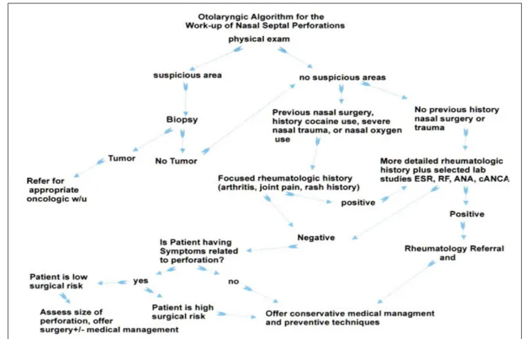
In diagnostic work up of septal perforation, several detailed laboratory investigations can be performed that are espe-cially useful to detect medical causes of septal perforations. an algorithm such as that recently proposed by Batniji may be used, but for patients who use cocaine (often not admit-ted) is absolutely mandatory to identify on their urine the presence of cocaine catabolites in urine, or if possible and from hair; in these patients, it is advisable to not to perform surgery until after one year of cocaine cessation.
Diagnostic management of the nasal septum performation is shown in the flow chart in Fig. 7, according to that pro-posed by Rami K. Batniji in 2012 97.
Surgical procedures
Many surgical procedures have been proposed to repair the defect, either by endonasal approaches using muco-perichondrial 98-115 or combined flaps with interposition of
the graft using septal cartilage, uncinate process, middle turbinate, auricular or costal cartilage and temporal fas-cia 116-139, also utilizing a backwards extraction-reposition
technique of the quadrangular cartilage 140 or using other
non-autologous tissues or synthetic materials 141-147.
In large perforations, four Authors have suggested the use of labial and oral flaps 148-151.
Thane open approach is considered more likely to dom-inate the edges of the perforation and to ensure routine surgical dissection and reconstruction of the defect 152-165.
However, it is often not possible to obtain flaps that are large enough to cover the defect. To overcome this, some authors have suggested combining reductive rhinosepto-plasty to use the excess of mucosa that arises as a fabric to repair the septal perforation 166-169.
Other authors have also proposed the use of expanders po-sitioned under the mucopericondrium to achieve the same result 170-172.
Radiological work-up
In many of our surgical cases, radiological specific work-up and elaboration techniques of imaging was performed before and after surgery for septal perforation to obtain
as much information as possible regarding nasal struc-ture and framework; the method was applied to identify the loss of bone and mucosal layers, and to quantify and obtain exact measures of the defect to help in planning reconstruction. Some anatomic structures of the parana-sal sinuses, and especially naparana-sal septum which is of main interest in our cases, are not optimally visualized to plan surgical procedures by computed tomography (CT) and 2D MRI (Magnetic Resonance Imaging) with standard image reconstruction in the axial and coronal planes. Elaboration of CT scans was performed, in addition to virtual 3D endoscopy (Fig. 8) using a work station fur-nished by the CT or MRI manufacturer. Multidetector CT scanners with 16 detector rows (or more) allow the acqui-sition of volumetric data sets that can be electronically reconstructed in any plane. Recent 1.5T (or 3.0T) MRI scanners equipped with new 3D acquisition sequences al-low the acquisition of volumetric data sets as well, with the advantage (compared to CT) of better visualization of soft tissues with no radiation exposure.
These techniques give radiologists and surgeons the op-portunity to visualize anatomic structures in greater detail and may help increase diagnostic accuracy and therapeu-tic planning of diseases of the paranasal sinuses and nasal septum.
Multidetector CT
CT examination of the paranasal sinuses is performed at our institution using a multidetector scanner (Sensation 16; Siemens, Erlangen, Germany). The scanner is equipped with 16 rows of detectors. Scanning is performed in the standard axial plane with a helical technique (120 kV, 100 Eff. mAs, pitch of 0.55, rotation time of 0.75 second, sec-tion thickness of 0.75 mm, and a 512 × 512 matrix). The subject’s head is placed in a neutral position, without chin tilt. The image data set is reconstructed with an indi-vidual voxel size of 0.75 × 0.75 × 0.75 mm.
The images included in the present article were recon-structed at a standard workstation (Wizard; Siemens) and at a standard PACS Workstation (Synapse, Fujifilm Medi-cal Systems, equipped with Voxar 3D 6.3 software). The time required for multiplanar reconstruction of the CT im-age data set was about 3-5 min per reconstruction.
Purpose
Each reconstruction was tailored to better depict the struc-ture of clinical interest, namely the nasal septum and pa-ranasal sinuses. The anatomic location and orientation of the structures to be evaluated were confirmed on images in three orthogonal (axial, coronal, and sagittal) planes of reference. Moreover, 3D VRT (volume rendering tech-nique) and animated three3D reformatting images were obtained to achieve the best overview of morphology and
local anatomy, integrating 2D information, thus providing an excellent preoperative “road map” and planning tool. Oblique and curved planes of section were defined to op-timally depict any given structure.
Surgical tecnique
According to Foda 1999 157, the major goal in septal
perfo-ration surgery is not only to repair the perfoperfo-ration, but also to restore normal form and function to the nose. The in-creased surgical exposure provided by an open approach not only facilitates repair of large and posterior perfora-tions, but also allows contemporary rhinoplasty. On the basis of these observations, in a large series of our patients (14 patients of the 87 observed over 8 years), reductive rhinoseptoplasty was performed with the principal aim to obtain more tissue in the same operatory field and to have the possibility of increasing the transfer and movement of flaps to cover the defect.
In 1995, Kridel affirmed that the possibility of correct-ing septal perforation and nasal external deformities in a unique set presents technical difficulties, and only chal-lenging cases in which perforation is small and a dorsal hump can be removed are appropriate for synchronous correction.
In our opinion, due to the general features of the perfora-tion, an oval form with a cranio-caudal diameter is gener-ally less important than the antero-posterior one. It is thus more useful to reduce the perforation with contemporary reductive rhinoseptoplasty.
In the cases we present herein, an open external approach was adopted in the majority of cases with an extramucosal internal technique and removal of the hump; extracorpor-eal treatment of the septum was performed with model-ling and reconstruction using the hump crushed and the covering of the obtained graft with fascia of compatible biological origin (pericardial fascia) or membrane ob-tained by hetereologous auricular biocompatible carti-lage. If the hole was completely covered with the graft, we employed the fascia. Otherwise, we utilized the carti-laginous membrane which is more consistent, and in our opinion provides a higher possibility of success.
In some cases from our experience, we used an endona-sal approach with a reductive rhinoseptoplasty. In these cases, the septal was reassembled with an extracorporeal
technique and repositioned inside between the two sutu-red layers of mucosa.
After the initial experience, an open approach was rou-tinely performed to obtain better exposition.
The sequence of step in the procedure generally adopted is detailed in Figs. 11-14.
Conclusions
The main objective in management of septal perforations is to restore the nasal framework and to close the defect to obtain complete healing of mucosa, and consequently less crusting and bleeding, which in some cases may persist despite the surgical effort.
Our aim was to repair the largest perforations by mainly utilizing the tissue present in the nasal field, and for this reason we began employing the hump to reassemble the septal defect; it is true that on occasion only transfer of mucosa and interpositioning of fascia may be success-ful, but for large defects we prefer a more consistent tis-sue to create more favourable healing and migration of epithelium. As affirmed by Foda 157, the disadvantages
of surgery are that the difficulties in effectively closing a septal perforation are directly proportional to the size of perforation. Nonetheless, we also believe that the relative dimension of the external nose is important, regardless of the presence or absence of the hump that could be used for reconstruction.
For better surgical planning a precise diagnostic work up with laboratory exams is useful if the perforation may have a medical origin or if cocaine use is suspected or confirmed; regarding radiological work-up, we have long-standing collaborations with radiologists that have provided excellent results. Additional applications and developments could further improve both results and fu-ture simulations.
Repair of septal perforation is now a challenging lengthy procedure, and only few expert surgeons have dedicated themselves to such procedures. To better understand the actual efficacy of these procedures, more experienced surgeons should share their results to improve knowledge and to obtain larger study groups, with particular atten-tion to the use of biological material utilized, which will increase the safety, efficacy and ease of the procedures for repair of septal perforations.
References
1 Lanier B, Kai G, Marple B, et al. Pathophysiology and
pro-gression of nasal septal perforation. Ann Allergy Asthma Immunol 2007;99:473-9;quiz 480-1,521.
2 Younger R, Blokmanis A. Nasal septal perforations. J Oto-laryngol 1985;14:125-31.
3 Diamantopoulos II, Jones NS. The investigation of nasal
sep-tal perforations and ulcers. J Laryngol Otol 2001;115:541-4. 4 Vignes S, Chaillet M, Cabane J, et al. [Nasal septal
perfora-tion and systemic disease]. Rev Med Interne 2002;23:919-26.
5 Døsen LK, Haye R. Nasal septal perforation 1981-2005:
changes in etiology, gender and size. BMC Ear Nose Throat Disord 2007;7:1.
6 Murray A, McGarry GW. The clinical value of septal
7 Stoksted P, Vase P. Perforations of the nasal septum
follow-ing operative procedures. Rhinology 1978;16:123-38. 8 Schønsted-Madsen U, Stoksted PE, Outzen KE.
Septorhi-noplastic procedures versus submucous resection of the sep-tum, using septum perforation as an indicator. Rhinology 1989;27:63-6.
9 Li Y, Xu G. [Complications of nasal endoscopic surgery]. Zhonghua Er Bi Yan Hou Ke Za Zhi 1998;33:142-5.
10 Ketcham AS, Han JK. Complications and management of
septoplasty. Otolaryngol Clin North Am 2010;43:897-904. 11 Topal O, Celik SB, Erbek S, et al. Risk of nasal septal
perfo-ration following septoplasty in patients with allergic rhinitis. Eur Arch Otorhinolaryngol 2011;268:231-3.
12 Amin M, Glynn F, Phelan S, et al. Silver nitrate cauterisation,
does concentration matter? Clin Otolaryngol 2007;32:197-9. 13 Bent JP 3rd, Wood BP. Complications resulting from
treatment of severe posterior epistaxis. J Laryngol Otol 1999;113:252-4.
14 von Schoenberg M, Robinson P, Ryan R. Nasal packing
after routine nasal surgery--is it justified? J Laryngol Otol 1993;107:902-5.
15 Dowley AC, Strachan DR. Allergy to Merocel nasal packs
causing septal perforation and inferior turbinate necrosis. J Laryngol Otol 2001;115:735.
16 Bundgaard N, Tveterås K. [Perforation of the nasal
sep-tum caused by a miniature battery]. Ugeskr Laeger 1989;151:3323-4.
17 Kharoubi S. Rhinolithiasis associated with septal perforation.
A case report. Acta Otorhinolaryngol Belg 1998;52:241-5. 18 García Callejo FJ, Martínez Beneito MP, Ortega Navarro
MC. [Body piercing complications in otorhinolaryngology]. Acta Otorrinolaringol Esp 1998;49:338-9.
19 Umaria N, Chavda SV, Pahor AL. An unusual cause of nasal
septum perforation. J Laryngol Otol 1999;113:1020-1. 20 Karkos PD, Karagama YG, Manivasagam A, et al. Magnetic
nasal foreign bodies: a result of fashion mania. Int J Pediatr Otorhinolaryngol 2003;67:1343-5.
21 Lehman DA, Roy S. Septal perforation caused by nasal
mag-netic foreign bodies. Ear Nose Throat J 2005;84:266-7. 22 Greenberg M, Magit A. Magnetic nasal foreign bodies in a
9-year-old male: opposites attract when it comes to nasal foreign bodies. Int J Pediatr Otorhinolaryngol 2005;69:981-2.
23 Shermetaro C, Charnesky M. Pediatric nasal septal
perfora-tion secondary to magnet misuse: a case report. Ear Nose Throat J 2007;86:675-6.
24 Bao XL, Cai KM. [One case with perforation of nasal
sep-tum and palate caused by iatrogenic foreign body in nose and rhinolith]. Zhonghua Er Bi Yan Hou Tou Jing Wai Ke Za Zhi 2010;45:559.
25 Baum ED, Boudousquie AC, Li S, et al. Sarcoidosis with
nasal obstruction and septal perforation. Ear Nose Throat J 1998;77:896-8, 900-2.
26 Patey O, Bonnieux P, Roucayrol AM, et al. Sarcoidosis of
the nose: report of a case with nasal perforation. Sarcoidosis 1990;7:123-4.
27 Mann W, Bumb P, Märker-Hermann E. [Chronic
rhinosinus-itis with septal perforation. Differential diagnostic consid-erations]. HNO 2008;56:1129-34.
28 Kasifoglu T, Cansu D, Korkmaz C. Clinical images:
perfora-tion of the nasal septum and palate due to Wegener’s granu-lomatosis. Arthritis Rheum 2008;58:2564.
29 Vachtenheim J., Grossmann J. Letter: Nasal-septum
per-foration in systemic lupus erythematosus. N Engl J Med 1974;291:51.
30 Snyder GG 3rd, McCarthy RE, Toomey JM, et al. Nasal
sep-tal perforation in systemic lupus erythematosus. Arch Oto-laryngol 1974;99:456-7.
31 Bach GL. [Nasal septum perforation in disseminated lupus
erythematosus, case report and literature review]. Z Rheu-matol 1980;39:22-7.
32 Lerner DN. Nasal septal perforation and carotid cavernous
aneurysm: unusual manifestations of systemic lupus erythe-matosus. Otolaryngol Head Neck Surg 1996;115:163-6. 33 Rahman P, Gladman DD, Urowitz MB. Nasal-septal
per-foration in systemic lupus erythematosus--time for a closer look. J Rheumatol 1999;26:1854-5.
34 Mascarenhas R, Tellechea O, Oliveira H, et al. Nasalseptum
perforation as the presenting sign of lupus erythematosus. Dermatol Online J 2005;11:12.
35 Matsumoto FY, Clivati Brandt HR, Costa Martins JE, et al.
Nasoseptal perforation secondary to lupus vulgaris. J Der-matol 2007;34:493-4.
36 Garg A, Wadhera R, Gulati SP, et al. Lupus vulgaris of
exter-nal nose with septal perforation--a rarity in antibiotic era. Indian J Tuberc 2010;57:157-9.
37 Lai TY, Liu PJ, Chan LP. Primary nasal tuberculosis
pre-senting with septal perforation. J Formos Med Assoc 2007;106:953-5.et al S, Cumberworth V. Tuberculosis with secondary vasculitis presenting as a nasal septal perfora-tion. J Coll Physicians Surg Pak 2011;21:631-3.
39 Rejali SD, Simo R, Saeed AM, et al. Acquired immune
defi-ciency syndrome (AIDS) presenting as a nasal septal perfo-ration. Rhinology 1999;37:93-5.
40 Colebunders R, De Roo A, Benimadho S, et al. A nasal
sep-tum perforation caused by a Varicella zoster infection in an AIDS patient. Acta Otorhinolaryngol Belg 1997;51:49-50. 41 Bachmeyer C, Laurette F, Coutarel P, et al. [Perforation of
the nasal septum in hemorrhagic rectocolitis]. Presse Med 1996;25:1890.
42 Kryssia RC, Henry ZB. Nasal septum perforation: rare
manifestation of Crohn’s disease. Indian J Gastroenterol 2006;25:214.
43 Sari S, Dalgic B, Yilmaz M, et al. Nasal septal perforation in
an adolescent girl with Crohn’s disease: a rare extraintesti-nal manifestation. Dig Dis Sci 2007;52:1285-7.
44 Goral V. Rare and new extraintestinal complication of
ulcer-ative colitis: nasal septal perforation. Inflamm Bowel Dis 2012;18:E397-8.
45 Mathews JL, Ward JR, Samuelson CO, et al. Spontaneous
nasal septal perforation in patients with rheumatoid arthri-tis. Clin Rheumatol 1983;2:13-8.
46 El Biaz S, Naji Y, Tijani A, et al. [Nasal septum perforation:
initial presentation in a patient with rheumatoid arthritis]. Presse Med 2010;39:982-3.
47 Akar S, Dogan E, Goktay Y, et al. Nasal septal perforation in
a patient with Takayasu’s arteritis; a rare association. Intern Med 2009;48:1551-4.
48 Banerjee SS, Lammin K, Carpentier J. Nasal septal
perfora-tion and antiphospholipid syndrome (Hughes syndrome). J Laryngol Otol 2007;121:1197-200.
49 Tsuda T, Nakajima A, Baba S, et al. A case of relapsing
poly-chondritis with bilateral sensorineural hearing loss and per-foration of the nasal septum at the onset. Mod Rheumatol 2007;17:148-52.
50 Avcin T, Silverman ED, Forte V, et al. Nasal septal
perfo-ration: a novel clinical manifestation of systemic juvenile idiopathic arthritis/adult onset Still’s disease. J Rheumatol 2005;32:2429-31. Erratum in: J Rheumatol 2006;33:199-200.
51 Amini J. [Perforation of the nasal septum caused by
leishma-niasis]. Ann Otolaryngol Chir Cervicofac 1976;93:691-4. 52 Vellin JF, Russier M, Mougeot G, et al. [Nasal
leishma-niasis: a case report]. Ann Otolaryngol Chir Cervicofac 2005;122:100-4.
53 Brahn E, Pegues DA, Yao Q, et al. Mucocutaneous
leish-maniasis masquerading as Wegener granulomatosis. J Clin Rheumatol 2010;16:125-8.
54 Vignes S, Chaillet M, Cabane J, et al. [Nasal septal
perfora-tion and systemic disease]. Rev Med Interne 2002;23:919-26.
55 Smith I, Smith M, Mathias D, et al. Cryoglobulinaemia and
septal perforation: a rare but logical cause. J Laryngol Otol 1996;110:668-9.
56 Medina-Banegas A, Pastor-Quirante FA, Osete-Albaladejo J, et al. Nasal septal perforation in a patient with subclinical celiac disease: a possible new association. Eur Arch Otorhi-nolaryngol 2005;262:928-31.
57 Ruiz N, Fernandez-Martos C, Romero I, et al. Invasive
fun-gal infection and nasal septum perforation with bevacizum-ab-based therapy in advanced colon cancer. J Clin Oncol 2007;25:3376-7.
58 Kuo WT, Lee TJ, Chen YL, et al. Nasal septal perforation
caused by invasive fungal sinusitis. Chang Gung Med J 2002;25:769-73.
59 Bennett AM, Patel N, Kotecha B, et al. Septal perforation
secondary to Mycobacterium kansasii infection. J Laryngol Otol 2003;117:992-4.
60 Vilensky W. Illicit and licit drugs causing perforation of the
nasal septum. J Forensic Sci 1982;27:958-62.
61 Schwartz RH, Grundfast KM. Nasal septal perforation from
illicit drug use. Am Fam Physician 1986;34:187-8.
62 Gendeh BS, Ferguson BJ, Johnson JT, et al. Progressive
sep-tal and palasep-tal perforation secondary to intranasal cocaine abuse. Med J Malaysia 1998;53:435-8.
63 Yewell J, Haydon R, Archer S, et al. Complications of
intra-nasal prescription narcotic abuse. Ann Otol Rhinol Laryngol 2002;111:174-7.
64 Goodger NM, Wang J, Pogrel MA. Palatal and nasal
necro-sis resulting from cocaine misuse. Br Dent J 2005;198:333-4. 65 Blaise G, Vanhooteghem O, De La Brassinne M.
[Perfora-tion of the nasal septum in cocaine abusers]. Rev Med Liege 2005;60:845-8.
66 Simsek S, de Vries XH, Jol JA, et al. Sino-nasal bony and
cartilaginous destruction associated with cocaine abuse, S. aureus and antineutrophil cytoplasmic antibodies. Neth J Med 2006;64:248-51.
67 Harper SJ, Jones NS. Cocaine: what role does it have in
cur-rent ENT practice? A review of the current literature. J Lar-yngol Otol 2006;120:808-11.
68 Medina R, Espinós MA, Bartumeus P, et al J. [Septal
perfo-ration in cocaine abusers: utility of the computed tomogra-phy]. Radiologia 2009;51:90-2.
69 Miller FF. Occurrence of nasal septal perforation with
use of intranasal dexamethasone aerosol. Ann Allergy 1975;34:107-9.
70 Soderberg-Warner ML. Nasal septal perforation associated
with topical corticosteroid therapy. J Pediatr 1984;105:840-1.
71 Isaksson M, Bruze M, Wihl JA. Contact allergy to
budeson-ide and perforation of the nasal septum. Contact Dermatitis 1997;37:133.
72 Cervin A, Andersson M. Intranasal steroids and septum
per-foration--an overlooked complication? A description of the course of events and a discussion of the causes. Rhinology 1998;36:128-32.
73 Deepak D, Panjabi C, Gudwani S, et al. Nasal septal
perfo-ration in a patient with allergic bronchopulmonary aspergil-losis and rhinitis on long term corticosteroids. Asian Pac J Allergy Immunol 2001;19:287-90.
74 Chiang MY, Shah P. Nasal septal perforation enlargement
related to topical ocular steroids. Br J Clin Pharmacol 2005;60:664-5.
75 Keyserling HF, Grimme JD, Camacho DL, et al. Nasal septal
perforation secondary to rhinitis medicamentosa. Ear Nose Throat J 2006;85:376,378-9.
76 Sanz P, Moline JL, Sole D, et al. Nasal septum
perfora-tion in chromate-producing industry in Spain. J Occup Med 1989;31:1013-4.
77 Lin SC, Tai CC, Chan CC, et al. Nasal septum lesions caused
by chromium exposure among chromium electroplating workers. Am J Ind Med 1994;26:221-8.
78 Williams N. Nasal septal ulceration and perforation in
jig-gers. Occup Med (Lond) 1998;48:135-7.
79 Castano R, Thériault G, Gautrin D. Categorizing nasal
sep-tal perforations of occupational origin as cases of corrosive rhinitis. Am J Ind Med 2007;50:150-3.
80 Adler D, Ritz E. Perforation of the nasal septum in patients
with renal failure. Laryngoscope 1980;90:317-21.
81 Fakih MG, Lombardo JC. Bevacizumab-induced nasal
sep-tum perforation. Oncologist 2006;11:85-6.
82 Traina TA, Norton L, Drucker K, et al. Nasal septum
perfora-tion in a bevacizumab-treated patient with metastatic breast cancer. Oncologist 2006;11:1070-1.
83 Burkart CM, Grisel JJ, Hom DB. Spontaneous nasal septal
perforation with antiangiogenic bevacizumab therapy. La-ryngoscope 2008;118:1539-41.
84 Bengrine-Lefevre L, Afchain P, Chibaudel B, et al. [Nasal
septum perforation and bevacizumab]. Rev Med Interne 2011;32:e43-5.
perfora-tion in a breast cancer patient treated with bevacizumab. Ann Oncol 2009;20:1901-2.
86 Power DG, Kemeny NE. Nasal septum perforation and
beva-cizumab. Med Oncol 2011;28:89-93.
87 Ramiscal JA, Jatoi A. Bevacizumab-induced nasal septal
perforation: incidence of symptomatic, confirmed event(s) in colorectal cancer patients. Acta Oncol 2011;50:578-81. 88 Petrelli F, Cabiddu M, Barbara C, et al. A patient presenting
nasal septum perforation during bevacizumab-containing chemotherapy for advanced breast cancer. Breast Cancer 2011;18:226-30.
89 Lee SL, Neskey D, Mouzakes J. Potential predisposition for
nasal septal perforation with methotrexate use: report of 2 cases and literature review. Ear Nose Throat J 2009;88:E12-4. 90 Tan TH, Stevenson B, Yip D. Docetaxel-induced nasal septal
perforation. Intern Med J 2006;36:471-2.
91 Fairbanks DN, Fairbanks GR. Nasal septal perforation:
pre-vention and management. Ann Plast Surg 1980;5:452-9. 92 Eng SP, Nilssen EL, Ranta M, et al. Surgical management of
septal perforation: an alternative to closure of perforation. J Laryngol Otol 2001;115:194-7.
93 Kridel RW. Considerations in the etiology, treatment, and
re-pair of septal perforations. Facial Plast Surg Clin North Am 2004;12:435-50,vi.
94 Tanuma F, Mishima H, Kase Y, et al. [A clinical treatise
up-on the nasal septal perforatiup-on]. Nihon Jibiinkoka Gakkai Kaiho 1999;102:878-82.
95 Rettinger G, Hosemann W. Measuring the size of nasal
sep-tal perforations. A simple radiological method. Rhinology 1988;26:157-9.
96 Frank DA, Kern EB, Kispert DB. Measurement of large or
irregular-shaped septal perforations by computed tomogra-phy. Radiol Technol 1988;59:409-12.
97 Rami K Batniji. Septal Perforation - Medical
Aspects Treat-ment & ManageAspects Treat-ment. Medscape Reference Feb. 2012 - emedicine.medscape.com
98 Virte M, Tisserant J, Simon C, et al. [Current status of the
treatment of nasal septal perforations]. Ann Otolaryngol Chir Cervicofac 1986;103:603-7.
99 Jahn AF. How I do it: a simple eversion flap for repair of
small septal perforations. J Otolaryngol 1994;23:69-70. 100 Schultz-Coulon HJ. Experiences with the bridge-flap
tech-nique for the repair of large nasal septal perforations. Rhi-nology 1994;32:25-33.
101 Woolford TJ, Jones NS. Repair of nasal septal perforations
using local mucosal flaps and a composite cartilage graft. J Laryngol Otol 2001;115:22-5.
102 Ayshford CA, Shykhon M, Uppal HS, et al. Endoscopic
re-pair of nasal septal perforation with acellular human dermal allograft and an inferior turbinate flap. Clin Otolaryngol Al-lied Sci 2003;28:29-33.
103 Zhang Q, Zhang J, Li S. Endoscope-assisted repair of large
nasal septal perforation using a complex mucoperichondrial flap and free tissue graft. Chin Med J (Engl) 2003;116:157-8.
104 Friedman M, Ibrahim H, Ramakrishnan V. Inferior turbinate
flap for repair of nasal septal perforation. Laryngoscope 2003;113:1425-8.
105 Huang Q, Zhou B, Han DM, et al. [Endoscopic surgery for
nasal septal perforation]. Zhonghua Er Bi Yan Hou Tou Jing Wai Ke Za Zhi 2005;40:579-81.
106 Tasca I, Compadretti GC. Closure of nasal septal
perfora-tion via endonasal approach. Otolaryngol Head Neck Surg 2006;135:922-7.
107 André RF, Lohuis PJ, Vuyk HD. Nasal septum perforation
repair using differently designed, bilateral intranasal flaps, with nonopposing suture lines. J Plast Reconstr Aesthet Surg 2006;59:829-34.
108 Lee HR, Ahn DB, Park JH, et al. Endoscopic repairment of
septal perforation with using a unilateral nasal mucosal flap. Clin Exp Otorhinolaryngol 2008;1:154-7.
109 Islam A, Celik H, Felek SA, et al. Repair of nasal septal
per-foration with “cross-stealing” technique. Am J Rhinol Al-lergy 2009;23:225-8.
110 Islam A, Felek S, Celik H, et al. [Repair of nasal
sep-tal perforation with different intranasal flap techniques and their outcomes]. Kulak Burun Bogaz Ihtis Derg 2009;19:232-8.
111 Teymoortash A, Werner JA. Repair of nasal septal
perfora-tion using a simple unilateral inferior meatal mucosal flap. J Plast Reconstr Aesthet Surg 2009;62:1261-4.
112 Neumann A, Morales-Minovi CA, Schultz-Coulon HJ.
[Clo-sure of nasal septum perforations by bridge flaps]. Acta Otorrinolaringol Esp 2011;62:31-9.
113 Raol N, Olson K. A Novel Technique to Repair
Moderate-Sized Nasoseptal Perforations. Arch Otolaryngol Head Neck Surg 2012;16:1-3.
114 Yousef-Mian M. Repair of nasal septal perforation. Am J Rhinol 1997;11:35-40.
115 Castelnuovo P, Ferreli F, Khodaei I, et al. Anterior ethmoidal
artery septal flap for the management of septal perforation. Arch Facial Plast Surg 2011;13:411-4.
116 Lopatin AS, Ovchinnikova EV .[Peculiarities of the surgical
treatment of nasal septum perforations]. Vestn Otorinolarin-gol 2012;2:13-7.
117 Matton G. Re: Ohlsén: Closure of nasal septal perforation
with a cutaneous flap and a perichondrocutaneous graft. Ann Plast Surg 1990;24:98.
118 Meyer R, Mayer B, Perko D. [Concept and technique for
closure of septum defects]. Handchir Mikrochir Plast Chir 1991;23:296-300.
119 Meyer R. Nasal septal perforations must and can be closed. Aesthetic Plast Surg 1994 Fall;18:345-55.
120 Nuñez-Fernández D, Vokurka J, Chrobok V. Bone and
tem-poral fascia graft for the closure of septal perforation. J Lar-yngol Otol 1998;112:1167-71.
121 Mobley SR, Boyd JB, Astor FC. Repair of a large septal
perforation with a radial forearm free flap: brief report of a case. Ear Nose Throat J 2001;80:512.
122 Pedroza F, Patrocinio LG, Arevalo O. A review of 25-year
ex-perience of nasal septal perforation repair. Arch Facial Plast Surg 2007;9:12-8.
123 Presutti L, Alicandri Ciufelli M, Marchioni D, et al. Nasal
septal perforations: our surgical technique. Otolaryngol Head Neck Surg 2007;136:369-72.
autogenous graft for the closure of nasoseptal perforations. Acta Otolaryngol 2011;131:983-8.
125 Ma YX, Tao AZ, Lu C, et al. [Endoscopic repair of nasal
septal perforation with acellular dermal matrix and pedicled mucoperichondrial flap]. Zhonghua Er Bi Yan Hou Tou Jing Wai Ke Za Zhi 2011;46:455-8.
126 Dosen LK, Haye R. Surgical closure of nasal septal
per-foration. Early and long term observations. Rhinology 2011;49:486-91.
127 Kazkayasi M, Yalcinozan ET. Uncinate process in the repair
of nasoseptal perforation. Aesthetic Plast Surg 2011;35:878-81.
128 Vuyk HD, Versluis RJ. The inferior turbinate flap for
clo-sure of septal perforations. Clin Otolaryngol Allied Sci 1988;13:53-7.
129 Friedman M, Ibrahim H, Ramakrishnan V. Inferior turbinate
flap for repair of nasal septal perforation. Laryngoscope 2003;113:1425-8.
130 Stoor P, Grénman R. Bioactive glass and turbinate flaps in
the repair of nasal septal perforations. Ann Otol Rhinol Lar-yngol 2004;113:655-61.
131 Liu B, Zhao R, Kong W. [To repair perforation of nasal
sep-tum with free mucosa of inferior turbinate]. Lin Chuang Er Bi Yan Hou Ke Za Zhi 2004;18:468-9.
132 Kazkayasi M, Tuna E, Kilic C. Bullous middle turbinate flap
for the repair of nasal septal perforation. J Otolaryngol Head Neck Surg 2010;39:203-6.
133 Mansour HA. Repair of nasal septal perforation using
infe-rior turbinate graft. J Laryngol Otol 2011;125:474-8. 134 Tastan E, Aydogan F, Aydin E, et al. Inferior turbinate
com-posite graft for repair of nasal septal perforation. Am J Rhi-nol Allergy 2012;26:237-42.
135 Martins M. [A contribution to the closing of perforations of
the nasal septum (author’s transl)]. HNO 1978;26:183-4. 136 Chua DY, Tan HK. Repair of nasal septal perforations using
auricular conchal cartilage graft in children: report on three cases and literature review. Int J Pediatr Otorhinolaryngol 2006;70:1219-24.
137 Taskin U, Yigit O, Sisman SA. Septal perforation repairing
with combination of mucosal flaps and auricular interpo-sitional grafts in revision patients. Otolaryngol Head Neck Surg 2011;145:828-32.
138 Guyuron B, Afrooz PN. Correction of cocaine-related nasal
defects. Plast Reconstr Surg 2008;121:1015-23.
139 Giacomini PG, Ferraro S, Di Girolamo S, et al. Large nasal
septal perforation repair by closed endoscopically assisted approach. Ann Plast Surg 2011;66:633-6.
140 Sarandeses-García A, Sulsenti G, López-Amado M, et al.
Septal perforations closure utilizing the backwards extrac-tion-reposition technique of the quadrangular cartilage. J Laryngol Otol 1999;113:721-4.
141 Chhabra N, Houser SM. Endonasal repair of septal
perfora-tions using a rotational mucosal flap and acellular dermal interposition graft. Int Forum Allergy Rhinol 2012;2:392-6. 142 Parry JR, Minton TJ, Suryadevara AC, et al. The use of fibrin
glue for fixation of acellular human dermal allograft in sep-tal perforation repair. Am J Otolaryngol 2008;29:417-22. 143 Ambro BT, Zimmerman J, Rosenthal M, et al. Nasal septal
perforation repair with porcine small intestinal submucosa. Arch Facial Plast Surg 2003;5:528-9.
144 Jasso-Victoria R, Olmos-Zuñiga JR, Gutierrez-Marcos LM, et al. Usefulness of bovine pericardium as interpositional graft in the surgical repair of nasal septal perforations (ex-perimental study). J Invest Surg 2003;16:209-17.
145 Deng C, Li R, Yang J, et al. [Repairing large perforation of
nasal septum with titanium membrane and local pedicled mucoperiosteum flap]. Lin Chuang Er Bi Yan Hou Ke Za Zhi 2006;20:358-9.
146 Daneshi A, Mohammadi S, Javadi M, et al. Repair of large
nasal septal perforation with titanium membrane: report of 10 cases. Am J Otolaryngol 2010;31:387-9.
147 Boenisch M, Nolst Trenité GJ. Reconstruction of the nasal
septum using polydioxanone plate. Arch Facial Plast Surg 2010;12:4-10.
148 Rettinger G, Masing H, Heinl W. [Management of septal
perforations by rotationplasty of the septal mucosa]. HNO 1986;34:461-6.
149 Heller JB, Gabbay JS, Trussler A, et al. Repair of large
na-sal septal perforations using facial artery musculomucona-sal (FAMM) flap. Ann Plast Surg 2005;55:456-9.
150 Kogan L, Gilbey P, Samet A, et al. Nasal septal perforation
repair using oral mucosal flaps. Isr Med Assoc J 2007;9:373-5.
151 Hirshowitz B, Moscona R, Eliachar I. Closure of septal
per-foration in Osler-Weber-Rendu’s disease by bilateral labial-buccal flaps. Case report. Plast Reconstr Surg 1978;62:296-9. 152 Romo T 3rd, Foster CA, Korovin GS, et al. Repair of nasal
septal perforation utilizing the midface degloving technique. Arch Otolaryngol Head Neck Surg 1988;114:739-42. 153 Goodman WS, Strelzow VV. The surgical closure of
na-soseptal perforations. Laryngoscope 1982;92:121-4.
154 Kridel RW, Foda H, Lunde KC. Septal perforation repair
with acellular human dermal allograft. Arch Otolaryngol Head Neck Surg 1998;124:8.
155 Romo T 3rd, Sclafani AP, Falk AN, et al. A graduated
ap-proach to the repair of nasal septal perforations. Plast Re-constr Surg 1999;103:66-75.
156 Kridel RW. Septal perforation repair. Otolaryngol Clin North Am 1999;32:695-724.
157 Foda HM. The one-stage rhinoplasty septal perforation
re-pair. J Laryngol Otol 1999;113:728-33.
158 Li Z, Zhang S. [With periosteal “sandwich” suturing and
repairing septonasal perforation by nasal columella deglov-ing]. Lin Chuang Er Bi Yan Hou Ke Za Zhi 1999;13:483-4. 159 Newton JR, White PS, Lee MS. Nasal septal perforation
re-pair using open septoplasty and unilateral bipedicled flaps. J Laryngol Otol 2003;117:52-5.
160 Kridel RW. Considerations in the etiology, treatment, and
re-pair of septal perforations. Facial Plast Surg Clin North Am 2004;12:435-50,vi.
161 Watson D, Barkdull G. Surgical management of the septal
perforation. Otolaryngol Clin North Am 2009;42:483-93. 162 Wong S, Raghavan U. Outcome of surgical closure of nasal
septal perforation.
163 Rokkjær MS, Barrett TQ, Petersen CG. Good results after
endonasal cartilage closure of nasal septal perforations. Dan Med Bull 2010;57:A4196.
164 Yildirim G, Onar V, Sayin I, et al. The reconstruction of nasal
septal perforation with high density porous polyethylene cov-ered with fascia lata: an experimental study on rabbit model. Clin Exp Otorhinolaryngol 2011;4:137-41.
165 Yenigun A, Meric A, Verim A, et al. Septal perforation
re-pair: mucosal regeneration technique. Eur Arch Otorhi-nolaryngol 2012;269:2505-10.
166 Strelzow VV, Goodman WS. Nasoseptal perforation--closure
by external septorhinoplasty. J Otolaryngol 1978;7:43-8. 167 Kridel RW, Appling WD, Wright WK. Septal perforation
closure utilizing the external septorhinoplasty approach. Arch Otolaryngol Head Neck Surg 1986;112:168-72.
168 Foda HM, Magdy EA. Combining rhinoplasty with septal
perforation repair. Facial Plast Surg 2006;22:281-8. 169 Ribeiro JS, da Silva GS. Technical advances in the
correc-tion of septal perforacorrec-tion associated with closed rhinoplasty. Arch Facial Plast Surg 2007;9:321-7.
170 Romo T 3rd, Jablonski RD, Shapiro AL, et al. Long-term
nasal mucosal tissue expansion use in repair of large na-soseptal perforations. Arch Otolaryngol Head Neck Surg 1995;121:327-31.
171 Van Damme PA. Long-term nasal mucosal tissue expansion
use. Arch Otolaryngol Head Neck Surg 1996;122:898. 172 Romo T. III : Septal Perforation – Surgical Aspects.
Med-scape Reference Jun 2012 emedicine.medMed-scape.com
Address for correspondence: Mocella Stelio, via 4 Novembre 28, 37126 Verona, Italy. E-mail: [email protected], [email protected]
S. Mocella et al.
10
28
Fig. n 1
Septal perforation of one cm in a
patient
cocain
e
user
. , iI
I
t is possible to note the circular configuration
,
the
infection and
the isch
a
emic aspect of mucosa near the margin of the lesion .
FIG. N.2
Patient with a large septal perforation due to chromic
acid exposition
.
it is visibleT
the superior part of perforation
is
visible
near the middle turbinate
and anshowing
important
vascularisation near the edges
.
Fig. 1. Septal perforation of one cm in a cocaine user. It is possible to note the circular configuration, infection and the ischaemic aspect of mucosa near the margin of the lesion.
28
Fig. n 1
Septal perforation of one cm in a
patient
cocain
e
user
. , iI
I
t is possible to note the circular configuration
,
the
infection and
the isch
a
emic aspect of mucosa near the margin of the lesion .
FIG. N.2
Patient with a large septal perforation due to chromic
acid exposition
.
it is visibleT
the superior part of perforation
is
visible
near the middle turbinate
and anshowing
important
vascularisation near the edges
.
Fig. 2. Patient with a large septal perforation due to chromic acid expo-sition. The superior part of perforation is visible near the middle turbinate showing important vascularisation near the edges.
Fig. n. 3 Patient with a small perforation due to previous surgery
W w
ith
a the
contemporary presence of nasal atrophia and secrections
Fig . n. 4-5 Patient with a septal perforation
of
almost 2 cm
with
irregularities of margins due to
previous
iatrogenic
previous
surgery
Fig. 3. Patient with a small perforation due to previous surgery with the contemporary presence of nasal atrophia and secrections.
29
Fig. n. 3 Patient with a small perforation due to previous surgery
W w
ith
a the
contemporary presence of nasal atrophia and secrections
Fig . n. 4-5 Patient with a septal perforation
of
almost 2 cm
with
irregularities of margins due to
previous
iatrogenic
previous
surgery
Fig. 4, 5. Patient with a septal perforation of almost 2 cm with irregularities of margins due to previous iatrogenic surgery.
30
FIG. n. 6 Patient
with perforation of septum and
destruction of
the
columella
.
Fig. 6. Patient with perforation of septum and destruction of the columella.
29
W w
ith
a the
contemporary presence of nasal atrophia and secrections
Fig . n. 4-5 Patient with a septal perforation
of
almost 2 cm
with
11
Fig n. 8 Virtual 3D
eE
ndoscopy of a patient
e
with septal perforation
with a high resolution imag
eine
of the nasal fossa
, turbinates
and margins
of perforation.
Fig. 7. [per autore: inserire didascalia]
31
Fig N. 7
Fig n. 8 Virtual 3D
eE
ndoscopy of a patient
e
with septal perforation
with a high resolution imag
eine
of the nasal fossa
, turbinates
and margins
of perforation.
Fig 8. Virtual 3D endoscopy of a patient with septal perforation with a high resolution image of the nasal fossa, turbinates and margins of perforation.
12
32
In the fig. 9 are showed theFigure 9.
Ss
election of reconstruction on
the rendering of the facea facial rendering.
Fig. n. 10 Lateral sagittal selected view of a patient with a middle
perforation
;,
the exact location of perforation is visible
and along with
the
relationship with other bone structure
s.
Fig. 10. Lateral sagittal selected view of a patient with a middle perfora-tion; the exact location of perforation is visible along with the relationship with other bone structures.
34
Fig. 12, 13. The hump removed is placed in the place of perforation dur-ing extracorporeal septoplasty, the septum is reassembled and modelled and covered with fascia.
32
In the fig. 9 are showed theFigure 9.
Ss
election of reconstruction on
the rendering of the facea facial rendering.
Fig. n. 10 Lateral sagittal selected view of a patient with a middle
perforation
;,
the exact location of perforation is visible
and along with
the
relationship with other bone structure
s.
Fig. 9. Selection of reconstruction on a facial rendering.
Fig. 11 Endoscopic view
:
after complete removal of the septum with
suture
perforation
the suture
of the mucopericondrial layers
is
performed
beginning posteriorly and advancing anteriorly in each side
Fig. 11. Endoscopic view after complete removal of the septum with suture perforation of the mucopericondrial layers performed beginning posteriorly and advancing anteriorly in each side
Innovative tecnique for large septal perforation repair and radiological evaluation
13
37
during
the
extracorporeal septoplasty
, the septum is reassembled and
modelled and covered with fascia.
Fig. n. 14 The septum is inserted in the middle
during an open approach
and fixed at the spine
pericondriumperichondrium
and
on the
dorsal vault
Fig. 14. The septum is inserted in the middle during an open approach and fixed at the spine perichondrium and dorsal vault.
Fig. n. 15-16 Endoscopic view of an iatrogenic septal perforation of
a
perforation larger thanmore than
3 cm
,
perforation
and
the a
normal
sagittal view with measures of perforation
Fig. 17-18 Elaborated
CT
reconstruction with the method
illustrated
described
in the text
,
and post-operative
same
reconstruction with
the
repair of the perforation
. The and visible the
biological material utilized
in
the
reconstruction and
the repair of the
perforation
repaired is visible.
S. Mocella et al.
14
38
Fig. n. 15-16 Endoscopic view of an iatrogenic septal perforation of
a
perforation larger thanmore than
3 cm
,
perforation
and
the a
normal
sagittal view with measures of perforation
Fig. 17-18 Elaborated
CT
reconstruction with the method
illustrated
described
in the text
,
and post-operative
same
reconstruction with
the
repair of the perforation
. The and visible the
biological material utilized
in
the
reconstruction and
the repair of the
perforation
repaired is visible.
Fig. 17, 18. Elaborated CT reconstruction with the method described in the text, and post-operative reconstruction with repair of the perforation. The bio-logical material utilized in reconstruction and repair of the perforation is visible.