Does rapid maxillary expansion induce adverse effects in growing subjects?
Roberta Lione
a; Lorenzo Franchi
b; Paola Cozza
cABSTRACT
Objective: To assess the scientific evidence that rapid maxillary expansion (RME) causes Adverse Effects on the midpalatal suture, vertical dimension, dental and periodontal structures in growing subjects.
Materials and Methods: Electronic databases were searched for articles dated through December 2011. The quality of the studies was ranked on a 13-point scale in which 1 was the low end of the scale and 13 was the high end.
Results: Thirty relevant articles were identified. The amount of midpalatal suture opening ranged from 1.6 to 4.3 mm in the anterior region and from 1.2 to 4.4 mm in the posterior region. At the end
of the active phase, RME resulted in slight inferior movement of the maxilla (SN-PNS +0.9 mm;
SN-ANS +1.6 mm), increased tipping of anchored teeth from 3.4u to 9.2u, and bending of the
alveolar bone from 5.1u to 11.3u. In the long term, RME did not modify the facial growth patterns, and no significant changes on dentoalveolar structures were observed. Of the 30 studies, 2 were medium-high quality, 8 were medium quality, and 20 were low quality.
Conclusions: RME always opened the midpalatal suture in growing subjects. The vertical changes were small and transitory. In the long-term evaluation, an uprighting of anchored teeth was observed and periodontal structures were not compromised. (Angle Orthod. 0000;00:000– 000.)
KEY WORDS: Rapid maxillary expansion; Orthopedic treatment; Growing subjects
INTRODUCTION
Rapid maxillary expansion (RME) is a common ortho-pedic procedure indicated for the treatment of maxillary transverse deficiency. The orthopedic expansion occurs in growing subjects with immature skeletal development when the force applied to the teeth and the maxilla
exceeds the limits needed for tooth movement.1 The
applied force causes widening and gradual opening of the midpalatal suture, compression of the periodontal ligament, bending of the alveolar processes, and dental tipping.2,3
Although RME has been recognized as a safe and reliable orthopedic procedure that allows correction of the maxillary transverse deficiency in growing patients,
some investigations4–10
have focused on the unwanted consequences of heavy forces on sutures, periodon-tal alveolar bone, and denperiodon-tal structures identified as
‘‘Adverse Effects’’ (AEs). Moreover, other authors11,12
have reported that RME with conventional appliances promotes the anterior and inferior displacement of the maxilla with a consequent posterior-inferior rotation of the mandible.
The present systematic review was undertaken to answer the following questions on RME in growing subjects: Is RME always effective in opening the midpalatal suture? Does RME increase the vertical skeletal dimension? Does RME produce detrimental effects on dental and periodontal structures?
MATERIALS AND METHODS
The PubMed, Embase, Cochrane Central Register of Controlled Clinical Trials, Web of Knowledge, Ovid, and Scopus databases were searched, and hand re-search using other sources, such as orthodontic books, was conducted for studies dated through December 2011 in order to identify articles reporting
aPostdoctoral Student, Department of Orthodontics, Univer-sity of Rome, Rome, Italy.
bAssistant Professor, Department of Orthodontics, University of Florence, Florence, Italy.
cProfessor and Chair, Department of Orthodontics, University of Rome, Rome, Italy.
Corresponding author: Dr Lorenzo Franchi, University of Florence, Orthodontics, Via del Ponte di Mezzo 46-48, Florence, 50127, Italy
(e-mail: [email protected]).
Accepted: May 2012. Submitted: April 2012. Published Online: July 23, 2012
G0000 by The EH Angle Education and Research Foundation, Inc.
possible AEs of RME on dentoalveolar and skeletal structures in growing subjects. The search strategy used in this review is shown in Table 1.
Based on data from the titles and abstracts of the retrieved studies, two investigators selected articles that met the following inclusion criteria:
—Studies on human growing subjects (maximum chronological age of 17 years), published in English, Italian, French, or German;
—Studies on orthopedic maxillary expansion;
—Studies that included clear descriptions of the materials and technique applied;
—Prospective and retrospective original studies with a minimum of 10 subjects in the study sample. The exclusion criteria were studies with orthodontic and surgical techniques, case reports, reviews, ab-stracts, author debates, summary articles, and studies on animals or adults. The reference lists of these articles were perused, and references related to the articles were followed up. If there was disagreement between the investigators, inclusion of the study was confirmed by mutual agreement.
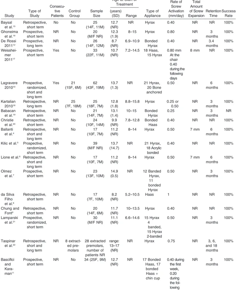
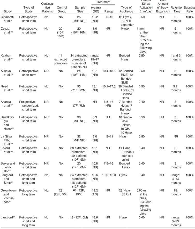
From the identified articles, the investigators inde-pendently extracted data referring to the year of publication, type of study, sample size, chronological age of subjects at the start of treatment, type of appliance, rate of activation, amount of expansion, duration of retention, and success rate (Table 2).
According to the Centre for Reviews and Dissemi-nation and to the PRISMA (Preferred Reporting Items for Systematic Reviews and Meta-Analyses) state-ment, evaluation of methodological quality gives an indication of the strength of evidence provided by the study because flaws in the design or conduct of a study can result in bias. However, no single approach for assessing methodological soundness is appropri-ate to all systematic reviews.13,14
Quality assessment, performed independently by the investigators, com-prised evaluation of the sample selection process, sample size estimation, adequacy of outcome mea-sures, adequacy of method error estimation, and adequacy of statistical analysis. If there was disagree-ment between the investigators, consensus was reached after discussion. The quality of the studies
was ranked on a 13-point scale and assessed as follows: high quality, a total score of 12 or 13 points; medium-high quality, a total score of 10 or 11 points; medium quality, a total score of 8 or 9 points; and low quality, a total score equal to or below 7 points. Statistical Analysis
A meta-analysis of the results of the studies that used comparable techniques of maxillary expansion was planned. Heterogeneity of the studies was
assessed first by calculating the I2 index. According
to the recommendation of the Cochrane Collaboration,
if heterogeneity is high (I2 . 75%), a meta-analysis
might produce misleading results, and omitting it from
a systematic review should be considered.13,14
RESULTS
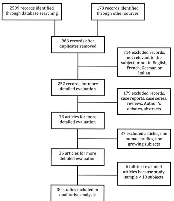
A search using the U.S. National Library of Medicine’s MeSH (medical subject headings) terms yielded the following results: PubMed yielded 146 publications; Embase, 546 publications; Cochrane Central, 92 publications; ISI Web Knowledge, 617 publications; and Ovid, 423 publications; Scopus, 685 publications. In addition, 173 records were identified through hand research. There was overlap among the databases. Application of the inclusion and exclusion criteria and follow-up on the referred studies identified 30 relevant publications (Figure 1).3–12,15–34
Heterogeneity of the results of the investigations with similar technique of maxillary expansion was high (.85%). A meta-analysis was not performed for this reason. Twenty-one studies3,4,6,8,9,15,18–20,22–26,28–34 were
retrospective, five studies7,11,12,16,17 were prospective,
and four studies5,10,21,27were prospective and
random-ized. Six studies10,18,22–24,33compared the results with a
control group of the same age, and eight stud-ies4,6,10,12,22,32–34 analyzed the long-term effects of
RME. Only two studies10,17
specified that the subjects of the study groups were selected consecutively. Mean age at the start of orthopedic expansion in the evaluated samples ranged from 7 to 17 years. Overall, in 24 studies3,4–7,9–11,15–19,21,22,24–27,30–34
the sample com-prised teenagers; in six studies,8,12,20,23,28,29the sample
was younger than 10 years (mean age, 8.6 years). The devices applied were bonded with acrylic coverage on the occlusal surface of posterior upper teeth or banded with the 2 or 4 anchored tooth (Hyrax or Haas type maxillary expanders). The methods used to detect the treatment effects were different:
two studies3,5used the dental casts before and after
treatment, 11 studies9,11,12,21,23,25–29,31 used
bidimensio-nal radiographic techniques, 10 studies4,6–8,10,15–18,20
used tridimensional radiographic techniques, 5 studies22,24,30,32,34
used histological evaluation, and 2 Table 1. Search Strategy
1. Orthodontic
2. Palatal expansion techniques OR palatal expansion technique 3. Rapid maxillary expansion
4. Rapid palatal expansion 5. Orthopedic expansion 6. Maxillary expansion
7. Palatal expansion techniques AND adverse effects 8. 2 OR 3 OR 4 OR 5 OR 6
Table 2. Characteristics of the Samples, Expansion Techniques, and Outcomes in the Included Studiesa Study Type of Study Consecu-tive Patients Control Group Sample Size Age at Start of Treatment Type of Appliance Rate of Screw Activation (mm/day) Total Amount of Screw Expansion Retention Time Success Rate (years) (SD) Range Baysal et al.15 Retrospective, short term No No 25 (14F, 11M) 12.7 (NR) NR Hyrax 0.40 NR NR 100% Ghoneima et al.16 Prospective, short term NR No 20 (M/F NR) 12.3 (1.9) 8–15 Hyrax 0.80 NR 3 months 100% De Rossi 201112 Prospective, long term NR No 26 (14F, 12M) 8.7 (NR) 6.9–10.9 Bonded Hyrax 0.40 NR 3.4 months 100% Weisshei-mer 201117 Prospective, short term Yes No 33 (22F, 11M) 10.7 (NR) 7.2–14.5 18 Haas, 15 Hyrax 0.80 mm At the chair 0.40 during the following days 8 mm NR 100% Lagravere 201010 Prospective, randomized, short and long term Yes 21 (15F, 6M) 62 (43F, 19M) 13.7 (1.3) NR 21 Hyrax, 20 Bone anchored 0.50 NR 6 months 100% Kartalian 201018 Retrospective, long term NR 25 (7F, 18M) 25 (18F, 7M) 12.8 (1.8) 8.8–15.8 Hyrax 0.25 or 0.50 NR 3 months 100% Babacan et al.19 Retrospective, short term NR No 21 (14F, 7M) 13.1 (1.4) 10–15 Bonded Hyrax 0.25 NR 3 months NR Christie et al.20 Retrospective, short term NR No 24 (10F, 14M) 9.9 (NR) 7.8–12.8 Bonded Haas 0.40 NR NR 100% Ballanti et al.4 Retrospective, short and long term NR No 17 (10F, 7M) 11.2 (NR) 8–14 Hyrax 0.50 7 mm 6 months 100%
Kilic et al.5 Prospective,
randomized, short term NR No 39 (M/F NR) 13.7 (14.7) NR 21 Hyrax, 18 Acrylic bonded 0.40 NR NR 100%
Lione et al.6 Retrospective,
short and long term NR No 17 (10F, 7M) 11.2 (NR) 8–14 Hyrax 0.50 7 mm 6 months 100% Olmez et al.7 Prospective, short term NR No 23 (13F, 10M) 14.9 (0.5) NR 12 Banded Hyrax, 11 bonded Hyrax 0.50 NR 3 months 100% da Silva Filho et al.8 Retrospective, short term NR No 17 (7F, 10M) 8.2 (NR) 5.2–10.5 Haas 1 NR NR 100% Chung and Font9 Retrospective, short term NR No 20 (14F, 6M) 11.7 (NR) 10–13.5 Hyrax 0.40 NR NR 100% Lamparski et al.21 Prospective, randomized, short term NR No 30 (M/F NR) 11.1 (NR) 6.6–14.6 15 Hyrax 4 banded, 15 Hyrax 2-banded 0.50 NR 3 months 100% Taspinar et al.22 Retrospective, short and long term NR 8 extract-ed pre-molars 28 extracted premolars, number of patients NR range: 13–17 (NR) NR Hyrax 0.75 NR 3, 6, and 18 months 100% Basciftci and Kara-man11 Prospective, short term NR No 34 (25F, 9M) 12.7 (NR) NR 17 Bonded Haas, 17 bonded Haas+ chin cup 0.40 during the first week, 0.20 during the fol-lowing days NR 3 months 100%
Study Type of Study Consecu-tive Patients Control Group Sample Size Age at Start of Treatment Type of Appliance Rate of Screw Activation (mm/day) Total Amount of Screw Expansion Retention Time Success Rate (years) (SD) Range Ciambotti et al.3 Retrospective, short term No No 25 (M/F NR) 10.2 (NR) 6–10 12 Hyrax, 13 NiTi expander 0.50 NR 3 months 100% Cozza et al.23 Retrospective, short term No 20 (10F, 10M) 20 (10F, 10M) 8.0 (NR) NR Hyrax 1 mm at the chair, 0.75 dur-ing the following days NR 6 months 100% Kayhan et al.24 Retrospective, short term No 11 extracted premolars 34 extracted premolars, number of patients NR range: 15–17 (NR) NR Bonded Haas 0.50 NR 1 and 3 months 100% Akkaya et al.25 Retrospective, short term NR No 24 (10F, 14M) 12.1 (NR) 10.4–13.5 12 Bonded RME, 12 Bonded SME 0.50 NR 3 months 100% Reed et al.26 Retrospective, short term No No 93 (71F, 22M) 13.1 (NR) 10.1–17.5 38 Banded Hyrax, 55 Bonded Haas 0.50 NR 5.2 months 100% Asanza et al.27 Prospective, randomized, short term NR No 14 (7F, 7M) NR 8.5–16 (NR) 7 Bonded Hyrax, 7 Banded Hyrax 0.40 NR 3 months 100% Sandikcio-glu and Hazar28 Retrospective, short term NR No 30 (M/F NR) 8.9 (NR) NR 10 remov-able plates, 10 QH, 10 Hyrax 0.50 NR 3 months 100% da Silva Filho et al.29 Retrospective, short term NR No 32 (M/F NR) 8.0 (NR) 5–11 Haas 0.80 NR NR 100% Erverdi et al.30 Retrospective, short term NR No 38 extracted premolars, 19 patients (10F, 9M) 15.1 (NR) NR 11 Haas, 9 Haas+ cast cap splint 0.40 NR 3 months 100% Sarver and John-ston31 Retrospective, short term NR No 20 (14F, 6M) 10.8 (NR) 7.5–16 Bonded Hyrax 0.40 NR 3 months 100% Langford and Sims32 Retrospective, short and long term No No 34 extracted premolars, 18 patients (12F, 6M) 13.6 (NR) 10.6–16.3 Hyrax 0.40 NR range: 3–13 months 100% Greenbaum and Zachris-son33 Retrospective, long term No 28 (23F, 5M) 61 (42F, 19M) 13.2 (1.9) NR 28 Haas, 33 QH 0.90 mm at the chair, 0.45 dur-ing the following days NR 15 months 100% Langford34 Retrospective, short and long term No No 18 (12F, 6M) 13.6 (NR) NR Hyrax 0.40 NR range: 3–13 months 100%
aNR indicates not reported; F, females; M, males; RME, rapid maxillary expansion; SME, slow maxillary expansion; QH, Quadhelix.
studies19,33 used other methods, such as probe and
intraoral measure of pulpal blood flow.
RME was used to treat transverse maxillary defi-ciency, and the device was maintained in situ as a passive retainer for a minimum of 3 months and a maximum of 18 months. The screw was activated twice a day in 25 studies,3–7,9–12,15,17,18,20,21,24–34 once a
day in 1 study,19 and more than twice a day in 4
studies.8,16,22,23 All the included studies used a clinical
evaluation, such as molar contact or overcorrection of 2–3 mm, to assess the required expansion. Only three investigations4,6,17
reported that the required expansion corresponded
to a precise amount of screw expansion of 7 mm4,6
and
8 mm.17
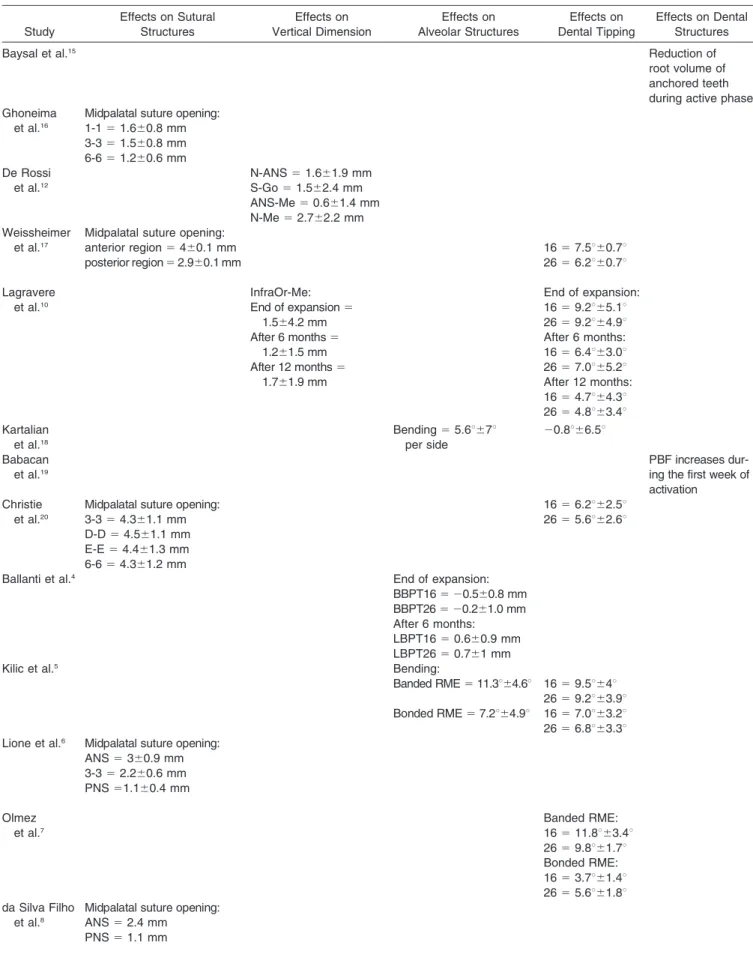
The effects of RME are shown in Table 3.
Quality Analysis
Research quality or methodological soundness was
medium-high in 2 studies10,17 (6.7%), medium in 8
studies4–7,12,16,18,20 (26.7%), and low in 20
stud-ies3 , 8 , 9 , 1 1 , 1 5 , 1 9 , 2 1 – 3 4
(66.7%). Twenty-one stud-ies3,4,6,8,9,15,18–20,22–26,28–34were retrospective, five studies
were prospective,7,10,11,16,17and only four studies5,10,21,27
were prospective and randomized. Withdrawals
(drop-outs) were declared in only one study.19
The most recurrent shortcomings were small sample size with no
consecutive cases, except for two studies,10,17implying
low power, problems of bias and confounding vari-ables, lack of method error analysis, blinding in measurements, and deficiency or lack of statistical Figure 1. PRISMA flow diagram. Flow chart illustrating the selection of relevant articles.
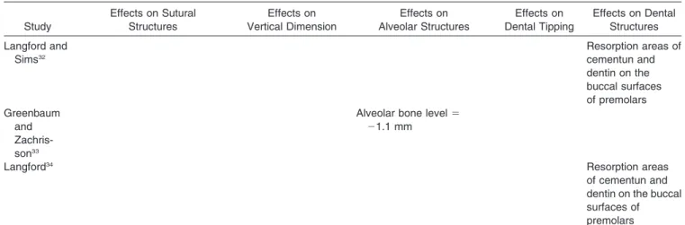
Table 3. Effects Evaluated in the Included Studiesa Study Effects on Sutural Structures Effects on Vertical Dimension Effects on Alveolar Structures Effects on Dental Tipping Effects on Dental Structures
Baysal et al.15 Reduction of
root volume of anchored teeth during active phase Ghoneima
et al.16
Midpalatal suture opening: 1-1 5 1.660.8 mm 3-3 5 1.560.8 mm 6-6 5 1.260.6 mm De Rossi et al.12 N-ANS 5 1.661.9 mm S-Go 5 1.562.4 mm ANS-Me 5 0.661.4 mm N-Me 5 2.762.2 mm Weissheimer et al.17
Midpalatal suture opening:
anterior region 5 460.1 mm 16 5 7.5u60.7u
posterior region 5 2.960.1 mm 26 5 6.2u60.7u
Lagravere et al.10 InfraOr-Me: End of expansion 5 1.564.2 mm After 6 months 5 1.261.5 mm After 12 months 5 1.761.9 mm End of expansion: 16 5 9.2u65.1u 26 5 9.2u64.9u After 6 months: 16 5 6.4u63.0u 26 5 7.0u65.2u After 12 months: 16 5 4.7u64.3u 26 5 4.8u63.4u Kartalian et al.18 Bending 5 5.6u67u per side 20.8u66.5u Babacan et al.19 PBF increases dur-ing the first week of activation
Christie et al.20
Midpalatal suture opening: 3-3 5 4.361.1 mm D-D 5 4.561.1 mm E-E 5 4.461.3 mm 6-6 5 4.361.2 mm 16 5 6.2u62.5u 26 5 5.6u62.6u
Ballanti et al.4 End of expansion:
BBPT16 5 20.560.8 mm BBPT26 5 20.261.0 mm After 6 months:
LBPT16 5 0.660.9 mm LBPT26 5 0.761 mm
Kilic et al.5 Bending:
Banded RME 5 11.3u64.6u 16 5 9.5u64u 26 5 9.2u63.9u Bonded RME 5 7.2u64.9u 16 5 7.0u63.2u 26 5 6.8u63.3u Lione et al.6 Midpalatal suture opening:
ANS 5 360.9 mm 3-3 5 2.260.6 mm PNS 51.160.4 mm Olmez et al.7 Banded RME: 16 5 11.8u63.4u 26 5 9.8u61.7u Bonded RME: 16 5 3.7u61.4u 26 5 5.6u61.8u da Silva Filho et al.8
Midpalatal suture opening: ANS 5 2.4 mm
Study Effects on Sutural Structures Effects on Vertical Dimension Effects on Alveolar Structures Effects on Dental Tipping Effects on Dental Structures Chung and Font9 SN-MP 5 1.7u61.2u N-Me 5 3.461.6mm PP-MP 5 1.6u61.7u Lamparski et al.21
Midpalatal suture opening: Anterior region 5 4.061.1 mm Posterior region 5 1.861 mm Taspinar et al.22 Fibrotic changes, increasing vessel diameter in the 3-month group Basciftci and Karaman11
SN-MP 5 1.8u61.4u Bending 5 5.1u63.8u PP-MP 5 1.2u61.4u
N-ANS 5 1.362.4mm ANS- Me 5 2.663.1mm Ciambotti
et al. 20013
Bending 5 5.1u65.4u 6.1u66.3u per side
Cozza et al.23 SN-PP 5 1.7u62.6u
N-Me 5 162.2 mm SN-ANS 5 1.662.3 mm Kayhan
et al.24
Fibrotic changes in the 3-month group Akkaya
et al.25
SN-MP 5 1.3u PP-MP 5 1.6u
Reed et al.26 Banded RME:
GoGn-SN 5 0.6u61.6u Anterior lower facial height
52.762.6 mm Anterior total facial height
54.463.5 mm Bonded RME:
Anterior lower facial height 52.2 62.4mm Anterior total facial height
54.363.1 mm Asanza et al.27 Banded RME: SN-PNS 5 1.9 mm SN-ANS 5 1.5 mm SN-MP 5 2.2u Bonded RME: SN-PNS 5 0.3 mm SN-ANS 5 1.7 mm SN-MP 5 1.5u Banded RME: 16 5 4.0u 26 5 3.7u Bonded RME: 16 5 2.6u 26 5 4.2u Sandikcioglu and Hazar28 SN-GoGn 5 1.5u61.3u SN-ANS 5 1.561.4 N-Gn 5 3.8 61.4 mm ANS-Gn 5 2.661.9 mm da Silva Filho et al. 199529
Midpalatal suture opening: 1-1 5 4.8 mm et al. 199430 Resorption areas of cementun and dentin on the buccal surfaces of premolars Sarver and Johnston31 Banded RME: SN-PNS 5 0.960.1 mm Bonded RME: SN-PNS 5 0.460.2 mm Table 3. Continued
methods. Furthermore, no study declared any power
analysis and only seven studies4–6,10,17,18,20 discussed
the possibility of a type II error occurring. Finally, six studies20,22,24,30,32,34 did not perform a statistical
analy-sis, focusing instead on a qualitative description of the results (Table 4).
DISCUSSION
The aim of the present systematic review was to assess whether RME causes AEs on dentoskeletal structures in growing subjects. With respect to previous reviews35,36
on RME, all studies that used computed tomography, which provides a more accurate three-dimensional assessment of changes induced by ortho-pedic forces, were searched and perused.
This systematic review included both retrospective and prospective studies, of which only four5,10,21,27
were randomized. The methodology of these investigations was generally of low and medium quality (Table 4); therefore, the findings should be interpreted with caution. A very serious limitation of most studies was the lack of an adequate untreated control group.
The analysis of the 30 articles3–12,15–34
included in the present review suggests that RME is an effective procedure that always produces transverse skeletal effects on the maxilla by opening the midpalatal suture in growing subjects, regardless of the type of palatal expander. Results showed that RME treatment was able to induce significantly more favorable skeletal changes in the transverse plane when it was initiated before the pubertal peak in skeletal growth.6,8,16,17,20,29,37
Indeed, the studies that reported a wider opening of
midpalatal suture were those20,29 in which the study
sample was younger, thereby confirming the role of treatment timing in the determination of skeletal modifications following RME therapy. In all studies
the amounts of midpalatal suture opening were greater in the anterior region than the posterior region, except
in the study by Christie et al.,20
who found a parallel manner of midpalatal suture opening.
RME treatment has been associated with downward movement of the maxillary posterior teeth and the maxilla.1–3,9,11
As reported by several articles included in the present review,9–12,23,25–28,31RME resulted in slight
inferior movement of the maxilla, as demonstrated by changes in the measurements SN-PNS (+0.9 mm) and
SN-ANS (+1.6 mm). Several authors9–12,23,25–28,31
point-ed out that this downward movement of the maxilla and premature dental contacts are responsible for the mandible rotation in a downward and backward direction with a mean increase in the following variables: SN-MP, 1.7u; PP-MP, 1.5u; SN-PP, 1.6u;
SN-GoGn, 1.1u.9–12,23,25–28,31Changes in vertical
dimen-sion before and after treatment with RME were less than 2 mm or 2u, and they may be not considered clinically relevant.38
In the short-term evaluation the only variable with a greater increase was the total anterior facial height (N-Me: 3.2 mm), which could be the transitory result of occlusal interferences.9,12,23,26
Though the maxilla was displaced downward, the facial growth patterns and/or the direction of mandib-ular growth were not modified 6 months after RME appliance removal, as observed in the only two
long-term studies, those of De Rossi et al.12and Lagravere
et al.10
These findings corroborate the results of other studies39,40
that also evaluated longitudinally the vertical effects associated with the RME, although in those cases RME was followed by other orthodontic
therapy.39,40 Moreover, Lagravere et al.10 and Cozza
et al.23 reported that, compared with an untreated
control group, changes in vertical dimension were negligible.10,23
The bonded RME showed less inferior Study Effects on Sutural Structures Effects on Vertical Dimension Effects on Alveolar Structures Effects on Dental Tipping Effects on Dental Structures Langford and Sims32 Resorption areas of cementun and dentin on the buccal surfaces of premolars Greenbaum and Zachris-son33
Alveolar bone level 5 21.1 mm
Langford34 Resorption areas
of cementun and dentin on the buccal surfaces of premolars
a1-1 indicates central incisor level; 3-3, canine level; D-D, first deciduous molar level; E-E, second deciduous molar level; 6-6, first molar level;
ANS, anterior nasal spine; PNS, posterior nasal spine; SN, Sella-Nasion line; S-Go, Sella gonion; PP, palatal plane; MP, mandibular plane; LBPT, lingual bone plate thickness; BBPT, buccal bone plate thickness; 16, right first molar; 26, left first molar; PBF, pulpal blood flow. Table 3. Continued
movement of PNS with respect to the banded RME.26,27,31 This might be due to the interocclusal
acrylic acting as an intrusive force to the basal bone of the maxilla by violating the freeway space and causing a passive stretch of the elevator musculature. The outcomes of the retrieved studies indicated an element
of vertical control using a bonded RME to counteract
the AEs seen with other expansion devices.26,27,31
Seven articles3,5,7,10,17,20,27showed that heavy forces
produced an increased buccal inclination of anchored teeth at the end of active phase regardless of the type of expanders.3,5,7,10,17,20,27
Ciambotti et al.3
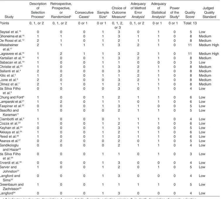
and Kilic Table 4. Description of Quality Score Assignment
Study Description of Selection Processa Retrospective, Prospective, or Randomizedb Consecutive Casesc Sample Sized Choice of Outcome Measuree Adequacy of Method Error Analysisf Adequacy of Statistical Analysisg Power of the Studyh Quality Scorei Judged Quality Standard Points 0, 1, or 2 0, 1, or 2 0 or 1 0 or 1 0, 1, 2, or 3 0, 1, or 2 0 or 1 0 or 1 Total: 13
Baysal et al.15 0 0 0 1 3 0 1 0 5 Low
Ghoneima et al.16 1 1 0 1 3 1 1 0 8 Medium
De Rossi et al.12 2 1 0 1 2 1 1 0 8 Medium
Weissheimer et al.17
2 1 1 1 3 2 1 0 11 Medium High
Lagravere et al.10 1 2 1 1 3 2 1 0 11 Medium High
Kartalian et al.18 1 0 0 1 3 2 1 0 8 Medium
Babacan et al.19 1 0 0 1 1 0 0 0 3 Low
Christie et al.20 1 0 0 1 3 2 1 0 8 Medium
Ballanti et al.4 2 0 0 0 3 2 1 0 8 Medium
Kilic et al.5 1 2 0 1 1 2 1 0 8 Medium
Lione et al.6 2 0 0 0 3 2 1 0 8 Medium
Olmez et al.7 2 1 0 1 2 1 1 0 8 Medium
da Silva Filho et al.8
0 0 0 0 3 0 1 0 4 Low
Chung and Font9 1 0 0 1 2 1 1 0 6 Low
Lamparski et al.21 1 2 0 1 1 0 1 0 6 Low
Taspinar et al.22 0 0 0 1 3 1 0 0 5 Low
Basciftci and Karaman11
1 0 0 0 2 1 1 0 5 Low
Ciambotti et al.3 1 0 0 0 1 1 1 0 4 Low
Cozza et al.23 1 0 0 1 2 1 1 0 6 Low
Kayhan et al.24 0 0 0 1 3 1 0 0 5 Low
Akkaya et al.25 1 0 0 1 2 1 1 0 6 Low
Reed et al.26 1 0 0 1 2 1 1 0 6 Low
Asanza et al.27 0 2 0 0 2 0 1 0 5 Low
Sandikcioglu and Hazar28 0 0 0 0 2 1 1 0 4 Low da Silva Filho et al.29 0 0 0 1 1 0 1 0 3 Low
Erverdi et al.30 0 0 0 1 3 0 0 0 4 Low
Sarver and Johnston31 1 0 0 1 2 0 1 0 5 Low Langford and Sims32 0 0 0 1 3 0 0 0 4 Low Greenbaum and Zachrisson33 1 0 0 1 1 1 1 0 5 Low Langford34 0 0 0 1 3 0 0 0 4 Low
a0 indicates inadequate description; 1, some details of sample selection missing; 2, in-depth description of sample selection. b0 indicates retrospective; 1, prospective; 2, prospective randomized.
c0 indicates sample comprised nonconsecutive patients or no information about the sample was included; 1, sample comprised consecutive patients. d0 indicates smaller than 20 subjects; 1, equal to or greater than 20 subjects.
e0 indicates inadequate outcome measure; 1, partially adequate outcome measure; 2, adequate outcome measure using two-dimensional
techniques; 3, adequate outcome measure using three-dimensional techniques or histologic evaluation.
f0 indicates method error not evaluated; 1, partially adequate method error analysis; 2, adequate method error. g0 indicates inadequate statistical analysis; 1, adequate statistical analysis.
h0 indicates power of the study not evaluated; 1, power of the study evaluated.
iThe quality of the studies was ranked on an 13-point scale and assessed as follows: 12–13 points indicates high; 10–11 points, medium-high;
et al.5
observed in the short term not only the dental tipping but also the effects of RME on alveolar structures; they found that the alveolar halves splayed buccally and carried the teeth with them. In growing subjects, anchored teeth and the alveolar bone are moved at the same time with the same magnitude and direction.3,5The inclination of the posterior teeth in the
bonded RME group appeared to be more stable for the
action of the acrylic coverage.5,7 Lagravere et al.10
reported that 12 months after the end of active expansion the dental tipping appeared reduced by
about half because of expansion relapse.10Kartalian et
al.18 compared measurements made on cone-beam
CT scans between patients with RME treatment and controls after a mean interval of 17.9 months. The RME group showed that the alveolus tipped buccally by 5.6u but the angulation of the dentition remained constant (20.8u) before and after treatment, probably because of postexpansion relapse of dental axial angulation.18
Greenbaum and Zachrisson33 demonstrated that,
after a retention period of 3 months, expansion groups exhibited minimal differences in periodontal condition
compared with the control group.33 Ballanti et al.,4 by
using computed tomography axial scans, reported that orthopedic forces in prepubertal subjects did not affect the alveolar bone palatal and buccal thickness after a 6-month retention period. A 6-month period was necessary to allow recovery of the alveolar plate when
the movement of anchored teeth is completed.4
Five articles evaluated histopathologically the vas-cular changes and root resorption of anchor extracted premolars after RME therapy.22,24,30,32,34
Kayhan et al.24
and Taspinar et al.22
observed the greatest fibrotic changes in periodontal bone in the 3-month group, less in the 6-month group, and very slight in the 18-month group. These fibrotic changes may represent the result of increasing duration of excessive force application, or they may occur as a result of the start of tooth
movement.22,24 Taspinar et al.22 found significantly
increased vessel diameter inside the pulp, which
disappeared 18 months after RME. Babacan et al.19
observed increased pulpal blood flow (PBF) during the first week of activation and PBF similar to initial values during the retention period, indicating that the vascular
changes are reversible.19 Langford,34 Langford and
Sims,32 and Erverdi et al.30 demonstrated that
contin-uous heavy forces as well as relapse forces capable of causing significant root resorption operate for up to 3 months after RME.Active resorption slowed signifi-cantly after about 3 months, with an evident trend to increased filling with cellular cementum of resorptive
defects.30,32,34 Finally, Baysal et al.15 performed cone
beam computed tomography to assess the root vo-lume and reported that in a study group with a mean
age of 12.7 years all investigated roots showed statistically significant volume loss after RME, with no difference between anchored and non-anchored teeth.15
The highest mean volume loss, 18.6 mm3
, was recorded for the mesiobuccal root of the first
molars, while Zachrisson41
reported that 2-mm apical root shortening was not detrimental to the function of dentition.15,41
CONCLUSIONS
N Most of the studies presented with methodological
problems: small sample size, bias and confounding variables, lack of method error analysis, blinding in measurements, and deficient or missing statistical methods. The quality level of the studies was not sufficient enough to draw any evidence-based conclusions.
N RME is an effective procedure that is able to produce
always transverse skeletal effects on the maxilla by opening the midpalatal suture in growing subjects regardless of the type of palatal expander.
N The vertical changes found after RME treatment,
although statistically significant, were small and proba-bly transitory. The bonded maxillary expansion appli-ance could be a viable option for correcting a narrow maxilla, regardless of the patient’s vertical problems or facial pattern.
N In growing subjects, heavy forces in the short-term
evaluation moved anchored teeth and the alveolar bone at the same time and with the same magnitude and direction. In the long-term evaluation, an upright-ing of anchored teeth was observed. Vascular changes after RME are reversible, and active root resorption appeared along with increased filling with cellular cementum after 3 months.
REFERENCES
1. Wertz RA. Skeletal and dental changes accompanying rapid midpalatal suture opening.Am J Orthod. 1970;58:41–64. 2. Haas AJ. Palatal expansion: just the beginning of dentofacial
orthopedics.Am J Orthod. 1970;57:219–255.
3. Ciambotti C, Ngan P, Orth C, Durkee M, Kohli K, Kim H. A comparison of dental and dentoalveolar changes between rapid palatal expansion and nickel-titanium palatal expan-sion appliances.Am J Orthod Dentofacial Orthop. 2001;119: 11–20.
4. Ballanti F, Lione R, Fanucci E, Franchi L, Baccetti T, Cozza P. Immediate and post-retention effects of rapid maxillary expansion investigated by computed tomography in growing patients.Angle Orthod. 2009;79:24–29.
5. Kilic N, Kiki A, Oktay H. A comparison of dentoalveolar inclination treated by two palatal expanders.Eur J Orthod. 2008;30:67–72.
6. Lione R, Ballanti F, Franchi L, Baccetti T, Cozza P. Treatment and posttreatment skeletal effects of rapid maxillary ex-pansion studied with low-dose computed tomography in
growing subjects.Am J Orthod Dentofacial Orthop. 2008;134: 389–392.
7. Olmez H, Akin E, Karacay S. Multitomographic evaluation of the dental effects of two different rapid palatal expansion appliances.Eur J Orthod. 2007;29:379–385.
8. Da Silva Filho OG, Lara TS, da Silva HC, Bertoz AF. Post expansion evaluation of the midpalatal suture in children submitted to rapid palatal expansion: a CT study. J Clin Pediatr Dent. 2006;31:142–148.
9. Chung C-H, Font B. Skeletal and dental changes in the sagittal, vertical, and transverse dimensions after rapid palatal expansion. Am J Orthod Dentofacial Orthop. 2004; 126:569–575.
10. Lagravere MO, Carey J, Heo G, Toogood RW, Major PW. Transverse, vertical, and anteroposterior changes from bone-anchored maxillary expansion vs traditional rapid maxillary expansion: a randomized clinical trial.Am J Orthod Dento-facial Orthop. 2010;137:304.e1–304.e12.
11. Basciftci FA, Karaman AI. Effects of a modified acrylic bonded rapid maxillary expansion appliance and vertical chin cap on dentofacial structures.Angle Orthod. 2002;72: 61–71.
12. De Rossi M, De Rossi A, Abrao J. Skeletal alterations associated with the use of bonded rapid maxillary expansion appliance.Braz Dent J. 2011;22:334–339.
13. Moher D, Liberati A, Tetzlaff J, Altman DG. The PRISMA group (2009) Preferred Reporting Items for Systematic Reviews and Meta-analyses: the PRISMA statement.PLoS Med 6(7):e1000097. Doi:10.1371/journal.pmed.1000097. 14. The Cochrane Collaboration. In: Higgins JPT, Green S, eds.
Cochrane Handbook for Systematic Reviews of Interven-tions. Chichester, UK: Wiley-Blackwell; 2008:276–282. 15. Baysal A, Karadede I, Hekimoglu S, Ucar F, Ozer T, Veli I,
Uysal T. Evaluation of root resorption following rapid maxillary expansion using cone-beam computed tomogra-phy.Angle Orthod. 2011 August 15. [Epub ahead of print]. 16. Ghoneima A, Abdel-Fattah E, Hartsfield J, El-Bedwehi A,
Kamel A, Kula K. Effects of rapid maxillary expansion on the cranial and circummaxillary sutures.Am J Orthod Dentofa-cial Orthop. 2011;140:510–519.
17. Weissheimer A, de Menezes LM, Mezomo M, Dias DM, Santayana de Lima EM, Deon Rizzatto SM. Immediate effects of rapid maxillary expansion with Haas-type and hyrax-type expanders: a randomized clinical trial. Am J Orthod Dentofacial Orthop. 2011;140:366–376.
18. Kartalian A, Gohl E, Adamian M, Enciso R. Cone-beam computerized tomography evaluation of the maxillary dentoskeletal complex after rapid palatal expansion. Am J Orthod Dentofacial Orthop. 2010;138:486–492. 19. Babacan H, Doruk C, Bicakci AA. Pulpal blood flow changes
due to rapid maxillary expansion.Angle Orthod. 2010;80: 1136–1140.
20. Christie KF, Boucher N, Chung C-H. Effects of bonded rapid palatal expansion on the transverse dimensions of the maxilla: a cone-beam computed tomography study. Am J Orthod Dentofacial Orthop. 2010;137:S79–S85.
21. Lamparski DG, Rinchuse DJ, Close JM, Sciote JJ. Comparison of skeletal and dental changes between 2-point and 4-point rapid palatal expanders.Am J Orthod Dentofa-cial Orthop. 2003;123:321–328.
22. Taspinar F, Akgul N, Simsek G, Ozdabak N, Gundogdu C. The histopathologic investigation of pulpal tissue following heavy orthopaedic forces produced by rapid maxillary expansion.J Int Med Res. 2003;31:197–201.
23. Cozza P, Giancotti A, Petrosino A. Rapid palatal expansion in mixed dentition using a modified expander: a cephalo-metric investigation.J Orthod. 2001;28:129–134.
24. Kayhan F, Kucukkeles N, Demirel D. A histologic and histomorphometric of pulpal reactions following rapid palatal expansion. Am J Orthod Dentofacial Orthop. 2000;117: 465–473.
25. Akkaya S, Lorenzon S, Ucem TT. A comparison of sagittal and vertical effects between bonded rapid and slow maxillary expansion procedures. Eur J Orthod. 1999;21: 175–180.
26. Reed N, Ghosh J, Nanda RS. Comparison of treatment outcomes with banded and bonded RPE appliances. Am J Orthod Dentofacial Orthop. 1999;116:31–40. 27. Asanza S, Cisneros GJ, Nieberg LG. Comparison of Hyrax
and bonded expansion appliances.Angle Orthod. 1997;67: 15–22.
28. Sandikcioglu M, Hazar S. Skeletal and dental changes after maxillary expansion in the mixed dentition. Am J Orthod Dentofacial Orthop. 1997;111:321–327.
29. da Silva Filho OG, do Prado Montes LA, Torelly LF. Rapid maxillary expansion in the deciduous and mixed dentition evaluated through posteroanterior cephalometric analysis. Am J Orthod Dentofacial Orthop. 1995;107:268–275. 30. Erverdi N, Okar I, Kucukkeles N, Arbak S. A comparison of
two different rapid palatal expansion techniques from the point of root resorption.Am J Orthod Dentofacial Orthop. 1994;106:47–51.
31. Sarver DM, Johnston MW. Skeletal changes in vertical and anterior displacement of the maxilla with bonded rapid palatal expansion appliances. Am J Orthod Dentofacial Orthop. 1989;95:462–466.
32. Langford SR, Sims MR. Root surface resorption, repair, and periodontal attachment following rapid maxillary expansion in man.Am J Orthod. 1982;81:108–115.
33. Greenbaum KR, Zachrisson BU. The effect of palatal expansion therapy on the periodontal supporting tissues. Am J Orthod. 1982;81:12–21.
34. Langford SR. Root resorption extremes resulting from clinical RME.Am J Orthod. 1982;81:371–377.
35. Lagravere MO, Major PW, Flores-Mir C. Long-term skeletal changes with rapid maxillary expansion: a systematic review.Angle Orthod. 2005;75:1046–1052.
36. Lagravere MO, Major PW, Flores-Mir C. Long-term den-tal arch changes after rapid maxillary expansion treat-ment: a systematic review. Angle Orthod. 2005;75: 155–161.
37. Baccetti T, Franchi L, Cameron CG, McNamara JA Jr. Treatment timing for rapid maxillary expansion. Angle Orthod. 2001;71:343–350.
38. Cozza P, Baccetti T, Franchi L, DeToffol L, McNamara JA Jr. Mandibular changes produced by functional appliances in Class II malocclusion: a systematic review.Am J Orthod Dentofacial Orthop. 2006;129:599.e1–599.e12.
39. Chang JY, McNamara JA, Herberger TA. A longitudinal study of skeletal side effects induced by rapid maxillary expansion. Am J Orthod Dentofacial Orthop. 1997;112: 330–333.
40. Velazquez P, Benito E, Braco La. Rapid maxillary expan-sion. A study of the long term effects. Am J Orthod Dentofacial Orthop. 1996;109:361–367.
41. Zachrisson BU. Iatrogenic tissue damage following ortho-dontic treatment.Trans Eur Orthod Soc. 1975:488–501.