Development and external validation of new
ultrasound-based mathematical models for preoperative
prediction of high-risk endometrial cancer
C. VAN HOLSBEKE*†, L. AMEYE‡, A. C. TESTA§, F. MASCILINI§, P. LINDQVIST¶,
D. FISCHEROVA**, F. FR ¨UHAUF**, S. FRANSIS††, E. DE JONGE†, D. TIMMERMAN* and
E. EPSTEIN¶
*Department of Obstetrics and Gynaecology, University Hospitals Leuven, Leuven, Belgium; †Department of Obstetrics and Gynaecology, Ziekenhuis Oost-Limburg, Genk, Belgium; ‡Department of Development and Regeneration, KU Leuven - University of Leuven, Leuven, Belgium; §Istituto di Clinica Ostetrica e Ginecologica, Universit `a Cattolica del Sacro Cuore, Roma, Italy; ¶Department of Obstetrics and Gynecology, Karolinska University Hospital, Stockholm, Sweden; **Gynecological Oncology Center, Department of Obstetrics and Gynecology, First Faculty of Medicine and General University Hospital, Charles University, Prague, Czech Republic; ††Department of Pathology, Ziekenhuis Oost-Limburg, Genk, Belgium
K E Y W O R D S: endometrial cancer; high risk; mathematical models; prediction
ABSTRACT
Objectives To develop and validate strategies, using new
ultrasound-based mathematical models, for the prediction of high-risk endometrial cancer and compare them with strategies using previously developed models or the use of preoperative grading only.
Methods Women with endometrial cancer were
prospec-tively examined using two-dimensional (2D) and three-dimensional (3D) gray-scale and color Doppler ultrasound imaging. More than 25 ultrasound, demo-graphic and histological variables were analyzed. Two logistic regression models were developed: one ‘objective’ model using mainly objective variables; and one ‘subjec-tive’ model including subjective variables (i.e. subjective impression of myometrial and cervical invasion, preop-erative grade and demographic variables). The following strategies were validated: a one-step strategy using only preoperative grading and two-step strategies using preop-erative grading as the first step and one of the new models, subjective assessment or previously developed models as a second step.
Results One-hundred and twenty-five patients were
included in the development set and 211 were included in the validation set. The ‘objective’ model retained preoperative grade and minimal tumor-free myometrium as variables. The ‘subjective’ model retained preoperative grade and subjective assessment of myometrial invasion. On external validation, the performance of the new
Correspondence to: Dr C. Van Holsbeke, Department of Obstetrics and Gynaecology, Z.O.L. Genk, Belgium Schiepse Bos 6, B-3600 Genk, Belgium (e-mail: [email protected])
Accepted: 17 September 2013
models was similar to that on the development set. Sensitivity for the two-step strategy with the ‘objective’ model was 78% (95% CI, 69–84%) at a cut-off of 0.50, 82% (95% CI, 74–88%) for the strategy with the ‘subjective’ model and 83% (95% CI, 75–88%) for that with subjective assessment. Specificity was 68% (95% CI, 58–77%), 72% (95% CI, 62–80%) and 71% (95% CI, 61–79%) respectively. The two-step strategies detected up to twice as many high-risk cases as preoperative grading only. The new models had a significantly higher sensitivity than did previously developed models, at the same specificity.
Conclusion Two-step strategies with ‘new’
ultrasound-based models predict high-risk endometrial cancers with good accuracy and do this better than do previously developed models. Copyright 2013 ISUOG. Published by John Wiley & Sons Ltd.
INTRODUCTION
Endometrial cancer is the most common gynecologic malignancy in developed countries, with an incidence of 12.9 per 100 000 women and a mortality rate of 2.4 per 100 000 women1. Prognosis in the early stages
of the disease (Stage I–II) depends on age, histological type and grading, depth of myometrial invasion, cervical invasion and lymph-node metastases. Most women with endometrial cancer are diagnosed at an early stage when the prognosis is excellent2. However, women with
high-risk endometrial cancer (high grade, deep myome-trial invasion or cervical invasion) have a worse prognosis. These women are at a higher risk of recurrence and might benefit from extended surgical staging with systematic pelvic and para-aortic lymphadenectomy in order to tailor adjuvant therapy. On the other hand, it is important to avoid overtreatment, as most of these women are elderly with comorbidities. Lymphadenectomy shows no survival benefit in low-risk endometrial cancer patients and, in fact, results in increased complications and higher morbidity3–7.
Thus far, preoperative risk stratification is mainly based on the use of preoperative grading (classifying Grade 3 endometrioid and the non-endometrioid histological subtypes (clear cell or serous carcinomas and carcinosar-comas) as high risk) and computed tomography scans to screen for enlarged lymph nodes. In most countries preop-erative sonographic assessment of the uterus to evaluate myometrial infiltration and cervical invasion is not yet standard practice. However, several studies have assessed subjective assessment of myometrial invasion using transvaginal ultrasound, with sensitivities of 68–93% and specificities of 82–83%8–11. There are fewer publications
on the use of ultrasound to assess cervical invasion, but the results are generally very good8,10,12. The accuracy of ultrasonography seems to be comparable with that of magnetic resonance imaging (MRI)13–16. Nevertheless, at
present, MRI is less accessible because it is more expen-sive and time consuming and, in most centers, the waiting lists are quite long. Therefore, if access to MRI is lim-ited, priority could be given to patients with poor image quality during ultrasound as a result of obesity, fibroids, adenomyosis or the anatomic position of the uterus.
Currently, most studies that use ultrasound to predict high-risk endometrial cancer or deep myometrial invasion are based on the performance of subjective assessment or ultrasound parameters only. Our hypothesis was that mathematical models combining ultrasound data with other available preoperative information could improve the ability to identify high-risk endometrial cancer.
The primary aim of this study was to assess whether new mathematical models that include ultrasound, demo-graphic variables and grade may improve the preoperative identification of women with high-risk endometrial can-cer. The secondary aim was to validate the new models and compare them with previously published models17,18.
METHODS
This was a prospective international multicenter study that included patients with endometrial cancer confirmed histologically between January 2007 and May 2009. The four centers that contributed to this study were the gynecologic ultrasound unit at the Department of Obstet-rics and Gynecology, Lund University Hospital, Sweden; the Gynecologic Oncology Unit, Catholic University of the Sacred Heart, Rome, Italy; the Department of Obstetrics and Gynecology, Leuven University Hospitals, Leuven, Belgium and the Department of Obstetrics
and Gynecology of Ziekenhuis Oost-Limburg, Genk, Belgium; and the Gynecological Oncology Center and Department of Obstetrics and Gynecology, Charles University, Prague, Czech Republic. Ethical approval was obtained from the Ethics Committee of Lund University (LU-412-07) and from the local Ethics Committees of the Catholic University of the Sacred Heart (Rome, Italy), University Hospitals Leuven (Leuven, Belgium) and Ziekenhuis Oost-Limburg (Genk, Belgium).
We included patients with a histologically proven diag-nosis of endometrial cancer obtained by pipelle, dilata-tion and curettage (D&C) or hysteroscopy. All patients underwent a standardized ultrasound examination by an experienced ultrasound examiner (C.V.H., E.E., A.C.T. or D.F.) following a strict protocol. The two-dimensional (2D) and three-dimensional (3D) gray-scale and vascular pattern criteria were defined at a consensus reading session including all examiners using the International Endome-trial Tumor Analysis (IETA) terminology19. The objective
ultrasound variables that were assessed during real-time 2D examination included: tumor size in three perpendic-ular diameters; minimal tumor-free margin (the minimal thickness of the tumor-free myometrium, i.e. the area at which the tumor is invading most (measured perpen-dicular to the serosa)); the tumor/uterine anteroposterior (AP) diameter ratio; tumor echogenicity; the endome-trial/myometrial border (regular/irregular); the presence and number of fibroids; the distance from the outer cervi-cal os to the caudal border of the lesion; and vasculariza-tion characteristics. The amount of tumor vascularizavasculariza-tion was classified subjectively using a ‘color score’ (absent (= 1), minimal (= 2), moderate (= 3) or high (= 4)), as introduced by the International Ovarian Tumor Analysis (IOTA) group20. From the stored 3D volumes we
calcu-lated the tumor volume, the uterine volume and the tumor volume/uterine volume ratio, and we assessed vascular morphology using GE 4D View (GE Healthcare, Little Chalfont, UK) or Philips QLAB (Koninklijke Philips N.V., Amsterdam, The Netherlands), using 30◦rotational steps. The examiners also reported on their subjective assess-ment of myometrial and cervical infiltration. Subjective assessment is the personal impression of the ultrasound examiner of depth of invasion and cervical invasion whilst scanning the uterus. Patients were classified as high risk when the ultrasound examiner believed that the tumor invaded one half or more of the myometrium or when cervical stromal invasion was suspected.
The ultrasound equipment used in Lund was a GE Voluson E8 ultrasound system with a RIC5-9 transducer or a Philips IU22 Ultrasound system with a 3D9-3v transducer. In the hospitals in Rome, Leuven and Genk, the GE Voluson E8 with a RIC5-9 transducer was used. Static images with all measurements, videoclips with and without power Doppler, and 3D volumes with and without power Doppler were collected for every patient.
All women underwent an extrafascial abdominal hysterectomy with bilateral salpingoophorectomy and systematic pelvic and para-aortic lymphadenectomy, if appropriate, according to the local protocols. The
International Federation of Gynecology and Obstetrics (FIGO) stage that was assigned to each case was based on final histology. In each center, the surgical specimens were examined by a dedicated pathologist with substantial experience in gynecologic oncology using a predetermined protocol. The histopathological variables that were assessed were: histological subtype (endometrioid/non-endometrioid; based on preoperative biopsy and on hysterectomy); grade of differentiation (based on preoperative biopsy and on hysterectomy); tumor size, macroscopic growth pattern and macroscopic estimation of infiltration; number of fibroids; microscopic infiltration; minimal tumor-free myometrium; cervical invasion; extension to parametria; and FIGO classifica-tion. The tumors were classified according to the criteria recommended by the World Health Association (WHO) and the pathological tumor stage according to the criteria recommended by the International Union Against Cancer (TNM Classification of malignant tumors)21. The
FIGO 2009 staging criteria were used22. Only epithelial
malignant tumors and carcinosarcomas (malignant mesodermal mixed tumors (MMMTs)) were included. Endometrial adenocarcinoma was classified into three grades (Grade 1, well differentiated; Grade 2, moderately differentiated; and Grade 3, poorly differentiated). Other histological subtypes, such as serous/seropapillary, clear cell and carcinosarcoma, were classified as Grade 3. The final allocation of high-risk status of a tumor was based on the microscopic findings in the hysterectomy specimen (i.e. Stage Ib or more, or Grade 3/non-endometrioid histotype (serous, clear cell or carcinosarcoma)).
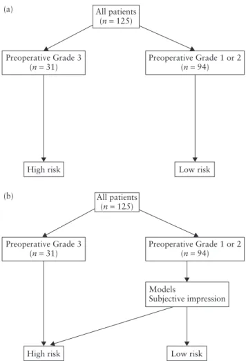
The prediction of high-risk cancer was performed using either the preoperative grading only (i.e. the ‘one-step strategy’) or a ‘two-step strategy’ (Figure 1). In the first phase of the study, all patients with a poorly differentiated carcinoma or a non-endometrioid cancer (serous, clear cell or carcinosarcoma) on preoperative biopsy were automatically classified as high-risk cancers. For the remaining patients (Grade 1 and Grade 2) we created mathematical models to predict high-risk cancer, ultimately adding the results from both groups. In the second phase we externally validated the new models used in the two-step strategy and compared their performance with previously published strategies.
The gold standard was the FIGO classification that was assigned after final histopathological analysis of the specimen.
For the development of a new two-step model we used 125 women examined in Lund, Leuven/Genk and Rome. Among these, 94 had a Grade 1 or a Grade 2 tumor based on preoperative histology. The external validation was based on 211 new consecutive cases examined in Prague that had not contributed to the development set; 163 of these cases had a Grade 3 tumor on preoperative histology and were used for model validation.
We developed two new logistic regression models. The ‘objective model’ considered all the variables listed in the univariate analysis and subjective assessment of cervical invasion as possible predictors. The ‘subjective
All patients (n = 125) Preoperative Grade 3 (n = 31) (a) (b)
High risk Low risk
High risk Low risk
Preoperative Grade 1 or 2 (n = 94) All patients (n = 125) Preoperative Grade 3 (n = 31) Preoperative Grade 1 or 2(n = 94) Models Subjective impression
Figure 1 Flow charts for the prediction of high-risk endometrial cancer using one-step (a) and two-step (b) strategies.
model’ considered subjective variables (i.e. subjective impression of myometrial and cervical stromal invasion, preoperative grading and all demographic variables) as possible predictors.
Old models in the validation included:
• The Karlsson criteria. Karlsson defined the following criteria to predict deep myometrial infiltration; the maximal AP thickness of the endometrial lesion measured in the sagittal plane (d1) divided by the AP uterine diameter (d2) (Figure 2). More than 50% myometrial infiltration was defined as d2/d1≥ 50%; less than 50% myometrial infiltration was defined as d2/d1 < 50%10. Cervical infiltration was suspected
when there was no clear demarcation of the endometrial lesion toward the cervical canal17.
• The logistic regression model of De Smet et al. This mathematical model was developed to predict deep myometrial invasion (> 50% infiltration), and variables selected included preoperative grade, number of fibroids, endometrial thickness and endometrial volume (calculated from three measurements of the endometrium in two perpendicular planes)18.
Statistical analysis
All ultrasound and demographic variables were first assessed in a univariate analysis. Differences between high
– = d2 × × × × × × (a) (b) (c) × × × – × = d1 – = d2 × – × = d1 – = d2 × – × = d1 × ×
Figure 2 Schematic representation, according to the criteria of Karlsson17, to demonstrate: (a) deep myometrial infiltration (d2/d1≥ 50%), myometrial infiltration of less than 50% (d2/d1 < 50%) and (c) cervical infiltration.
risk and low risk in continuous variables were assessed using a t-test and the Mann–Whitney U-test; differences in categorical variables were assessed using the chi-square test and Fisher’s exact test.
As described previously, we decided to create two new mathematical models: one containing mainly objective measurements, ‘the objective model’, and the other also including subjective assessment of cervical and myometrial invasion. By the use of stepwise logistic regression we selected variables for the models. Only variables with a univariate P < 0.2 were considered as possible predictors in the multivariate model.
The different prediction strategies were tested at two different cut-off levels: 0.40 and 0.50. The cut-off level of 0.50 gave the best sensitivity for an acceptable level of specificity. The cut-off level of 0.40 was tested to evaluate the level of specificity when increasing sensitivity. The cut-off levels represent values for the mathematical model and are not related to absolute risk. To compare differences in sensitivity and specificity between the different strategies, the McNemar test was used. All statistical analyses were performed with SAS 9.3 (SAS Institute Inc., Cary, NC, USA). All P-values were two-sided and P < 0.05 was considered significant.
RESULTS
Of the 144 patients initially included in this study, 19 were excluded because no information on preoperative grade was available. The demographic and clinical characteristics for women with high- and low-risk cancer from development and validation sets, according to final histology, are presented in Table 1. Women with high-risk
cancer were significantly older than were women with low-risk cancer.
The preoperative histology, final histology and final staging of development and validation sets are presented in Table 2. Around half of the tumors were Stage IA, in both development and validation sets. On the preoperative biopsy, 25% of cases in both development and validation sets had a high-grade tumor. The ultrasound examiner assumed that there was cervical invasion in 17% (21/125) of cases and deep myometrial invasion in 41% (51/125).
Table S1 shows the univariate analysis of the 2D and 3D gray-scale and color Doppler ultrasound variables for the women with Grades 1 and 2 endometrial cancer according to preoperative biopsy included in the develop-ment set (n= 94). The retained variables for the objective model were preoperative grade and minimal tumor-free myometrium. The subjective model used subjective assessment of myometrial invasion, preoperative grade and age as variables.
The probability of having high-risk cancer is equal to
y1/(1+ e–z), where z is −0.4468 + 1.2921 × preoperative
grading –0.2292 × minimal-free myometrium (for the objective model) and−2.6276 + 1.1458 × preoperative grading+ 2.2514 × subjective evaluation of myometrial invasion (for the subjective model).
Table 3 shows the results for predicting high-risk cancer for: a one-step strategy using Grade 3 only; the combined two-step strategies as described above; and the performance of the new ultrasound-based mathematical models and subjective assessment alone (when tested only on the preoperative Grade 1 and 2 patients). Of the 31 women classified as high risk according to preoperative grading, one woman was later found to be low risk
Table 1 Demographic and baseline characteristics of patients included in the total development set (n= 125 = 94 + 31) and the total validation set (n= 211 = 163 + 48) stratified by ‘low-risk’ and ‘high-risk’ cancer according to final pathology
Development set Validation set
Characteristic Total (n= 125) Low risk (n= 55) High risk (n= 70) P Total (n= 211) Low risk (n= 90) High risk (n= 121) P Age (years) 66± 10 63± 9 68± 10 0.006 65± 9 62± 9 66± 9 <0.001 66 (37–92) 64 (37–82) 67 (44–92) 64 (41–89) 61 (41–81) 66 (44–89) BMI* 29± 7 29± 6 29± 7 0.89 31± 8 32± 8 31± 7 0.34 27 (19–55) 27 (19–46) 27 (20–55) 30 (18–61) 30 (18–55) 30 (20–61) Postmenopausal No 8 (6) 3 (5) 5 (7) 1 17 (8) 12 (13) 5 (4) 0.02 Yes 117 (94) 52 (95) 65 (93) 194 (92) 78 (87) 116 (96) Hormone treatment No 112 (90) 48 (87) 64 (91) 0.56 199 (94) 83 (92) 116 (96) 0.37 Yes 13 (10) 7 (13) 6 (9) 12 (6) 7 (8) 5 (4) Current low-potency estrogen use No 112 (90) 47 (85) 65 (93) 0.24 210 (99.5) 89 (99) 121 (100) — Yes 13 (10) 8 (15) 5 (7) 1 (0.5) 1 (1) —
Current tamoxifen use
No 122 (98) 52 (95) 70 (100) 0.08 209 (99) 90 (100) 119 (98) — Yes 3 (2) 3 (5) — 2 (1) — 2 (2) Family history Breast cancer No 106 (85) 47 (85) 59 (84) 1 200 (95) 86 (96) 114 (94) 0.76 Yes 19 (15) 8 (15) 11 (16) 11 (5) 4 (4) 7 (6) Gynecological cancer No 107 (86) 45 (82) 62 (89) 0.31 198 (94) 80 (89) 118 (98) 0.02 Yes 18 (14) 10 (18) 8 (11) 13 (6) 10 (11) 3 (2) Diagnosis by: Pipelle biopsy 21 (19) 12 (27) 9 (14) 0.14 — — — 0.58 Hysteroscopy only 19 (17) 4 (9) 15 (23) 93 (46) 42 (48) 51 (44) D&C only 52 (48) 22 (49) 30 (47) 82 (40) 36 (41) 46 (40)
Hysteroscopy and D&C 17 (16) 7 (16) 10 (16) 29 (14) 10 (11) 19 (16)
No data/not performed/ other method (tru-cut biopsy/biopsy of meta)
16 10 6 7 2 5
Values are given as n, n (%), mean± SD or median (range). *Data for BMI missing in three low-risk and 12 high-risk cases in the development set. BMI, body mass index; D&C, dilatation and curettage.
according to final histology. Subjective assessment alone performed as well as any model. A two-step strategy with the ‘subjective’ or ‘objective’ model as the second step achieved a significantly higher sensitivity in comparison with preoperative grading only. All two-step strategies using models had similar specificities, indicating that ultrasound in the combination with grade will improve the correct identification of high-risk cases. The two-step strategies detected up to twice as many high-risk cases as did the one-step strategy based on grade only. Differences when using different cut-offs are explained below.
In Tables S2 and S3 we compared the P-values for the difference in sensitivity and specificity between the two-step strategy using models or subjective assessment, and between the use of different cut-offs in the development set. The only difference between the models and subjective assessment using the 0.5 cut-off was a significantly lower specificity for the objective model (P= 0.03), and when using the 0.4 cut-off both objective and subjective models had a lower specificity (both P < 0.001), the subjective model having a higher sensitivity (P= 0.03) compared with subjective assessment. Thus, the 0.5 cut-off achieved
a result more similar to subjective assessment, but the use of a 0.4 cut-off detected more high-risk cases at a cost of lower specificity.
Table 4 shows the external validation of preoperative grading only and the two-step strategies with our ‘new’ models, subjective assessment or previously published models (of Karlsson and coworkers and De Smet and co-workers) as the second step. As in Table 3 the performance is presented for the models individually or for the whole two-step strategy. Of the 48 women classified as high risk according to preoperative grading, four were later found to be low risk according to final histology. The ultrasound examiner assumed that there was cervical invasion in 11% (24/211) cases and deep myometrial invasion in 52% (109/211). The performance of the models in the validation set was very similar to the results from the development set.
Tables S4 and S5 compare the P-values for the differences in sensitivity and specificity between two-step strategies using new and old models as the second step and subjective assessment using different cut-off levels and subjective assessment for the validation set. The
Table 2 Histological characteristics of patients included in the total development set (n= 125 = 94 + 31) and the validation set (n = 211= 163 + 48)
Development set Validation set
Total Grade 3 Grade 1 or 2 Total Grade 3 Grade 1 or 2
Characteristic (n= 125) (n= 40) (n= 85) (n= 211) (n= 51) (n= 160) Stage, FIGO 200922 IA 64 (51) 11 (27.5) 53 (62) 106 (50) 17 (33) 89 (56) IB 28 (22) 9 (22.5) 19 (22) 58 (27) 10 (20) 48 (30) II 11 (9) 4 (10.0) 7 (8) 19 (9) 9 (18) 10 (6) IIIA 3 (2) 3 (7.5) — 6 (3) 3 (6) 3 (2) IIIB 2 (2) 1 (2.5) 1 (1) 1 (<1) 1 (2) — IIIC 11 (9) 6 (15.0) 5 (6) 16 (8) 7 (14) 9 (6) IVA — — — — — — IVB 6 (5) 6 (15.0) — 5 (2) 4 (8) 1 (<1)
Development set (n= 125) Validation set (n= 211) Type and grade preoperatively
Endometrioid
Grade 1 65 (52) 89 (42)
Grade 2 29 (23) 74 (35)
Grade 3 20 (16) 31 (15)
Non-endometrioid 11 (9) 17 (8)
Type and grade postoperatively Endometrioid
Grade 1 51 (41) 100 (47)
Grade 2 34 (27) 60 (28)
Grade 3 19 (15) 35 (16)
Non-endometrioid 21 (17)* 16 (8)†
Values are given as n (%). *Non-endometrioid histotypes, after final surgery, in the development set (four carcinosarcoma, three clear cell, three mixed, three seropapillary and eight serous). †Non-endometrioid histotypes, after final surgery, in the validation set (four clear cell, two mixed, seven serous, two undifferentiated, one squamous).
sensitivity for any of the new models and for subjective assessment performed significantly better compared with the old models at the same level of specificity.
In Figure 3a and b, pie charts present and compare the performance of all strategies on the development and the validation set.
Figure 4 shows the percentage of additional women with a correct diagnosis compared with the one-step strat-egy using preoperative Grade 3 only in the validation set. The subjective models and subjective assessment improved the accuracy by 16%. In the development set the accuracy was improved by up to 29% (for subjective assessment and the subjective model (at the cut-off of 0.5)).
DISCUSSION
Good preoperative risk stratification is of utmost impor-tance. Surgical staging to identify high-risk cancers and to decide whether to proceed with a lymphadenectomy is based on several factors: histological tumor type; grade of tumor differentiation; depth of myometrial invasion; and cervical extension. The first two factors can be determined on the preoperative endometrial biopsy, although the tumor grade might have been underestimated as becomes clear on the final microscopic analysis of the hysterec-tomy specimen. However, depth of myometrial invasion and cervical extension to a certain extent can only be assessed during surgery and therefore are not available to guide appropriate referral23.
In this study we prospectively developed and tested new mathematical models aiming to preoperatively identify women with high-risk endometrial cancer. We found that a two-step strategy combining Grade 3 with either one of the models as a second step or subjective assessment detected up to almost twice as many high-risk cases as preoperative grade alone (78–83% for two-step strategies
vs 36% for preoperative grade alone) at the comparable
level of specificity (68% for the objective model, 72% for the subjective model and 71% for subjective assessment)
vs 96% (validation set, cut-off level= 0.50 for both
models). Subjective assessment performed as well as any of the models. Based on the preoperative grading, 75% or 77% (development and validation sets, respectively) of the women were considered as low risk. Among these low-risk patients, 42% (40/94) and 47% (77/163) were classified as high risk according to the final surgical staging, indicating a need for repeat surgery with lymphadenectomy or complementary treatment with radiochemotherapy. By introducing the two-step strategy one could improve the correct classification of the patients by up to 16% (Figure 4). Looking at the whole group, preoperative grading only would miss 64% of the high-risk patients, the models would miss 17–22% of the high-risk patients and subjective assessment would miss 17%.
We tested two different cut-off levels for the models to determine which would yield the most favorable clinical outcome in terms of trade-off between sensitivity and specificity. The 0.5 cut-off gave results that were very
Table 3 Ability of the two-step strategy to detect high-risk endometrial cancer through combining preoperative Grade 3 with ultrasound-based mathematical models or subjective assessment in the development set (n= 125)
Preoperative Grade 3 (n= 31) Model
Preoperative Grade 1 or 2 (n= 94) Total (n= 125) PPV= 30/31 Objective model AUC 0.76 (0.66–0.86) Cut-off 0.5 Sensitivity (%) 63 (46–77) [25/40] 79 (68–87) [55/70] Specificity (%) 74 (60–85) [40/54] 73 (60–83) [40/55] LR+ 2.41 (1.45–4.02) 2.88 (1.84–4.51) LR– 0.51 (0.33–0.78) 0.30 (0.18–0.48) Cut-off 0.4 Sensitivity (%) 70 (53–83) [28/40] 83 (72–90) [58/70] Specificity (%) 61 (47–74) [33/54] 60 (47–72) [33/55] LR+ 1.80 (1.22–2.66) 2.07 (1.47–2.91) LR– 0.49 (0.29–0.83) 0.29 (0.16–0.50) Subjective model AUC 0.78 (0.68–0.87) Cut-off 0.5 Sensitivity (%) 63 (46–77) [25/40] 79 (68–87) [55/70] Specificity (%) 85 (73–83) [46/54] 84 (72–91) [46/55] LR+ 4.22 (2.13–8.35) 4.80 (2.61–8.84) LR– 0.44 (0.29–0.67) 0.26 (0.16–0.41) Cut-off 0.4 Sensitivity (%) 75 (59–87) [30/40] 86 (76–92) [60/70] Specificity (%) 65 (51–77) [35/54] 64 (50–75) [35/55] LR+ 2.13 (1.42–3.19) 2.36 (1.64–3.39) LR– 0.39 (0.22–0.68) 0.22 (0.12–0.41) Subjective impression Sensitivity (%) 63 (46–77) [25/40] 79 (68–87) [55/70] Specificity (%) 85 (73–93) [46/54] 84 (72–91) [46/55] LR+ 4.22 (2.13–8.35) 4.80 (2.61–8.84) LR– 0.44 (0.29–0.67) 0.26 (0.16–0.41)
All low risk
Sensitivity (%) 0 (0–9) [0/40] 43 (32–55) [30/70]
Specificity (%) 100 (93–100) [54/54] 98 (90–100) [54/55]
LR+ 23.57 (3.32–167.48)
LR– 0.58 (0.47–0.72)
Values in round parentheses are 95% CI and those in square parentheses are n/n. AUC, area under the curve; LR+, positive likelihood ratio; LR–, negative likelihood ratio; PPV, positive predictive value.
close to that of subjective impression in both development and validation sets. There seems to be no benefit in use of the 0.4 cut-off.
The variables selected by the objective model were preoperative grade (Grade 1 or 2) and minimal tumor-free myometrium, which are not directly related to the subjective assessment of the examiner.
In the past, several attempts have been made to improve the current preoperative risk stratification. The aim of Karlsson’s criteria was to predict deep myometrial infiltration, not high-risk cancer patients. Using his criteria, as described above, he achieved a sensitivity of 79% and a specificity of 100%17. Because Karlsson also
described cervical invasion as one of the criteria, we could assess Karlsson’s criteria as a two-step strategy also for the prediction of high-risk cancers. Alcazar et al. assessed myometrial infiltration by assessing stored 3D volumes. In each plane the minimal tumor-free myometrial thickness was measured. The study showed that the best cut-off to predict more than 50% myometrial infiltration was a minimal tumor-free myometrial margin of 9 mm, resulting
in a sensitivity of 100% and a specificity of 61%24. De Smet et al. developed a logistic regression model with degree of differentiation, number of fibroids, endometrial thickness and tumor volume as variables. He achieved, after validation, a sensitivity of 50% to predict deep infiltration and a specificity of 75%18.
We compared our new models with those previously developed models (Karlsson and De Smet) and found that the new models performed significantly better, the sensitivity being significantly better for the new model at the same level of specificity. Nevertheless, there is still room for improvement of the performance of our models because we did not achieve optimal results for sensitivity and specificity. Perhaps new biochemical tumor markers, new histological tumor markers or new ultrasound techniques can improve this performance, but we conclude that, at present, the current 3D ultrasound variables are not able to improve performance and 2D ultrasound assessment is sufficient at this stage.
Because De Smet et al. did not assess cervical invasion, which inevitably upgrades the stage to Stage II, the
Table 4 External validation of the new and previously developed models and subjective assessment (validation set, n= 211) Preoperative Grade 3 (n= 48) Model Preoperative Grade 1 or 2 (n= 163) Total (n= 211) PPV= 44/48 Objective model AUC 0.73 (0.66–0.81) Cut-off 0.5 Sensitivity (%) 65 (53–75) [50/77] 78 (69–84) [94/121] Specificity (%) 71 (60–80) [61/86] 68 (58–77) [61/90] LR+ 2.23 (1.55–3.23) 2.41 (1.76–3.30) LR– 0.49 (0.35–0.69) 0.33 (0.23–0.47) Cut-off 0.40 Sensitivity (%) 73 (61–82) [56/77] 83 (75–88) [100/121] Specificity (%) 58 (47–69) [50/86] 56 (45–65) [50/90] LR+ 1.74 (1.31–2.31) 1.86 (1.46–2.38) LR– 0.47 (0.31–0.70) 0.31 (0.20–0.48) Subjective model AUC 0.77 (0.70–0.85) Cut-off 0.5 Sensitivity (%) 71 (60–81) [55/77] 82 (74–88) [99/121] Specificity (%) 76 (65–84) [65/86] 72 (62–80) [65/90] LR+ 2.93 (1.97–4.35) 2.95 (2.09–4.15) LR– 0.38 (0.26–0.55) 0.25 (0.17–0.38) Cut-off 0.4 Sensitivity (%) 71 (60–81) [55/77] 82 (74–88) [99/121] Specificity (%) 76 (65–84) [65/86] 72 (62–80) [65/90] LR+ 2.93 (1.97–4.35) 2.95 (2.09–4.15) LR– 0.38 (0.26–0.55) 0.25 (0.17–0.38) Subjective assessment Sensitivity (%) 73 (62–81) [56/77] 83 (75–88) [100/121] Specificity (%) 74 (64–83) [64/86] 71 (61–79) [64/90] LR+ 2.84 (1.93–4.18) 2.86 (2.05–4.00) LR– 0.37 (0.25–0.54) 0.24 (0.16–0.37) De Smet Sensitivity (%) 51 (40–62) [39/77] 69 (60–76) [83/121] Specificity (%) 79 (69–87) [68/86] 76 (66–83) [68/90] LR+ 2.42 (1.52–3.86) 2.81 (1.91–4.12) LR– 0.62 (0.49–0.80) 0.42 (0.31–0.56) Karlsson Sensitivity (%) 47 (42–65) [36/77] 66 (57–74) [80/121] Specificity (%) 78 (68–86) [67/86] 74 (65–82) [67/90] LR+ 2.12 (1.33–3.36) 2.59 (1.78–3.76) LR– 0.68 (0.54–0.87) 0.46 (0.35–0.60)
All low risk
Sensitivity (%) 0 (0–5) [0/77] 36 (28–45) [44/121]
Specificity (%) 100 (96–100) [86/86] 96 (89–98) [86/90]
LR+ 8.18 (3.05–21.94)
LR– 0.67 (0.58–0.77)
Values in round parentheses are 95% CI and those in square parentheses are n/n. AUC, area under the curve; LR+, positive likelihood ratio; LR–, negative likelihood ratio; PPV, positive predictive value.
comparison between the two-step strategy using De Smet’s model as the second step and the other two-step strategies is not completely fair and the two-step strategy with De Smet’s model might be performing less well, for this reason. Another weakness in this study is that a lymphadenectomy was not performed in all patients because patients were treated according to local protocols and, in several centers, patients with low-risk cancer do not undergo a lymphadenectomy. Nevertheless, the new FIGO staging requires lymphadenectomy and therefore the staging of the patients might be underestimated.
In several centers MRI is the standard preoperative imaging for risk stratification, but this is certainly not the
case in all countries; therefore, we decided to develop and evaluate strategies with ultrasound-based models rather than with MRI, although a comparison with MRI would have been interesting.
It remains to be shown whether a two-step strategy containing subjective ultrasound assessment or objective ultrasound measurements will have the highest repro-ducibility and if any of the strategies work as well in the hands of less-experienced examiners.
The issues that need to be considered when we compare the debate of the preoperative prediction of malignancy in adnexal masses with that of the prediction of high-risk endometrial cancers, are completely different.
One-step strategy (a)
(b)
Two-step strategy
Subjective impression Subjective model cut-off 0.50 Objective model cut-off 0.50 Preoperative grading only 40 30 54 55 40 55 55 15 15 46 46 9 9 1 One-step strategy Two-step strategy Objective model cut-off 0.50 Preoperative grading only Subjective model cut-off 0.50 No statistical difference in sensitivity and specificity No statistical difference in sensitivity and specificity Subjective impression De Smet
New models and subjective impression have a significantly better sensitivity at a similar level of specificity than do ‘old’ models
Karlsson 77 44 86 29 94 27 61 65 26 100 21 64 22 41 80 67 23 83 38 68 99 22 25 4 15 15
Figure 3 Pie charts demonstrating performance of one-step and two-step strategies on the development set (n= 125) (a) and on the whole validation set (n= 211) (b). , true positive; , false positive;
, true negative; , false negative.
The main reason is that there is no general international consensus on the treatment strategy of patients with endometrial cancers, more specifically on which patients should be treated with a lymphadenectomy and which patients should receive adjuvant radiotherapy. Therefore, depending on the clinical setting of the local hospital and the local protocol that is followed, the cut-off that is used by the model could be changed. Hospitals that offer easy access to a team with special skills in lymphadenectomy
12% 9% 16% 16% 16% 10% 8% 0 4 8 Improvement (%) 12 16 20 Obj. model cut-off 0.50 Obj. model cut-off 0.40 Subj. model cut-off 0.50 Subj. model cut-off 0.40 Subj.
impressionDe Smet Karlsson
Figure 4 Percentage improvement, using validation set (n= 211), in preoperative classification of endometrial cancers using
ultrasound-based models (objective (Obj.) and subjective (Subj.) models with two different cut-offs, subjective impression, and the older models of De Smet and Karlsson), compared with
preoperative grading only.
could opt for the cut-off that gives the highest sensitivity for an acceptable false-positive rate, thus reducing the number of patients who undergo unnecessary radiother-apy. On the other hand, patients with endometrial cancer are quite often obese with a substantial degree of comor-bidity, making lymphadenectomy a technically difficult procedure with substantial risks of complications.
In conclusion, based on this study we have shown that a two-step strategy combining preoperative grade and ultrasound-based mathematical models can help us iden-tify twice as many women with high-risk cancer compared with a strategy based on preoperative grade alone, and can improve the correct classification of patients by 29%. Fur-ther research is needed to evaluate wheFur-ther referral based on this adapted strategy will improve patient outcome.
ACKNOWLEDGMENT
The external validation study was supported by the Internal Grant Agency of the Ministry of Health, the Czech Republic (grant NT 13070), and by Charles University in Prague (UNCE 204024 and PRVOUK-P27/LF1/1).
REFERENCES
1. Jemal A, Bray F, Center MM, Ferlay J, Ward E, Forman D. Global cancer statistics. CA Cancer J Clin 2011; 61: 69–90. 2. Lewin S, Herzog T, Barrena Medel N, Deutsch I, Burke
W, Sun X, Wright J. Comparative performance of the 2009 international Federation of gynecology and obstetrics’ staging system for uterine corpus cancer. Obstet Gynecol 2010; 116: 1141–1149.
3. Morrow CP, Bundy BN, Kurman RJ, Creasman WT, Heller P, Homesley HD, Graham JE Relationship between surgical-pathological risk factors and outcome in clinical stage I and II carcinoma of the endometrium: a Gynecologic Oncology Group study. Gynecol Oncol 1991; 40: 55–65.
4. Brown AK, Madom L, Moore R, Granai CO, DiSilvestro P. The prognostic significance of lower uterine segment involvement in surgically staged endometrial cancer patients with negative nodes. Gynecol Oncol 2007; 105: 55–58.
5. Nag S, Erickson B, Parikh S, Gupta N, Varia M, Glasgow G. The American Brachytherapy Society recommendations for high-dose-rate brachytherapy for carcinoma of the endometrium. Int J Radiat Oncol Biol Phys 2000; 48: 779.
6. Kitchener H, Swart AM, Qian Q, Amos C, Parmar MK. Efficacy of systematic pelvic lymphadenectomy in endometrial cancer (MRC ASTEC trial): a randomised study. Lancet 2009; 373: 125–136.
7. Blake P, Swart AM, Orton J, Kitchener H, Whelan T, Lukka H, Eisenhauer E, Bacon M, Tu D, Parmar MK, Amos C, Murray C, Qian W. Adjuvant external beam radiotherapy in the treatment of endometrial cancer (MRC ASTEC and NCIC CTG EN.5 randomised trials): pooled trial results, systematic review, and meta-analysis. Lancet 2009; 373: 137–146.
8. Akbayir O, Corbacioglu A, Numanoglu C, Guleroglu FY, Ulker V, Akyol A, Guraslan B, Odabasi E. Preoperative assessment of myometrial and cervical invasion in endometrial carcinoma by transvaginal ultrasound. Gynecol Oncol 2011; 122: 600–603. 9. Alcazar JL, Galvan R, Albela S, Martinez S, Pahisa J, Jurado M, Lopez-Garcia G. Assessing myometrial infiltration by endometrial cancer: uterine virtual navigation with three-dimensional US. Radiology 2009; 250: 776–783.
10. Savelli L, Ceccarini M, Ludovisi M, Fruscella E, De Iaco PA, Salizzoni E, Mabrouk M, Manfredi R, Testa AC, Ferrandina G. Preoperative local staging of endometrial cancer: transvaginal sonography vs. magnetic resonance imaging. Ultrasound Obstet Gynecol 2008; 31: 560–566.
11. Akbayir O, Corbacioglu A, Goksedef BP, Numanoglu C, Akca A, Guraslan H, Bakir LV, Cetin A. The novel criteria for predicting pelvic lymph node metastasis in endometrioid adenocarcinoma of endometrium. Gynecol Oncol 2012; 125: 400–403.
12. Sawicki W, Spiewankiewicz B, Stelmach ´ow J, Cendrowski K. The value of ultrasonography in preoperative assessment of selected prognostic factors in endometrial cancer. Eur J Gynaecol Oncol 2003; 24: 293–298.
13. Pete I, God´eny M, T ´oth E, Rad ´o J, Pete B, Pulay T. Prediction of cervical infiltration in Stage II endometrial cancer by different preoperative evaluation techniques (D&C, US, CT, MRI). Eur J Gynaecol Oncol 2003; 24: 517–522.
14. Haldorsen IS, Berg A, Werner HM, Magnussen IJ, Helland H, Salvesen OO, Trovik J, Salvesen HB. Magnetic resonance imaging performs better than endocervical curettage for preoperative prediction of cervical stromal invasion in endometrial carcinomas. Gynecol Oncol 2012; 126: 413–418. 15. Savelli L, Ceccarini M, Ludovisi M, Fruscella E, De Iaco PA, Salizzoni E, Mabrouk M, Manfredi R, Testa AC, Ferrandina G.
Preoperative local staging of endometrial cancer: transvaginal sonography vs. magnetic resonance imaging. Ultrasound Obstet Gynecol 2008; 31: 560–566.
16. Sala E, Rockall AG, Freeman SJ, Mitchell DG, Reinhold C. The added role of MR imaging in treatment stratification of patients with gynecologic malignancies: what the radiologist needs to know. Radiology 2013; 266: 717–740.
17. Karlsson B, Norstr ¨om A, Granberg S, Wikland M. The use of endovaginal ultrasound to diagnose invasion of endometrial cancer. Ultrasound Obstet Gynecol 1992; 2: 35–39.
18. De Smet F, De Brabanter J, Van den Bosch T, Pochet N, Amant F, Van Holsbeke C, Moerman P, De Moor B, Vergote I, Timmerman D. New models to predict depth of infiltration in endometrial carcinoma based on transvaginal sonography. Ultrasound Obstet Gynecol 2006; 27: 664–671.
19. Leone F, Timmerman D, Bourne T, Valentin L, Epstein E, Goldstein S, Marret H, Parsons A, Gull B, Istre O, Sepulveda W, Ferrazzi E, Van den Bosch T. Terms, definitions and measurements to describe the sonographic features of the endometrium and intrauterine lesions: a consensus opinion from the International Endometrial Tumor Analysis (IETA) group. Ultrasound Obstet Gynecol 2010; 35: 103–112. 20. Timmerman D, Valentin L, Bourne TH, Collins WP, Verrelst H,
Vergote I. Terms, definitions and measurements to describe the sonographic features of adnexal tumors: a consensus opinion from the International Ovarian Tumor Analysis (IOTA) Group. Ultrasound Obstet Gynecol 2000; 16: 500–505.
21. World Health Organization Classification of Tumours. Tumours of the uterine corpus. In Pathology and Genetics. Tumours of the Breast and Female Genital Organs, Tavassoli FA, Devilee P (eds). IARC Press: Lyon, 2003; 217–287. 22. Pecorelli S. Revised FIGO staging for carcinoma of the vulva,
cervix, and endometrium. Int J Gynaecol Obstet 2009; 105: 103–104.
23. Ramondetta L, Burke T, Broaddus R, Jhingran A. Treatment of endometrial cancer. In The MD Anderson Cancer Care Series: Gynecologic Cancer, Eifel PJ, Gershenson DM, Kavanagh JJ, Silva EG (eds). Springer-Verlag: New York, NY, 2006; 152–153.
24. Alcazar JL, Galvan R, Albela S, Martinez S, Pahisa J, Jurado M, Lopez-Garcia G. Assessing myometrial infiltration by endometrial cancer: uterine virtual navigation with three-dimensional US. Radiology 2009; 250: 776–783.
SUPPORTING INFORMATION ON THE INTERNET
The following supporting information may be found in the online version of this article:
Table S1 Univariate analysis of the two-dimensional (2D) and three-dimensional (3D) gray-scale and color
Doppler ultrasound variables of the endometrial cancers included in the model building dataset (n= 94)
Tables S2 and S3 Difference in sensitivity (Table S2) and specificity (Table S3) between two-step strategies
using models as the second step and subjective assessment, and according to different cut-offs (values are given as P) (development set, n= 125)
Tables S4 and S5 Difference in sensitivity (Table S4) and specificity (Table S5) between two-step strategies
using models as the second step and subjective assessment, and according to different cut-offs (values are given as P) (validation set, n= 211)