Alma Mater Studiorum – Università di Bologna
DOTTORATO DI RICERCA IN
SCIENZE VETERINARIE
Ciclo XXVIII
Settore Concorsuale di afferenza: 07/ H4
Settore Scientifico disciplinare: Vet 08
TITOLO TESI
Evaluation of the ABCB1 genotype and risk factors in dogs affected
by refractory idiopathic epilepsy
Presentata da: Dott.ssa Teresa Gagliardo
Coordinatore Dottorato
Relatore
Chiar.mo Prof. Arcangelo Gentile
Prof. Gualtiero Gandini
Esame finale anno 2017
ABSTRACT
Despite the advances in the treatment of Idiopathic Epilepsy, still a relevant percentage of dogs
don’t achieve an adequate seizure control despite the association of different anti-epileptic drugs (AEDs) at appropriate dosage. This condition, know as Refractory Epilepsy, is considered one of the most frustrating condition for pets, owners and neurologists. In human and veterinary medicine, great effort is spent in trying to elucidate the mechanisms underlying responsiveness and refractoriness to AEDs treatment.
Recently, attention has been focused on the attempt to identify risk factors to predict the outcome of the disease and to understand the exact pathophysiological mechanisms of RIE. A key role seems to have P-glycoprotein encoded by the ABCB1 gene. It has been supposed that an overexpression of these efflux transporters, due to an ABCB1 mutation, may inhibit AED penetration in epileptic foci resulting in a reduced efficacy of antiepileptic treatment. A similar mechanism was hypothesized also in veterinary medicine, indeed a single nucleotide variation (SNV) of ABCB1 gene (c.-6-180T>G) has been associated with phenobarbital-resistance in a population of idiopathic epileptic Border collie.
In the present study, data from a population of Refractory Idiopathic Epileptic dogs (RIE-dogs) were statistically compared with a control group of AED-responsive dogs to identify clinical risk factors associated with RIE and the frequency of the ABCB1 c.-6-180T>G SNV were assessed in this multi-breed population affected by RIE. The present study confirmed that clinical risk factors for RIE include the early onset of seizures and the experience of cluster seizure and identified a higher risk to develop RIE in the Cane Corso and in the Border Collie breed. Furthermore, the study confirmed the presence of the c.-6-180T>G polymorphism in several breeds and failed to identify any association with RIE.
INDEX
S
UMMARY5
C
HAPTERI
C
ANINE EPILEPSY1.1
HISTORY
OF
EPILEPSY
7
1.2
SEIZURE
PATHOPHYSIOLOGY
8
1.3
CLASSIFICATION OF SEIZURES
11
1.3.1 CLASSIFICATION OF SEIZURES BASED ON FREQUENCY
12
1.3.2 CLASSIFICATION OF EPILEPSY BASED ON AETIOLOGY
12
1.3.3 CLASSIFICATION OF EPILEPSY BASED ON SEMIOLOGY
13
1.3.4 PHASES OF GENERALIZED SEIZURE
17
1.4
APPROACH TO EPILEPTIC DOG
18
1.4.1 MAIN FEUTURES OF IDIOPATHIC EPILEPSY
19
1.4.2 MAIN FEUTURES OF STRUCTURAL EPILEPSY
20
1.4.3 MAIN FEUTURES OF REACTIVE EPILEPSY
21
1.5
TREATMENT OF EPILEPSY
23
1.5.1 PHENOBARBITAL
27
1.5.2 IMEPITOIN
29
1.5.3 BROMIDE
29
1.5.4 BENZODIAZEPINES
31
1.5.5 ZONISAMIDE
31
1.5.6 LEVETIRACETAM
32
1.5.7 GABAPENTIN AND PREGABALIN
33
1.5.8 TOPIRAMATE
34
1.5.9 THERAPEUTIC MONITORING OF AEDs
34
1.5.10 DISCONTINUATION OF AEDs
35
C
HAPTERII
R
EFRACTORY IDIOPATHIC EPILEPSY2.1
DEFINITION OF REFRACTORY EPILEPSY
47
2.1.1 GENETIC FACTORS
49
2.1.2 CLINICAL FACTORS
50
2.1.3 PSEUDORESISTANCE
52
2.2
MECHANISMS OF REFRACTORINESS
53
2.2.1 DRUG TARGET-HYPOTHESIS
53
2.2.2 MULTIDRUG TRANSPORTER- HYPOTHESIS
References
54
56
C
HAPTERIII
EXPERIMENTAL PART
3.1
INTRODUCTION AND AIMS
64
3.2
MATERIALS AND METHODS
66
3.2.1 ABCB1 GENOTYPING
67
3.2.2 STATISTICAL METHODS
68
3.3
RESULTS
69
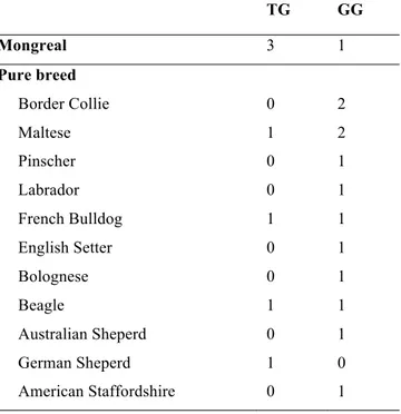
3.3.1 GENOTYPE RESULTS
73
3.3.2 STATISTICAL RESULTS
74
3.4
DISCUSSION
75
4.1
CONCLUSION
78
References
79
SUMMARY
Despite the advances in the treatment of Idiopathic Epilepsy, still a relevant percentage of dogs
don’t achieve an adequate seizure control with the association of different anti-epileptic drugs (AEDs) at appropriate dosage (Trepanier et al., 1998; Schwartz-Porsche et al., 1985; Podell and Fenner, 1993). This condition, know as Refractory Epilepsy is considered one of the most frustrating condition for pets, owners and neurologists (Muñana, 2013). In human and veterinary medicine, great effort is spent in trying to elucidate the mechanisms underlying responsiveness and refractoriness to AEDs treatment (Remy and Beck, 2006; Schmidt and Löscher, 2005; Muñana et al., 2012; Alves et al., 2011).
Recently attention has been focused on the attempt to identify risk factors to predict the outcome of the disease and to understand the exact pathophysiological mechanisms of RIE. A key role seems to have the P-glycoprotein encoded by the ABCB1 gene (Kwan and Brodie, 2005). It has been supposed that an overexpression of these efflux transporters, due to an ABCB1 mutation, may inhibit AED penetration in epileptic foci, resulting in a reduced efficacy of antiepileptic treatment (West and Mealey, 2007). A similar mechanism was hypothesized also in veterinary medicine, indeed, a single nucleotide variation (SNV) of ABCB1 gene (c.-6-180T>G) has been associated with phenobarbital-resistance in a population of idiopathic epileptic Border collie (Alves et al., 2011).
The aims of this multicentric study consisted in: providing a detailed description of the clinical presentation aimed to identify possible clinical risk factors and assessing the frequency of the ABCB1 c.-6-180T>G SNV in a multi-breed population of dogs affected by RIE
Three parts compose this thesis. According to recent literature, the first part describes the seizure pathophysiology, the classification system of epilepsy and the clinical, diagnostic and therapeutic approach to the epileptic dog (Chapter I).
The second part focuses on the definition of refractory epilepsy, on clinical and genetic factors associated with a poor drug-response and on main hypothesis of the pathogenetic drug-resistance mechanisms (Chapter II).
In the Chapter III (the experimental part) data from a population of Refractory Idiopathic Epileptic dogs (RIE-dogs) were statistically compared with a control group of AED-responsive dogs to identify clinical risk factors associated with RIE. Furthermore, the frequency of the ABCB1 c.-6-180T>G SNV was assessed, as possible genetic risk factor in this multi-breed population affected by RIE.
C
HAPTER
I
1.1 H
ISTORY OF EPILEPSYThe word epilepsy derives from the Greek word epilambanein, which means “to be taken”.
This definition perfectly reflects the feutures of a seizure; it suddenly appears and spontaneously stops. For these characteristics of unpredictability and inexplicability, in ancient times, epilepsy was called the “sacred disease”, and humans suffering from epilepsy have been thought to be insane or possessed by demons.
The Greek philosopher Hippocrates (460-377 BC) was the first person to think that epilepsy starts in the brain. Afterward, Galen viewed the epileptic seizure as a symptom of intracranial dysfunction or systemic disease, caused by an accumulation of mucous in the arterial system.
During the Middle Ages, the bilief that people with epilepsy were under a demonic or other spiritual possession came back. Therefore, the treatment of epilepsy included practices as exorcisms and bloodletting.
In the late 19th century, the physician John Hughlings Jackson came to the conclusion that epileptic seizures were due to an abnormality of "excessive neuronal discharge" in the cortical gray matter. This finding led to a series of studies that investigated the origin of focal cortical changes as a cause of seizures.
At the beginning of the XX century, there was a futher evolution with the introduction of new therapeutic approaches based on Jackson's theories. In 1912, Alfred Hauptmann discovered the anticonvulsant activity of the phenobarbital.
Some years later, in 1924, Hans Berger, a German psychiatric, succeeded in recording the first human elettroencephalogram that allowed a better understanding of the epileptogenesis mechanisms.
1.2 SEIZURE
PATHOPHYSIOLOGY
The epileptic seizure has been defined as a transient occurrence of signs due to abnormal, excessive or synchronous neuronal activity in the brain (Fisher et al., 2005). The main feature of all epileptic syndromes is a persistent increase of neuronal excitability (Platt, 2014a). The epiletic activity results from an imbalance between excitatory and inhibitory mechanisms due to a modification in the normal neuronal circuit (March, 1998).
In order to properly understand the pathophysiology of seizures is of paramount importance having a good knowledge of the normal neuronal properties.
The neuron is a polarized cell. It shows an unequal distribution of sodium ions (Na+) and potassium ions (K+) across the membrane. This different distribution produces a resting membrane potential (RMP) of approximatelly -70 millivolts. The RMP is maintained by the selective permeability of the plasma membrane to some ions and by an energy dependent sodium-potassium pump. The pump, extruding actively the sodium out of the cell and pumping the potassium (in a ratio of 3:2) inside keeps the internal of the cell more negative (Klein and Cunningham, 2013a). After the release of a neurotransmitter from a presynaptic axon terminal, a local reversal in membrane potential occurs. According to the function of the neurotransimitter released, a the local change in the permeability of the cell membrane results in an influx of positive or negative charged ions and, respectively, in a state of depolarization or hyperpolarization. The depolarization is the consequence of an excitatory postsynaptic potential (EPSP) and the hyperpolarization of an inhibitory postsynaptic potential (IPSP) [(March, 1998)]. EPSP and IPSP are both local events.
When the simultaneous (“Spatial summation”) and/or the rapid succession (“Temporal summation”) release of depolarizing neurotransmitters reach a certain threshold (-55mV) at the axon hillock, an Action potential is generated (King, 1999). The action potential is the result of the sequential opening of voltage-gated ion channels in the cell membrane, first to the sodium and then to the potassium. The dramatic influx of Na+ ions that accompanies action potential depolarization of the initial segment’s membrane, results in the passive spread of positive charges toward the adjacent resting segment of the axonal membrane. This positive charge migration allows the transmission of the nervous impulse. (Klein and Cunningham, 2013b).
Conversely, spatial or temporal summation of IPSPs results in hyperpolarization of the cell membrane and the maintenance of a resting state.
The principal inihibitory neurotransmitter in the cerebral cortex is the γ-Aminobutyric acid (GABA), acting on two types of GABA receptors: GABAA e GABAB receptors. GABAA receptors
are ligand-gated ion channels that hyperpolarize the neuron by increasing inward chloride conductance. GABAB receptors hyperpolarize the neuron by increasing potassium conductance and
decreasing calcium entry (Treiman, 2001). The predominant excitatory neurotransmitter is the glutamate, which acts on several receptors: the α-amino-3-hydroxy-5-methyl-4-isoxazolepropionic acid receptor (AMPAr), crucial for fast excitatory neurotrasmission, the N-methyl-D-aspartate receptor (NMDAr), mediating the slow post synaptic excitatory potentials and Kainate receptor (KAr) which role has not yet been entirely understood (Barker-Haliski and White, 2015).
An imbalance between excitatory and inhibitory neurotrasmission underlies the mechanism of the epileptic seizures. The seizures develop when the balance shifts towards excessive excitation. In the epileptic patient, the excessive excitation and/or the lack of inhibition lead to the paroxysmal depolarization of neurons without normal regulatory feedback mechanisms. This is particularly true in certain brain areas more sensitive to depolarization (trigger or epileptic foci). The result is a paroxysmal depolarization shift of the neuronal aggregate. In response to this sudden change in brain activity, the local surrounding inhibitory zones try to prevent the spread of this epileptogenic activity. When the inhibition is unsuccessful, other neuronal aggregates are excited through thalamocortical recruitment. The recruitment of a critical number of areas with synchronized depolarization may lead to a seizure (Podell, 2013).
The sequence of events converting a normal neuronal network in a hyperexcitable network is called “epileptogenesis” (Patterson, 2013). These can be induced by several causes, such as brain lesions or genetic factors. Brain lesions (e.g. trauma, hypoxia, and ischemia) can produce epileptic foci through the synaptic reorganization of the neuronal circuits. Indeed, after a brain injury, excitatory axons may sprout new collateral axons producing aberrant connections and breaking down the inhibitory circuits (March, 1998). Among the genetic epilepsies in human medicine, one of the possible causes of epileptogenesis is the dysfunction of mutated voltage- or ligand-gated ion channels (“channelopathies”) or mutation of the neurotransmitters (Avanzini et al., 2007).
Schematic representation of the neuronal circuitry in the cerebrum responsible for feed-forward excitation and feed-forward inhibition. An imbalance in the levels of excitation and inhibition can lead to seizure discharges (from March, 1998).
1.3 CLASSIFICATION OF SEIZURES
In veterinary medicine, the terms used to classify seizure and epilepsy have been debated for a long time. In the last three decades, several classifications have been proposed, sometimes reflecting the authors’ preferences. For this reason, they lacked uniformity in the definitions (Schwartz-Porsche, 1994; Berendt and Gram, 1999; Licht et al., 2002; Berendt, 2004; Podell, 2004; Thomas, 2010). With the aim to uniform the terminology concerning seizures and epilepsy, the veterinary classification of seizures has progressively tried to be more compliant with the guidelines proposed by ILAE (International Legue Against Epilepsy) for the human medicine.
ILAE is a renowned association of “physicians and other health professionals working towards a world where no persons’ life is limited by epilepsy” (ILAE homepage www.ilae.org). ILAE provides a universally recognized vocabulary to facilitate the communication among the scientific community, allowing the comparison between clinical and basic research on epilepsy (Engel, 2001). The first classification of the Commission on Classification and Terminology of the ILAE was published in 1969, updated in 1981 for seizures (Commission, 1981) and in 1989 for epilepsies (Commission, 1989). Afterwards, a new consensus document was produced approximately every 5 years, on the basis of the growing knowledge on the disease.
Despite the veterinary terminology is largely compliant with ILAE classification, many significant differences between humans and animals have been considered to avoid the passive acquisition of terms unfit to properly define seizures and epilepsy in animals. Unlike humans, in pets the recognition of seizure occurrence and its clinical manifestations is mainly dependent on the owner’s observation; electroencephalographic (EEG) data are usually not available (De Risio, 2014a), and, sometimes, the owners because of financial reasons refuse diagnostic investigation. In 2014 the International Veterinary Epilepsy Task Force (IVETF) was founded in order to produce consensus statements on veterinary epilepsy, (Volk, 2015). IVETF has proposed a classification system reflecting the human ILAE guidelines, taking into consideration the well-accepted terminology (Berendet et al., 2015).
According to the recent IVETF publications, epileptic seizure is “a transient occurrence of signs due to abnormal excessive or synchronous neuronal activity in the brain” (Berendet et al., 2015; Fischer et al., 2005, De Risio, 2014a) and epilepsy “a disease of the brain characterized by an enduring predisposition to generate epileptic seizures. This definition is usually practically applied as the occurrence of two or more unprovoked epileptic seizures at least 24 h apart” (Fisher et al., 2014; Berendt et al., 2015).
According to this definition, not all the seizures are associated with epilepsy. Seizures occurring as a natural response of the normal brain to a transient disturbance, such as a metabolic or toxic insult, and that disappear when the cause is solved are called reactive seizure. In this case, the patient does not suffer from epilepsy (De Risio, 2104a).
The IVETF system of classification is based on seizure frequncy, etiology and semiology (Berendt et al., 2015).
1.3.1 CLASSIFICATION OF SEIZURES BASED ON FREQUENCY
Epileptic seizures are classified as single seizure, if is a seizure occurs only once in a 24-hours period, and cluster seizures, if two or more seizures occur within 24 hours with full recovery of consciousness between seizures. Furthermore, Status epilepticus is a condition characterized by continuous seizure activity for five or more minutes, or two or more discrete seizures within 24 hours without full recovery of consciousness between seizures (Berendt et al., 2015).
1.3.2 CLASSIFICATION OF EPILEPSY BASED ON AETIOLOGY
Recently, ILAE has adopted a new classification defining three categories: genetic epilepsy, structural/metabolic epilepsy and epilepsy of unknow origin (Berg et al., 2010).
In veterinary medicine, the earlier terminology classified epilepsy based on etiology, distinguishing: idiopathic (or primary), symptomatic (or secondary) and probably symptomatic (or criptogenic) epilepsy (Gandini, 2015a).
According to the new ILAE classification, in 2015 IVETF proposed to consider: idiopathic epilepsy, structural epilepsy and epilepsy of unknow causes (Berendt et al., 2015).
suspected or confirmed genetic basis.
The term idiopathic does not imply a disorder of unknown cause, referring to a recognized clinical syndrome with typical clinical features. These include the age at onset (between 6 months and 6 years), normal interictal behaviour, normal physical and neurological examination, and the exclusion of metabolic, toxic and structural cerebral disorders after proper diagnostic investigations (Platt and De Risio, 2014).
Idiopathic epilepsy includes three subcategories:
ü genetic –when a causative gene for epilepsy has been identified/confirmed genetic background
ü suspected genetic – when a genetic influence is strongly suspected but the causative gene/s has/ve not been discovered. Suspected genetic epilepsy is the term used in presence of a high breed prevalence (>2%), positive pedigree analysis and/or familial accumulation of epileptic individuals.
ü epilepsy of unknown cause - when the nature of the underlying cause is unknown and there is lack of evidence of structural epilepsy (Berendet et al., 2015).
Structural epilepsy is characterized by epileptic seizures provoked by intracranial/cerebral pathology including vascular, inflammatory/infectious, traumatic, anomalous/developmental, neoplastic and degenerative diseases, confirmed by diagnostic imaging, cerebrospinal fluid examination, DNA testing or post mortem findings (Berendet et al., 2015).
Unkown causes epilepsy refers to recurrent seizures caused by a strongly suspected underlying brain disease, which cannot be identified despite adequate investigation (De Risio, 2014a; Mariani, 2013; Podell, 2013).
1.3.3 CLASSIFICATION OF EPILEPSY BASED ON SEMIOLOGY
generalized-onset seizure.
Focal-onset epileptic seizures (previously named as partial seizure) are characterized by lateralized and/or regional signs due to an abnormal electrical activity arising in a localized group of neurons or network within one hemisphere. The old nomenclature classified focal seizures in simple or complex focal seizures based on the consciousness impairment. Unfortunately, during a focal seizure, the animal can be awake but disoriented and/or not able to recognize the owner nor to respond to commands. For these reasons, the state of consciousness cannot be assessed objectively. Therefore, the new terminology avoids subclassification of focal seizures based on consciousness. Rather, it is now suggested to describe the clinical expression of the seizure.
The clinical expression of a focal-onset epileptic seizure reflects the functions of the part of the brain involved. For this reason, focal-onset epileptic seizures can present tehmselves as:
• Focal Motor seizure, consisting of abnormal movements of a body part, such as facial twitches, repeated jerking head movements, rhythmic blinking, twitching of facial musculature or repeated rhythmic jerks of one extremity (Berendet et al., 2015). Focal-onset motor seizures are presumed to arise from a seizure focus near a primary motor area in the frontal cortex contralateral to the observed involuntary motor activity (De Risio, 2014a). • Focal Autonomic seizure is characterized by parasympathetic and epigastric components as
dilated pupils, hypersalivation or vomiting.
• Focal Behavioural seizure in humans can present psychic and/or sensory seizure phenomena, while in animals it results in a short lasting episodic change in behaviour such as anxiousness, restlessness, unexplainable fear reactions or abnormal attention seeking/‘clinging’ to the owner (Berendet et al., 2015).
Any type of focal-onset seizure can evolve into a generalized seizures. They are charaterized by a focal ictus consistent with the location of the seizure focus, that in seconds to minutes spreads, involving both cerebral hemispheres and resulting in bilaterally symmetrical motor disturbances (usually tonic-clonic), autonomicdysfunction and (commonly) altered consciousness. The focal onset may occur so rapidly that could be undetected and the seizure is misclassified as a generalized-onset seizure (Berendet et al., 2015; De Risio, 2014a).
In generalized-onset epileptic seizures, the clinical expression supports the immediate and simultaneous involvement of both cerebral emispheres. The motor manifestations are bilateral and consciousness alteration may frequently occur (Thomas, 2010). The main types of generalized
seizures are tonic, clonic or tonic-clonic. Less frequently, myoclonic, atonic or absence seizures can occur.
• Tonic-clonic seizures are the most common type of generalized-onset seizure in dogs (Berendt and Gram, 1999; Licht et al., 2002). The prodromal phase and the aura is not always recognized by the owner. The ictal phase is characterized by generalized contraction of the body muscles resulting in a rigid extension of the limbs and opisthotonos, usually lasting 10 to 60 s (tonic phase). The animal falls on its side and often loses consciousness. Breathing is frequently irregular and the animal might become cyanotic. The tonic phase is followed by the clonic phase, which is characterized by rhythmic muscular contractions resulting in jerking movements of the limbs. These stages are often associated to autonomic signs, such as hypersalivation, urination, defecation and mydriasis (De Risio, 2014a).
• Tonic seizures are characterized by an increased muscle contraction without clonic motor activity. During this type of seizure the consciousness might impaire and autonomic manifestations might be present (De Risio, 2014a).
• Clonic seizures consist in a motor activity with no tonic component (Thomas, 2010). • Myoclonic seizures are characterized by sudden, brief, involuntary, shock-like contractions
that can be generalized (Potschka et al., 2013).
• Atonic seizures are characterized by a sudden and generalized loss of muscle tone, usually appearing as episodes of collapse (Berendet et al., 2015).
• Absence seizures are characterized by a transient and brief impairment of consciousness (Poma et al., 2010).
1.3.4 PHASES OF GENERALIZED SEIZURES
Generalized seizures are tipically characterized by four phases: prodrome, aura, ictus and post ictal phase. The prodrome is a period that may occur within hours preceding the ictal phase. During the prodromic phase, the animal displays altered behavior: may hide, appear nervous, or seek out their owners (Lorenz et al., 2011).
The aura is the initial manifestation of a seizure, lasting usually just a few seconds. Aura is described in people as a subjective sensation, such as dizziness, tingling, and anxiety. In animals aura may be recorded as an increased or decreased attention seeking, stereotypical sensory or motor behaviour (e.g. licking, pacing) or autonomic manifestations (e.g. salivating, vomiting, urinating) (Berendt et al., 2015). The definition of aura in veterinary medicine has generated several controversies. Currently, the aura is considered the focal-onset phase of a secondary generalized seizure.
The ictus is the seizure itself, reflecting the paroxistic activation of neurons and, according to the semiology, may consist of generalized epileptic seizure, a focal epileptic seizure, or a focal epileptic seizure evolving into a generalized seizure.
The postictal phase may be absent, short or lasting several hours to days depending on the number and severity of the seizures experienced bu the patient. Typically, the animal is disoriented, may have behavioural abnormalities such as repetitive vocalisation, compulsive locomotion, bumping into obstacles. Furtehrmore, owner descriptions of post ictal phases include ataxia, excessive hunger or thirst, urination, defecation or exhaustion and excessive sleep. Postictal blindness or aggression may also be present (Berendt et al., 2015).
1.4 APPROACH TO EPILEPTIC DOG
The recognition of a seizure in canine and feline patients is sometimes challenging, since in most cases is based upon owners’ observation. A detailed and accurate history is essential to identify an epileptic seizure and distinguish disorders mimicking seizure activity (Moore, 2013). Questions to the owners are aimed to understand if the patient has really experienced an epileptic seizure. If the description of the event is vague or difficult to interpret, it’s helpful to ask the owner to make a video of the paroxysmal episode (Gandini, 2015a).
The signalment itself may give clues about seizure aetiologies. Generally, puppy and young animals are more likely to develop infective and congenital diseases. Conversely, elder patients suffer more often from neoplastic lesions. Dogs with seizure onset between 6 month and 6 years, especially some canine breeds, commonly suffer from idiopathic epilepsy (De Risio, 2014b).
The collection of information about the signalment might be followed by a detailed medical history. The owner should describe the episode, reporting its duration and frequency.
He should tell if the events occur at a certain time of the day and/or in association with specific situations, such as feeding or exercise, and if there are any interictal abnormalities, including changes in behavior, gait, appetite, weight, or sleep habits.
Furthermore, the physician should focus on previous medical history, including earlier occurrence of head trauma, febrile disorder, vaccination diet, exposure to toxins.
A careful phisical examination should precede the neurological examination. By doing so, illness underlying the cause of seizures or abnormalitis of cardiac or respiratory origin that might mimic a seizure, could be detected (Thomas and Dewey, 2016).
The neurological examination must be complete, including the evaluation of mentation, gait and posture, cranial nerves, postural reactions, spinal reflexes and spinal palpation. The neurological examination is focused to identify clinical signs suggesting a structural disease (Moore, 2013). According to the signalment, age at seizure onset, presence or absence of other clinical abnormalities (in addition to the seizures), onset, course and distribution of the other neurological abnormalities (if present), the clinician produces a list of differential diagnosis and chooses the most appropriate diagnostic tests (De Risio, 2014b).
A complete blood count, a serum biochemistry analysis should be always performed to rule out metabolic causes and to obtain information about the patient's metabolic status useful for a possible diagnostic anesthesia and for therapy. Magnetic resonance imaging (MRI) is the diagnostic imaging modality of choice for the evaluation of the brain in animals with seizures. MRI is indicated any
time structural epilepsy is suspected and, in case of a normal scan, to support the diagnosis of idiopathic epilepsy (De Risio, 2014b; Gandini, 2015a).
The cerebrospinal fluid (CSF) analysis is recommended when an inflammatory/infectious cause of seizure is highly suspected. Survey radiography of the thorax and ultrasonography of the abdomen should be performed in animals with suspected neoplastic disease. Abdominal ultrasound is also indicated to investigate certain metabolic disorders causing seizures such as insulinoma-associated hypoglycaemia and hepatic encephalopathy due to a portosystemic shunt (De Risio, 2014b).
To properly approach a dog with suspect idiopathic epilepsy, IVETF has recently described a three-tier system, progressively more accurate in establishing the presence of IE (De Risio et al., 2015). According to the tier I confidence level the diagnosis of IE occurs in case of a history of two or more unprovoked epileptic seizures happening at least 24 h apart, with an age at epileptic seizure onset of between 6 months and 6 years, an unremarkable interictal physical and neurological examination, and no significant abnormalities on minimumdata base (MDB) blood tests and urinalysis. The MDB blood tests should include: complete blood cell count and serum biochemistry profile (sodium, potassium, chloride, calcium, phosphate, alanine aminotransferase - ALT), alkaline phosphatise (ALP), total bilirubin, urea, creatinine, total protein, albumin, glucose, cholesterol, triglycerides, and fasting bile acids and/or ammonia. Urinalysis should include specific gravity, protein, glucose, pH, and sediment cytology. Additional tests vary according to the differential diagnosis.
The tier II confidence level includes unremarkable fasting and postprandial bile acids, brain magnetic resonance imaging (MRI), and cerebrospinal fluid (CSF) analysis. If the results of routine CSF analysis are abnormal additional test on CSF for infectious diseases should be performed. The tier III confidence level includes, on electroencephalography, the identification of characteristic electroencephalographic abnormalities for seizure disorders.
1.4.1 MAIN CLINICAL FEATURES OF IDIOPATHIC EPILEPSY
Idiopatic epilepsy (IE) is the most common cause of epilepsy in dogs (Thomas and Dewey, 2016,). The prevalence of IE has been extimated varying from 0.5 to 5% in the general canine population (Thomas, 2016; Platt and De Risio, 2014).
In several breeds the prevalence documented is much higher. For example in the Belgian Shepherd Tervueren has been reported to be 9,5%, in the Irish Wolfhounds 18,3%, in the Border Terrier 13,1%, in the Petit Basset Griffon Vendeen 8,9%, in the Labrador Retriever 3,1%, in the Finnish Spits Dog 5,4 %, in the Spinone Italiano 5,3 % (Berendt et al., 2008; Casal et al., 2006; Kloene et al., 2008; Gullov et al., 2011; Berendt et al., 2002; Vitmaa et al., 2013; De Risio et al., 2015a). This higher rate in some breeds compared to the general population, is one of the reasons why a genetic component is highly suspected (Hülsmeyer et al., 2015).
Most dogs with IE suffer their first seizure between 6 months and 6 years of age, although seizures occasionally start before 6 months or as late as 10 years of age (Podell et al., 1995; Heynold et al., 1997; Jaggy and Bernardini, 1998; Berendt and Gram, 1999; Thomas, 2010).
In the past, generalized tonic-clonic seizures were considered the most common type of seizure in dogs with idiopathic epilepsy (Heynold et al., 1997). However, more recent studies have shown that the most common seizure type in dogs with IE is the focal seizure with secondary generalization (Platt and De Risio, 2014). Dogs with IE may present cluster seizures (CS) and/or status epilepticus (SE). According to previous studies, the frequency of idiopathic epileptic dogs suffering CS varies from 41 to 49%, the SE range from 2,5-15% (Monteiro et al., 2012; Packer et al., 2016). Several breeds seem to be predisposed to CS. In Monteiro study, German Shepherd Dogs and Boxers were found more likely to suffer from CS than Labrador Retrievers (Monteiro et al., 2012). On the other hand, a more recent study has confirmed the predisposition of Border Collie to CS, as previously reported, and has identified further breeds, including the Cavalier King Charles Spaniel and the Staffordshire Bull Terriers (Hülsmeyer et al., 2010; Packer et al., 2016).
The diagnosis of IE is made by ruling out other possible causes and is based on the age at epileptic seizure onset, unremarkable inter-ictal physical and neurological examinations, and the exclusion of metabolic, toxic and structural cerebral diseases.
1.4.2 MAIN CLINICAL FEATURES OF STRUCTURAL EPILEPSY
Structural epilepsy is a condiction characterized by repeated seizures due to a known and identifiable structural forebrain disorder such as vascular, inflammatory/infectious, traumatic, anomalous/developmental, neoplastic and degenerative diseases (De Risio, 2014c).
According to previous studies, the prevalence of structural epilepsy in dogs and cats ranges from 25–38% in dogs and 34–87% in cats (Quesnel et al., 1997; Bateman and Parent, 1999; Platt and Haag, 2002; Pákozdy et al., 2008; Schriefl et al., 2008; Zimmermann et al., 2009; Steinmetz et al., 2013; De Risio, 2014c).
Animals with structural epilepsy usually present an abnormal interictal neurological examination. They could show prosencephalic signs, such as disorientation, aggression, compulsive walking, head pressing, and circling.
During a posture examination, clinicians might sometimes observe pleurothotonus (usually ipsilateral to the affected forebrain side).
Tipically, gait evaluation does not identify remarkable abnormalities, but propriocetive deficits could be present. On cranial nerves examination, the only alteration detected might be the menace response and to cotton ball tests (De Risio, 2014c; Gandini; 2015a).
However, focal lesions in “clinically silent” areas of the brain (including olfactory bulb, frontal, and pyriform lobes) can result in seizure activity without any other neurological sign (Foster et al., 1988; Smith et al., 1989, De Risio et al., 2015b). In a study evaluating the risk factors for development of epileptic seizures in dogs with intracranial neoplasia, epileptic seizure was the first sign noted by the owners in 76 % of dogs (Schwartz et al., 2011).
The combination of information concerning signalment, history, disease onset and course help to formulate the differential diagnosis list. On the basis of the differential diagnosis, the physician will choose the most appropriate diagnostic investigations. For those patients in which structural epilepsy is highly suspected an MRI and CSF analysis should be strongly recommended and represent the core of the diagnostic work-up (Moore, 2013)
Treatment of structural epilepsy is aimed to treat the underlying aetiology and control the seizures with antiepileptic medications (De Risio, 2014c; Moore, 2013).
1.4.3 MAIN CLINICAL FEATURES OF REACTIVE SEIZURES
Reactive seizures are the reaction of a normal brain to a systemic metabolic, nutritional or exogenous toxic disorder (Podell et al., 1995). They can result from a variety of metabolic disturbances (e.g., hypoglycaemia, electrolyte disorders, portosystemic shunt resulting in hepaticvencephalopathy) or intoxications (e.g., carbamates, organophosphates, lead poisoning,
ethylene glycol toxicity, metaldehyde, strychnine) (De Risio et al., 2015b).
Clinical presentation in animals affected by metabolic/toxic disorders is variable depending on the underlying aetiology (De Risio, 2014d). Toxic disorders tipically have an acute (< 24 h) onset and neurological signs may be preceded or accompanied by gastrointestinal, cardiovascular or respiratory signs. Metabolic disorders can present with an acute, subacute, or chronic onset and may be progressive or characterized by waxing and waning signs. Systemic clinical abnormalities can often be detected on general physical examination. The neurological examination generally reveals diffuse, bilateral and often symmetrical neurological deficits, however seizures can sometimes be the only neurological abnormality (De Risio et al., 2015b).
In a study investigating metabolic and toxic causes of canine seizures, the most frequent cause of reactive seizures were intoxications (39 %, 37/96 of dogs) and hypoglycaemia (32 %, 31/96 of dogs). Metaldehyde (19%, 7/37) and organophosphate or carbamate poisoning (16%, 6/37) were the most frequent intoxications (Brauer et al., 2011). In this study, 41 % (39/96) of dogs were presented in status epilepticus (Brauer et al., 2011). According to another study, dogs with reactive seizures have a significantly higher risk of developing status epilepticus, particularly as first manifestation of a seizure disorder, than dogs with other seizure aetiologies (Zimmerman et al., 2009).
For patients in whom a reactive cause of seizures is suspected, initial diagnostic workup should include a complete bloodcount, chemistry profile (including glucose and electrolytes), measurement of preprandial and post- prandial bile acids, and urinalysis (Moore, 2013).
Most of these conditions are reversible depending on the underlying disease; therefore permanent antiepileptic drug therapy should only be initiated when the seizures are uncontrolled despite therapy or when an emergency situation such as status epilepticus occurs (Boggs, 1997).
1.5 TREATMENT OF EPILEPSY
In veterinary medicine, the mainstay of epilepsy treatment is the administration of antiepileptic drugs (AEDs). Seizure eradication in dogs is a target unlikely to achieve. More realistically, the main goal is the reduction of seizure frequency, duration and severity with limited and acceptable AED adverse effects to maximize the dog and owner’s quality of life (Bhatti et al., 2015). This goal should be clearly explained to the pet owner to avoid non-commensurate expectations. A good compliance with the owner is the key to a successfull treatment (Gandini, 2015b).
To date, there is no evidence-based data on when to start antiepileptic treatment in dogs (De Risio, 2014e). The decision is taken on a case-by-case basis, considering information about the general health of the patient, the owner’s lifestyle, financial limitations and comfort with the proposed therapeutic regimen (Muñana, 2013). Recently, a consensus statment on seizure management in dogs was produced on the basis of a careful meta-analysis of all the literature concerning seizures and epilepsy including the proceedings of Annual Congresses of the European Society and College of Veterinary Neurology (ESVN ⁄ ECVN) and the American College of Veterinary Internal Medicine (ACVIM)[(Podell et al., 2016)].
According to the careful meta analysis of the literature, the “2015 ACVIM Small Animal Consensus Statement on Seizure Management in Dogs” has established guidelines for a predetermined, concise and logical sequential approach to seizure management, reviewing decision-making, treatment strategies, focusing on issues related to chronic AED treatment response and monitoring, and concluding with guidelines to enhance patient response and quality of life (Podell et al., 2016).
The “2015 ACVIM Small Animal Consensus Statement on Seizure Management in Dogs”, in agreement with the International Epilepsy Veterinary Task Force statements, has suggested starting the treatment when the animal has:
• Identifiable structural lesion or a prior history of brain disease or injury
• Interictal period of ≤ 6 months (i.e. 2 or more epileptic seizures within a 6 month period) • Status epilepticus or cluster seizures
• Particularly severe postictal signs (e.g. aggression, blindness) or lasting longer than 24 hours
• An increased epileptic seizure frequency and/or duration and/or deterioration over 3 interictal periods of seizure severity (Bhatti et al., 2015, Podell et al., 2016).
Concerning the choice of the AED, several factors should be taken into account: AED-specific factors (e.g. regulatory aspects, safety, tolerability, adverse effects, drug interactions, frequency of administration), dog-related factors (e.g. seizure type, frequency and aetiology, underlying pathologies such as kidney/hepatic/gastrointestinal problems) and owner-related factors (e.g. lifestyle, financial circumstances) (Bhatti et al., 2015).
Until recently, primary treatment options for dogs and cats with epilepsy were limited to phenobarbital and bromide. In 1857, Sir Charles Locock used bromide to treat “hysterical” epileptic fits in women. Phenobarbital was discovered in 1912 (Podell, 2013). Although these two drugs are still the most widely used in veterinary practice, over the past 20 years in human medicine several new antiepileptic drugs have been developed. Some of them are now used in the treatment of canine and feline epilepsy. According to their appearance, they have been categorized into first, second, third and next generation AEDs (Podell, 2013).
Among the first generation AEDs, the most used in veterinary medicine are: bromide, phenobarbital and benzodiazepines.
The second generation AEDs include Zonisamide, Levetiracetam, Topiramate, Gabapentin and Pregabalin. In 2013, a new AED, the Imepitoin, was introduced in Europe for the treatment of generalized-onset seizures in idiopathic epileptic dog (Tipold et al, 2015).
Anti-epileptic drugs categorized by generation of development (form De Risio, 2014e).
The 2015 ACVIM consensus, after the careful evaluation of the literature identified 4 levels of recommendation based on the evidence of efficacy for the first-line treatment of IE. According to the data resulting from the meta-analysis, Phenobarbital and Imepitoin are highly recommended for
the first-line antiepileptic monotherapic treatment. Bromide has moderate scientific evidence and levetiracetam and zonisamide have poor evidence to be recommended as first-line drugs used in monotherapy for the treatment of IE.
At the onset of the treatment is recommended the use of a single AEDs rather than a combination of drugs. The monotherapy improves the compliance and limits the cost, the adverse effects and pharmacokinetic/dynamic interactions. If seizures are not very frequent, the anti-epileptic treatment can be started at the lower dose and increased gradually based on efficacy, tolerability and, for those drugs requiring it, serum concentration monitoring. Conversely, dogs with frequent and severe seizures should be administered a loading dose to achieve quickly the therapeutic level (De Risio, 2014e).
The decision to add a second AED is based-on seizures frequency, severity (duration, cluster activity, postictal effects), and overall quality of life. Strict criteria for decision-making strategy on starting a second AED are lacking in veterinary medicine. Several factors should be considered when deciding on a second AEDs. Selection of an AED with a different mechanism of action, minimizing drug-drug interactions, avoiding additive toxicity, and determination of risk-benefit of polypharmacy versus quality of life are important considerations (Podell et al., 2016). The 2015 ACVIM consesus reported good evidence to recommend phenobarbital, bromide, zonisamide and levetiracetam as add-on AEDs and imepitoine as not reccomanded (Podell et al., 2016).
1.5.1 PHENOBARBITAL
Phenobarbital (Pb) has the longest history of chronic use of all AEDs in veterinary medicine (Podell et al., 2016). It is generally the first drug of choice in epileptic dogs and cats (De Risio, 2014f). PB is reported to be effective in reducing or eradicating seizure activity in 60 to 85 per cent of dogs with idiopathic epilepsy, if serum concentration is maintained within the therapeutic range (Farnbach, 1984; Morton and Honhold, 1988; Schwartz-Porsche et al., 1985; Boothe et al., 2012). The superior efficacy as a first-line antiepileptic drug in dogs was demonstrated in a randomized controlled trial comparing to bromide, in which 85% of dogs treated with phenobarbital became seizure-free compared with 52% of dogs treated with potassium bromide.
Pb enhances the postsynaptic inibition increasing responsiveness inhibitory neurotransmitter (GABAA receptor). Specifically, it enhances receptor-mediated chloride currents by prolonging the opening of postsynaptic chloride channels, resulting in increased intracellular chloride concentration and subsequent hyperpolarization of the cell membrane. At higher concentrations, it also causes a presynaptic reduction of calcium dependent action potentials, which might also contribute to its antiepileptic effect (Muñana, 2013).
It is rapidly absorbed after oral administration in dogs and achieves the peak serum concentrations approximately 4− 8h after oral administration in dogs (Ravis et al., 1984).
The elimination half-life in normal dogs has been reported to range from 37− 73hours after multiple oral dosing (Ravis et al., 1984). Plasma protein binding is approximately 45% in dogs (Frey et al., 1979). PB is metabolized primarily by the liver and approximately 25% is excreted unchanged in the urine. PB is a potent inducer of cytochrome P450 enzyme activity in the liver (Hojo et al., 2004) and, for this reason, is contraindicated in dogs with hepatic dysfunction. The induction of cytochrome P450 activity leads to increased clearance resulting in the reduction of PB serum concentrations and possible therapeutic inadequacy. Therefore, monitoring of serum PB concentrations is mandatory for proper dose modulation over time (Bhatti S et al., 2015).
Simultaneous PB administration with other drugs metabolized by cytochrome P450 subfamilies and/or bound to plasma proteins, can alter drugs pharmacokinetics and, as a consequence, may decrease the therapeutic effect of other AEDs (levetiracetam, zonisamide, and benzodiazepines) as well as corticosteroids, cyclosporine, metronidazole, voriconazole, digoxin, digitoxin, phenylbutazone and some anaesthetics (Bhatti S et al., 2015). Conversely, the co-administration of PB with drugs that inhibit the cytochrome P450 such as cimetidine, omeprazole, lansoprazole, chloramphenicol, trimethoprim, fluoroquinolones, tetracyclines, ketoconazole, fluconazole,
itraconazole, fluoxetine, felbamate and topiramate may inhibit PB metabolism increasing the serum concentration (Bhatti S et al., 2015).
The recommended oral starting dose of PB in dogs is 2.5− 3 mg/kg BID. The serum concentration should be measured 14 days after starting therapy once steady state concentrations are achieved ad after the change of the dose.
The therapeutic range of PB in serum is 15 mg/l to 40 mg/l (Bhatti S et al., 2015).
Most of the adverse effects due to PB are dose-dependent, occur at the beginning of the treatment or after an increase in the dose and, generally, disappear or decrease in the subsequent weeks. The most frequent adverse effects are: sedation, ataxia, polyphagia, polydipsia and polyuria (Podell et al., 2016). Less frequently, the administration of Pb may lead to idiosyncratic reaction including hepatotoxicity (Bunch et al., 1982; Dayrell-Hart et al., 1991; Gaskil and Cribb, 2005; Müller et al., 2000), haematologic abnormalities (anaemia, and/or thrombocytopenia, and/or neutropenia) (Jacob et al., 1999; Bersan E., 2012), superficial necrolytic dermatitis (March PA et al., 2004), potential risk for pancreatitis (Gaskill and Cribb, 2000), dyskinesia, anxiety (Kube et al., 2006) and hypoalbuminaemia (Gieger et al., 2000). Usually they resolve after therapy discontinuation (Bhatti S et al., 2016).
1.5.2 IMEPITOIN
Imepitoin is a new AED approved in the Europe in 2013 for the treatment of canine idiopathic epilepsy.
It was initially developed for the treatment of anxiety and epilepsy in man, but, then, it has not been used in humans because in smokers it had a different pharmacokinetic (Rundlfeldt et al., 2015). Based on promising findings of imepitoin in a preclinical seizure model in dogs, it was decided to develop this drug for the treatment of canine epilepsy (Loscher et al., 2004). Imepitoin acts as a low affinity partial agonist at the benzodiazepine site of the GABAA receptor (Rundlfeldt and Loscher, 2014).
It is administered orally and achives the maximal plasma levels after 2− 3. The elimination half-life is approximately 1.5 to 2 hour. It is metabolized mainly in the liver (Podell et al. 2016). There is no information on pharmacokinetic interactions between imepitoin and other drugs (De Risio, 2014m).
The most common adverse effects reported include: ataxia and polyphagia, followed by sedation, hyperactivity, increased serum creatinine activity, vomiting and diarrhoea, disorientation and polydipsia. Less commonly, polyuria, anxiety, tachypnea, hypersalivation, decrease in sight and motor activity, prolapsed nictitating membrane and increased sensitivity to sound (Charalambous et al., 2016). Recently, has been described a cutaneous adverse reaction associated with chronic administration of imepitoin in a Jack Russel terrier (Royaux et al., 2016).
The IVETF recommend starting the treatment with imepitoin at the dose of 10− 20 mg/kg q 12 hours. If seizure control is not satisfactory after at least 1 week of treatment at this dose, it can be increased up to a maximum of 30 mg/kg q 12 hours. Reference range of plasma or serum imepitoin concentrations is unknown and there are no therapeutic monitoring recommendations for imepitoin (Bhatti et al., 2015).
1.5.3 BROMIDE
Bromide (Br) is a salt used the first time for the treatment of human epilepsy in 1857 (Locock, 1857). It is thought to exert its antiepileptic activity by passing through neuronal chloride ion
channels with subsequent neuronal hyperpolarization, raising the seizure threshold and stabilizing neurons against excitatory input from epileptic foci (Baird-Heinz et al., 2012).
After oral administration, the bioavailability of Br is approximately 46 % (Trepanier and Babish, 1995). Due to its long elimination hal-flife the steady state is reached approximately 2.5–3 months after treatment initiation at maintenance dosage (Podell and Fenner, 1993; Trepanier and Babish, 1995; Podell, 1998; Ducoté, 1999; March et al., 2002).
Br is not metabolised in the liver, therefore is a good option in dogs with hepatic dysfunction (Bhatti et al., 2015). It is excreted unchanged by the kidneys, where it is filtered by the glomeruli and then undergoes tubular reabsorption in competition with chloride (Dewey, 2006). An increase intake of chloride leads to increase the renal elimination of Br with subsequently decrease of serum concentration and potentially loss of seizure control (Baird-Heinz et al., 2012).
On serum chemestry the chloride concentrations are often falsely increased because the assays cannot distinguish between chloride and Br ions (Trepanier, 1995)
Common adverse effects of Br in dogs include sedation, ataxia and pelvic limb weakness, polydipsia/polyuria, and polyphagia with weight gain (Baird-Heinz et al., 2012; Dewey, 2006; Podell et al., 2016). These adverse effects seem to be dose-dependent. Commonly, they occur at the biginnig of treatment and partly or completely subside after the achivement of the steady-state concentrations (De Risio, 2104g). Potassium bromide is a mucosal irritant, and it may cause nausea, vomiting and/or diarrhoea. In order to prevent gastrointestinal irritation it is prefered administer Br with food and share the daily dose into two or more doses (Baird-Heinz et al., 2012). Uncommonly adverse effects are personality changes (like aggressive behaviour, irritability, and hyperactivity), persistent cough, increased risk of pancreatitis and megaoesofagus (Bhatti et al., 2015). The recommended starting dose of Br is 15 mg/kg q 12 hours when used as adjunctive therapy, 20 mg/kg q 12 hours when used as a monotherapy (Bhatti et al., 2015; De Risio, 2014g). The reported therapeutic range is 810 mg/l - 2000 mg/l when administered with phenobarbital, 2000mg/l to 3000mg/l when administered alone (Podell et al., 2016). To reach more rapidly the steady state concentration, due for instance to severe seizures, or because phenobarbital must be rapidly discontinued due to life-threatening adverse effects, it is possible to administer a loading dose of Br. The IVETF suggests two protocols: 625 mg/kg given over 48h and divided into eight or more doses or 125 mg/kg/day divided in three to four daily administrations for 5 consecutive days (Bhatti et al., 2015).
1.5.4 BENZODIAZEPINES
Benzodiazepines exert their anticonvulsant effects by enhancing GABA activity in the brain (Dewey, 2006). In veterinary medicine diazepam is the most used drug especially for the treatment of emergency seizures by intravenous and per rectum administration (Podell, 2013).
Diazepam is not used as an oral maintenance anticonvulsant in dogs because of its short halflife of elimination (2–4 hours) and the tendency for dogs to develop tolerance to its anticonvulsant effect (Dewey, 2006). On the contrary, it could be used in cats, but unfortunately its oral use has been associated with acute fatal hepatic (Center et al., 1996; Hughes et al., 1996).
In human, midazolam has been shown to be more effective and safer for the control of seizures than comparable doses of diazepam (Koul et al., 1997; Nordt and Clark, 1997). In dogs and cats, its use is poorly documented. It is used especially in the treatment of status epilepticus. Midazolam can be administered by intravenous bolus, continuous intravenous infusion or intra muscular injection at the raccomanded dose 0,07 - 0,2 mg/kg (Platt, 2014b).
1.5.5 ZONISAMIDE
Zonisamide is a sulphonamide-based anticonvulsant approved for use in humans. It acts with multiple mechanisms of action, including blockage of calcium channels, enhancement of GABA release, inhibition of glutamate release, and inhibition of voltage-gated sodium channels (Bhatti et al., 2015). In dogs, zonisamide is well absorbed after oral administration. It is metabolized predominanltly by hepatic enzime. The elimination half-life is approximately 15hours in dogs (Podell et al., 2016). In people, it has been shown that the elimination half-life of zonisamide is dramatically shorter in patients already receiving drugs that stimulate hepatic enzymes in comparison with patients who are not receiving such drugs. A similar phenomenon seems to occur in dogs (Dewey, 2006).
The recommended starting dose of zonisamide in dogs is 3− 7 mg/kg q 12 hours and 7− 10 mg/kg q 12 hours in dogs in wich inducer hepatic enzymes, such as phenobarbital, are coadminister. In human the terapeutic target range is 10 - 40 µg/ml, it can be used as a guidance regarding effective
concentrations that can be targeted in dogs. Serum concentrations of zonisamide should be measured 1 week after treatment initiation or after every dosage adjustment (De Risio, 2014h). The efficacy on the use of zonisamide in dogs is limited to three studies. The first one valued the efficacy of oral zonisamide as a monotherapy (Chung et al., 2012), while the other two studies evaluated the efficacy as an add-on treatment in dogs with refractory idiopathic epilepsy (Dewey et al., 2004; von Klopmann et al., 2007).
The most common adverse effects reported in dogs include: sedation, vomiting, ataxia, and loss of appetite (Chung et al., 2012; Dewey et al., 2004; von Klopmann et al., 2007). Other adverse effects reported are hepatotoxicity (it has been described in 2 dogs receiving zonisamide monotherapy), renal tubular acidosis (in a dog receiving zonisamide monotherapy), and erythema multiforme (Schwartz et al., 2011; Miller et al., 2011; Cook et al., 2011; Ackermann et al., 2016).
Zonisamide should be used with caution in dogs with renal or hepatic impairment
(Bhatti et al.,
2015).
1.5.6 LEVETIRACETAM
Levetiracetam was approved for use in 1999 for the treatment of partial-onset seizures in humans (Muñana, 2013). It acts modulating the release of neurotransmitters by binding to the presynaptic vescicle protein (SVA2) (Volk, 2008). It is rapidly absorbed after oral administration and is primarily excreted in the urine (De Risio, 2014i). Levetiracetam has a minimal hepatic metabolism, so it is recommended in animals with hepatic dysfunction. The oral maintenance dose of levetiracetam in dogs is 20 mg/kg q 8-6 hours (Bhatti et al., 2015). In human, the concomitant administration of AEDs inducing cytochrome P450 metabolism (as phenobarbital), may increase the clearance resulting in a lower plasma concentrations (Contini et al., 2004). This effect has also been demonstrated in healthy dogs. In this situation, it is advisable to increase the oral dose of levetiracetam (Moore, 2011).
In veterinary medicine, the serum concentrations of levetiracetam are not routinely measured. The reference range has not been established. As a guidance regarding effective concentrations, the human target range (12− 46 µg/l) can be used (Bhatti et al., 2016).
appetite and vomiting, adverse effects are very rarely described in dogs (Muñana et al., 2012; Volk et al., 2008).
1.5.7 GABAPENTIN AND PREGABALIN
Gabapentin has been approved for people in Europe since 1993 for adjunctive treatment of focal seizures with or without secondary generalisation and for the treatment of post-herpetic neuralgia (Bhatti et al., 2015).
The precise mechanism of action is unclear, is believed that it acts binding to a specific modulatory protein of voltage-gated calcium channels, resulting in presynaptic inhibition of calcium influx, subsequent inhibition of excitatory neurotransmitter release and attenuation of postsynaptic excitability (De Risio, 2014l). Gabapentin is well absorbed after oral administration, it achives the maximum blood concentration within 2 hours. In dogs, gabapentin is excreted by the kidneys after a partial hepatic metabolism. The elimination half-life is 3-4hours (Muñana, 2013). In litterature there are only two studies that have evaluated the efficacy of oral gabapentin as an adjunct to other AEDs (Govendir et al., 2005; Platt et al., 2006) but none of these have demonstrated an increased likelihood of a successfully response with gabapentin (Charalambous et al., 2016).
The recommended oral dosage of gabapentin in dogs is 10 to 20 mg/kg q 8 hours, although dose reduction may be necessary in patients with reduced renal function. Sedation and ataxia were the most common side effects reported in dogs (Bhatti et al., 2015).
Pregabalin is a GABA analogue that is structurally similar to gabapentin. It was approved in 2004 for the treatment of adults with peripheral neuropathic pain and as adjunctive treatment for adults with focal seizures with or without secondary generalization (Bhatti et al., 2015). In veterinary medicine there is only a study evaluating the efficacy of oral administration of pregabalin as an adjunct to phenobarbital and bromide in dogs. The oral dose in dogs is 3− 4 mg/kgq 12-8 hours. The most common adverse effects reported included sedation, ataxia and weakness (Dewey et al., 2009).
1.5.8 TOPIRAMATE
Topiramate is an AED used in adult and paediatric human patients for the treatment of focal (partial) and generalized seizures (Platt, 2014c). It acts enhancing GABA-ergic activity and inhibiting voltage sensitive sodium and calcium channels, kainate-evoked currents and carbonic anhydrase isoenzymes (Vuu et al., 2016).
In human, topiramate is well absorbed. It is not enterly metabolized, the 70-80 % of an administered dose is eliminated unchanged in the urine (Lyseng-Williamson and Yang, 2007). Also in dogs, topiramate is not enterly metabolized and is primarily eliminated unchanged in the urine. However, a significant biliary excretion is present following topiramate administration (Caldwell et al., 2005). It has an elimination half-life of 2-4h. The dose of topiramate should be reduced by 50% in patients with renal impairment; instead dosage reductions are not necessary in patients with hepatic impairment. The drug has a relatively low potential for clinically relevant interactions with other medications (Platt, 2014c).
To date, there are no studies about the use of topiramte in monotherapy in dog. Its efficacy has been evaluated as an adjunct to phenobarbital, bromide, and levetiracetam in 10 dogs (Kiviranta et al., 2013). In this study the proposed dose it was recommended to start at a low dosage first (2.0 mg/kg q 12 hours) and increase to a higher dose (5-10 mg/kg q 8-12 hours), in order to minimize the adverse effects. The most common were: sedation, ataxia and weight loss (Kiviranta et al., 2013).
1.5.9 THERAPEUTIC MONITORING OF AEDs
The therapeutic drug monitoring is the measurement, for clinical use, of AED concentrations in body fluids, usually serum (Johannessen and Johannessen Landmark, 2008). The drug monitoring is a mandatory necessity for some first generation AEDs, such as phenobarbital and bromide, to assess:
• the effective drug concentrations after initiation of successful treatment
• if treatment failure is caused by poor compliance or an insufficient or changed drug concentration
• the most proper AED and dosage. • to prevent toxic effects
• to aid with individualization of treatment (Podell et al. 2016). It should be measured:
ü After initiating treatment, once steady-state concentrations are achieved ü After each dosage adjustment, once steady-state concentrations are achieved ü When seizured are not adequately controlled
ü When there is concern about possible drug-related toxicity
ü At 6- to 12-month intervals to screen for any changes in drug disposition over time (Muñana, 2013).
The therapeutic ranges of drug monitoring represent only an approximation, since they are based-on retrospective data from a small number of patients. Although most responders attain levels within the expected range, some do well below the lower limit whereas others obtain benefit at levels above the upper limit without toxicity (Thomas, 2010).
Therapeutic monitoring of new generation AEDs is not routinely performed and it may be of limited value because there is no established correlation between serum AED concentration and therapeutic efficacy or toxicity (De Risio, 2014e).
1.5.10 DISCONTINUATION OF AEDs
In most cases, the treatment for canine idiopathic epilepsy involves a long-term or lifelong AED administration (Geselle et al., 2015). There are different reasons to discontinue treatment, including the remission of seizures and life-threatening adverse effects. In case of remission of seizures, the decision to gradually taper the dose of an AED should be taken carefully, after a seizure free period of at least 1− 2 years (Bhatti et al., 2015).
In people with prolonged seizure remission (generally 2 or more years), the decision to discontinue AED treatment is done on an individual basis, considering relative risks and benefits (Bhatti et al., 2015). In humans, the chance to remain seizure-free after AED discontinuation is higher in patiens without structural brain lesion, with a short duration of epilepsy, with few seizures before pharmacological control, and with AED monotherapy (O'Dell and Shinnar, 2001; Shih and Ochoa, 2009). In human medicine, a seizure relapse rate ranging from 12% to 67% has been reported after
AED withdrawal (Shih and Ochoa, 2009).
In dogs, there is still little information on risk factors associated with seizure relapse after treatment discontinuation. In a recent paper (Geselle et al., 2015), the consequences of AED withdrawal were studied in dogs with epilepsy. In 11 cases out of 138, the therapy had been stopped after a seizure free period for a median time of 1 year. After therapy discontinuation the majority of these dogs suffered again from seizure (63.6%),
In order to prevent withdrawal seizures or status epilepticus, especially when on PB treatment it is advised to decrease the dose with 20% or less on a monthly basis. In case of life-threatening adverse effects, instant cessation of AED administration under 24h observation is necessary. In these cases, loading with an alternative AED should be initiated promptly in order to achieve target serum concentrations (Bhatti et al., 2015).