Alma Mater Studiorum – Università di Bologna
DOTTORATO DI RICERCA in
Biologia Cellulare e Molecolare
Ciclo: 29
Settore Concorsuale di afferenza:O5/E1
Settore Scientifico disciplinare:BIO/10
TITOLO TESI
Mitochondrial DNA deletions:
Quantitative evaluation of single and multiple
deletions generation and expansion in single cells
Presentata da: Selena Trifunov
Coordinatore Dottorato
Relatore
Prof. Giovanni Capranico
Prof. Valerio Carelli
Correlatore
Prof. ssa Michela Rugolo
ABSTRACT
Mitochondria are unique organelles, containing their own, maternally inherited genome. Human mitochondrial DNA (mtDNA) is small, circular and intronless, encoding 37 genes, precisely 13 proteins of the respiratory chain, 22 transfer RNAs (tRNAs) and 2 ribosomal RNAs. Each mitochondrion contains several mtDNA copies. Mitochondrial DNA deletions are pathogenic mutations that remove various portions of mtDNA genome, leading to shorter mtDNA molecules. In patients, there are two classes of mtDNA deletions: single, large-scale deletions that are present from the birth, whereas multiple deletions accumulate with age secondarily to mutations in nuclear genes involved in mtDNA maintenance. Every deleted mtDNA, coexists with other wild type mtDNAs, in an intracellular pool of mitochondrial genomes, determining the condition known as heteroplasmy. Pathogenic mtDNA mutations may become prevalent in certain cells; this process is defined as intracellular clonal expansion. This study investigates clonal expansion patterns of mtDNA deleted genomes, applying for the first time the droplet-digital polymerase chain reaction (ddPCR) approach on single muscle cells collected by laser-capture microdissection from muscle biopsies of patients with different paradigms of mitochondrial disease with single and multiple mtDNA deletions accumulation. The results of this study indicate different patterns of accumulation of clonally expanded mtDNA deletions in patients with single and multiple deletions. It was found that single deletions patients have clonally expanded deletion in all single muscle cell populations, suggesting that the original deletion event occurred at early stage of embryonic development or even along the maternal germline transmission. Importantly, we distinguish localized clonal expansion of mtDNA deletions in patients with mutations in the OPA1 fusion gene. In conclusion, the ddPCR is a promising new technique for the investigation of clonal expansion byaccurate quantifying of the mtDNA heteroplasmy levels.
INDEX
INTRODUCTION……… 1
1. MITOCHONDRIA………. 2
1.1 THE BEGININGS……… 2
1.2 MORPHOLOGY AND ACTIVITY……… 3
1.3 LIFE CYCLE OF THE MITOCHONDRIA: MODERN NETWORKING ORGANELLS……….. 4
1.4OXPHOS SYSTEM………. 6
2. MITOCHONDRIAL GENETICS……… 8
2.1 STRUCTURE OF MITOCHONDRIAL DNA: THE MAGIC CIRCLE……… 8
2.2 MITOCHONDRIAL REPLICATION, TRANSCRIPTION AND TRANSLATION……… 11
2.2.1 MODELS FOR MITOCHONDRIAL DNA REPLICATION……….. 11
2.2.2 MITOCHONDRIAL DNA REPLICATION- THE REPLIOSOM………. 13
2.2.3 TRANSCRIPTION………. 15
2.2.4 TRANSLATION………... 17
2.3 INHERITANCE OF MITOCHONDRIAL DNA……… 18
2.4 MITOCHONDRIAL GENETICS AND HUMAN DISEASE……… 19
3. MITOCHONDRIAL DNA DELETIONS……… 20
3.1MITOCHONDRIAL DNA DELETIONS HETEROPLASMY AND CLONAL EXPANSION ……… 22
3.2 SINGLE MITOCHONDRIAL DNA DELETIONS……….. 24
3.3 INHERITANCE OF SINGLE MTDNA DELETIONS………. 26
3.4 MULTIPLE MITOCHONDRIAL DNA DELETIONS……… 28
3.5 MITOCHONDRIAL DNA MAINTENANCE GENES AND MULTIPLE MTDNA DELETIONS……….... 30
3.6 MECHANISMS OF MTDNA DELETIONS FORMATION……….. 35
3.7 METHODS FOR QUANTITATIVE ANALYSIS OF MTDNA HETEROPLASMY……….. 38
AIMS………. 41
MATERIAL AND METHODS………. 43
1. Patients………. 44
2. Southern blot……… 48
3. Preparation of recombinant plasmid for the standard curve ……….. 50
5. Transversal and longitudinal muscle biopsy sectioning……….. 51
6. Histochemical analysis and classification of individual muscle fibres……….. 51
7. Laser Capture Microdissection……… 52
8. Lysis of the single cells……….. 52
9. Droplet Digital PCR……….. 52
10. Single cell long range PCR……….. 54
11.Statistical analysis……… 54
RESULTS………. 55
1. Validation of Droplet Digital PCR……… 56
2. Proportion of COX negative and intermediate fibres throughout different patient groups…….. 59
3. Levels of mtDNA deletions in patients with OPA1 mutations……….. 61
4. Levels of mtDNA deletions in patients with POLG mutations……….. 62
5. Levels of mtDNA deletions in patients with single deletions………. 62
6. Comparison of mtDNA deletion accumulation in all patient groups……….. 62
7. Deletion levels in longitudinal biopsy sections……….. 65
8. MtDNA copy number levels show mtDNA proliferation in certain fibre types……… 67
9. MtDNA deletions presence by single cell long range PCR……….. 68
DISCUSSION……… 70
1
2
1. MITOCHONDRIA 1.1 THE BEGININGS
The very first evidence of intracellular structures that probably represent mitochondria dates back to the 1840s, only a few years after the discovery of the cell nucleus (Brown, R. 1833).
In 1890, Altman first appreciated the recurrence of these structures that he named "bioblasts". His description was: "elementary organisms" living inside cells and carrying out vital functions. The name mitochondrion first appeared in 1898, used by Benda, and it emerges from the Greek "mitos" (thread) and "chondros" (granule), referring to the appearance of these structures during spermatogenesis (Ernster and Schatz, 1981).
Today, more than a century from Altmann’s observations, we know that mitochondria are necessary organelles, powerhouses of eukaryotic cells. They produce ATP through the oxidative phosphorylation (OXPHOS). They are unique for containing their own maternally inherited circular genome (Giles, 1980). But, mitochondria have not always been cytoplasmic organelles, once upon a time they were free facultative aerobic bacteria (Ernster and Schatz, 1981).
According to the endosymbiotic theory, first proposed by Lynn Margulis in the 1970s, mitochondria were originally bacteria, engulfed by ancestral cells but not digested. In return for security of host environment, mitochondria became the power supplier of the cell (Margulis, 1975).
When exactly the symbiosis between eukaryotic cell and mitochondrion emerged? Probably between 1.5 and 2 billion years ago (Lane, 2014; Martin, 2015). However, it was at the beginning of the eukaryotic life, which developed with multicellular organisms thanks to the energy produced through mitochondria.
3
1.2 MORPHOLOGY AND ACTIVITY
From the bacterial ancestor mitochondria inherited, along with many other features, the double membrane system. The outer mitochondrial membrane (OMM) is the boundary to cytoplasm and also a place where mitochondria make contacts with their environment. In that sense, OMM is not just a simple barrier but has multiple roles that are vital for mitochondrial life and death. Maybe the most important is the interaction of OMM with endoplasmic reticulum (ER) at specific sites called mitochondria-associated ER membranes (MAMs). Mitochondria require a regular and synchronized replenishment of membrane lipids to perform the physiological processes and maintain their membrane integrity. OMM and its ER connected sites are involved in many other important roles: calcium signalling, apoptosis, fission, and inflammation (Marchi, 2014).
Voltage dependent anion channel (VDAC), a multispanning β-barrel protein often called mitochondrial porin, which provides high permeability to small molecules and ions, is placed at OMM. Overall, OMM operates like exchange hub for mitochondria.
Between the two mitochondrial membranes there is the inter-membrane space (IMS), hosting a quite large pool of hydrophilic proteins (Marchi, 2014).
The second membrane, the inner mitochondrial membrane (IMM) acts more privately, being impermeable to the majority of ions and smaller molecules.
IMM is the site where energy conversion occurs and there are two structural regions to consider: the “boundary membrane” and the cristae, which are defined by narrow tubular sections named cristae junctions (Frey and Mannella, 2000).
Structurally, a multitude of proteins are embedded in the IMM, most being constituents of the oxidative phosphorylation system (OXPHOS). The core of mitochondria is the matrix, a gel like structure containing enzymes of the tricarboxylic acid cycle and of β-oxidation. The oxidation-reduction reactions occurring mostly in association to the OXPHOS metabolism engage a large number of mitochondrial proteins. The
4
global mitochondrial proteome is currently estimated to contain up to 1500 proteins, and this number is constantly growing (Mao and Holt, 2009).
As mentioned above, the IMM is highly invaginated to form structures called cristae (Palade, 1952). The tips of these invaginations are juxtaposed to the OMM, held together by the IMM integral protein OPA1. This organisation is important for the activation of apoptosis, because when the mitochondrial membrane potential declines or the intrinsic pathway of apoptosis is activated, the IMM proteases become activated and cleave OPA1. This results in the enlargement of the cristae junctions leading to release of luminal cytochrome c into the IMS (Burke, 2015; Frezza, 2006). However, not only apoptosis is dependent on cristae organization, but recent research highlights how respiratory efficiency and assembly of respiratory supercomplexes are tightly correlated with the cristae morphology (Cogliati, 2013).
1.3 LIFE CYCLE OF THE MITOCHONDRIA: MODERN NETWORKING ORGANELLS
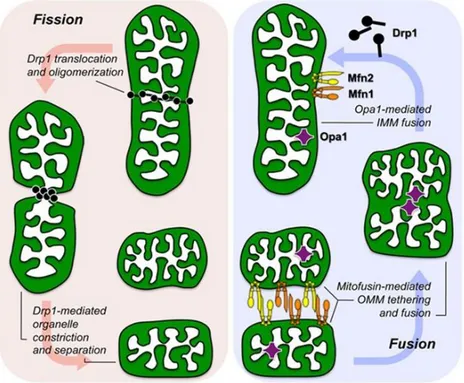
The idea of a discrete mitochondrion, isolated from the rest of the cell by the two membranes, seemed the right one for a long time. The endosymbiotic mitochondria, considered as discrete foreign entities, misled many to think mitochondria to stand alone within the cytoplasm of the cell. By means of tridimensional imaging techniques emerged the evidence that mitochondria are in fact very social organelles (Chan, 2012). Interconnected in highly dynamic networks, they move fast and can be in spotlight of cellular needs for energy. This network allows mitochondria to travel long distances, like along axons, and to be called up at active synapses. In conditions of oxidative stress, or nutrient starvation, the mitochondrial network is hyperfused, opposite to the G1/S cell phase transition, when mitochondria are elongated. So-called “mitochondria-shaping” proteins are in charge for regulation of organelles fusion and fission, two processes that are essential for managing the demanding mitochondrial network (Griparic and van der Bliek, 2001). Mitochondrial fusion is the process by which two mitochondria join to form a single mitochondrion, whereas fission is the process through which a
5
single mitochondrion divides into two mitochondria (Fig.1) (Chan, 2012). In the OMM, mitofusins (MFN) 1 and 2, dynamin-related proteins that exhibit high homology, in tight cooperation with OPA1, orchestrate fusion (Santel and Fuller, 2001; Chen, 2003; Cipolat, 2004).
The cytoplasmic dynamin-related protein 1 (Drp1) translocates to mitochondria upon dephosphorylation, regulating the process of mitochondrial fission (Cogliati, 2013; Smirnova, 2001). Recently, it has been shown that mitochondria can even be electrically coupled, without physical exchange of their matrix content, which normally happens during fusion, and this could be a way to simplify the energy transmission (Santo-Domingo, 2013). Further analysis of mitochondrial interactions points out that neighbouring mitochondria interact through specific inter-mitochondrial junction (IMJ) sites, where cristae membranes become structured into coordinated pairs across organelles (Picard, 2015).
This further level of interaction among mitochondria may provide a structural basis for events of electrochemical inter-mitochondrial communication, but further research in this exciting area is needed.
Figure 1. Molecular mechanism of mitochondrial fission and fusion. The three molecular drivers of fission and fusion are shown as they associate with a normal mitochondrion. Fission (left) is initiated by recruitment of cytosolic dynamin-related protein 1 (Drp1) to the organelle, Drp1 oligomerization, and constriction of the parent into two daughters. Fusion (right) requires initial mitofusin 1 (Mfn1)/Mfn2-mediated OM tethering followed by fusion, and finally optic atrophy 1 (Opa1)–mediated IM fusion (from Dorn and Kitsis, 2015).
6
The maintenance of mitochondrial function is crucial for cellular homeostasis.Balance between fission and fusion is closely related to the homeostatic adjustment of mitochondrial mass to the metabolic needs of different tissues or sensing specific functional needs of cells (Mishra, 2014). Loss of this balance closely relates to regulation of apoptosis and cell death. Thus, maintenance of mitochondrial homeostasis is regulated by mitochondrial biogenesis, which provides newly synthesized organelles, as opposed to the autophagic elimination of damaged mitochondria, called mitophagy (Youle and Narendra 2011). Impaired mitochondrial function caused by different mechanisms may result in a decrease in mitochondrial membrane potential. Depolarized mitochondria become fusion incompetent, they fragment through mitochondrial fission and can be selectively digested by lysosomes (Sarraf, 2013;Dorn and Kitsis, 2015). Mitophagy is part of the quality control system of cells to avoid the accumulation of damaged mitochondria that could eventually lead to apoptosis (Carelli, 2015). In the process of mitophagy, the PTEN-induced kinase 1 (PINK1) and Parkin, a cytosolic E3 ubiquitin ligase, play a crucial role (Narendra, 2010). Mitochondrial fusion is necessary for complementation of mutated mitochondrial DNA (mtDNA) (Chen, 2010). Fusion between damaged and healthy mitochondria has the potential to dilute out the damaged components, thus repairing the damaged organelle through functional complementation (Youle and Van der Bliek, 2012). However, there is always the opposite side of the coin, thus mitochondrial fusion may be considered as a mechanism leading to the pollution of healthy mitochondria, with damaged ones. Thus, it is important to bear in mind that fusion may also be the mechanism for dissemination of mtDNA mutations (Dorn II, 2015).
1.4 OXPHOS
Energy generation and consumption are prerequisite for life itself. This process is elegant, sophisticated and highly complex. In eukaryotes, respiration is a task carried out by the mitochondria.
7
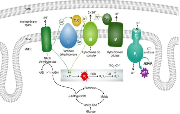
The OXPHOS system is composed of five enzymatic complexes (CI-V) (Fig.2). It is built up by nearly 100 polypeptides, of which only 13 are encoded by mtDNA, whereas the remaining are coded by the nuclear DNA (nDNA), and only subsequently imported within mitochondria (Carelli V., Chan D., 2014).
Mitochondrial respiratory complexes (CI to IV) are responsible for the oxidation of the reducing equivalents, in the form of NADH or FADH2, originating from different metabolic pathways (glycolysis,
fatty acid oxidation or the Krebs cycle). Oxidation of NADH and FADH2 is coupled with the pumping of
protons into the IMS, and the resulting proton gradient is used by the ATPase (complex V) to generate utilizable energy in the form of ATP molecules. NADH reducing equivalents enter the mitochondrial electron transport chain (mtETC) through complex I, whereas FADH2 through complex II or other
dehydrogenases. The electrons are then delivered to coenzyme (CoQ), and subsequently to complex III, cytochrome c, and complex IV, where the final acceptor oxygen is reduced to water (Acin-Perez and Enriquez, 2014). Altogether this processes is called respiration (Mitchell, 1961). Respiratory complexes are not present within the IMM as single entities but are organized in supercomplexes or respirasomes. Studies on isolated bovine heart mitochondria revealed a supercomplex consisting of one copy of complex I, one complex III dimer, and one or more complex IV monomer (Lapuente-Brun, 2013). This drives the path of electrons from NADH via the iron-sulfur clusters of complex I to ubiquinol, then to the prosthetic groups of complex III, and finally to molecular oxygen at complex IV. Genetic evidence provides strong support for the existence of respirasomes in vivo (Mileykovskaya, 2012; Koopman, 2013).
The OXPHOS pathway is integrally coupled to the creation of reactive oxygen species (ROS). Under standard conditions, both cytosolic and mitochondrial ROS amounts are controlled by mitochondrial and cytosolic antioxidant systems and utilize a signalling function. Still, in case the ROS levels exceed the harmless limits, bypassing the antioxidant systems, DNA, proteins and lipid molecules are exposed to being damaged. If this exposure takes place over the years it leads to the slow decline of mitochondrial and overall cellular integrity and function (Koopman, 2013).
8
Figure 2. The mitochondrial electron transport chain and its association to ROS production. All respiratory complexes are coloured and specified with roman numbers (I-V). Transfer of electrons along the complexes (dotted arrows), proton (H+) transport (dashed arrows) in direction from the matrix to the
IMS. Proton flow through ATP synthase (complex V) converts ADP to ATP. In normal conditions oxygen is the final electron acceptor from complex IV. However, damaged mitochondria may have electron leak from complexes I or III which produces toxic reactive oxygen species (ROS) (from Dorn G.W., 2015).
2. MITOCHONDRIAL GENETICS
2.1 STRUCTURE OF MITOCHONDRIAL DNA: THE MAGIC CIRCLE
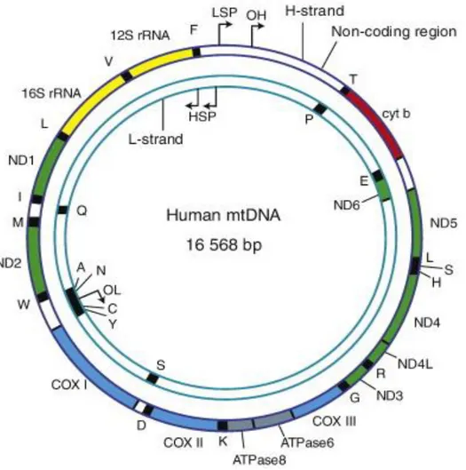
Whenever one thinks of mitochondria it must be kept in mind their bacterial origin, which helps to understand many distinguishing features of these organelles. Main characteristic, granting mitochondria to follow their own rules, is the existence of mtDNA. This little genome is maternally inherited and nowadays is just a 16.659 base pair long, circular, double stranded molecule that underwent various changes during evolution. Still, unorthodox, it remains as extra DNA in the eukaryotic cells (Carelli and Chan, 2014).
The mtDNA contains 37 genes. Each mitochondrion contains several copies of this genome (Fig. 3) and this number varies between individuals and tissue types. The mtDNA codes for the 13 most important
9
OXPHOS polypeptides. These include seven from nearly 45 polypeptides composing the respiratory complex I (ND1-3, ND4L, ND4-6), one of the 11 polypeptides of complex III (cytochrome b, cytb), three of the 13 polypeptides of complex IV, and two of the 15 polypeptides of complex V (ATP6 and 8). Complex II (succinate dehydrogenase) is the only complex that has no polypeptides coded by mtDNA. Additionally, the mtDNA encodes the mitochondrial 16S and 18S rRNAs, and 22 tRNAs for mitochondrial protein synthesis (Wallace, 2013). mtDNA has two strands, denoted as ‘heavy’ (H) and ‘light’ (L) strand, based upon their distinct base compositions, H strand is guanine rich while L strand is cytosine rich. Both strands of mtDNA are transcribed as long, polycistronic molecules, with transcription initiated from the heavy-strand promoters (HSPs) and light-heavy-strand promoter (LSP), respectively (Wallace, 2007). The mechanism of mtDNA replication is still unresolved and has been lively debated in the past twenty years. Presently, three mechanisms for replication are proposed and will be discussed later on. Compactness of mtDNA is one more extraordinary characteristic, with no introns, two pairs of protein coding regions overlapping and only two non-coding regions. The first 1kb long non-coding region (NCR) of mtDNA is also a control region (CR), containing the origin for replication of H (OH) strand, as well as promoters for transcription of both strands HSP and LSP. A very enigmatic structure, residing in the control region, is the so-called D-loop (Displacement D-loop) structure. In this region there is incorporated a third linear DNA strand referred as 7S DNA due to its sedimentation properties (Fig.3). The nomenclature of 7S DNA often induces confusion, since this is not one molecule but a group of molecules slightly different in size and with 5’ end that may start at various positions. The exact function of this region is still unknown; however it has been speculated that because it encompasses the origin of heavy strand replication and is positioned in the control region of mtDNA, is possibly involved in mtDNA maintenance, by control of dNTP pool and/or anchoring the mtDNA molecule (Nicholls and Minczuk, 2014).
The second non-coding region is substantially smaller and contains the origin of L-strand replication (OL). It is located in a cluster of five tRNA genes approximately two thirds of the mtDNA length from the OH. The two origins divide the genome into roughly two-thirds and one-third sections, with the larger portion
10
denoted as the ‘major arc’, and the smaller portion called the ‘minor arc’ (Anderson et al., 1981; Fernandez-Silva et al., 2003).
The circular molecule of mtDNA has a contour length of ∼5 μm and each mitochondrion, wide approximately ∼0.5 μm, contains numerous mtDNA circular molecues. Therefore, mtDNA has to be arranged to fit within a mitochondrion. This is accomplished by shaping the mtDNA in ellipsoidal structures termed nucleoids that present basic organizational units of mtDNA not wider than few hundred nanometers (Kukat, 2011; Kukat, 2015).
Taking advantage of new stimulated emission depletion (STED) nanoscopy, electron microscopy and electron cryo-tomography (cryo-ET), it has been elucidated that most mammalian mitochondrial nucleoids contain a single mtDNA copy compacted by mitochondrial transcription factor A (TFAM) aggregation and cross- strand binding. TFAM is very abundant, present at approximately thousand copies per mtDNA molecule and besides being responsible for compacting mtDNA it has essential role in mtDNA replication as well as in transcription (Kukat, 2015). Expression of TFAM also governs the mtDNA copy number in cells, therefore this protein is irreplaceable for mtDNA maintenance. However, TFAM is not the only protein that affects the morphology and the organization of the nucleoids (Bogenhagen, 2012). Other proteins crucial for mtDNA maintenance are additional components of nucleoids: mitochondrial polymerase gamma (Polγ), mitochondrial single-stranded DNA binding protein, replicative helicase TWINKLE, mitochondrial transcription factors B1 and B2 (TFB1M and TFB2M), mitochondrial transcription termination factor (mTERF), along with others (Bogenhagen, 2008;Kolesnikov, 2016).
11
Figure 3. The human mitochondrial genome. The human mitochondrial genome consists of 16569 base pairs and contains a heavy (H-strand) and light-strand (L-strand). Complex I NADH dehydrogenase (ND) genes are presented in green; Complex III cytochrome b (Cytb) gene is presented in red; Complex IV cytochrome c oxidase (COX) genes are presented in light blue; Complex V ATP synthese (ATPase) genes are presented in grey. Transfer RNA genes are in black and ribosomal RNA genes (rRNA) are in yellow (from Wanrooij and Falkenberg, 2010).
2.2 MITOCHONDRIAL REPLICATION, TRANSCRIPTION AND TRANSLATION
All components needed for mtDNA transcription and replication are encoded in the nuclear genome, same as the mitochondrial translation machinery (Peralta S., 2012).
2.2.1 MODELS FOR MITOCHONDRIAL DNA REPLICATION
Strand displacement model
Replication and turnover of mtDNA occurs independently from the cell cycle. The molecule of mtDNA is small and replicates within few hours. In the early 1980’s an extensive body of work has been done to define the rules of mtDNA replication (Bogenhagen, 1979; Nass, 1969; Tapper and Clayton, 1981). From
12
these studies the mtDNA replication model generated was denominated as strand-displacement model. This model implies that mtDNA replication starts with synthesis of the H strand near to the origin of heavy strand replication. Replication of heavy strand would than proceed and parental strand will be displaced. At about two thirds of genomic distance is the origin of light strand replication and when replication fork exposes this region the replication of the lagging strand may begin. This operational asynchrony of the two origins of mtDNA replication results in segregation of two different progeny circles, one of them requires single-strand gap filling before closure (Clayton, 1982). Strand- displacement model assumes that replication is unidirectional, without formation of Okazaki fragments, asynchronous and asymmetrical (McKinney and Oliveira, 2013)
RITOLS (RNA Incorporated Through Out the Lagging Strand) model
In the early 2000’s new replicative intermediates have been discovered with use of two-dimensional agarose gel electrophoresis (2DAGE). These new intermediates are essentially wide segments of RNA that are incorporated into the lagging strand hybridized to leading strand (Yang, 2002; Yasukawa, 2006). With use of antibodies specific to RNA-DNA hybrids it was shown that replicating mtDNA’s are duplexes, with RNA being present in long tracts, and these findings were confirmed with transmission electron microscopy (Pohjoismaki, 2010).
The strand-coupled model
This model was proposed from Holt et al. in the 2000. Essentially it assumes that mtDNA replication is more like to the one seen in bacteria, due to existence of theta like structures, double DNA replication intermediates found after artificially induced mtDNA depletion. By this model, replication starts from the broad zone and proceeds bidirectional (Bowmaker, 2003). It would be necessary, in this model, that Okazaki fragments are present and that two polymerases function simultaneously, and to date these are still not found. However, there are indications within the field that both RITOLS and strand-coupled model could represent alternative modes of mtDNA replication (Brown and Clayton, 2006).
13
2.2.2 MTDNA REPLICATION- REPLIOSOM
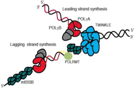
Mitochondria contain enzymatic systems responsible for mtDNA replication, diverse from those in the nucleus, however all these proteins are encoded by nuclear genes (Figure 4). To date, just three of these proteins have been identified operating at the mtDNA replication fork. Ahead of the fork, the replicative helicase Twinkle translocate on one DNA strand in 5’ to 3’ direction, unwinding double-stranded DNA into single-stranded DNA that now may serve as a template for DNA polymerase performing DNA synthesis. For the protection from nucleolysis, mtDNA bounds to the mitochondrial single-stranded DNA-binding protein (mtSSB). Replication is primed by extension of processed RNA transcripts laid down by the mitochondrial RNA polymerase (Reyes, 2013). Part of replication enzymes, POLRMT, the catalytic subunit of mtDNA polymerase (POLγA), and the replicative mitochondrial helicase (TWINKLE) are similar to proteins encoded by the T-odd lineage of bacteriophages and not related to eubacteria (Shutt and Gray, 2006).
Figure 4. The mtDNA replication machinery. The TWINKLE helicase (blue) moves in a 5' to 3' direction while unwinding dsDNA. The mtSSB protein (dark green) stabilizes the single stranded conformation and
14
stimulates the DNA synthesis by the POLγ (red (A) and gray (B)). POLRMT (light green) synthesizes the RNA primer (yellow line) needed for lagging strand DNA synthesis (from Wanrooij andFalkenberg, 2010).
Mitochondrial DNA polymerase ϒ
The only DNA polymerase found in mammalian mitochondria is the POLγ or POLG1 the enzyme promoting mtDNA replication and repair (Kaguni, 2004).
The enzyme belongs to the family A group of DNA polymerases and human catalytic subunit POLγA has a molecular mass of 140 kDa. The catalytic subunit POLγA is associated with a smaller protein, the mtDNA polymerase γ accessory subunit (POLγB), which has molecular mass of 55 kDa (Gray and Wong, 1992). POLγA and POLγB form a heterotrimer (POLγAB2) in mammalian cells (Yakubovskaya, 2006); for sake of simplicity, later on the holoenzyme will be denoted simply as POLG. POLγB subunit significantly enhances the catalytic activity and the processivity of main subunit POLγA. To some extent this may be explained with enhanced binding of POLγA to mtDNA in the holoenzyme formation and also with increased nucleotide binding. Altogether this suggests that the accessory subunit has fundamental influence on the function of the catalytic subunit and allows for the most efficient binding of substrate (Falkenberg, 2007). POLγA subunit possess the enzyme’s 5’-3’ polymerase, 3’-5’ exonuclease and 5’ deoxyribose-5 phosphate lyase activities, and is probably the most studied polypeptide of the mtDNA replisome at the clinical, biochemical and structural levels (Stumpf and Copeland, 2011). Given that it is the only mtDNA polymerase, which functions in both mtDNA replication and repair, it is not surprising that research has been focused to disease-associated POLG mutations. Since the first POLG mutations were found to be associated with progressive external ophthalmoplegia (PEO) (Van Goethem, 2001), over 200 mutations in the POLG1 and POLG2 genes have been detected in association with various mitochondrial diseases (Akhmedov and Marin-Garcıa, 2015).
15
TWINKLE
Mutations in the human Twinkle gene (C10orf2) were for the first time reported as a cause of autosomal dominant PEO, associated with multiple mtDNA deletions (Spelbrink, 2001). Later, biochemical studies showed the NTPase, 5’-3’ ssDNA translocase and dsDNA unwinding activities of the enzyme, identifying Twinkle as the replicative mtDNA helicase (Korhonen, 2004).
Mitochondrial single-stranded DNA binding (mtSSB)
This protein resembles Escherichia coli SSB and is the only protein of three replication-fork associated proteins that has no similarities with phage proteins (Tiranti, 1993). MtSSB binds to single-stranded DNA as a tetramer composed of four 16 kDa subunits (Yang, 1997). The protein stimulates synthesis of mtDNA by facilitating POLG primer recognition and enhancing POLG processivity (Genuario and Wong, 1993). MtSSB also stimulates the dsDNA unwinding activity of TWINKLE (Korhonen, 2003).
Recent study of mtSSB protein showed this protein is highly abundant being present at more than two thousand units per mtDNA. It was demonstrated that mtSSB is actively recruited to nucleoids during mtDNA replication in vivo. MtSSB covers displaced single strands of replicative intermediates (Van Tuyle and Pavco, 1985). It was found that mtSSB is not evenly distributed over the genome, and that it associates exclusively with the H strand. Highest levels of mtSSB are downstream of the D-loop region, at the 3’ end of 7S DNA, and mtSSB levels gradually reduce towards OriL. A second, minor, peak of mtSSB take place just after OriL, but it weakens away going towards the D-loop region (Fuste, 2014). All these findings strongly favour strand displacement mode as primary mtDNA replication mechanism.
2.2.3 TRANSCRIPTION
In human cells, transcription initiation depends from promotor regions, and each strand has its own promoters: the light-strand promoter (LSP) and the two heavy-strand promoters (HSP). Transcription
16
from these promoters finally results in polycistronic precursor RNAs. These primary transcripts are processed to produce the individual mRNA, rRNA, and tRNA molecules (Montoya, 1981; Ojala, 1981; Clayton, 1991).
Two heavy strand promoters HSP1 and HSP2 have been identified at the H strand (Micol, 1997). The HSP1 and HSP2 promoters are located very close, spaced by about 100 bp in the D-loop region and transcribed in the same direction (Martin, 2005). Transcription from the HSP1 promoter is prematurely terminated downstream of the 16S rRNA, transcribing only for the tRNAVal, tRNAPhe and the 2 rRNAs. Premature termination is result of a site-specific binding of the mitochondrial termination factor MTERF1 (Roberti M, 2009). Transcription initiated from the HSP2 promoter produces a full-length polycistronic transcript covering the 2rRNAs (12S rRNA and 16S rRNA), 12mRNAs and 13 tRNAs. All the protein-coding and rRNA genes are flanked by tRNA genes, which need to be excised in order to have mature mRNA and rRNA molecules (Falkenberg, 2007).
Transcription from the LSP produces the RNA primers required for initiation of mtDNA replication at the origin of H-strand DNA replication (OH) (Clayton, 1991; Chang and Clayton, 1985). The molecular mechanism stopping the transcription and allowing the switch to primer formation is still not completely explained. However, very recent research showed that transcription and replication of mtDNA cannot go in parallel without interfering with each other. It was proposed that in presence of human transcription elongation TEFM, mitochondrial RNA polymerase (mtRNAP) efficiently transcribes through termination sites of transcription (Agaronyan, 2015).
As mentioned above, within the control region there are three conserved sequence blocks (CSBI- III) downstream of LSP (Walberg and Clayton, 1983). CSB II increases the stability of an RNA-DNA hybrid, and transitions from the RNA primer to the newly synthesized DNA have been mapped near CSBII (Falkenberg, 2007). Termination of transcription is still under investigation, since it is known just one exact termination site. This termination site is located at the end of the 16S rRNA gene, when
17
transcription starts from the HSP1 end (Christianson and Clayton, 1988). Transcription termination at this site is determined by the mitochondrial transcription termination factor (mTERF).
The mitochondrial ribosome is shaped when mitochondrial ribosomes, 12SrRNA and 16S, form a complex with the mitochondrial ribosomal proteins (Pietromonaco, 1991).It is still unclear if 5S rRNA, which is imported within mitochondria, also assembles with mitochondrial ribosomes (Smirnov, 2011).
2.2.4 TRANSLATION
The components of mitochondrial translation are different from the cytosolic counterparts and, at the same time, there are also differences with the bacterial translation machinery. These differences concern not only the structure of the mitoribosome, which is more similar to the bacterial one, but in particular the number of associated proteins that is higher when compared to both the cytosolic and bacterial ribosomes. The role of most of these proteins is not assigned, but it is thought they may serve as a protection of rRNA from ROS insults (Lightowlers, 2014). Translation of the mtDNA-encoded protein transcripts results in nascent polypeptides key for OXPHOS, while the other polypeptides of this system are translated in the cytosol. Not surprisingly the two translation systems are synchronized during mitochondrial biogenesis in yeast (Couvillion, 2016). Mito-translation takes place through the following major steps: formation of initiation complex, elongation of the rising polypeptide chain, and termination, and recycling of the mitoribosome is considered as the final step of the process (Boczonadi and Horvath, 2014). The initiation complex consists from: 28S small subunit, mRNA, fMET-tRNA and IF2/3mt. When this complex is formed the mRNA enters into the IF3mt:28S subunit complex. The supposed role for IF3mt is to provide the right position of mRNA in binding the small subunit at the peptidyl (P) site of the mitoribosome. When the suitable start codon is matched, the formylmethionin -tRNA can bind to the first codon with the assistance of IF2mt. The assembly of the mitoribosome signals for release of initiation factors and the elongation proceeds on the 55S ribosome (Boczonadi and Horvath, 2014).
18
The mitochondrial elongation factor (EF-Tumt-GTP) and an amino-acylated tRNA occupy the A-site of the mitoribosome where truthful codon anticodon pairing takes place. If codon-anticodon pairing is correct than the elongation factor leaves the mitoribosome while the aminoacyl-tRNA moves to the P position where formation of peptide bond is catalysed leading to extension of the growing polypeptide chain. When the stop codon (UAA, UAG, AGA or AGG) comes across at the A-site, elongation stop is terminated (Boczonadi and Horvath, 2014). Stop codon is recognized, by the mitochondrial release factor (mtRF1a), which binds to the mitoribosome, inducing hydrolysis of the peptidyl-tRNA bond in the A-site, thus releasing the mature protein from the site (Richter, 2010).
2.3 INHERITANCE OF MITOCHONDRIAL DNA
Among many peculiarities of mtDNA, inheritance is maybe the most intriguing. The mtDNA is inherited in a uniparental fashion, meaning that all the copies of mtDNA are passed to next generation solely from the mother (Giles, 1980). This implies that recombination of mitochondrial genetic material, if occurring, involves identical mtDNA molecules, raising the question why is there uniparental inheritance and how is this accomplished (Lane, 2012)? This question is still a matter of scientific debate, and a few recent studies may shed some light. First, the mtDNA sequence is highly variable even amongst individuals of the same mammalian specie and certain sequence polymorphisms can have influence even on acidity of the mitochondrial matrix (Gómez-Durán, 2010; Kazuno, 2006). As a consequence, mixing two distinct sequences, with different influences on certain physiological conditions, and with different variants in the same OXPHOS polypeptides may have harmful effects (Wallace, 2007). Indeed, a recent study showed that if two normal, but different mtDNA haplotypes are artificially mixed in the same mice model, an incompatibility manifests as the heteroplasmic state is unstable, resulting in a pathological phenotype with behavioural abnormalities (Sharpley, 2012). Second, different mtDNA haplotypes support equal
19
rates of ATP synthesis but differ in reactive oxygen-species (ROS) leak and mtDNA copy number. ROS leak seems to optimize ATP synthesis by stimulating mtDNA copy increase (Moreno-Loshuertos, 2006). Thus, for various possible reasons, the uniparental transmission of mtDNA is evolutionary conserved and favoured (Lane, 2011). But, how human paternal DNA gets eliminated? Two models are considered in the field: passive and active elimination (Carelli, 2015). First, the passive or “dilution” model reasons that paternal mtDNA, much inferior in copy number, is basically watered down away by the excess of oocyte mtDNA and consequently it is hardly detectable in the offspring (Sato and Sato, 2012). In accordance to this model, study done on mice showed paternal mtDNA elimination prior to fertilization, while infrequent not eliminated paternal mtDNA enters in the egg and could be found in newborn mice (Luo, 2013). However, when this model was tested in humans, by examining heteroplasmy in buccal derived DNA from the children, confirmation for paternal mtDNA transmission was not found (Pyle, 2015). Second, the active elimination model predicts that paternal mtDNA is selectively eliminated, by ubiquitination or other still elusive mechanisms, in order to prevent its transmission to offspring. Mechanisms involved in this elimination are diverse, and may act at different pre- or post-fertilization levels (Carelli, 2015).
2.4 MITOCHONDRIAL GENETICS AND HUMAN DISEASE
Mitochondrial dysfunction underlies a large and diverse array of human diseases. The list of diseases in which mitochondrial dysfunction is recognized as a cause or a contributor to the pathogenic mechanism has been constantly growing in the past decade (Wallace and Chalkia, 2013). Many of the functions that mitochondria carry out in eukaryotic cell are key to survival, and have implications far beyond the energy supply. Independently from the energy production capacity, mitochondria affect a number of other cellular processes interfering with the expression of several thousands of genes linked to diverse cellular functions (Elstner and Turnbull, 2012; Latorre-Pellicer, 2016). The mtDNA has 100-1000 higher mutation
20
rate than nDNA and each cell variably contains 100-1000 copies of mtDNA. A subset of mtDNA molecules may carry pathogenic mutations, leading to the condition of intracellular heteroplasmy. Nuclear genes implicated in mitochondrial functions are estimated to range from 1000 to 2000 (Pitchard, 2016). Thus, mitochondrial function may be of relevance for any complex disease. Individuals can accumulate different mtDNA mutations over time, impinging on the energetic capacity of the cell, and such clusters of mtDNA mutations are relevant for aging and cancer. Epidemiological surveys estimated that incidence of the most common pathogenic mtDNA mutations is one in 5000 (Schaefer, 2008). A second study of newborn cord bloods revealed that one in 200 infants harbour one of the 10 most common pathogenic mtDNA mutations (Chinnery, 2012). In general, mitochondrial diseases can be defined as a clinically heterogeneous group of disorders related with OXPHOS dysfunction. Mitochondrial disorders typically affect postmitotic and energy-demanding tissues, such as brain, heart or muscle, can appear sporadically or be maternally inherited (Schon, 2012). There is no cure for mitochondrial disease and therapy is mostly limited to manage symptoms. Despite the fact that it is now well established that overall mitochondrial disorders cannot be considered rare, our understanding of the pathogenic mechanisms remains limited.
3. MITOCHONDRIAL DNA DELETIONS
The first pathogenic mtDNA mutations, deletions and point mutations, were discovered nearly three decades ago (Holt, 1988; Wallace, 1988). Both large-scale rearrangements (deletions or duplications) and point missense mutations of mtDNA have been linked to a vast array of clinical phenotypes. MtDNA deletions are pathogenic mutations that remove various portions of mitochondrial genome thus resulting in shorter molecules of mtDNA. Circular deleted mtDNA is smaller than wild-type, due to the missing section of the genome, but it remains in a closed circular structure. The mtDNA deleted molecules are transcribed, but there is no evidence that the ”fusion” transcript crossing the deletion breakpoint is translated into a chimeric protein. On the other hand, mtDNA deletions remove a certain
21
number, depending on size and position of the deletion, of indispensable tRNA genes consequently disrupting the translation of key mtDNA-encoded respiratory chain polypeptides (Nakase, 1990). Thus, the deleterious effect of mtDNA deletions is out of any doubt. The phenotypic heterogeneity observed in patients carrying mtDNA deletions, imply however further elements to be considered, specifically the heteroplasmy and clonal expansion. In patients, we may distinguish two classes of mtDNA deletions: single and multiple deletions. These classes are based on inheritance pattern and the variety of deletions present. Furthermore, mtDNA sequence surrounding the deletion breakpoints may be used for classification of the mtDNA deletions. In fact, deletions may occur flanked by two direct repeats with identical sequences (class I deletions), flanked by imperfect repeats (class II deletions) and those that have no direct repeats (class III deletions).The deletions may vary in size from in the range from 2 kb to 10 kb and may cover any mtDNA gene. Regardless of that, distribution of mtDNA deletions is not random; the mtDNA deletions are prevalent in the major arc of mtDNA, between two origins of the replication. Only 1.4% of all deleted regions, published and described in largest database of mtDNA deletions- Mitobreak (www.mitobreak.portugene.com), is located within the minor arc of mtDNA(Damas,2014).In single deletions the size of the deletion is associated with the severity of clinical phenotype, because larger the deletion is more mtDNA genes are removed, and consequently OXPHOS is more compromised (Grady, 2014). The location for the majority of deletions within the major arc explains the length constraint; the length of the major arc (11,230 nt) creates an upper limit for the size of the deletions. The central region of the major arc is often deleted, with the one ranging from positions 7,946 to 15,158 being missed in more than half of cases and that ranging from positions 10,936 to 12,893, including tRNA-His (TRNH) and tRNA-Ser (TRNS2) genes, being missed in 90% of cases (Damas, 2014). A few studies identified a significant number of minor arc mtDNA deletions in sporadic inclusion body myositis patients (sIBM) and in elder people studied in ageing research (Rygiel, 2015; Bank, 2000). The two regions that cannot be deleted are the origin of heavy-strand replication and the light-strand promoter, as these regions are necessary for replication of both the deleted and wild-type mtDNA molecules (Moraes,
22
2003). The predominant deletion in the patient’s population is a 4977-bp deletion (“common deletion”), surrounding several mitochondrial genes encoding for structural proteins and tRNAs, and bordered with a long 13-bp direct repeat on both breakpoints (Schon, 1989).
3.1 MITOCHONDRIAL DNA DELETIONS HETEROPLASMY AND CLONAL EXPANSION
Every deleted mtDNA molecule exists mixed with wild type mtDNA molecules in an intracellular pool of mitochondrial genomes, making this pool non homogenous, and defined as heteroplasmic. When a new mtDNA mutation, in this case a deletion, arises it is just one mutant mtDNA molecule coexisting amongst thousands of non-mutant mtDNA molecules. Under these circumstances, the wild-type mtDNA easily compensates for the few mutant mtDNA molecules. Thus, low levels of mtDNA deletions can be easily tolerated, but once the mutant mtDNA molecules overcome a critical threshold level, a biochemical defect arises. This threshold level significantly varies between tissues and is distinct for different mutations. In the case of mtDNA deletions, the threshold level is proposed to be around 80%. In rapidly dividing cells such as hair or blood cells heteroplasmy of rearranged mtDNA is markedly shifted in favour of wild-type mtDNA (Moraes, 2003). A genetic drift during the germ-line bottleneck, characterized by a drastic drop in the number of mtDNA segregating units during oogenesis, can remarkably change the heteroplasmy levels between generations (Rebolledo-Jaramillo, 2014).
Over time, the pathogenic mtDNA mutations (i.e. deletions) may become prevalent in certain cells, due to their clonal expansion, which may take place in both the germline and somatic cells. The mechanism beyond the clonal expansion is still poorly understood. Three major hypothesis deal with potential forces leading to clonal expansion of mtDNA deletions: replicative advantage of a smaller molecule, lover levels of ROS production and random genetic drift.
The replicative advantage mechanism argues that deleted mtDNA molecules, due to being smaller size, are replicated faster than wild-type mtDNA, thus allowing for the mtDNA deletion to accumulate
23
(Wallace, 1992; Corral-Debrinski, 1992). This mechanism would provide a plausible explanation if two prerequisites are fulfilled. First, mtDNA replication needs to take significant amount of time in compared with time needed for mtDNA turnover, thus allowing for smaller molecules to accumulate. Second, this would imply that replicative advantage would favour larger deletions (Campbell, 2014). Indeed, in agreement with this second prerequisite, two studies found that larger deletions accumulate in greater extent (Diaz, 2002; Fukui and Moraes, 2008, 2009), but this accumulation is observed only under relaxed copy number conditions and when no biochemical defect was observed, respectively. Another valuable study performed on patient tissues reported that there is no preferential accumulation of larger deletions over smaller deletions, under normal copy-number control. Given that mtDNA replication takes from one to two hours and time of mitochondrial half-life is estimated to be from one to three weeks, replication can hardly be rate limiting the turnover (Campbell, 2014). However, mtDNA point mutations are also accumulated by clonal expansion, and they cannot have any influence on the size of mtDNA molecule, thus replicative advantage of shorter molecules cannot be interpretation for the clonal expansion (Elson, 2001).
Survival of the slowest hypothesis argues that because of decline in respiratory chain function, provoked by mtDNA damage, free radical production is also reduced. This way the compromised mitochondria manage to be spared from mytophagy and apoptosis as they are not considered as producers of toxic free radicals. Consequently the deleted mitochondria will predominate within a cell (De Grey, 1997). This hypothesis has not been supported by experimental evidence.
The random genetic drift hypothesis implies there is no selective advantage for deleted molecules (Elson, 2001). Random genetic drift model is established on validated rules of mtDNA replicative dynamics, which assume a relaxed mtDNA replication unrelated to the cell cycle, and is based on the mathematical models (Elson, 2001). On the other hand, evidences for significant levels of mtDNA deletions in the most of dopaminergic neurons ofsubstantia nigra from older persons (Kraytsberg, 2006) contrasts with the predicted low levels of age-related COX-deficient cells, which should be less than 5% if only age driven.
24
Furthermore, this model does not explain the preferential accumulation of mtDNA deletions in certain tissues, under the assumption of a replicative turnover under relaxed replication and copy number control (Campbell, 2014).
None of the proposed mechanisms fully explains the prevalent accumulation of mtDNA deletions in certain tissues and the consequent occurrence of a clinical phenotype.
In muscle fibres, clonally expanded mtDNA genomes are found in specific, more or less restricted domains. These domains presumably represent areas with extended expansion process. Segmental nature of mtDNA clonal expansion is suggesting that mitochondrial fusion is restricted to some extent, and this is visible in elongated cells like muscle fibres. It seems likely and has been proposed that the dynamic processes of fusion and fission would affect the dimensions of segmental domains where deleted mtDNA expanded, becoming prevalent (Carelli and Chan, 2015).
No matter the mechanism for arise of clonal expansion, it leads to local shifts in heteroplasmy rates, within cells or even within cell domains as in the case of skeletal muscle. Such heteroplasmy can vary between cells and tissues, making the phenotypic expression of a mitochondrial disorders difficult to predict. Thus, clonal expansion is the driving force leading to prevalent populations of deleted mtDNAs, ultimately causing the deleterious effect on mitochondrial OXPHOS and other metabolic pathways. Waiting to understand the mechanisms of clonal expansion, the distribution pattern of mtDNA heteroplasmy remains an unpredictable aspect of mitochondrial genetics (Wallace, 2013; Carelli, 2015). 3.2 SINGLE MITOCHONDRIAL DNA DELETIONS
Clinical features
The first mtDNA mutations discovered in 1988, were in fact single mtDNA deletions and the Leber’s hereditary optic neuropathy point mutation, proving for the first time that mtDNA mutations cause human disease (Holt, 1988; Wallace, 1988). In patients with single mtDNA deletions, there is one prevalent deletion species exhibiting a wide tissue distribution. Single, large scale, deletions often cause a sporadic disease, which may be present from early childhood such as Pearson and Kearns-Sayre
25
syndromes or, later in life with chronic progressive external ophthalmoplegia (CPEO). As stated above, KSS, CPEO, and PS are sporadic disorders meaning that detectable mtDNA deletions are not found in other family members (mothers and maternal siblings)do not have detectable mtDNA deletions. Opposed to the single mtDNA deletions, large-scale mtDNA duplications are often maternally transmitted (Ballinger, 1992, 1994; Manfredi, 1997). Single deletions account for about a third of all patients with mtDNA-related disease (Mancuso M, 2015).
Pearson's bone marrow-pancreas syndrome is a severe disorder, with high incidence of mortality. It involves the hematopoietic system, kidneys, exocrine pancreas and liver. The hallmark feature is sideroblastic anaemia, not typical for other mitochondrial diseases. The single type of mtDNA deletion is consistently found in all tissues, but in the blood it seems to be even more abundant and in neonatal period certain symptoms may differ from classical presentation (Manea, 2009). Pearson syndrome is often fatal in infancy. Individuals with Pearson syndrome who survive beyond infancy later on develop the neurological symptoms of Kearns-Sayre syndrome (Lee, 2007).
Kearns-Sayre Syndrome (KSS) is a multisystem disorder affecting predominantly the central nervous system, skeletal muscle, and heart. Onset is usually in childhood, with ptosis, ophthalmoplegia, or both. Exercise intolerance and impaired night vision due to retinal dystrophy may be early symptoms (Auré, 2007). A single mtDNA deletion is again found in all tissues. The disease usually progresses to death in young adulthood.
Progressive external ophthalmoplegia (PEO) is a mitochondrial myopathy with drooping of the eyelids (ptosis), paralysis of the extraocular muscles (ophthalmoplegia), and variably severe proximal limb weakness. A few individuals with PEO have other manifestations of KSS and this situation is termed PEO plus. The disorder is quite benign, not affecting a normal life span. The single deletion occurrence is mostly restricted to skeletal muscle (Zeviani and Carelli, 2003).
26
3.3 INHERITANCE OF SINGLE MTDNA DELETIONS
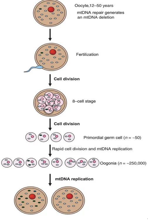
Single, large-scale mtDNA deletions are in most cases considered sporadic events occurring in the early stages of embryonic development of an individual (Zeviani and Carelli, 2003). However, there are studies showing that mtDNA deleted molecules may be present in the oocyte where mtDNA is sustained for long time up until fertilization. A few studies reported evidence of low levels (no more than 0.1%) mtDNA deletions in human oocytes (Chen, 1995; Brenner, 1998). If an oocyte containing a certain amount of mtDNA deletion is fertilized, the deleted molecules most probably will be lost by negative selection or purifying selection during the early stages of embryonic development. However, the genetic drift potentially occurring during the germ-line bottleneck may occasionally lead to prevalent mtDNA deletion, as result of the severe reduction in mtDNA copy number during oogenesis (Rebolledo-Jaramillo, 2014). After fertilization, mtDNA variants are distributed among cells according to mitotic segregation, taking place upon fertilization, and it is assumed this process is leading to random distribution of mitochondria and mtDNAs during cell divisions (Poulton, 2010). Even if most times single mtDNA deletions are believed to be sporadic De Novo events, it cannot be excluded that they may in fact result from mtDNA bottleneck. The mtDNA germ-line bottleneck may provide a plausible explanation in the case of maternally inherited single mtDNA deletions, as the mother and maternal siblings of affected individual are healthy. It is possible that a low-level mtDNA deletion segregates through the bottleneck and gets the opportunity of high expansion, eventually populating the primary oocyte (Krishnan, 2008) (Fig.5). In support of the bottleneck-derived germline transmission of deleted mtDNA there are a few cases of maternal inheritance in single mtDNA deletions that have been reported (Blakely, 2004; Shanske, 2002; Bernes, 1993). A good example is the case of a woman with sporadic CPEO who harboured a single large-scale mtDNA deletion, which was transmitted to her infant son, affected with PS. The single deletion of 5,355-bp in mtDNA, without flanking direct repeats, was the only abnormal species of mtDNA identified in both patients (Shanske, 2002). In another case reported, a woman, again with CPEO, and her son who also had a Pearson-like syndrome, both harboured the 4,977-bp “common”
27
mtDNA deletion. Although this case was similar to the previous one, the experimental analysis did not rule out the presence of duplicated mtDNA, which could have explained the transmission from mother to child (Bernes, 1993). In a third reported case, a woman and her daughter both had CPEO, but mother and daughter harboured different single mtDNA deletions (Ozawa, 1989).
It is important to point out that these few cases with documented maternal inheritance of a single mtDNA deletion cannot be considered as definitive proof for the not sporadic nature of these mutations. In order to confirm maternal transmission, more cases should be thoroughly investigated. An important feature of mtDNA deletions is that they are usually not detectable in blood, except in the cases of Pearson syndrome, and muscle biopsy sampling of clinically unaffected mothers are ethically problematic. Therefore the development of highly sensitive approaches for detection of mtDNA deletions would be of outmost importance for understanding potential transmission of these deleterious mutations, and consequently would help in genetic counselling.
28
.
Figure 5. Illustration of a model for existence of sporadic mtDNA deletions in the oocyte. Formation of mtDNA deletion due to repair ofoocyte mtDNA. The segregation of mtDNA deletion after fertilization and cell division. If the segregation was at the level of primordial cell than extensive synthesis of mtDNA could lead to the prevalence of mtDNA deletion in the oocyte. If this oocyte is fertilized in future, a child born would be at risk of being born with high levels of an mtDNA deletion (from Kirshnan, 2008).
3.4 MULTIPLE MITOCHONDRIAL DNA DELETIONS
Shortly after the discovery of single, sporadic mtDNA deletions, the accumulation of multiple mtDNA-deleted species was observed in families with recurrent cases of CPEO transmitted as an autosomal
29
dominant trait (Zeviani, 1989). Mendelian inheritance of mtDNA mutations appeared puzzling, since this genome is transmitted in a strictly maternal fashion. However, this observation was explained as the consequence of a dominant mutation in a nuclear gene leading to altered structural integrity of mtDNA (Zeviani, 1990). This hypothesis proved to be correct, as it led to the discovery that many genes responsible for familial CPEO were associated with multiple mtDNA deletions. Later one, it was determined that multiple mtDNA deletions may arise secondarily from mutations in nuclear genes involved in mtDNA maintenance. The list of genes involved in mtDNA maintenance, and thereby associated with multiple deletion formation, is constantly growing. Thus, the inheritance of these mtDNA deletions follows the classic Mendelian rules.
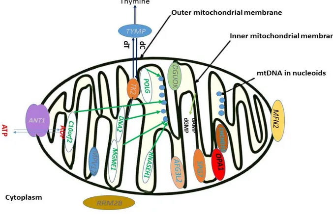
These genes can be involved directly in mtDNA replication (POLG, POLG2, TWINKLE, RNASEH1, MGME1, and DNA2), but also in nucleotide pool balance (TYMP, SLC25A4, TK2, DGUOK, RRM2B, and MPV17) and, quite surprisingly, in mitochondrial dynamics (OPA1 and MFN2) (Sommerwille, 2014;Copeland, 2012). Also, mutations in genes difficult to categorize in aforementioned groups, are being correlated with multiple deletions like SPG7- paraplegin mutations and its partner protein AFG3L2, previously associated with dominant spinocerebellar ataxia type 28 disease (Gorman, 2015). Most recently, the CHCHD10 gene of unknown function was also found mutated in a few families with accumulation of mtDNA multiple deletions in muscle (Bannwarth, 2014). Thus, it is important to emphasize that these are not all mtDNA maintenance genes (Fig.6). Mutations in the nuclear encoded mitochondrial maintenance genes may cause two molecular phenotypes: mtDNA multiple deletions (“mtDNA multiple deletion disorders”) and reduction of mtDNA copy number (“mtDNA depletion syndromes”, MDS). The two molecular phenotypes may co-exist and are known collectively as mtDNA maintenance disorders (Kornblum, 2013). They can be either autosomal-recessive or -dominant mitochondrial disorders, in most cases with CPEO as the common clinical feature (Reyes, 2015).
To complicate the scenario, mtDNA multiple deletions arise and accumulate also spontaneously as age-dependent by-product of mtDNA replication in post-mitotic tissues. This somatic accumulation of mtDNA
30
multiple deletions characterizes certain subpopulations of neurons in neurodegenerative disorders, such as the dopaminergic neurons of substantia nigra in Parkinson disease (PD), as well as in elderly subjects (Carelli, 2015; Chan, 2015). Questions that still remain open are why in PD the accumulation of mtDNA deletions leads to detrimental consequences when the levels of deletion loads are similar in aged individuals? A very recent study reported that in elderly subjects the mtDNA copy number increased significantly with age and correlates with level of mtDNA deletions, whereas this compensatory affect fails in PD patients (Dolle, 2016).
3.5 MITOCHONDRIAL DNA MAINTENANCE GENES AND MULTIPLE MTDNA DELETIONS
Mutations in the DNA polymerase-γ (POLG) gene are a major cause of clinically heterogeneous mitochondrial diseases, associated with mtDNA depletion and multiple deletions.
POLG is the only polymerase responsible for mtDNA replication, and knockout mouse for POLG show embryonic lethality (Hance, 2005). POLG-related disorders are currently defined by at least five major neurological phenotypes, that include: childhood myocerebrohepatopathy spectrum (MCHS), myoclonic epilepsy myopathy sensory ataxia (MEMSA), Alpers-Huttenlocher syndrome (AHS), the ataxia neuropathy spectrum (ANS), and chronic progressive external ophthalmoplegia (CPEO), some of them being fatal in early childhood (Wong, 2008). In individuals presenting with CPEO and multiple mtDNA deletions most likely the laboratory tests will find pathogenic mutations in POLG. Psychiatric symptoms together with Parkinsonism are particularly prevalent in patients harbouring mutations of POLG. Other clinical manifestations include POLG-related encephalopathy and lactic acidosis and stroke-like episodes syndrome (MELAS)-like phenotype (Sommerwille, 2014; Hudson, 2006). In summary, both mtDNA depletion and accumulation of mtDNA deletions are important consequences of POLG mutations, but their reciprocal impact is variable among patients (Carelli, 2015).
31
C10orf2 (twinkle) encodes the mitochondrial protein twinkle, as mentioned above, this protein is DNA
helicase that unwinds double stranded mtDNA in the 5’ to 3’ direction. This process is dependent on ATP and essential for mtDNA replication (Spelbrink, 2001). Two Twinkle mutations have been expressed in mice, dup352–364 and A359T, associated with CPEO in patients. The accumulation of mtDNA deletions was observed in the mouse muscle and brain tissues. The number of deleted molecules increased with age, along with the progressive respiratory chain dysfunction and myopathy (Tyynismaa, 2005). Dominant and recessive forms of PEO were reported in patients with C10orf2 mutations. Phenotypic spectrum seen in patients with Twinkle mutations is very diverse, including gait disturbance, exercise intolerance, diabetes mellitus, visual impairment, diplopia, hearing loss, ataxia, and seizures. Psychiatric symptoms are particularly common in these patients and may be any of the following, or combination of more symptoms, like: more or less severe depression and dementia, Alzheimer’s disease, avoidant personalities and other. Presence of mtDNA deletions is a hallmark of these mutations (Spelbrink, 2001; Van Hove, 2009;Sommerwille, 2014).
Recently RNASEH1 was identified as another gene crucial for mtDNA maintenance. RNASEH1, encoding ribonuclease H1 (RNase H1), is an endonuclease that is present in both the nucleus and mitochondria and digests the RNA component of RNA-DNA. Patients first present with CPEO and exercise intolerance followed by muscle weakness, dysphagia, and spino-cerebellar signs with impaired gait coordination, dysmetria, and dysarthria (Reyes, 2015). Mouse knockout of RNASEH1 shows embryonic lethality due to failure to synthetize mtDNA (Carittelli, 2003). Deleterious RNASEH1 mutations slow down and stall mtDNA replication, causing both mtDNA depletion and deletions and ultimately leading to mitochondrial disease (Reyes, 2015).
MGME1 (mitochondrial genome maintenance exonuclease 1), encodes a mitochondrial RecB-type
exonuclease belonging to the PD-(D/E) XK nuclease superfamily. MGME1 cleaves single-stranded DNA and processes DNA flap substrates, also necessary for maintaining mitochondrial 7SDNA levels. By date only homozygous mutations in the gene are causing clinical phenotype characterized by ptosis in
32
childhood, followed by CPEO, diffuse skeletal muscle wasting and weakness, profound emaciation, and respiratory distress. Interestingly, mtDNA deletions from patients with MGME1 mutations were unusually large and detectable in DNA derived from blood (Kornblum, 2013).
The molecular pathway responsible for the involvement of DNA2 in mtDNA is still under investigation. However, DNA2 encodes a helicase/nuclease family member that is most likely involved in mtDNA replication, as well as in the long-patch base-excision repair (LP-BER) pathway. Patients present with lower limb weakness, ophthalmoplegia, diplopia, myalgia and progressive myopathy. Multiple mtDNA deletions were reported in muscle (Ronchi, 2013).
TYMP encodes thymidine phosphorylase which catalyses the conversion of thymidine to thymine and
deoxyuridine to uracil (El-Hattab, 2017). Depletion of thymidine phosphorylase disturbs dNTP supply affecting mtDNA replication. Mutations of the TYMP cause mitochondrial neurogastrointestinal encephalopathy (MNGIE). This is a multisystem disorder and gastrointestinal dysmotility is the main symptom. Other common symptoms are CPEO, cachexia,muscle weakness, muscle atrophy and hearing loss. One of the hallmarks of MNGIE is somatic alteration of mtDNA, namely, depletion, multiple deletions, and site-specific point mutations (Valentino, 2007).
SLC25A4 (ANT1) encodes ADP/ATP translocase1, as the name of this protein suggests it is necessary in
the exchange of ATP generated within mitochondria and ADP present in the cytoplasm. Phenotype manifestation is associated to adCPEO and Senger's syndrome. Patients often have ptosis and neuromuscular symptoms, if present, are usually restricted to the facial muscles, mostly extra ocular. There were reports with psychiatric symptoms in patients (Sommerwille, 2014).
TK2 encodes thymidine kinase 2, which phosphorylates thymidine, deoxycytidine, and deoxyuridine and
is essential for dNTP generation for mtDNA replication. Thus, TK2 is an enzyme of the mitochondrial nucleotide salvage pathway and its loss-of-function mutations have shown to underlie the early-infantile myopathic form of mtDNA depletion syndrome (MDS). Heterozygous mutations lead to arCPEO, with
33
multiple deletions. Clinical presentation is with mild late-onset CPEO together with myopathy and central nervous system involvement (Tyynismaa, 2012).
DGUOK encodes deoxyguanosine kinase, enzyme necessary for the first step of salvage of
deoxyribonucleotides in the mitochondria. This function is essential for mtDNA maintenance. Heterozygous mutations with very late onset CPEO and ptosis were described to have multiple mtDNA deletions (Ronchi, 2012). Interestingly, the same mutations were found in in early onset mitochondrial disease patients with mtDNA depletion (Sommerwille, 2014).
RRM2B encodes ribonucleoside-diphosphate reductase subunit M2 B (p53R2), protein that hasessential role for dNTP supply for mtDNA replication, and consequently to its role it is involved in both mtDNA maintenance and repair. Mutations in RRM2B, after POLG and TWINKLE, represent the third major number of adult-onset CPEO patients, and patients may have a broad spectrum of symptoms including milder CPEO, ptosis may occur but not necessary, exercise intolerance, muscle weakness, muscle atrophy, fatigue andhearing loss. Multiple mtDNA deletions are a common molecular finding in these patients (Sommerwille, 2014).
MPV17 is a mitochondrial inner membrane protein, and its absence or dysfunction may cause OXPHOS failure and mtDNA depletion in affected individuals and in the Mpv17 knockout mice. Severe mtDNA depletion manifests as early childhood-onset failure to thrive, hypoglycemia, encephalopathy, and hepatopathy progressing to liver failure (Spinazzola, 2006). Homozygous mutation in MPV17 was found in Navajo Indian patients with infantile-, childhood-, or juvenile-onset neurohepatopathy (Spinazzola, 2008). Heterozygous mutation was reported only in one patient, initially diagnosed with Charcot-Marie-Tooth disease. Later in life patient developed other symptoms: progressive proximal limb weakness, exercise intolerance, diabetes mellitus, ptosis, ophthalmoparesis, hearing loss, gastrointestinal dysmotility and depression. MtDNA deletions are found only in one patient, thus MPV17 mutations manifest preferentially as mtDNA depletion disorders (Garone, 2012).