A
A
l
l
m
m
a
a
M
M
a
a
t
t
e
e
r
r
S
S
t
t
u
u
d
d
i
i
o
o
r
r
u
u
m
m
–
–
U
U
n
n
i
i
v
v
e
e
r
r
s
s
i
i
t
t
à
à
d
d
i
i
B
B
o
o
l
l
o
o
g
g
n
n
a
a
DOTTORATO DI RICERCA IN
SCIENZE MEDICHE GENERALI E SCIENZE DEI SERVIZI
Ciclo 30°
Settore Concorsuale: Area 06 Scienze Mediche 06/H Clinica ginecologica
Settore Scientifico Disciplinare:
Area 06 - Scienze mediche > 06/H - Clinica ginecologica >
06/H1 Ginecologia e ostetricia
TRANSPERINEAL ULTRASOUND IN WOMEN WITH RECTAL
ENDOMETRIOSIS: COULD SONOGRAPHIC PARAMETERS BE
CORRELATED WITH BOWEL SYMPTOMS?
Presentata da:
Valentina Martelli
Coordinatore Dottorato
Supervisore
Prof Fabio Piscaglia
Prof Maria Cristina Meriggiola
TRANSPERINEAL ULTRASOUND IN WOMEN WITH RECTAL ENDOMETRIOSIS: COULD SONOGRAPHIC PARAMETERS BE CORRELATED WITH BOWEL SYMPTOMS?
ABSTRACT
STUDY OBJECTIVE: to compare levator hiatal area and anorectal angle at rest and after maximal contraction, at transperineal 2D/3D/4D ultrasound between patients with rectal endometriosis and asymptomatic healthy women and, secondly, to find any association between sonographic findings and bowel symptoms.
DESIGN: pilot, prospective study conducted between September 2015 and December 2016. SETTING: tertiary level referral Center of Minimally Invasive Gynecologic Surgery.
PATIENTS: 96 nulliparous patients with symptomatic rectal endometriosis scheduled for laparoscopic surgery (study group) were compared to 88 nulliparous asymptomatic healthy women (control group). Patients had never undergone surgery for deep endometriosis and had not assumed hormonal therapy before the enrollment.
INTERVENTIONS: transperineal ultrasound for evaluation of levator hiatal area and anorectal angle was performed in all patients at rest. Data were analyzed offline with a dedicated software (4DView 14.4; GE Healthcare) by an investigator blinded to clinical data. Bowel symptoms were collected using a validated questionnaire (Knowles-Eccersley-Scott-Symptom Questionnaire). Comparisons of mean values between controls and cases were performed with Student's t-test. Correlations between sonographic parameters and KESS questionnaire’s items were analyzed using Spearman’s correlation. P values <0.05 were considered significant.
MEASUREMENTS AND MAIN RESULTS: major demographic and anthropometric data were homogeneous for the groups. Compared to the control group, patients with rectal endometriosis
show a significantly narrower levator hiatal area at rest and after maximal contraction; patient with rectal endometriosis show a narrower anorectal angle at rest (109.8±10.8 grade versus 113.7±13.0 grade, p=0.03). Moreover, in the study group we found a significant association between severity of dyschezia at KESS questionnaire and dimension of anorectal angle (p < 0.001). In the study group, Patients with constipation had a narrower anorectal angle compared to endometriotic patients without constipation .
CONCLUSION: women with rectal endometriosis had a significantly narrower levator hiatal area and anorectal angle than healthy controls, suggesting pelvic floor hypertone. Pelvic floor dysfunctions in women with rectal endometriosis seem to be associated to bowel complaints, particularly dyschezia and constipation. Transperineal ultrasound may be a useful, inexpensive and non-invasive tool to detect pelvic floor dysfunctions in sympomatic patients affected by deep endometriosis.
INTRODUCTION
Endometriosis is a chronic and recurrent disease defined as the presence and proliferation of endometrial glands and stroma outside the uterine cavity. The ovary is the most common site involved, accounting for 80% of cases of endometriosis, but it can also involve other organs such as rectum, bladder and ureters (1). Rectal endometriosis belongs to a particular clinical condition: deep infiltrating endometriosis (DIE) that is defined as the presence of endometrial-like tissue (glands and stroma) >5 mm under the peritoneum (2). The major clinical problem of endometriosis is the pain syndrome, described as chronic pelvic pain, dysmenorrhea, dyspareunia, dysuria and dyschezia, affecting negatively women’s health and quality of life (1) , Noteworthy, deep lesions are associated also to sexual, urinary and rectal dysfunctions (3-6). In particular, sigestive complaints reported by women presenting with deep endometriosis infiltrating the rectum can be partially explained by cyclic micro-hemorrhages and inflammation into the rectal wall, anterior fixation of the rectum to the uterine cervix or vaginal fornix, and rectal stenosis (7). Moreover, recent studies demonstrated that women with DIE have an increased prevalence of pelvic floor muscle (PFM) dysfunctions (8)., which can play an important role in the pathophysiology of dyschezia and rectal symptoms itself (9).
Transperineal ultrasound 2D, 3D and 4D imaging was demonstrated as valid, inexpensive, reliable and non-invasive tool for assessing of pelvic floor function (10-13). Our group investigated PFM tone and strength through transperineal ultrasound in women with deep endometriosis and demonstrated in these patients a hyper-tone of PFM (represented by lower levator hiatal area) and a low strength of contraction (smaller changes in levator hiatal aerea narrowing during PFM contraction) (14).
Anorectal angle dimensions have been associated with evacuation difficulty revealed with defecography findings (16) and measurements of anorectal angle's excursions have been used widely as a proxy of PFM strength in women with incontinence and pelvic organ prolapsed (17). The aim of our study is to evaluate, static and dynamic amplitude of levator hiatal area and anorectal angle in women affected by rectal endometriosis, in comparison to asymptomatic healthy women using 2D-3D-4D transperineal ultrasound. Furthermore, we analyze any correlation between sonographic data (anorectal angle and levator hiatal area at transperineal ultrasound) and digestive symptoms reported by the patients of study group through a validated questionnaire (18).
MATERIALS AND METHODS Participants
This pilot prospective study was conducted between September 2015 and December 2016 at our tertiary level referral Center of Minimally Invasive Gynecologic Surgery. Ninety-six consecutive nulliparous women with diagnosis of rectal endometriosis were recruited in the study group. Diagnosis of rectal endometriosis is based on clinical and transvaginal/transabdominal ultrasound examinations and, when necessary, magnetic resonance All patients did not show a significant narrowing of rectal lumen (narrowing < 50%). All patients in the study group were scheduled for laparoscopic surgery and diagnosis of rectal endometriosis was confirmed by histological examination. Eighty-eight nulliparous asymptomatic healthy volunteers were enrolled in control group. Women in control group did not show any clinical or ultrasonographic signs of endometriosis and had to report no history of recurrent abdominal pain. For each women demographic and anthropometric data (age, body mass index, pain symptoms using a numerical rating scale from 0 to 10, previous surgery) were collected. In study group, surgical data and histological findings were also collected and KESS questionnaire was handed over to the subjects. The questionnaire includes eleven questions about bowel symptoms, in particular constipation, with a total scores ranging from 0 (no symptoms) to 39 (high symptoms severity). A cut-off score of ≥
11 indicates constipation (18, 19).
Exclusion criteria included: age less than 18 years or greater than 45 years, current or previous pregnancy, post-menopausal status, rectal endometriosis with more than 50% stenosis of bowel lumen, other cause of pelvic pain or pelvic floor dysfunctions (acute or chronic pelvic inflammatory disease, irritable bowel disease, vulvodynia, active urinary tract infection, congenital or acquired abnormalities of the pelvis or pelvic floor, diagnosis of genital malignancy, pelvic organ prolapse) and hormonal therapy within 3 months before the enrollment .
Patients gave informed written consent to participate to our study. The study protocol obtained approval from the local ethics committee.
Procedure
Information about pelvic floor anatomy and physiology was given to each participant. Transperineal ultrasound examinations were performed in both groups as previously described (14). In particular, levator hiatal area, antero-posterior diameter (AP diameter), left-right transverse diameter (LR diameter) and anorectal angle were evaluated at rest and at maximum pelvic floor contraction. (Figure 1, Figure 2, Figure 3)
The anorectal angle was defined as the angle between the posterior wall of the rectal ampulla and the anal canal. During PFM contraction, the anorectal angle becomes more acute and it moves cranially. (10).
All scans from both groups were obtained by the same experienced operator using a Voluson E6 system (GE Healthcare, Zipf, Austria) with RAB 8-4-MHz volume transducer for all acquisitions. Measurements were evaluated offline with a dedicated software (4DView 14.4; GE Healthcare, Zipf, Austria) by an experienced investigator blinded to clinical data. These measured parameters have already been studied for their properties demonstrating good test-retest, intra-observer and inter-observer reliability (17, 20-23).
All groups completed Knowles-Eccersley-Scott-Symptom Questionnaire (KESS) questionnaire. The questionnaire includes eleven questions about bowel symptoms, in particular constipation, with a total scores ranging from 0 (no symptoms) to 39 (high symptoms severity). A cut-off score of ≥ 11 indicates constipation (18, 19).
Statistical analysis
Continuous data were expressed in terms of mean ± SD or median (range). Categorical variables were expressed as numbers and percentages. Student’s t-test was used to compare continuous parametric variables. The comparison of KESS items and anorectal angle was performed using Spearman’s correlation because Kolmogorov – Smirnov test failed to show normal distribution (p < 0.001) for KESS items and for KESS total score. A correlation of 0.10 to 0.29 was considered slight, 0.30 to 0.49 modest, and 0.50 to 1.0 as good. A P-value of <0.05 was considered significant for all tests. Statistical analysis was carried out using the Statistical Package for the Social Sciences (SPSS) software version 24.0 (IBM Corp., Armonk, NY, USA).
RESULTS
Baseline characteristics of the control and study groups did not differ significantly and are reported in Table 1. Pain symptoms and endometriotic localizations of the study group are reported in Table 2. Transperineal ultrasound was successfully performed in all women, and no patients were removed from the study as a result of discomfort. The Outcomes of hiatal area (cm2) for DIE group and for control group respectively are: at rest 10.90 + 2.69 and 13.02 + 2.58, p<0.0001; after maximal contraction 8.55 + 1.85 and 9.45 + 2.11, p=0.002. The delta of hiatal area between contraction and rest for DIE group and for control group respectively are: 2.34 + 2.02 and 3.56 + 1.84, p<0.0001. The AP diameter of hiatal area (cm) for DIE group and for control group respectively are: at rest 4.67 + 0.69 and 4.92 + 0.63, p= 0.01; after maximal contraction 3.82 + 0.59 and 3.99 + 0.56, p= 0.04. The LR diameter of hiatal area (cm) for DIE group and for control group respectively are: at rest 3.29 + 0.46 and 3.63 + 0.53, p<0.0001; after maximal contraction 3.03 +
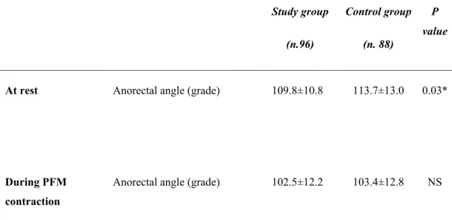
0.39 and 3.20 + 0.48, p= 0.007. Compared to the control group, patients with rectal endometriosis showed a significantly narrower anorectal angle at rest (109.8±10.8 grade versus 113.7±13.0 grade, p=0.03); anorectal angle after contraction did not significantly differ between the two groups (Table 3).
In the study group we found a significant association between severity of dyschezia at KESS questionnaire and grade of anorectal angle (p < 0.001). No further associations were detected concerning the other items of the KESS questionnaire and sonographic parameters.
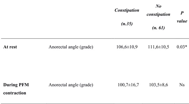
In the study group 35 women (36.5%) reported constipation according to the results of KESS questionnaire. In this particular group, anorectal angle was significantly narrower than women with rectal endometriosis without constipation (106,6±10,9 grade versus 111,6±10,5 grade p=0,03). Results are shown in Table 4 and Table 5.
DISCUSSION
Women presenting with pelvic endometriosis frequently report gastrointestinal complaints of increased intensity during menstruation (24).
This is the first study evaluating the correlations between pelvic floor muscle sonographic findings at transperineal ultrasound and bowel symptoms in women with deep endometriosis.
Our analysis of the PFM morphometry showed a narrower levator hiatal area and anorectal angle at rest and after maximal PFM contraction in patients with DIE rather than control women, suggesting a higher PFM tone. This result is consistent with our previous publications (14; MABROUK ET AL.). Like other visceral pain syndromes responsible for chronic pelvic pain, DIE may be the cause of PFM hypertone through central and peripheral sensitization and lowering of nociceptive thresholds, resulting in neuropathic upregulation, hypersensitivity and allodynia (25).
Noteworthy, it has been shown that floor hypertonic dysfunctions can be an additional causal factor of a patient’s pelvic pain, determining pelvic dysfunctions and worsening chronic pelvic pain (26).
In accordance with this opinion, in the study group, we found a correlations between anorectal angle at rest and dyschezia and constipation.
The prevalence of gastrointestinal symptoms, expecially constipation, is higher in patients with endometriosis (27); this symptom could be related to PFM hypertone. Patients with posterior DIE often experience dyschezia associated to constipation (28).
CONCLUSION
The use of transperineal ultrasound could represent a pain free methodology for assessing the PFM
function in women with DIE, In particular assessing anorectal angle can represent an important
method to recognize symptomatic patients with DIE in order to start properly rehabilitative
REFERENCES
1. Dunselman GA, Vermeulen N, Becker C, Calhaz-Jorge C, D'Hooghe T, De Bie B et al. ESHRE guideline: management of women with endometriosis. Hum Reprod 2014;29:400-12.
2. Cornillie FJ, Oosterlynck D, Lauweryns JM, Koninckx PR. Deeply infiltrating pelvic endometriosis: histology and clinical significance. Fertil Steril 1990;53:978-83.
3. Di Donato N, Montanari G, Benfenati A, Monti G, Leonardi D, Bertoldo V et al. Sexual function in women undergoing surgery for deep infiltrating endometriosis: a comparison with healthy women. J Fam Plann Reprod Health Care 2015;41:278-83.
4. Ballester M, Dubernard G, Wafo E, Bellon L, Amarenco G, Belghiti J et al. Evaluation of urinary dysfunction by urodynamic tests, electromyography and quality of life questionnaire before and after surgery for deep infiltrating endometriosis. Eur J Obstet Gynecol Reprod Biol 2014;179:135-40.
5. Spagnolo E, Zannoni L, Raimondo D, Ferrini G, Mabrouk M, Benfenati A et al. Urodynamic evaluation and anorectal manometry pre- and post-operative bowel shaving surgical procedure for posterior deep infiltrating endometriosis: a pilot study. J Minim Invasive Gynecol 2014;21:1080-5.
6. Mabrouk M, Montanari G, Di Donato N, Del Forno S, Frasca C, Geraci E et al. What is the impact on sexual function of laparoscopic treatment and subsequent combined oral contraceptive therapy in women with deep infiltrating endometriosis? J Sex Med 2012;9:770-8.
7. Roman H, Vassilieff M, Gourcerol G, Savoye G, Leroi AM, Marpeau L et al. Surgical management of deep infiltrating endometriosis of the rectum: pleading for a symptom-guided approach. Hum Reprod 2011;26:274-81.
8. Dos Bispo AP, Ploger C, Loureiro AF, Sato H, Kolpeman A, Girao MJ et al. Assessment of pelvic floor muscles in women with deep endometriosis. Arch Gynecol Obstet 2016;294:519-23.
9. Stratton P, Khachikyan I, Sinaii N, Ortiz R, Shah J. Association of chronic pelvic pain and endometriosis with signs of sensitization and myofascial pain. Obstet Gynecol 2015;125:719-28.
10. Morin M, Bergeron S, Khalife S, Mayrand MH, Binik YM. Morphometry of the pelvic floor muscles in women with and without provoked vestibulodynia using 4D ultrasound. J Sex Med 2014;11:776-85.
11. Stuge B, Saetre K, Braekken IH. The association between pelvic floor muscle function and pelvic girdle pain--a matched case control 3D ultrasound study. Man Ther 2012;17:150-6. 12. Thibault-Gagnon S, McLean L, Goldfinger C, Pukall C, Chamberlain S. Differences in the
Biometry of the Levator Hiatus at Rest, During Contraction, and During Valsalva Maneuver Between Women With and Without Provoked Vestibulodynia Assessed by Transperineal Ultrasound Imaging. J Sex Med 2016;13:243-52.
13. Dietz HP. Pelvic Floor Ultrasound: A Review. Clin Obstet Gynecol 2017;60:58-81.
14. Raimondo D, Youssef A, Mabrouk M, Del Forno S, Martelli V, Pilu G et al. Pelvic Floor Muscle Dysfunction at 3d/4d Transperineal Ultrasound in Patients with Deep Infiltrating Endometriosis: A Pilot Study. Ultrasound Obstet Gynecol 2016.
15. Dietz HP, Beer-Gabel M. Ultrasound in the investigation of posterior compartment vaginal prolapse and obstructed defecation. Ultrasound Obstet Gynecol 2012;40:14-27.
16. Beer-Gabel M, Teshler M, Schechtman E, Zbar AP. Dynamic transperineal ultrasound vs. defecography in patients with evacuatory difficulty: a pilot study. Int J Colorectal Dis 2004;19:60-7.
17. Braekken IH, Majida M, Engh ME, Bo K. Test-retest reliability of pelvic floor muscle contraction measured by 4D ultrasound. Neurourol Urodyn 2009;28:68-73.
18. Knowles CH, Eccersley AJ, Scott SM, Walker SM, Reeves B, Lunniss PJ. Linear discriminant analysis of symptoms in patients with chronic constipation: validation of a new scoring system (KESS). Dis Colon Rectum 2000;43:1419-26.
19. Knowles CH, Scott SM, Legg PE, Allison ME, Lunniss PJ. Level of classification performance of KESS (symptom scoring system for constipation) validated in a prospective series of 105 patients. Dis Colon Rectum 2002;45:842-3.
20. Youssef A, Montaguti E, Sanlorenzo O, Cariello L, Awad EE, Pacella G et al. A new simple technique for 3-dimensional sonographic assessment of the pelvic floor muscles. J Ultrasound Med 2015;34:65-72.
21. Youssef A, Montaguti E, Sanlorenzo O, Cariello L, Salsi G, Morganelli G et al. Reliability of new three-dimensional ultrasound technique for pelvic hiatal area measurement. Ultrasound Obstet Gynecol 2016;47:629-35.
22. Raizada V, Bhargava V, Jung SA, Karstens A, Pretorius D, Krysl P et al. Dynamic assessment of the vaginal high-pressure zone using high-definition manometery, 3-dimensional ultrasound, and magnetic resonance imaging of the pelvic floor muscles. Am J Obstet Gynecol 2010;203:172 e1-8.
23. Thompson JA, O'Sullivan PB, Briffa NK, Neumann P. Assessment of voluntary pelvic floor muscle contraction in continent and incontinent women using transperineal ultrasound, manual muscle testing and vaginal squeeze pressure measurements. Int Urogynecol J Pelvic Floor Dysfunct 2006;17:624-30.
24. Fauconnier A, Chapron C, Dubuisson JB, Vieira M, Dousset B, Bre ́art G. Relation between pain symptoms and the anatomic location of deep infiltrating endometriosis. Fertil Steril 2002;78:719–726.
25. Faubion SS, Shuster LT, Bharucha AE. Recognition and management of nonrelaxing pelvic floor dysfunction. Mayo Clin Proc 2012; 87: 187–193.
26. Butrick CW. Pathophysiology of pelvic floor hypertonic disorders. Obstet Gynecol Clin
North Am 2009; 36: 699–705
27. Karlbom U, Edebol Eeg-Olofsson K, Graf W, Nilsson S, Pahlman L. Paradoxical
puborectalis contraction is associ- ated with impaired rectal evacuation. Int J Colorectal Dis
1998;13:141–7
28. Riiskjaer M, Egekvist AG, Hartwell D, Forman A, Seyer-Hansen M, Kesmodel U.S. Bowel Endometriosis Syndrome: a new scoring system for pelvic organ dysfunction and quality of life. Human Reproduction 2017; 32(9):1812-18.
Figure 1
3D image of levator hiatal area
Figure 2
Figure 3
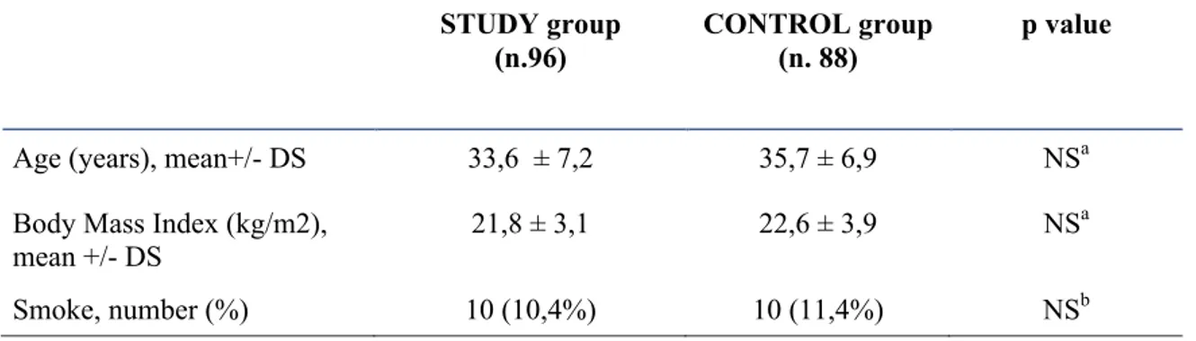
TABLE 1 Baseline characteristic of study and control group. STUDY group (n.96) CONTROL group (n. 88) p value
Age (years), mean+/- DS 33,6 ± 7,2 35,7 ± 6,9 NSa Body Mass Index (kg/m2),
mean +/- DS 21,8 ± 3,1 22,6 ± 3,9 NSa Smoke, number (%) 10 (10,4%) 10 (11,4%) NSb a Student’s t-test b Chi Square Test
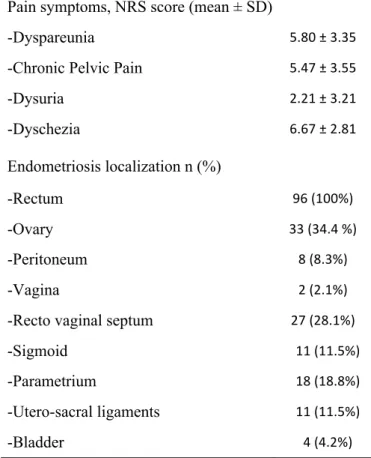
TABLE 2 Pain symptoms and endometriotic localizations (confirmed after laparoscopic excision)
of the study group (96 women).
Pain symptoms, NRS score (mean ± SD)
-Dyspareunia 5.80 ± 3.35 -Chronic Pelvic Pain 5.47 ± 3.55
-Dysuria 2.21 ± 3.21 -Dyschezia Endometriosis localization n (%) 6.67 ± 2.81 -Rectum 96 (100%) -Ovary 33 (34.4 %) -Peritoneum 8 (8.3%) -Vagina 2 (2.1%)
-Recto vaginal septum 27 (28.1%)
-Sigmoid 11 (11.5%)
-Parametrium 18 (18.8%)
-Utero-sacral ligaments 11 (11.5%)
-Bladder 4 (4.2%)
Table 3 Static (at rest) and dynamic (during contraction and during Valsalva manoeuvre) anorectal angle at transperineal ultrasound in study and control group. Values are expressed as mean (± standard deviation). * student’s T- test Study group (n.96) Control group (n. 88) P value
At rest Anorectal angle (grade) 109.8±10.8 113.7±13.0 0.03*
During PFM contraction
Table 4 Static (at rest) and dynamic (during contraction and during Valsalva manoeuvre) anorectal angle at transperineal ultrasound in women belonging to study group with or without constipation according to KESS results. Values are expressed as mean (± standard deviation).
* Student’s T- test Constipation (n.35) No constipation (n. 61) P value
At rest Anorectal angle (grade) 106,6±10,9 111,6±10,5 0.03*
During PFM contraction
KE S S IT E M 0 pt 1 pt 2 pt 3 pt 4 pt Re su lts o f st u d y g ro u p me an (± S D ) Spe ar m an co rr ela tio n val u es b et w ee n res u lts an d an or ec tal an gl e at res t p Du ra tio n of co n sti p at io n 0 – 18 mo nth s 18 m ont hs – 5 ye ar s 5 – 10 ye ar s 10 – 20 ye ar s > 2 0 y ea rs (o r all lif e) 1. 17 ± 1. 14 - 0. 160 Ns La xa tiv e u se No ne La xa tiv es p rn or for shor t dur ati on La xa tiv es re gu la r, lo ng dur ati on La xa tiv es lo ng dur ati on, in ef fe ctiv e 1.1 7 ± 1 .0 5 0. 044 Ns Fr eq u en cy of bo w el m ov em ent 1-2 tim es / 1-2 da ys 2 or le ss tim es / w ee k Le ss th an o nc e pe r w ee k Le ss th an o nc e pe r 2 w ee ks 1. 11 ± 1. 23 - 0. 003 Ns Un su cc es sfu l ev ac ua to ry at te m p ts Ne ve r / ra re ly Oc ca sio na lly Us ua lly Al wa ys = ma nu al ev acu at io n 0. 91 ± 1. 05 - 0. 005 Ns Fe eli n g in co m p le te ev acu at io n Ne ve r Ra re ly Oc ca sio na lly Us ua lly Al wa ys 1. 02 ± 1. 09 - 0. 021 Ns Ab d om in al p ain Ne ve r Ra re ly Oc ca sio na lly Us ua lly Al wa ys 1. 29 ± 1. 42 0. 005 Ns Bl oa tin g Ne ve r Pe rc eiv ed b y pa tie nt onl y Vi sib le to o th er s Se ve re c au sin g sa tie ty o r n au se a Se ve re w ith vom iti ng 1. 52 ± 1. 20 - 0. 070 Ns En em as / Di git at io n No ne En em ata / su pp osi to rie s oc ca siona lly En em ata /su pp os ito rie s r eg ula r Ma nu al ev acu at io n oc ca siona lly Ma nu al ev acu at io n al w ay s 1. 13 ± 0. 87 - 0. 167 Ns Ti m e ta k en in la va to ry /a tte m p t < 5 m in ute s 5-10 m inut es 10 -30 m inut es > 30 m in ute s 0. 98 ± 0. 88 - 0. 107 Ns Di ffi cu lty ev acu at in g ca u sin g a p ain fu l ev acu at io n ef fo rt Ne ve r Ra re ly Oc ca sio na lly Us ua lly Al wa ys 1. 57 ± 1. 26 - 0. 433 < 0. 001 St oo l c ons ist enc y (w ith ou t la xa tiv es ) So ft / lo os e / nor m al Oc ca sio na lly ha rd Al wa ys h ar d Al wa ys h ar d, us ua lly pe lle t-lik e 1. 27 ± 1. 05 - 0. 190 N s To ta l K ES S 13. 20 ± 7. 34 - 0. 155 Ns Tab le 5 K E S S Q ue sti onna ire re sul ts for s tudy group (96 w om en)