A
A
l
l
m
m
a
a
M
M
a
a
t
t
e
e
r
r
S
S
t
t
u
u
d
d
i
i
o
o
r
r
u
u
m
m
–
–
U
U
n
n
i
i
v
v
e
e
r
r
s
s
i
i
t
t
à
à
d
d
i
i
B
B
o
o
l
l
o
o
g
g
n
n
a
a
DOTTORATO DI RICERCA IN
Biologia Cellulare, Molecolare e Industriale
Progetto n. 2: Biologia Funzionale dei Sistemi Cellulari e Molecolari
Ciclo: XXII
Settore/i scientifico-disciplinare/i di afferenza: BIO-11
TITOLO TESI
THE IMMUNOGENICITY OF CHEMICALLY DERIVED
ZWITTERIONIC POLYSACCHARIDES
Dottorando:
Dott.ssa Simona GalloriniCoordinatore Dottorato
Relatore
Ch.mo Prof. Vincenzo Scarlato Dott. Andreas Wack
Index
Abstract 2
1 Introduction 4
1.1 Innate and adaptive immunity 4
1.2 Adjuvant and TLR 8
1.3 T-dependent and T-independent antigens 10
1.4 ZPS 13
1.5 GBS glycoconjugate vaccine 16
2 Aim of the study 19
3 Materials and Methods 21
3.1 In vitro 21
3.1.1 Purification of GBS capsular PS 21
3.1.2 Chemical modifications of GBS PS and conjugation to the carrier protein 23
3.1.3 Cell preparation and culture 24
3.1.4 Determination of cytokine and chemokine production 24
3.1.5 Plasmid production 26
3.1.6 HEK-293 stable transfectants 26
3.1.7 Proliferation assays 27
3.1.8 Enzymatic treatments 27
3.2 In vivo 28
3.2.1 Mice and Immunizations 28
3.2.2 Ag-specific T-cell cytokine responses 29
3.2.3 Determination of Ag-specific antibody by ELISA 29
3.2.4 Opsonophagocytosis assay 30
3.2.5 Statistical analysis 30
4 Results 31
4.1 ZPS immunogenicity in vitro 31
4.1.1 Chemical modifications of the native GBS PS 31
4.1.2 Chemically derived ZPS are able to activate APCs 33
4.1.3 The stimulatory activity of ZPS depends on the integrity of the zwitterionic motif
and is not extracted by phenol 36
4.1.4 APC activation by ZPS is mediated by TLR2 39
4.1.5 ZPS induce T cells activation in APC - T cell co-culture 43
4.2 ZPS immunogenicity in vivo. 45
4.2.1 Chemical conjugation of the ZPS to carrier protein 45
4.2.2 ZPS-conjugates are more immunogenic than the corresponding PS-conjugates 46
4.2.3 ZPS-conjugates activate bone marrow derived DCs (BM-DCs) 54
4.2.4 ZPS-conjugates confer enhanced protection against GBS infection 56
4.2.5 TLR2 is critical for ZPS-conjugate adjuvant activity in vivo 59
5 Discussion 62
Acknoledgments 70
Abstract
Bacterial capsular polysaccharides (PS) which naturally contain zwitterionic charge motifs (ZPS) possess specific immunostimulatory activity, leading to direct activation of antigen-presenting cells (APCs) through Toll-like receptor 2 (TLR2) and of T cells in co-culture systems. When administered intraperitoneally, ZPS and bacteria expressing them are involved in the induction or regulation of T-cell dependent inflammatory processes such as intra-abdominal abscess formation. Moreover it has been published that ZPSs are processed to low molecular weight carbohydrates and presented to T cells through a pathway similar to that used for protein antigens. These findings were in contrast with the paradigm according to which polysaccharides are T-independent antigens unable to be presented in association with MHC class II molecules and unable to induce a protective immune response. For this reason in glycoconjugate vaccines polysaccharides often need to be conjugated to a carrier protein to induce protection. The aim of our work was to generate vaccine candidates with antigen and adjuvant properties in one molecule by the chemical introduction of a positive charge into naturally anionic PS from group B streptococcus (GBS). The resulting zwitterionic PS (ZPS) has the ability to activate human and mouse APCs, and in mixed co-cultures of monocytes and T cells, ZPS induce MHC II-dependent T-cell proliferation and up-regulation of activation markers. TLR2 transfectants show reporter gene transcription upon incubation with ZPS and these stimulatory qualities can be blocked by anti-TLR2 mAbs or by the destruction of the zwitterionic motif. However, in vivo, ZPS used alone as vaccine antigen failed to induce protection against GBS challenge, a
result which does not confirm the above mentioned postulate that ZPS are T-cell dependent Ags by virtue of their charge motif. Thus to make ZPS visible to the immune system we have conjugated ZPS with a carrier protein. ZPS-glycoconjugates induce higher T cell and Ab responses to carrier and PS, respectively, compared to control PS-glycoconjugates made with the native polysaccharide form. Moreover, protection of mothers or neonate offspring from lethal GBS challenge is better when mothers are immunized with ZPS-conjugates compared to immunization with PS-conjugates. In TLR2 knockout mice, ZPS-conjugates lose both their increased immunogenicity and protective effect after vaccination. When ZPS are co-administered as adjuvants with unconjugated tetanus toxoid (TT), they have the ability to increase the TT-specific antibody titer. In conclusion, glycoconjugates containing ZPS are potent vaccines. They target Ag to TLR2-expressing APCs and activate these APCs, leading to better T cell priming and ultimately to higher protective Ab titers. Thus, rational chemical design can generate potent novel PS-adjuvants with wide application, including glycoconjugates and co-administration with unrelated protein Ags.
1 Introduction
1.1 Innate and adaptive immunity
The mammalian immune system is comprised of two branches: innate and adaptive. The innate immune system is the first line of host defense against pathogens and is mediated, among others, by phagocytes including macrophages and dendritic cells (DCs). The adaptive immune system is involved in elimination of pathogens in the late phase of infection as well as the generation of immunological memory. The adaptive immune system detects non-self through recognition of antigens using antigen receptors expressed on the surface of B and T cells. In order to respond to a wide range of potential antigens, B and T cells rearrange their immunoglobulin and T cell receptor genes to generate over 1011 different species of antigen receptors. Engagement of antigen receptors by the cognate antigen triggers clonal expansion of the T lymphocyte that produces cytokines and gives help to the B lymphocyte in the production of antigen-specific antibodies. The innate immune response is not completely nonspecific, as was originally thought, but rather is able to discriminate between self and a variety of pathogens. The innate immune system recognizes microorganisms via a limited number of germline-encoded pattern- recognition receptors (PRRs). This is in contrast to the large repertoire of rearranged receptors utilized by the adaptive system (1). A class of PRRs called Toll-like receptors (TLRs) has the ability to recognize pathogens or pathogen-derived products and initiate signaling events leading to activation of innate host defenses. The subfamily of TLR1, TLR2, and TLR6 recognizes
lipopeptides, whereas TLR3, TLR7, TLR8, and TLR9, a group of tightly related TLRs, recognize nucleic acids. However, TLRs are unusual in that some can recognize several structurally unrelated ligands. TLRs are expressed on various immune cells, including macrophages, dendritic cells (DCs), B cells, specific types of T cells, and even on nonimmune cells such as fibroblasts and epithelial cells. Expression of TLRs is not static but rather is modulated rapidly in response to pathogens, a variety of cytokines, and environmental stresses. Furthermore, TLRs may be expressed extra- or intracellularly. While certain TLRs (TLRs 1, 2, 4, 5, and 6) are expressed on the cell surface, others (TLRs 3, 7, 8, and 9) are found almost exclusively in intracellular compartments such as endosomes, and their ligands, mainly nucleic acids, require internalization to the endosome before signaling is possible. The transmembrane and membrane-proximal regions (TIR-domain) are important for the cellular compartmentalization of these receptors. Stimulation with their ligands recruits TIR-domain-containing adaptors including MyD88 to the receptor, leading to the formation of a complex of IRAKs, TRAF6, and IRF-5. This process induces the activation of NF-kB, a transcriptional factor, that translocates into the nucleus and initiates the expression of proinflammatory cytokine genes. Thus signaling by TLRs initiates acute inflammatory responses by induction of antimicrobial genes and inflammatory cytokines and chemokines. Subsequent events, such as recruitment of neutrophils and activation of macrophages, lead to direct killing of the microbes (2). The notion of TLRs being primary sensors of pathogens and responsible for orchestrating the innate responses is now widely accepted. In addition, there is accumulating evidence that TLRs contribute
significantly to activation of adaptive immune responses. Although T and B cells of the adaptive immune system express receptors of enormous diversity, activation of these cells depends on induction of co-stimulatory molecules and secretion of cytokines and chemokines by the cells of the innate immune system. In fact efficient priming of adaptive immune responses requires not only the presentation of antigen in the context of major histocompatibility complex (MHC) but also the induction of accessory signals (costimulators and cytokines) on antigen-presenting cells (APCs). TLRs expressed on APCs may regulate these accessory signals through their recognition of PAMPs and consequently control activation of antigen-specific adaptive immune responses (3, 4). Thus, we consider the innate and adaptive immune responses to be integrated in the vertebrate host as a single immune system, with the innate response preceding, and being necessary for, the adaptive immune response (Figure A) (5, 6).
Because of their capacity to bridge innate and adaptive immunity, Toll-like receptors (TLRs) have offered new opportunities for the development of immunostimulatory adjuvants (7, 8).
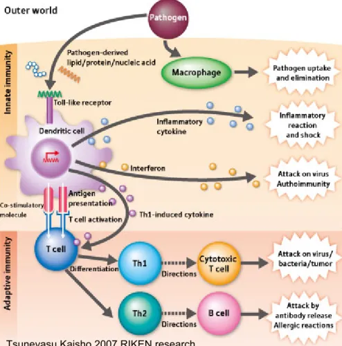
Figure A : TLRs link innate and adaptive immunity
Tsuneyasu Kaisho 2007 RIKEN research
FIGURE A. APCs like DCs recognize pathogens by Toll-like receptors and produce a wide variety
of cytokines. Cytokines serve to eliminate pathogens on one hand, and activate adaptive immunity on the other. Even when the same type of Toll-like receptor is involved, different signals are transmitted to cause different immune reactions depending on the type of dendritic cell in which the receptor is expressed.
1.2 Adjuvant and TLR
Adjuvants are molecules, compounds or macromolecular complexes that increase the potency and longevity of specific immune response to antigens, but cause minimal toxicity or long lasting immune effects on their own (9). The addition of adjuvants to vaccines enhances, sustains and directs the immunogenicity of antigens, effectively modulating appropriate immune responses, reducing the amount of antigen or number of immunizations required and improving the efficacy of vaccines in newborns, elderly or immuno compromised individuals (10). Traditional live vaccines based on attenuated pathogens typically do not require the addition of adjuvants. Likewise, vaccines based on inactivated viruses or bacteria are often sufficiently immunogenic without added adjuvants, although some of these can be formulated with adjuvants to further enhance the immune responses. By contrast, protein-based vaccines, although offering considerable advantages over traditional vaccines in terms of safety and cost of production, in most cases have limited immunogenicity and require the addition of adjuvants to induce a protective and long-lasting immune response. Adjuvants can be classified according to their component sources, physiochemical properties or mechanisms of action. Two classes of adjuvants commonly found in modern vaccines include: immunostimulants that directly act on the immune system to increase responses to antigens (Examples include: TLR ligands, cytokines, saponins and bacterial exotoxins that stimulate immune responses) and vehicles that present vaccine antigens to the immune system in an optimal manner, including controlled release and depot delivery systems, to increase the specific immune response to the
antigen. The vehicle can also serve to deliver the immunostimulants described in the previous point (Examples include: mineral salts, emulsions, liposomes, virosomes, biodegradable polymer, microspheres).
With few exceptions, aluminum salts (alum) are currently the only vaccine adjuvant approved for human use worldwide. Alum is effective at generating a strong antibody response to an antigen with a bias towards a Th2 type of immune response, and, as such, has been widely and effectively used in many vaccines, such as tetanus, diphtheria, pertussis and poliomyelitis vaccines (11, 12). The mechanism of immunopotentiation by alum involves inflammation and recruitment of antigen-presenting cells, retention of antigen at the injection site, uptake of antigen, dendritic cell maturation, T-cell activation and T-cell differentiation (13). Although alum adjuvants have proved their efficiency in a large number of applications, some limitations of alum have been reported. Thus, alum failed to confer satisfactory increase of the immune response in certain vaccines, such as typhoid fever and influenza vaccines. Reports have also demonstrated that alum displays limited ability to raise high antibody titers against small-size peptides. This calls for rational design of novel vaccine adjuvants that can establish protective immunity against different diseases. Advances in the design of efficient adjuvants based on the use of TLR agonists have been promising (although it should be noted that some of these were in development before the role of TLRs was identified) and some of these have reached advanced human trials and even registration. Thus we studied the chemical structure of TLR agonists in literature to
understand if we could find a new adjuvant, TLR agonist among in house compounds.
A very recent publication shows that the natural zwitterionic polysaccharide A (PSA) is a TLR2 agonist and able to activate a number of different APCs (14). In house we had many polysaccharides extracted from the capsule of different bacteria used to make vaccine. It has been demonstrated that the induction of antibodies specific to the capsular PS confers protection against infection. This is the reason why a number of vaccines against bacterial infections aim exclusively at the induction of antibodies against capsular polysaccharides.
1.3 T-dependent and T-independent Antigens
Polysaccharide antigens are large molecules consisting of repeating epitopes which are not processed by antigen-presenting cells (APC) but interact directly with B cells, inducing antibody synthesis without the need of T cell help or in the absence of T cell help. In addition, pure PS do not contain structures that are assumed visible to T cells, since the antigen receptor of T cells (TCR) recognizes peptide fragments derived from protein antigens. This is why PS from bacteria are considered to be T-independent Ags (15) able to activate B cells without a contribution of help by CD4+ T cells. T-dependent antigens are proteins or peptides that require immune stimulation from helper T cells to elicit an immune response. Such antigens are presented to T cells in the context of MHC molecules on macrophages, B cells or dendritic cells following bacterial or viral infection. The subsequent activation of T cells induces cytokine production and a range of
immunologic effects. TD antigens are effective at inducing a lasting immune response, forming memory B and T cells, and producing high affinity antibodies of multiple isotypes. In contrast, T-independent responses are restricted in a number of ways. Most importantly, they fail to induce significant and sustained amounts of antibody in young children below the age of 18 months. While polysaccharides are immunogenic in older children and adults, the characteristics of the antibody responses are rather restricted. They are dominated by IgM and IgG2, are relatively short lived, and a booster response cannot be elicited on repeated exposure. This failure to induce immunological memory is also reflected in the absence of demonstrable affinity maturation. In contrast to polysaccharides, antibody responses to protein antigens have an absolute requirement for T cells. The consequence of this T cell help is that antibody responses to protein antigens can be elicited in the very young and immunity is long lived due the generation of immunological memory. Antibody responses to protein antigens are dominated by the IgG1 and IgG3 subclasses and affinity maturation can be demonstrated over time.
The ability to enhance the immunogenicity of polysaccharide antigens was first noted by Avery & Goebel in 1929, who demonstrated that the poor immunogenicity of purified S. pneumoniae type 3 polysaccharide in rabbits could be enhanced by conjugation of the polysaccharide to a protein carrier (16). Their observations have formed the foundation for the modern development of conjugate vaccines. It is hypothesized that PS-specific B cells internalize the PS-carrier complex. Proteolysis of the carrier protein produces peptides that bind to class II MHC
molecules and activate helper T cells (17). As a result, PS-specific B cells can then mature to antibody producing plasma cells or into memory cells (18-20).
The carrier protein provides T cell help and consequently immunological memory (21). The precise nature of the molecular events that permit polysaccharides conjugated to protein carriers to be processed as T-dependent antigens remains unclear and more research is required. In contrast to this paradigm, natural ZPS such as PSA may have the characteristics of a T-dependent antigen, despite the lack of a protein component. It has been published that ZPSs are processed to low molecular weight carbohydrates and presented to T cells through the MHCII endocytic pathway. Furthermore these carbohydrates bind to MHCII inside APCs for presentation to T cells (22). Therefore ZPS should not need the conjugation to a carrier protein to induce an immune response (Figure B).
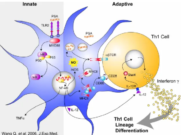
Figure B: PSA mechanism of action as TLR2 agonist and as ZPS T-dependent antigen.
Wang Q, et al. 2006, J.Exp.Med.
FIGURE B: The innate response begins with TLR2 recognition of PSA and the subsequent
stimulation of the MyD88-mediated pathway inside the APC. Meanwhile, NF-κB translocation also leads to up-regulation of MHCII and CD86, thus facilitating PSA processing and presentation by MHCII proteins. Presentation of PSA on the cell surface by MHCII leads to adaptive CD4+ T cell activation and T cell secretion of cytokines like IFN-γ.
1.4 ZPS
PSA is a capsular polysaccharide (PS) from Bacteroides fragilis which naturally contains both positive and negative charges in its repeating structure, thus it is a zwitterionic polysaccharide (ZPS). A number of publications have shown in the past that PSA or the ZPS extracted from the capsule of Staphylococcus aureus and type 1 Streptococcus pneumoniae (Sp1) are able to activate T cells and APCs (23-25). The structures of the natural ZPS, PSA and Sp1, have been resolved (26, 27) (Figure C).
The initial findings demonstrated that abscess formation in a rat model was induced by PS containing zwitterionic charge motives (28) and that abscess formation was T cell dependent and transferable with ZPS-activated T cells (29, 30).
The integrity of the zwitterionic motif was essential for this biological activity, as removal of one of the two charges also removed the ability to induce abscesses (28). In vitro experiments with unfractionated splenocytes or with mixed populations showed that natural ZPS were able to induce T cell activation in these conditions, while the co-culture of fixed APCs with T cells was not sufficient to induce proliferation (31). The alternative, but not mutually exclusive hypotheses to explain these findings are that ZPS require processing to activate T cells directly through TCR recognition of MHC class II –ZPS complexes (32) or that ZPS activate APCs to up-regulate MHC class II, co-stimulatory molecules and cytokines and thus generate conditions that favor the activation of T cells (31). MHC class II
blocking antibodies inhibit T cell activation, and up-regulation on APCs of a number of molecules involved in T cell activation has been demonstrated (25).
To summarize, ZPS seem to be not only TLR2 agonists but also a T-dependent antigens. Thus, ZPS could be a perfect vaccine with the antigen and the adjuvant in a single molecule. Thus we explored the structure of in-house better available pure bacterial capsular polysaccharides and found that the polysaccharide extracted from Group B Streptococcus (GBS) that is naturally anionic could become zwitterionic through a chemical modification. Based on the results cited above, we hypothesized that ZPS obtained in this way should perform well as an efficient vaccine against GBS and may be used as alternative to the already existing glycoconjugate vaccine.
Figure C: PSA and Sp1 ZPS structure.
FIGURE C. Sp1 and PSA consist of a trisaccharide and a tetrasaccharide with free amino and
carboxyl groups that confer zwitterionic characteristics to these polymers.
1.5 GBS Glycoconjugate vaccine
GBS is the foremost cause of life-threatening bacterial infections in newborns (33). In about 80% of cases, neonatal GBS infection is acquired during delivery by direct mother-to-baby transmission of the pathogen, which colonizes the anogenital
mucosa of 25 to 40% of healthy women (34). Despite the introduction of intrapartum antibiotic prophylaxis, in the United States GBS still causes 2500 cases of infection and 100 deaths annually among newborns in the first 3 months of life. About half of these cases occur in the first week after birth. Thus, it is commonly believed that effective vaccination will be the only way to reduce the incidence of GBS disease over the long term. GBS bacteria are encapsulated by complex branched polysaccharides, and variations in these sequences correspond to strain classifications. Each capsular polysaccharide (CPS) is derived generally from the same set of glycosyl residues; structural and immunological diversity arising from differences in linkage position and anomeric configuration (35). At present, nine serotypes of GBS (Ia, Ib, II–VIII) have been identified containing various arrangements of galactose, glucose, GlcNAc, and the most prevalent sialic acid (Sia) of humans, Nacetylneuraminic acid (Neu5Ac) (8–14). Neu5Ac residues of the GBS CPS are situated on the branching terminus of each repeating unit. These CPS are naturally anionic, as the otherwise neutral sugar backbone carries anionic groups such as carboxyls which are present as carboxylate ions at physiological pH.
The rationale for GBS vaccine development is supported by the observation that the risk of neonatal infection is inversely proportional to the maternal amounts of specific antibodies to the capsular polysaccharide (CPS) antigen that surrounds GBS (36, 37), implying that protective immunoglobulin G (IgG) antibodies are transferred from the mother to the baby through the placenta.
A glycoconjugate vaccine against GBS is currently being developed. This vaccine has been shown to confer serotype specific protection in mice and have been tested in clinical trials. In humans, addition of alum to the vaccine formulation did not further increase the immune response induced (38). In contrast, in animal models, adsorption to Al(OH)3 (Alum) enhances the immunogenicity of the glycoconjugate (39, 40), which may be explained by the possibility that animals are more naïve to GBS than humans. More generally, a number of commercially available glycoconjugate vaccines such as that against Meningococcus C, Haemophilus influenzae and the seven-valent Pneumococcus vaccine use Alum or aluminium phosphate as an adjuvant. Thus, it appears that the combination of glycoconjugates with adjuvants likely generates potent vaccines, able to activate APCs and induce strong Ag-specific T and B cell responses.
2 Aim of the study
The overall purpose of this study is to add, through rational chemical modification, biological function to vaccine Ags.
It has been described in the literature that bacterial capsular polysaccharides (PS) which naturally contain zwitterionic charge motifs (ZPS) possess specific immunostimulatory activity, leading to direct activation of antigen-presenting cells (APCs) through Toll-like receptor 2 (TLR2) and of T cells in co-culture systems. When administered intraperitoneally, ZPS and bacteria expressing them are involved in the induction or regulation of T-cell dependent inflammatory processes such as intra-abdominal abscess formation. Thus these natural polysaccharides have adjuvant properties, they are TLR-2 agonists, but they seem to be also T-dependent antigens. The majority of polysaccharides are naturally anionic and do not have these biological activities. To generate vaccine candidates with antigen and adjuvant properties in one molecule we have chemically introduced zwitterionic motifs into naturally anionic PS.
We chose the PS from type Ia, Ib and III of GBS, group B streptococcus. Positive charges were chemically introduced, and first of all the ability of these chemically derived ZPS to activate APCs and T cells were tested on a variety of human and mouse cell types. In a second step we asked if the in vitro activities of ZPS translate also into increased immunogenicity in vivo. Therefore, we tested the biological activities of ZPS in an animal model. The ZPS alone used as antigen to protect mice from GBS challenge showed little effect. This means that as T-dependent antigen the chemically modified ZPS was not efficient. Therefore, we
further esplored whether the ability to activate APCs shown in vitro translate into adjuvant activity in vivo. Thus, to render the ZPS visible to the immune system, we generated a glycoconjugate vaccine with ZPS in order to compare its immunogenicity with the glycoconjugate vaccine made with the native PS. The different glycoconjugates were injected into mice, and Ab titers, T cell responses, opsonophagocytosis and protection was measure. To test TLR dependence, immunogenicity and protection was compared in wt and TLR2 deficient mice.
This strategy may be applied to other polysaccharides and represents a new path for rational chemical design of novel adjuvants and glycoconjugate vaccines.
3 Materials and Methods
3.1
In vitro
3.1.1 Purification of GBS capsular PS
Capsular PS were prepared from Streptococcus agalactie bacteria using a modified version of the procedure previously published (41). Isolated PS were found to contain low concentrations of nucleic acids (<10 µg/mg), proteins (<10 µg/mg), Group B saccharides (<10 µg/mg), and LPS (< 0.001 UI/µg).
3.1.2 Chemical modifications of GBS PS and conjugation to the carrier protein
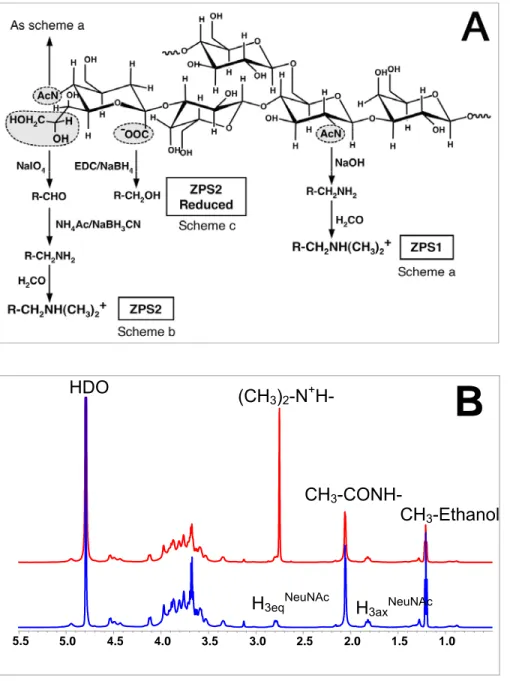
Cationic protonated forms of amines can be introduced into PS at (a) free N-acetyl groups, which are part of N-acetylneuraminic acid (NeuNAc) and Nacetylglucosamine residues, and at (b) the terminal aliphatic chain of NeuNAc (see Fig.1A). De-N-acetylation was achieved by basic hydrolysis (1 M NaOH for 60 min at 80°C) to make free amino groups available for further reaction (a in Fig.1A). Alternatively, chemical oxidation of the aliphatic chain from the terminal NeuNAc residue (b in Fig.1A) with sodium metaperiodate (Aldrich, 0.01 M NaIO4 for 90 min at room temperature) leaves an aldehyde group (42). Here, using NaIO4 as limiting (30%) or stoechiometric (100%) reagent of the reaction, the periodate oxidation selectively cleaves the C8-C9 bond between vicinal hydroxyl groups (-CHOHCH2OH) of NeuNAc residues, leaving an aldehyde group (-CHO) at C8. This group was converted to a cationic –NH3+ group by reductive amination using
300 mg/mL ammonium acetate (NH4Ac, Sigma) and 49 mg/mL sodium cyanoborohydride (NaBH3CN, Sigma) at pH 6.5 and T=37°C for 5 days (43). PS obtained by the reaction schemes a and b were treated with 37% formaldehyde (H2CO, Carlo Erba) in the presence of sodium cyanoborohydride in order to convert the generated free amino group to a tertiary dimethylamine such that it retained a positive charge (44). In order to remove the anionic charge on ZPS2 (Fig.1A, scheme c), the carboxyl group was reduced to alcoholic group by treatment with 1-ethyl-3-(3-dimethylaminopropyl) carbodiimide (EDC – Sigma) and sodium borohydride (NaBH4, Aldrich). Where indicated, a phenol extraction of GBS PS was conducted as described by Sen et al. (45). As for the native PS, contamination with proteins, nucleic acids, and group B saccharide was also determined in all ZPS preparations and found to be below 10µg/mg. Since reagents used for the modifications contain undetectable level of these contaminants, the purity of ZPS products should be higher than the native PS due to additional purification steps performed after the chemical treatments.
The covalent attachment of the carrier protein CRM197 or HSA to the zwitterionic PS was performed according to the protocol used for the conjugation of the native CPS (41). The crucial step in the zwitterionization, first, and in the conjugation, in a second time, is the initial oxidation that has to occur only for a 10-30% of NeuNAc residues. Therefore, the final glycoconjugate has a 10-30% of NeuNAc residues modified with a positive charge, a 10-30% of NeuNAc residues implicated in the covalent binding with the carrier protein and the rest of NeuNAc residues unaltered. ZPS-conjugates were generated using ZPS from serotype Ib and V.
ZPconjugates were purified by gel filtration chromatography on the Sephacryl S-300 HR column. Polysaccharide content of ZPS and ZPS-conjugate preparations was estimated by the colorimetric detection of sialic acid residues with the Svennerholm method (46). The microBCA kit assay (Pierce) was used to estimate the protein content of ZPS-conjugate sample. The polysaccharide/protein ratio for all glycoconjugates used here was measured to be 1:1 (w:w).
Nuclear Magnetic Resonance (NMR) was used to assess first of all the structural identity of the purified PS and the chemically derived ZPS molecules then the structural identity of the native PS-conjugate and the chemically derived ZPS-conjugate. NMR spectra were recorded at 25°C on a Bruker DRX 600 MHz spectrometer using a 5-mm triple-resonance NMR probe (Bruker). For data acquisition and processing, XWINNMR 2.6 software package (Bruker) was used. NMR samples were prepared by dissolving lyophilized product in 0.75 ml of deuterium oxide (D2O, Aldrich) to a uniform concentration and transferred to 5-mm NMR tubes (Wilmad). 1-D proton NMR spectra were collected using a standard one-pulse experiment and collecting 32 k data points over a spectral window of 6000 Hz. The complete relaxation of all nuclei was assured. The spectrum was Fourier-transformed after applying a 0.2 Hz line broadening function and referenced relative to mono-deuterated water (HDO) at 4.79 ppm.
3.1.3 Cell preparation and culture
PBMCs were collected by Ficoll Hypaque (Amersham Biosciences, Uppsala, Sweden) density gradient centrifugation.from buffy coats of healthy donors who had given written informed consent Highly purified (>98%) monocytes were obtained from PBMCs by positive selection of CD14+ cells using anti-CD14 coated magnetic microbeads and MACS technology (Miltenyi Biotec, Bergisch Gladbach, Germany).
Monocytes (2x10⁵ per well) were cultured for 24h in RPMI 1640 (GIBCO Invitrogen) supplemented with Penicillin (100U/ml), Streptomycin (100 µg/ml), Glutamine (2mM) solution (Invitrogen Life Technologies) (RPMI-PSG) and 5% Human Serum (Sigma) using U-bottom 96-well plates. T cells (>98%) were prepared from PBMCs by MACS by negative selection using the Pan T Cell Isolation kit (Miltenyi Biotec). T cells (2x10⁵ per well) were co-cultured with monocytes (1x10⁵ per well) for 6 or 8 days in the same conditions as described above for monocytes.
Immature Mo-DCs were obtained culturing monocytes for 6 days in RPMI-PSG supplemented with 10% Fetal Calf Serum (Hyclone, Logan. Utah) (complete medium) with IL-4 (10% of supernatant from IL-4 secreting cell line, provided by A. Lanzavecchia, Institute for Research in Biomedicine, Bellinzona, Switzerland) and 50 ng/ml of GM-CSF (Gentaur, Brussels, Belgium). Immature dendritic cells were washed and cultured for the experiments in complete medium, using 96 well flatbottom cell culture plates. Human cells were stained with FITC-conjugated CD14 or CD83, PE-conjugated CD80, allophycocyanin-conjugated
anti-CD86, PerCP-conjugated anti-HLA-DR (all Becton Dickinson). Rabbit serum was used as a blocking agent. After incubation for 20 min. on ice, cells were washed and analyzed on a FACSCalibur flow cytometer using CellQuest software (Becton Dickinson).
Mouse BM-DCs were generated culturing femoral bone marrow with recombinant murine GM-CSF (PeproTech) as described (47). At day 6, BM-DC were washed and cultured in complete medium with β-mercaptoethanol 50 µM (Sigma) and 100 U/ml mGM-CSF, using pro-bind U-bottom 96-well plates (Becton Dickinson).
Where indicated, cells were treated with LPS, macrophage-activating lipopeptide-2 (MALP-2) or N-Palmitoyl-S-[2,3-bis(palmitoyloxy)-(2RS)-propyl]-[R]-cysteinyl-[S]-seryl-[S]-lysyl-[S]-lysyl-[S]-lysyl-[S]-lysyl (Pam3CSK4) obtained from Alexis Biochemicals. The following purified mAbs were used in blocking experiments: anti-HLA-DR, DP, DQ (10 µg/ml) (BD Pharmingen) and anti-TLR2 clone T2.5 (50 µg/ml) (eBioscience). BM-DCs were stained with PE-conjugated anti-CD86, FITC-conjugated anti-MHC class II and allophycocyanin-FITC-conjugated anti-CD11c. Rabbit serum was used as a blocking agent. The acquisition was made on a LSR-II and data analyzed using DIVA software (BD).
3.1.4 Determination of cytokine and chemokine production
TNF-a production in culture supernatants was quantified by specific standard sandwich ELISA, using capture B154.9 and biotinylated B154.7 mouse monoclonal antibodies kindly provided by Dr. G. Trinchieri. Cytokine and chemokine secretion in supernatants was assessed by Bio-Plex analysis (Bio-Rad), according to the
manufacturer’s instructions using the mouse 23-Plex panel. The following soluble proteins are assayed: 1α, IL1-β, 2, 3, 4, 5, 6, 9, 10, IL-12(p40), IL-12(p70), IL-13, IL-17, Eotaxin, G-CSF, GM-CSF, IFN-γ, KC, MCP-1, Macrophage Inflammatory Protein (MIP)-1α, MIP-1β, RANTES, TNF-α.
3.1.5 Plasmid production
A DNA fragment coding for human TLR2 with no signal peptide was obtained by RT-PCR from human PBMC cDNA using TLR2 specific primers and then cloned in the pFLAG-CMV-1 vector (Sigma) to attach an N-terminal FLAG epitope to TLR2; the FLAG-TLR2 sequence was then subcloned in pcDNA3 .1 (Invitrogen). The NF-κB-regulated promoter of the Igκ-luc plasmid (a gift of Dr. Antonio Leonardi, University of Naples, Italy) was subcloned in pd2EGFP-1 vector (Clontech) upstream to the EGFP coding sequence to obtain pNFkB-d2EGFP plasmid; the hygro gene with an upstream SV40 promoter, excised from the pTK-Hygro vector (Invitrogen), was then inserted in an un-influential region to obtain pNFkB-d2EGFP-Hygro.
3.1.6 HEK-293 stable transfectants
HEK-293 cells were grown in DMEM supplemented with glucose (4500g/l) glutamine (2mM), 10% FCS, penicillin and streptomycin. Cells were transfected using lipofectamine 2000 (Invitrogen) with the pcDNA-FLAG-TLR2 construct and a stable clone was derived by Geneticin (Invitrogen) selection. FLAG-TLR2 expression was verified by surface staining with FLAG-M2 monoclonal antibody
(Sigma) and FACS analysis. This clone was then transfected with pNFkB-d2EGFP-Hygro and a stable clone was derived by selection with pNFkB-d2EGFP-Hygromycin B (Invitrogen). For experiments, cells were cultured for 24 hours with PS and controls, washed and analyzed by flow cytometry. The HEK-293 triple transfectants TLR4/MD-2/CD14 were obtained from Invivogen, and surface expression of CD14 was confirmed by FACS. In experiments comparing directly TLR2 and TLR4 agonist activity, the respective transfectant cell lines were incubated with PS for 20 hours, supernatant was obtained, and IL-8 content was determined using the FlexSet kit (Becton Dickinson) according to manufacturer’s instructions. IL-8 produced by the transfectants was normalized to the amount of IL-8 present in the supernatant of the respective unstimulated transfectant line.
3.1.7 Proliferation assays
T cell proliferation was assessed by [3H] thymidine incorporation using 2x10⁵ T cells and 1x10⁵ γ-irradiated monocytes (3000 rad) per well in round-bottom 96-well plates. After 6 days of culture, cells were pulsed with 0,5 µCi/well of [3H] thymidine, incubated for 18 hours, harvested onto filter plates (Packard Instruments), and counts were analyzed using a Top Count NXT β counter (Packard Instruments).
3.1.8 Enzymatic treatments
Treatments were adapted from (Mattern 1999). Prior to addition to cells, ZPS and controls were incubated in PBS with 200 µg/ml lipoprotein lipase (LPL) from bovine milk or from pseudomonas sp. (Sigma-Aldrich) at 37°C for 7 hours. Results shown
were obtained using bovine milk-derived LPL. Alternatively, immobilized trypsin (Pierce) or Proteinase K-acrylic beads (Sigma-Aldrich) were incubated with samples at a 1:20 ratio at 37°C for 1 hour. Proteases were removed from the reaction by centrifugation. The enzymatic treatments were performed on ZPS and controls which were concentrated 10x compared to their final concentration in the in vitro experiments.
3.2
In vivo
3.2.1 Mice and Immunizations
Groups of 6-8 female 6-week-old Balb/C, C57BL/6, CD1 outbred mice (Charles River) or C57BL/6 TLR2-/- (48) (kindly provided by Giuseppe Teti, Messina, Italy) were used for experiments reviewed and approved by the institutional review committees. Animals were immunized intraperitoneally at days 0, 21 and 35 with 1 µg of glycoconjugates made as indicated. Where indicated, Alum was used at 0.4mg AlOH3/dose. Serum and spleen samples were collected at 2 weeks following the third immunization. In adult mice, the challenges were performed injecting intraperitoneallystrain H36B (serotype Ib) at 1 x 10⁸ CFU at 2 weeks after the third immunization. For the neonatal challenge experiments, we first determined the 80% lethal doses (LD80) by titration in both wt and ko mice. H36B was administered at 1 x LD80 to the pups subcutaneously between 24 and 48 h after birth. Mortality was recorded daily for the 2days following challenge.
3.2.2 Ag-specific T-cell cytokine responses
Three mice per treatment were sacrificed, spleens were collected, and single cell suspensions were obtained. Red blood cells were lysed and splenocytes cultured in RPMI (Gibco) containing 2.5% FCS (Hyclone), beta-mercaptoethanol and antibiotics. Splenocytes were stimulated in the presence of anti-CD28 (1 µg/ml) (Becton–Dickinson [BD]) and the carrier protein CRM197 (30 µg/ml), or with anti-CD28 alone (unstimulated, <0.1% total cytokine-positive cells), or with anti-anti-CD28 plus anti-CD3 (0.1 µg/ml) (BD). After 4 h of stimulation, Brefeldin A (2.5 µg/ml)(Sigma Aldrich) was added for additional 12 h. Cells were washed and stained with LIVE/DEAD Fixable Aqua Dead Cell Stain Kit for 405 nm excitation (Invitrogen). Cells were fixed, permeabilized and stained with the following mAbs: allophycocyanin-Alexa750-conjugated anti-CD4 (Caltag), Pacific Blue-conjugated anti-CD3, Alexa700-conjugated anti-TNF-α, Peridinin chlorophyll protein cyanine5.5-conjugated anti-IFNγ, PE-conjugated anti-IL-5, Alexa488-conjugated anti-IL-2 (BD). Cells were acquired on a LSR-II (BD) and analyzed using FlowJo software (Tree Star). For each individual mouse, percentages of unstimulated samples were subtracted from the Ag-stimulated sample.
3.2.3 Determination of Ag-specific antibody by ELISA
For titration of IgG specific for the native polysaccharides, Maxisorp plates (Nunc, Roskilde, Denmark) were coated with 1 µg/ml (in PBS) of the glycoconjugate that
contains a different carrier protein to that used for the immunization, in order to detect only the antibodies specific for the polysaccharide. Antibody titers are those dilutions that gave an OD higher than the mean plus five times the SD of the average OD obtained in the pre-immune sera. The titers were normalized with respect to the reference serum assayed in parallel.
3.2.4 Opsonophagocytosis assay
Serum samples from mice immunized with serotype Ib glycoconjugates were tested for their in vitro ability to promote the opsonization of type Ib GBS strain H36B for phagocytosis and killing by differentiated HL60 cells in the presence of rabbit active complement. Results were expressed as the mean log10 reduction in GBS colony-forming units before and after 60 min of incubation at 37°C.
3.2.5 Statistical analysis
Statistical significance was determined using a two-tailed Student’s T-test analysis or by Fisher's exact test. Significance was reconfirmed using nonparametric statistical analysis.
4 Results
4.1 ZPS immunogenicity in vitro
4.1.1 Chemical modifications of the native GBS PS
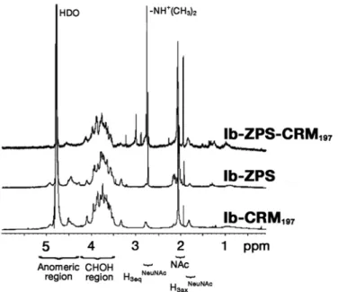
Figure 1A shows, as an example, the modifications introduced into the repeating unit of the capsular PS from GBS serotype III (GBS III). The same type of modifications was also introduced into GBS Ia and Ib. According to the annotations shown in Fig.1A, ZPS1 is the zwitterionic PS obtained from the chemical modification (a) that converts the N-acetyl groups to free amino groups. Chemical modification (b) generates the ZPS2 resulting from a periodate oxidation to generate an aldehyde group which is also converted to an amino group by a reductive amination reaction. Furthermore, all amino groups are converted to tertiary amines that retain their positive charges. Thus, in each repeating unit, ZPS1 molecules from all three serotypes contain two positive and one negative charge, while ZPS2 contain a balanced motif of one charge each. In order to remove the anionic charge from ZPS2, the carboxyl group of ZPS2 is reduced by a carbodiimide-mediated reaction with NaBH4 (modification c). All structural analyses of chemical modifications of the native PS were made using NMR spectroscopy (Fig.1B). In particular, the zwitterionic structure is confirmed by detecting the methyl group (CH3)2-N+H- which has been generated by the chemical modification scheme b (Fig.1A). All labeled signals have been assigned using bi-dimensional homo-nuclear (1H-1H) and hetero-nuclear 1(H-13C) NMR experiments (not shown). Additional saccharide contaminations (i.e. group B
polysaccharide) were also excluded by analyzing the NMR profiles. Only a little residual amount of ethanol used in the purification procedure has been detected.
Figure 1 : Chemical modifications of native GBS serotype III capsular PS A.
CH3-Ethanol 1.0 1.5 2.0 2.5 3.0 3.5 4.0 4.5 5.0 5.5 CH3-CONH-
H3eqNeuNAc H3axNeuNAc
(CH3)2-N+H- HDO
B
FIGURE 1.De-N-acetylation by basic treatment leading to ZPS1 (a), and periodate oxidation and
reductive amination leading to ZPS2 (b), of one repeating unit of GBS serotype III PS. The additional reaction with formaldehyde to obtain tertiary amines is also shown. (c) Reduction of the
carboxyl group leading to ZPS2 reduced. B, NMR spectra at 600 MHz and 25°C of the native form
(top line) and the ZPS2 modification (bottom line) of serotype Ib PS from GBS. The peaks
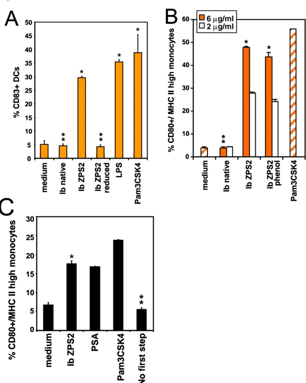
4.1.2 Chemically derived ZPS are able to activate APCs
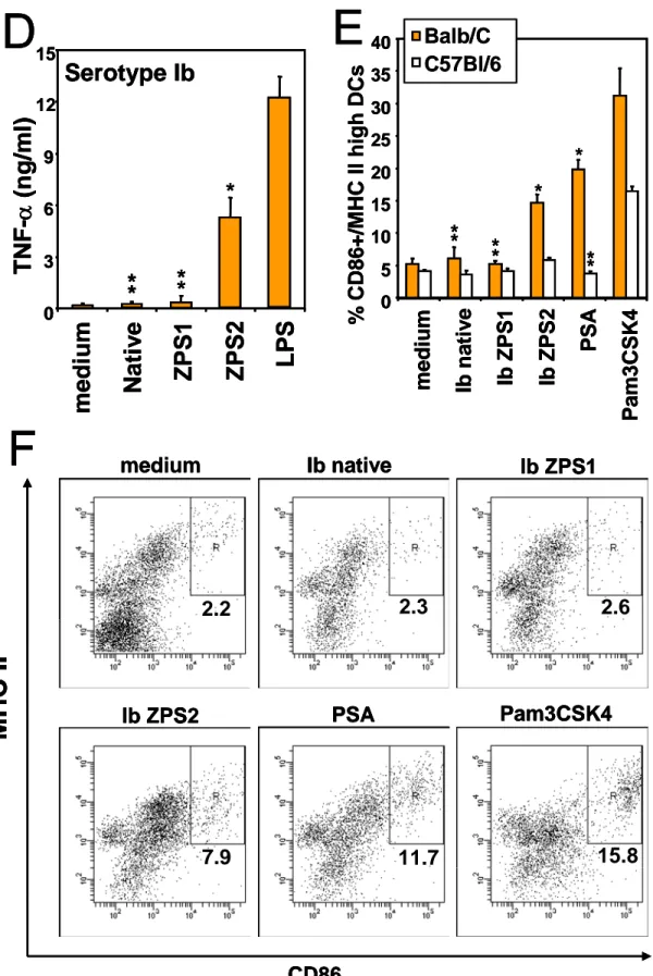
In order to test the ability of ZPS to activate APCs, purified human monocytes and Mo-DCs were incubated with either the natural ZPS purified from Bacteroides fragilis, the capsular PS A (PSA), or with the ZPS derived from GBS PS by the chemical modification a as indicated in figure 1A, leading to ZPS1, or by the modification b (ZPS2). The dot plots in figure 2A show that both PSA and the chemically derived ZPS2 are able to induce up-regulation of MHC class II and CD80 on human monocytes, while the native anionic GBS PS (Fig. 2A) or the ZPS1 derived by chemical modification a (not shown) do not activate monocytes. The dose response relation of this activation is shown in fig. 2B. Similarly, ZPS2 of all three GBS serotypes induce TNF-a production by purified human monocytes, while the native forms are inactive (Fig.2C). The amount of TNF-a induced in monocytes by PSA is comparable to that induced by serotype Ib ZPS2 (not shown). The production of TNFa is induced in human Mo-DC by the ZPS2 form but not the native or ZPS1 form of the GBS capsular PS, again indicating that the zwitterionic motif introduced is essential for the biological activities observed (Fig. 2D). Similar results were found for up-regulation of the maturation marker CD83 on human Mo-DCs (Fig. 3A). Finally, also mouse BM-DCs from Balb/C mice are activated by ZPS2 but not the native PS or ZPS1 (Fig. 2E). The dot plots and gates used for the determination of marker upregulation are shown in Fig. 2F. As previously reported (49), Balb/C mice are more responsive to TLR2 agonists than C57Bl/6 mice, and this is reflected in the stronger response of Balb/C BM-DCs to the positive control Pam3CSK4. In conclusion, these results indicate that the
chemical introduction of a zwitterionic charge motif into anionic PS confers the ability to activate a variety of human and mouse APCs.
Figure 2: ZPS activate human and mouse APCs.
medium
Ib native
Ib ZPS2
PSA
Pam3CSK4
LPS
MHC I
I
CD80
7.9
7.8
53.5
38.1
55.8
36.1
A
medium
Ib native
Ib ZPS2
PSA
Pam3CSK4
LPS
MHC I
I
CD80
7.9
7.8
53.5
38.1
55.8
36.1
A
0 5 10 15 20 25 30 35 40 45 m ve ve S2 S2 SA K4 PS 6 mg/ml 2 mg/ml 0,6 mg/mlB
% C D + monocy t * * * * * * * 8 0 / MH C II hig h e s TN F -α (ng/m l) 0 20 40 60 80 0 1 2 3 4 5 6 7 8 9C
* * * * Ia native Ib native III native Ia ZPS2 Ib ZPS2 III ZPS2 medium 0 5 10 15 20 25 30 35 40 45 m ve ve S2 S2 SA K4 PS 6 mg/ml 2 mg/ml 0,6 mg/ml 6 mg/ml 2 mg/ml 0,6 mg/ml 6 mg/ml 2 mg/ml 0,6 mg/mlB
% C D + monocy t * * * * * * * TN F -α (ng/m l) 0 20 40 60 80 0 1 2 3 4 5 6 7 8 9 0 1 2 3 4 5 6 7 8 9C
* * * * Ia native Ib native III native Ia ZPS2 Ib ZPS2 III ZPS2 medium Ia native Ib native III native Ia ZPS2 Ib ZPS2 III ZPS2 medium Ia native Ib native III native Ia ZPS2 Ib ZPS2 III ZPS2 medium e s h / MH C II hig 8 0medium Ib native 2.2 2.3 Pam3CSK4 15.8 Ib ZPS1 2.6 PSA 11.7 Ib ZPS2 7.9 CD86
MHC II
F
TNF-α
(ng/m
l)
0 3 6 9 12medi
um
Nati
v
e
ZPS1
ZPS2
LPS
Serotype Ib
D
15*
*
*
*
*
E
0 5 10 15 20 25 30 35 40 % CD86+/M HC II h igh DCs medium Ib na tiv e Ib Z P S1 Ib Z P S2 PSA Pam3CSK4 * * * * ** ** Balb/C C57Bl/6 medium Ib native 2.2 2.3 Pam3CSK4 15.8 Ib ZPS1 2.6 PSA 11.7 Ib ZPS2 7.9 CD86MHC II
F
medium Ib native 2.2 2.3 Pam3CSK4 15.8 Ib ZPS1 2.6 PSA 11.7 Ib ZPS2 7.9 CD86MHC II
F
TNF-α
(ng/m
l)
0 3 6 9 12medi
um
Nati
v
e
ZPS1
ZPS2
LPS
Serotype Ib
D
15*
*
*
*
*
E
0 5 10 15 20 25 30 35 40 % CD86+/M HC II h igh DCs medium Ib na tiv e Ib Z P S1 Ib Z P S2 PSA Pam3CSK4 * * * * ** ** Balb/C C57Bl/6TNF-α
(ng/m
l)
0 3 6 9 12medi
um
Nati
v
e
ZPS1
ZPS2
LPS
Serotype Ib
D
15*
*
*
*
*
*
*
*
*
E
0 5 10 15 20 25 30 35 40 % CD86+/M HC II h igh DCs medium Ib na tiv e Ib Z P S1 Ib Z P S2 PSA Pam3CSK4 * * * * * * ** **** Balb/C C57Bl/6 Balb/C C57Bl/6 Balb/C C57Bl/6FIGURE 2. Human CD14+ monocytes (A,B,C), Mo-DCs (D) or mouse BM-DCs (E,F) were
incubated with the indicated compounds for 24 hours (A,B,D,E,F) or as indicated (C), and up-regulation of the indicated surface markers was measured by flow cytometry (A,B,E,F) or TNF-α concentration in the culture supernatants was determined by ELISA (C,D). LPS 1µg/ml, Pam3CSK4 100 ng/ml; all PS in A,C-F 6µg/ml. Error bars indicate standard deviation of triplicate samples, results are representative of at least 3 experiments. *, p<0.05; **, p>0.05 compared to medium.
4.1.3 The stimulatory activity of ZPS depends on the integrity of the zwitterionic motif and is not extracted by phenol
The above results indicate that the chemical generation of ZPS leads to molecules with new biological activity. To confirm that the integrity of the zwitterionic motif is essential for the observed stimulatory abilities, we removed the negative charge from ZPS2 by modification C (see figure 1A) and tested whether the resulting cationic molecule can stimulate APCs. As shown in figure 3A, the ability to stimulate Mo-DCs disappears when the positive charge is removed from the molecules and thus the zwitterionic motif is destroyed. This result demonstrates that the zwitterionic motif is required for the ability of PS to stimulate APCs. A recent publication shows that the ability of Pneumococcus PS to induce strong immune responses in mice depends on associated TLR2 agonists which can be separated from the PS by phenol extraction (45). In order to ensure that the APC stimulatory abilities are not due to a lipophilic contamination, we subjected ZPS2 to phenol extraction and tested the residual biological activity. Figure 3B shows that the ability of ZPS2 to activate monocytes is not affected by phenol extraction. Similar results were obtained for the ability to stimulate Mo-DCs (not shown). In addition, limulus amoebocyte lysate tests performed on the native PS and on ZPS2 before and after phenol extraction resulted in extremely low endotoxin values, and no correlation was found between endotoxin content and the biological activity observed here (not shown). All subsequent experiments were done with ZPS2 that had undergone phenol extraction. As the native PS did not show any ability to activate APCs, it can be excluded that APC stimulation was due directly to
bacterial contaminants that remained after PS purification. In order to exclude also the possibility that contaminants were introduced during chemical modification, we subjected native PS to the whole chemical modification with the exception of the first step, thus not generating the ZPS molecule but allowing for all factors that may contribute to the introduction of contaminants (“no first step”). Fig. 3C shows that this preparation has no biological activity.
Figure 3: APC stimulatory capacity of ZPS depends on the zwitterionic motif and is not phenol extractable.
% C D 80+ / M H C II h ig h mon o cy tes 10 20 30 40 50 60 0 me dium Ib Z PS2 Ib nat iv e Pa m3 CS K 4 Ib Z PS2 ph en ol
B
6 µg/ml 2 µg/ml*
*
*
*
% C D 80+ / M H C II h ig h mon o cy tes 10 20 30 40 50 60 0 me dium Ib Z PS2 Ib nat iv e Pa m3 CS K 4 Ib Z PS2 ph en olB
6 µg/ml 2 µg/ml*
*
*
*
*
*
0 5 10 15 20 25 30 35 40 45 50 mediu m Ib n a tiv e Ib ZPS2 Ib ZPS2 reduced LPS Pa m3 CSK 4 % CD 8 3 + DC sA
*
*
*
*
*
*
*
0 5 10 15 20 25 30 35 40 45 50 mediu m Ib n a tiv e Ib ZPS2 Ib ZPS2 reduced LPS Pa m3 CSK 4 % CD 8 3 + DC sA
*
*
*
*
*
*
*
*
*
*
*
0 5 10 15 20 25 30 medi um Ib ZP S2 PSA Pa m 3 C S K 4 No fi rs t s te p % CD 80+/ M HC II high m onocy tesC
*
*
*
0 5 10 15 20 25 30 medi um Ib ZP S2 PSA Pa m 3 C S K 4 No fi rs t s te p % CD 80+/ M HC II high m onocy tesC
*
*
*
*
*
FIGURE 3. Human Mo-DCs (A) or purified monocytes (B,C) were incubated with the indicated compounds for 48 (A) or 24 (B,C) hours, and surface marker expression was measured by flow cytometry. (A) LPS 1µg/ml, Pam3CSK4 10 g/ml; (B,C) LPS 1µg/ml, Pam3CSK4 100 ng/ml; All PS in A and C 6µg/ml. Error bars indicate standard deviation of triplicate culture, results are representative of at least 3 experiments. *, p<0.05; **, p>0.05 compared to medium.
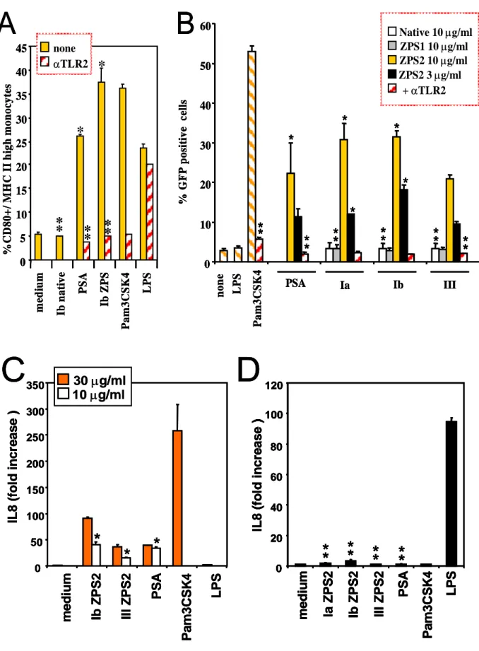
4.1.4 APC activation by ZPS is mediated by TLR2
The results shown in figure 2E suggest an involvement of TLR2 in the APC activating properties of ZPS. In addition, it was recently reported that the natural ZPS, PSA, is able to activate APCs through TLR2 (14). To analyze in detail TLR2 involvement, we tested whether anti TLR2 blocking antibody can inhibit the observed effects. Figure 4A shows that anti-TLR2 mAbs block the induction on human monocytes of MHC II and co-stimulatory molecules by both the natural and the chemically derived ZPS. The canonical TLR2 agonist Pam3CSK4 induces the same effects on monocytes which can also be blocked by the anti-TLR2 mAb. This mAb did not block APC activation induced by TLR4 (Fig. 4A) or TLR7/8 agonists (not shown), thus confirming the specificity of the reagent. ZPS2- and PSA-induced TNF-a production by monocytes was also blocked by this mAbs (not shown). An isotype-matched control antibody was not able to block the effects described here (not shown). To reconfirm the specific interaction with TLR2, we used stable TLR2 transfectants and observed reporter gene transcription upon incubation with both natural and chemically derived ZPS but not with the native or the ZPS1 form of the GBS PS (Fig. 4B). GFP expression induced by Pam3CSK4 and the natural and chemically derived ZPS was blocked by the anti-TLR2 mAb, confirming the specificity of this induction. As a further reconfirmation of absence of LPS contamination or TLR4 agonist activity of the ZPS2, we performed in parallel experiments using TLR2 and TLR4/MD-2/CD14 transfectants and assayed for the same read out, namely IL-8 production (Fig. 4C, D). While all natural and chemically derived ZPS induce significant IL-8 production in TLR2 transfectants at
the two different doses tested, none of these molecules did so in the triple transfectants at any of the doses tested. Hence, this very sensitive assay confirms that TLR4-mediated activation does not play a significant role in the phenomena described here. TLR2 transfectants were also used to exclude a number of other contaminants (Fig. 4E, F). To exclude lipopeptide contaminants in the preparations, ZPS were treated with LPL. As shown in fig. 4F, pre-treatment with LPL does not reduce the TLR2 agonist activity of ZPS while Pam3CSK4 activity is greatly reduced. Similarly, pretreatment with proteases does not alter the biological activity of ZPS, while Pam3CSK4 activity is reduced to different degrees by these two proteases (Fig.4E). In conclusion, this set of experiments indicates that the biological activity of ZPS is mediated by TLR2 and that LPS, lipopeptide or protein contaminations do not play a role in this activity.
Figure 4: ZPS-induced APC activation is mediated by TLR2. 35 30 25 20 5 10 15 40 PS A Ib Z P S 0 45 m edi um Ib n a ti v e Pa m 3 C S K 4 LP S % CD8 0 + / M H C I I hi g h m o no cy te s