UNIVERSITA‟ DEGLI STUDI DI VERONA
DIPARTIMENTO DIMEDICINA
SCUOLA DI DOTTORATO DI SCIENZE DELLA VITA E DELLA SALUTE
DOTTORATO DI RICERCA IN INFIAMMAZIONE, IMMUNITA’ E CANCRO
XXX CICLO / 2014
TITOLO DELLA TESI DI DOTTORATO
Role of Neutrophils in the Imiquimod (IMQ)-induced mouse model of Psoriasis S.S.D. MED/04
Coordinatore:
Prof./ssa Constantin Gabriela
Tutor:
Dott./ssa Scapini Patrizia
Dottorando:
Quest’opera è stata rilasciata con licenza Creative Commons Attribuzione – non commerciale Non opere derivate 3.0 Italia . Per leggere una copia della licenza visita il sito web:
Abstract
Psoriasis is a chronic skin disease associated with deregulated interplays between immune cells and keratinocytes. Neutrophil accumulation in the skin is one of the histological features that characterize psoriasis. However, the role of neutrophils in psoriasis development remains poorly understood. In this study, we utilized the imiquimod (IMQ)-induced mouse model of psoriasis to elucidate the specific contribution of neutrophils to psoriasis development. We report that neutrophils act as negative modulators of disease propagation and exacerbation by inhibiting
δ T cell effector functions via NADPH oxidase-mediated reactive oxygen species (ROS) production, as revealed by analysing disease development/progression in neutrophil-depleted mice. We also report that Syk functions as crucial molecule mediating neutrophil and δ T cell interactions. In support of the latter findings, we demonstrate that the selective impairment of Syk-dependent signalling in neutrophils only, is sufficient to reproduce the enhancement of skin inflammation and δ T cell infiltration observed in neutrophil-depleted mice. Overall, our findings add new insights into the specific contribution of neutrophils to disease progression in the IMQ-induced mouse model of psoriasis. Considering that, similarly to mouse psoriasis, the important role of IL-17 producing γδ T cells in human psoriasis has just started to emerge, it is likely that inhibitory crosstalk between neutrophils and γδ T cells may exist also in human psoriasis. Neutrophils may indeed act as unexpected negative players of disease development in specific types or clinical stages of human psoriasis. Consequently, also the utilization of therapeutic interventions targeted to inhibit neutrophil functions should be carefully evaluated.
Sommario
La psoriasi è un‟infiammazione cronica e recidivante della pelle, caratterizzata da un‟alterata interazione tra sistema immunitario e cheratinociti. Nonostante l‟infiltrazione dei granulociti neutrofili nella pelle sia una peculiarità della patologia psoriatica, il ruolo di queste cellule nella psoriasi non è ancora ben noto. In questa tesi, per studiare il ruolo dei neutrofili nella psoriasi abbiamo utilizzato un modello murino di psoriasi indotta dall‟applicazione topica di imiquimod (IMQ), insieme alla somministrazione di un anticorpo specifico per la deplezione dei neutrofili. I risultati ottenuti in questo studio, hanno mostrato che i neutrofili possono interferire con la progressione della patologia inibendo in maniera specifica i linfociti T γδ, principali mediatori di infiammazione in questo modello sperimentale di psoriasi. I dati da noi ottenuti dimostrano che i neutrofili inibiscono la proliferazione e la produzione di IL-17 da parte dei linfociti T γδ tramite la produzione di specie reattive dell'ossigeno (ROS), indotta dall'attivazione del complesso enzimatico NADPH ossidasi. Inoltre, abbiamo dimostrato che tale meccanismo inibitorio è contatto dipendente e richiede l‟interazione specifica neutrofilo-linfocita T γδ. Infine, abbiamo identificato la chinasi Syk quale molecola cruciale implicata nella modulazione di questa capacità dei neutrofili di inibire i linfociti T γδ. Infatti, l‟assenza specifica di Syk nei neutrofili è risultata sufficiente per riprodurre il fenotipo osservato con la deplezione completa dei neutrofili nel modello di psoriasi indotta da IMQ. L‟insieme dei dati ottenuti contribuiscono quindi alla comprensione del ruolo del neutrofilo nel modello murino di psoriasi indotta dall‟applicazione topica di imiquimod. Come per il modello murino, la possibile rilevanza patogenetica dei linfociti T γδ sta emergendo anche nella psoriasi umana. E‟ quindi ipotizzabile che interazioni inibitorie tra i neutrofili e i linfociti T γδ possano essere importanti anche nello sviluppo di psoriasi nell‟uomo. I neutrofili potrebbero infatti esercitare un inaspettato ruolo di modulazione negativa nell‟evoluzione dell‟infiammazione psoriatica. Conseguentemente, l‟utilizzo di terapie mirate all‟inibizione delle funzioni effettrici dei neutrofili nella psoriasi dovrebbe essere valutato con cautela.
Table of contents
1. Introduction 7
Psoriasis: general features 7
1.1 Epidemiology 7 1.2 Histological hallmarks 7 1.3 Triggering factors 8 1.3.1 Genetic triggers 8 1.3.2 Environmental triggers 8 1.3.3 Epigenetic in psoriasis 9 1.4 Pathogenesis 9 1.4.1 IL-23/IL-17A axis 13 1.4.2 γδ T Cells 13 1.4.3 Neutrophils 16
1.5 The imiquimod (IMQ) mouse model of psoriasis 19
2. Aim of the study 21
3. Materials and Methods 24
3.1 Mice 24
3.2 IMQ-induced psoriasis model 24
3.3 Neutrophil depletion 25
3.4 Cell preparation and flow cytometry 25
3.5 Quantitative real-time PCR 26
3.6 Skin histology 26
3.7 Isolation of peritoneal neutrophils 27 3.8 Proliferation and IL-17A production by γδ T cells 27 3.9 Quantification of reactive oxygen species 28
4. Results 30
5. Discussion 48
6. Additional studies 54
7
1. Introduction
Psoriasis: general features
1.1 Epidemiology
Psoriasis is a common immune-mediated chronic skin disease that occurs in genetically predisposed individuals with an overall prevalence of 2% to 3% of the world's population (Christophers 2001). Psoriasis exhibits a bimodal distribution with a peak between 15 and 30 years and another between 50 and 60 years
(Raychaudhuri & Gross 2000). Crucial traits of psoriasis, such as the high prevalence, chronicity, comorbidity and the resulting visible impact on physical appearance, make it a disorder with both a physical and psychological burden
(Kimball et al. 2005). Individuals with psoriasis have indeed also an increased risk of developing other chronic and serious health diseases. These so-called comorbid diseases include psoriatic arthritis, the metabolic syndrome, cardiovascular disorders, as well as numerous other diseases such as anxiety/depression, non-alcoholic fatty liver disease, Crohn‟s disease, or lymphoma (Griffiths & Barker 2007; Nestle et al. 2009).
1.2 Histological hallmarks
Psoriasis is characterized by different clinical phenotypes and its classification is complex and it is therefore out of the scope of this thesis. Chronic plaque psoriasis (psoriasis vulgaris) is the most common form of the disease, accounting for about 90% of cases (Boehncke & Schön 2015). Chronic plaque psoriasis typically manifests with sharply demarcated chronic erythematous plaques covered by silvery white scales, which most commonly appear on the elbows, knees, scalp, umbilicus, and lumbar area. Histopathological features of psoriatic skin include: epidermal achantosis (thickening of the viable epidermis layers do to an abnormal keratinocytes turnover), hyperkeratosis (thickened cornified layer), parakeratosis
8
(retention of nuclei in the stratum corneum) and exaggerated angiogenesis. There is also an inflammatory infiltrate within the skin layers consisting of T-lymphocytes, dendritic cells (DCs), macrophages, mast cells and neutrophils. The latter cells are responsible for the formation of subcorneal micro-abscesses, also referred to as Munro‟s microabscesses.
1.3 Triggering factors
Psoriasis is a chronic autoimmune skin disease, whose multifactorial pathogenesis is thought to result from a combination of genetic, environmental and immunologic factors (Deng et al. 2016).
1.3.1 Genetic triggers
The high concordance rates found in twins and families affected by psoriasis support the evidence for a strong genetic component in psoriatic disease (Bowcock
2005). Initial linkage analysis of several affected families allowed the
identification of at least 12 psoriasis-susceptibility (PSORS) loci (Pasić et al.
2009). More recently, genome-wide association studies (GWAS) focused on these
large genome regions unveiled susceptibility genes carrying single nucleotide polymorphisms (SNPs). A significant percentage of susceptibility genes is related to the immune system, spanning an array of functions that involve antigen presentation (HLA-Cw6, ERAP1, ERAP2, MICA), the IL-23 axis (IL12Bp40, IL23Ap19, IL23R, JAK2, TYK2), T-cell development and T-cells polarization (RUNX1, RUNX3, STAT3, TAGAP, IL4, IL13), innate immunity (CARD14, c-REL, TRAF3IP2, DDX58, IFIH1) and negative regulators of immune responses (TNIP1, TNFAIP3, NFKBIA, ZC3H12C, IL36RN, SOCS1) (Tsoi et al. 2012).
1.3.2 Environmental triggers
Genetics is only a part of the pathogenesis. Indeed, without certain environmental triggers, epigenetic modification and inflammatory responses, people with high genetic susceptibility may still fail to develop psoriasis, even though they are at significantly higher risk. Psoriasis is indeed known to be either triggered or
9
exacerbated also by non-specific exogenous factors, such as mild physical trauma (tattoos, sunburns, scratching) (Sagi & Trau 2011). Strong evidence also exists for psoriasis onset after tonsillar Streptococcus pyogenes infection (Gudjonsson et al. 2003). The use of various drugs, such as lithium, β-blockers, antimalarial agents, non-steroidal anti-inflammatory drugs, and angiotensin-converting enzyme inhibitors, has also been associated with onset or worsening of disease in psoriatic patients (Basavaraj et al. 2010). In addition, a possible association between vaccination and the new onset and/or exacerbation of psoriasis has been reported recently (Gunes et al. 2015).
1.3.3 Epigenetics in psoriasis
Increasing data suggest an emerging role of epigenetics in psoriatic disease as a plausible link between environmental exposure and psoriasis development. In this context, methylation of genes involved in the modulation of cell proliferation and apoptosis, such as SHP-1 and p16 respectively, was found abnormal in patients with psoriasis compared to healthy controls (Chen et al. 2008), (Ruchusatsawat et al. 2006). Abnormal expression of histone acetyltransferases (HATs) and histone deacetylases (HDACs) has also been observed in psoriatic skin samples (Tovar-Castillo et al. 2007). Zhang P and colleagues demonstrated that hypoacetylation of histone H4 is observed in peripheral blood of psoriatic patients, and that the degree of hypoacetylation of histone H4 inversely correlates to disease severity
(Zhang et al. 2011). Importantly, emerging evidence has indicated that HDAC inhibitors (HDAC-Is) can be used in the treatment of psoriasis (McLaughlin & La Thangue 2004). Another epigenetic mechanism found to play a role in the pathogenesis of psoriasis is RNA-associated silencing (Sonkoly et al. 2007).
1.4 Pathogenesis
In the past psoriasis was believed to be a disease primarily caused by intrinsic alterations of epidermal keratinocyte proliferation. Around 1980, however, several observations supported the central role of immune cells in the pathogenesis of psoriasis (Bos et al. 2005). Genetic data on human leukocyte (HLA) associations with psoriasis development, as well as data on the presence of oligoclonal T cells
10
in human psoriatic skin, further underline the importance of the immune system in
psoriasis pathogenesis (Eberle et al. 2016). Loss of tolerance against putative auto-antigens, including keratins, the antimicrobial peptide LL37, heat shock
proteins and the melanocytic antigen ADAMTSL5-like protein 5 (ADAMTSL5), have also been recognized as an important factor contributing to disease pathogenesis (Besgen et al. 2010; Arakawa et al. 2015). For many years, psoriasis was considered a Th1-type disease in which IFN-γ and TNF were the predominant pathogenic driver cytokines. More recently, deregulated axis involving the overproduction of IL-23, and the consequent activation of IL-17 producing T cell subsets (T17), has instead emerged as the central immune pathway driving psoriasis development (Zaba et al. 2009). IL-23 expressed by inflammatory DCs drives Th17 development and expansion. In turn, Th17-driven IL-17A and IL-22 act on keratinocytes to induce CC chemokine 20 (CCL20) and anti-microbial peptides production (Wilson et al. 2007) which sustains skin inflammation. This inflammatory loop is then amplified by the capability of keratinocyte-derived CCL20 to further recruit CCR6-bearing Th17 cells, which, in turn, further activate keratinocytes to produce CCL20 in a positive feedback manner (Chiricozzi et al. 2011). The overproduction of other inflammatory cytokines, such as IL-1, IL-36, TNFα and IL-22, is also known to trigger pivotal pathogenic pathways in human psoriasis (Johnston et al. 2017; Johnston & Gudjonsson 2014)(Figure 1).
11
From Di Meglio et al, 2014
Figure 1. Psoriasis immunopathogenesis. A pathogenic crosstalk between innate and
adaptive immune cells sustained by pro-inflammatory mediators, underlies the deregulated immune response seen in psoriasis. The three main cellular players and their products are depicted in this diagram. Keratinocytes (KCs) produce key cytokines, while activated DCs present yet undefined antigens and secrete mediators leading to the differentiation and activation of IL-17-producing T cells (T, here representing both αβ and γδ T cells). T cells, in turn, secrete cytokines that activate KC aberrant differentiation program and induce the production of further pro-inflammatory mediators, especially chemokines (CXCL1, CXCL8) recruiting neutrophils (not shown) or other immune cells (CXCL9, CXCL10, CCL20), as well as other antimicrobial peptides (AMPs) (not shown). Critical pro-inflammatory molecules, effectively targeted by biologic drugs, are shown in red.
12
Among the cellular mediators, today there is a widely accepted consensus that psoriatic lesions originate as a result of a deregulated crosstalk between keratinocytes and resident cutaneous immune cell types belonging to both the adaptive and the innate branch of the immune system. However, despite the large experimental knowledge already existing on psoriasis pathogenesis, there is still an on-going debate on which is/are the immune cell type/s primarily involved in disease initiation and progression. What is now emerging is that psoriasis is a dynamic disease in which specific cell types can dominate over the others and sustain the mounting inflammation during the different phases of disease, including disease development, progression and maintenance. As anticipated above, the crucial role of DCs (Jariwala 2007) and T17 [including T helper 17 (Th17) and, to a lower extent, T cells] (Diani et al. 2015; Papotto et al. 2017) has been widely studied in psoriatic patients as well as in preclinical models of psoriasis (Cai et al. 2011; C. Wohn et al. 2013).By contrast, the role of myeloid cells in diseases pathogenesis (such as neutrophils, monocytes and macrophages), which are also known to infiltrate the psoriatic plaques and to display abnormal functions in psoriatic patients, is less well characterized (Deng et al. 2016; Coimbra et al. 2012; Schön et al. 2017).Focus of this thesis has been to better characterize the role of neutrophils in psoriasis pathogenesis. Particular attention has been dedicated to better understand the ability of these cells to modulate the IL-23\T17 axis and T cells (an innate-like T cell type of IL-17 producing T cells) during psoriasis development.
13
1.4.1 IL-23/IL-17A axis
IL-23 is a heterodimer of a unique IL-23p19 and shared IL-12/23p40 chains produced by both resident dermal DCs and inflammatory mDCs, as well as macrophages in psoriasis (Zaba et al. 2009). A broad bunch of evidence suggests a pathogenic role for IL-23 in psoriasis. Indeed, expression of IL-23p19, IL-23p40 and IL-23R is increased in psoriasis lesional skin (Tonel et al. 2010), along with an increased number of Th17 cells in the dermis (Lowes et al. 2008) and in the blood. Moreover intradermal injection of IL-23 in mouse skin resulted in erythema with psoriasis-like histological features (Chan et al. 2006). The clinical success of biologic drugs targeting IL-12/IL-23p40 confirms the importance of this cytokine and its associated pathways, in psoriasis. In fact, IL-23 represents a crucial mediator in psoriatic inflammation, driving the development of IL-17 and IL-22 producing Th17 cells. Importantly, IL-23 along with its receptor and downstream transcription factor STAT3, are all genes that are associated with increased susceptibility to psoriasis development. Of note, activation of STAT3 and RORγδ, another Th17 characterizing transcription factor, is required for 17A and IL-17F expression. Th17, which largely infiltrate psoriatic skin dermis in humans, represent an important source of IL-17A, IL-17F, and IL-22 (Ortega et al. 2009; Cai
et al. 2011). IL-17A and IL-17F share high structural and functional homology and
primarily activate KCs to produce leukocyte recruiting chemokines, AMPs and many inflammatory mediators. IL-17A/F can also mobilize neutrophils, working as a link between adaptive and innate immunity (Pelletier et al. 2010), and further create a self-sustaining loop through IL-17A induced production of CCL20 by KCs. Besides Th17 cells, IL-17A and/or IL-22 are produced by other cell types of immune cells identified in psoriatic skin, including innate lymphoid cells (ILCs)3 and γδ T cells (Chien et al. 2014; Artis & Spits 2015).
1.4.2 γδ T Cells
γδ T cells account for approximately 3-5% of all lymphoid cells found in the secondary lymphoid tissue and the blood whereas they are highly enriched in different epithelial tissues, such us the intestinal mucosa, the skin and the reproductive tract. These cells represent, therefore, the first line of defence against
14
pathogens by providing a rapid cytokine response. γδ T cells are the first immune cells found in the fetus and provide immunity to new-borns prior to activation of the adaptive immune system (Sinkora et al. 2005). While αβ CD4+T cells mostly leave the thymus as 'naive' cells that differentiate upon activation in the periphery, γδ T lymphocytes are generated in the thymus from CD27+ γδ-cell-committed progenitor, and acquire their effector function during thymic development (Muñoz-Ruiz et al. 2017). After functional rearrangement and expression of the γ and δ-TCR chains, these cells become reprogrammed depending on the TCR signalling strength: strong agonists lead differentiation towards an IFN-γ -producing lineage, which can be identified on the basis of the expression of the co-stimulatory receptor CD27 (Haas et al. 2009). In contrast low TCR signalling leads to the development of IL-17-producing γδ T cells expressing RORγδ (Th17-lineage transcription factor) and other surface receptors including CD127 (IL7R) and CCR6 (Kisielow et al. 2008).
Several chemokine receptors, cytokine receptors and pattern recognition receptors (PRRs) are expressed by γδ T cells and have been showed to be involved in their activation. Production of IL-23, IL-1 and IL-18 by activated DCs and macrophages promotes IL-17 production by γδ T cells (Sutton et al. 2009).
Despite the fact that the activation of γδ T cells relies mainly on innate cytokines, it appears to be also regulated and „fine-tuned‟ by various accessory receptors such us programmed cell death protein 1 (PD-1), the B and T lymphocyte attenuator (BTLA) and the co-stimulatory receptor CD28 (Ribot et al. 2012).
Mouse skin harbours distinct subsets of γδ T cells which differ according to the skin layer. In the epidermis there is a large number of γδ T cells which, do to their marked dendritic morphology, have been named dendritic epidermal T cells (DECT) (Havran & Jameson 2010). DECT express the canonical Vγ5Vδ1 TCR and participate in tissue surveillance and wound healing producing keratinocyte growth factors and IFN-γ (Jameson et al. 2002). In the dermis, the Vγ4+ and Vγ6+ T cells are the most represented subsets and are responsible for pathogenic inflammatory responses mediated by the production of IL-17 (O‟Brien & Born 2015). Moreover dermal γδ T cells, but not epidermal γδ T cells, constitutively
15
Vγ6+
cells are long-lived, self-renewing, mostly radio-resistant cells that require a source of fetal thymocytes for reconstitution. On the contrary, infiltrating Vγ4+ cells migrate from the lymph nodes, are radiosensitive and can be reconstituted from bone marrow progenitors (Haas et al. 2012; Roark et al. 2007). Dermal γδ T cells have been shown to proliferate and secrete IL-17A, IL-17F and IL-22 when cultured in the presence of IL-23 and IL-1β (Cai et al. 2011; Gray et al. 2011). This pathogenic signature is critical for triggering the formation of psoriasiform plaques in mice, as genetic deletion of these cytokines substantially diminishes the disease severity in the imiquimod (IMQ)-induced mouse model of psoriasis (see IMQ mouse model below) (Pantelyushin et al. 2012). Furthermore, Cai Y. and colleagues showed that skin pathology was diminished in Tcrd-/- (T-cell receptor delta chain knock-out) mice treated with IMQ as opposed to Tcra-/- (T-cell receptor αβ knock-out) and wild-type mice, suggesting that dermal γδ T cells play a major role in driving the IMQ-induced skin inflammation (Cai et al. 2011). Using the same animal model, Ramirez-Valle F. and colleagues found an expansion of a
population of Vγ4+ Vδ4+ T cells that was mostly responsible for IL-17 production. IL-17 producing Vγ4+ Vδ4+ T cells were able to establish long-lived memory in
the skin, persisting in the dermis for long periods of time after the initial stimulation with IMQ. Moreover, experienced Vγ4+ Vδ4+ T cells showed enhanced effector functions and mediated and exacerbated secondary
inflammatory response (Ramírez-Valle et al. 2015; Hartwig et al. 2015). The observation that mice bearing a mutation in the gene encoding Sox13, which
fail to develop dermal Vγ4+
cells but can generate dermal Vγ6+ cells, had lower psoriasis-like dermatitis scores than those of Sox13+/+ mice upon IMQ treatment, further support the pathologic role of Vγ4+ Vδ4+ T cell population in psoriasis
(Gray et al. 2013). As discussed above, IL-17 producing dermal γδ T cells have been extensively studied in murine models of psoriasis. In contrast to mouse skin, γδ T cells are rare in healthy human skin. In fact, approximately 2-9% of all dermal cells are γδ T cells (Ebert et al. 2006). Furthermore, the role of these cells in human skin, and especially their role in IL-17 signalling, remains poorly characterized. There are however few evidence for a novel Vγ9+Vδ2+ T cell subset, expressing cutaneous lymphocytes antigen CCR6, which was found to
16
accumulate in psoriatic skin and to be reduced into the circulation of patients. This mobilization of CCR6+Vγ9+Vδ2+ T cells observed in psoriatic patients associates with disease severity and is normalized after successful treatment of psoriasis (Laggner et al. 2011).
1.4.3 Neutrophils
Neutrophils are innate immune cells belonging to the family of polymorphonuclear leukocytes, that are well recognized as one of the major players during acute inflammation (Mantovani et al. 2011). They are typically the first leukocytes to be recruited to an inflammatory site and are capable of eliminating pathogens by multiple mechanisms. In the circulation, mature neutrophils have an average diameter of 7–10μm, their nucleus is segmented and their cytoplasm is enriched with granules and secretory vesicles (Mestas & Hughes
2004). Mature neutrophils develop in the bone marrow from pluripotent
hematopoietic stem cells (HSC) in a process called “granulopoiesis”. Granulopoiesis is strictly dependent on the effects of several colony stimulating factors (CSFs), among which granulocyte CSF (G-CSF) is the most important, and cytokines, such as IL-6, IL-23 and IL-17 (Bugl et al. 2012). The release of neutrophils from the bone marrow to the circulation and peripheral tissue contributes to the maintenance of neutrophil homeostasis and, as such, is a tightly regulated process. Indeed, in physiological conditions the peripheral neutrophil population is maintained within a constant number by balancing the production, release and clearance of neutrophils from the circulation. The process is controlled by granulocyte colony stimulating factor (G-CSF) (Lieschke et al. 1994), which is produced in response to IL-17A synthesized by T cell populations (γδ and natural killer T cells) involved in regulating neutrophil homeostasis (Ley et al. 2006).
Release of IL-17A is, in turn, induced by IL-23 originating from tissue-resident macrophages and dendritic cells. During inflammation the number of neutrophils in the blood and tissues can increase up to 10-fold. With time the cells die by
apoptosis and are removed by macrophages and dendritic cells. This process results in down-regulation of IL-23 synthesis by those cells and thus
17
Neutrophils were always considered short-lived cells with a half-life in the circulation of approximately 1.5 and 8 hours in mice and humans, respectively. Recent studies challenged this concept, proposing that under basal conditions the average circulatory lifespan of neutrophils is up to 12.5 hours for mouse cells and 5.4 days for human neutrophils (Pillay et al. 2010). In this context, the notion that neutrophil longevity can increase several fold, both in vitro and in vivo, under inflammatory conditions has changed the view under which these cells have long been considered. Indeed, a longer lifespan may allow neutrophils to carry out more complex activities at the inflammatory sites, such as contributing to the resolution of inflammation or shaping adaptive immune responses. The classical view of neutrophils as “suicide” phagocytic cells capable only to release lytic enzymes and to produce reactive oxygen intermediates (ROI) with antimicrobial potential (Nathan 2006) has indeed been challenged by the demonstration that these cells can actually be induced to express several genes encoding key inflammatory mediators, including complement components, Fc receptors, chemokines and cytokines (Mantovani et al. 2011; Tecchio & Cassatella 2016).Thus, in response to different signals neutrophils express a vast and diverse repertoire of cytokines that are crucial to the role of neutrophils in innate and adaptive immune responses and to their role in defence and pathology. Accordingly, also the observation that neutrophils can infiltrate lymphoid organs, including spleen and lymph nodes, as well as the demonstration that neutrophils exhibit complex crosstalk with components of the innate and adaptive immune system (including DCs, T and B cells) (Scapini & Cassatella 2014) has renewed the interest in these cells within the immunology community. Importantly, locally activated neutrophils not only amplify the inflammatory process, but, surprisingly, can also actively participate in its resolution phase (Jones et al. 2016). In this context, relevant to this thesis, is the notion that the inflammatory environment can profoundly shape the functional status of neutrophils. Indeed, among the novel discoveries on neutrophil biology, concepts such as “neutrophil heterogeneity” and “neutrophil plasticity” have started to emerge, implying that, under pathological conditions, neutrophils may differentiate into discrete subsets defined by distinct phenotypic and functional profiles (Scapini et al. 2016; Silvestre-Roig et al. 2016).
18
Despite the fact that the origin and phenotype of the majority of these neutrophil subsets remain unclear, accumulating evidence supports the idea that these distinct neutrophil populations may play diverse, and even opposing roles in infection, inflammation and cancer immunology (Scapini et al. 2016; Silvestre-Roig et al. 2016). For instance, the controversial role of neutrophils in cancer pathogenesis is a subject of extensive investigation in the field. Indeed, evidence that neutrophils can display both immunosuppressive/pro-tumor and immunostimulatory/anti-tumor functions has been reported in both tumour-bearing mice and in cancer patients (Shaul & Fridlender 2017; Liang & Ferrara 2016). In this context, it has been suggested that neutrophils can polarize from an N1 pro-inflammatory and anti-tumoral phenotype, to an N2 anti-inflammatory and pro-tumoral phenotype (Granot & Jablonska 2015). Furthermore, it is now well established that myelopoiesis can be profoundly modified during inflammation and cancer, releasing altered mature myelocytes and myeloid-derived suppressor cells (MDSCs) that exert immunosuppressive and protumoral activity, mainly by inhibiting T cell functions (Moses & Brandau 2016; Scapini et al. 2016). Altogether, these novel aspects of neutrophil biology have shed a new light not only on the potential complex roles that neutrophils play during inflammation and immune responses, but also on the pathogenesis of several inflammatory disorders including infection, autoimmunity and cancer. In the context of psoriasis, as it will be described more in details in the aim of the study and in the discussion of this thesis, neutrophil accumulation in the skin is one of the histological features of this disease (Perera et al. 2012). In psoriatic skin, neutrophils initially infiltrate into the dermis at the early phase and later into the epidermis at the chronic phase. However, to date little is known about the pathogenic role of neutrophils in vivo, despite the fact that the role of IL-17 in neutrophil-mediated inflammation strongly suggests that neutrophils may also participate to psoriasis pathogenesis.
19
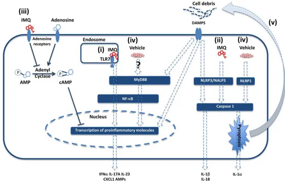
1.5 The imiquimod (IMQ) mouse model of psoriasis
Research on the pathogenesis of psoriasis has been severely hampered by the lack of a naturally occurring disorder in laboratory animals that mimics the complex phenotype and pathogenesis of the human disease (Bocheńska et al. 2017). However, a mouse model of psoriasiform dermatitis caused by the repeated topical application of Aldara™ cream [containing the Toll-like receptor 7/8 (TLR7/8) ligand IMQ (5%)] was described in 2009 (van der Fits et al. 2009). This model is currently broadly utilized to elucidate pathogenic mechanisms involved in psoriasis development as well as to evaluate possible new therapies for this disease. IMQ is a potent immune activator used for topical treatment of infections caused by Papilloma virus and other skin-associated abnormalities, including pre-cancerous skin lesions (Beutner & Ferenczy 1997). IMQ treatment is known to exacerbate the disease in patient already affected by psoriasis, also at distant sites, suggesting a possible systemic effect of IMQ (Wu et al. 2004). The primary mode of action of IMQ in mice is through ligation of TLR7 that activates NF-κB signalling in a MyD88-dependent fashion (Hemmi et al. 2002) (Figure 2). However, IMQ has been shown to have pro-inflammatory actions independently from TLR7 ligation. In fact, IMQ is a ligand for adenosine receptor for which it may act as an antagonist leading to inflammation development (Schön et al. 2006). Independently of TLR7 and MyD88 signalling, IMQ may also activate the inflammasome in keratinocytes via the NALP3 pathway, leading to activation of caspase 1 and production of IL-1β and IL-18 (Kanneganti et al. 2006). Repeated applications of IMQ-containing cream rapidly induces skin inflammation in mice with remarkable pathological and histological resemblance to human psoriasis, including the development of skin erythema and scaling, epidermal thickening (acanthosis), altered keratinocyte differentiation, neoangiogenesis and skin infiltration of immune cells (van der Fits et al. 2009). Interestingly, the involvement of a deregulated IL-23/IL-17 axis and the overproduction of other inflammatory cytokines, such as IL-1, IL-36 and IL-22, that are known to trigger pivotal pathogenic pathways involved in human psoriasis, also appears to be mirrored in the IMQ-induced psoriasis (Flutter & Nestle 2013a; Tortola et al. 2012; Rabeony et al. 2015). The murine cell populations that express high levels of TLR7 include
20
macrophages and DCs. IMQ-induced activation of Myd88 in these cells triggers the production of IL-23, which is required for the maximal T cell responses and inflammation (Costa et al. 2017; C. Wohn et al. 2013). Dermal γδ T cells are the
major IL-17-producing cells in the skin in response to IL-23 stimulation, while conventional αβ T cells do not contribute to the development of psoriasis lesions in this model (Kalyan et al. 2014). Overall these observations indicate that IMQ-induced psoriasis in mice closely resembles human psoriasis in terms of phenotypic skin changes and that lesion development is strictly dependent upon the IL-23/IL-17 axis (Cai et al. 2011; van der Fits et al. 2009).
From Flutter B and Nestle FO 2013
Figure 2. Mechanisms of action of AldaraTM. AldaraTM can activate immune responses via a number of different pathways including: (i) TLR7-dependent MyD88 pathway activation in immune cells, (ii) NALP3 activation of the inflammasome, by IMQ, (iii) antagonism of adenosine receptor signalling, (iv) direct activation of the inflammasome or MyD88 pathways through unknown receptors by the vehicle (v), imiquimod- or vehicle-driven cell death leading to the release of preformed IL-1α and cell debris.
21
2. Aim of the study
Neutrophils are the most abundant leukocytes in humans and play a major role in driving immune responses against most type of infections. Recently, it has become clear that the functions of neutrophils go far beyond the elimination of microorganisms and that these cells may contribute to the pathogenesis of numerous chronic inflammatory disorders (Kolaczkowska & Kubes 2013; Mócsai 2013). In this context, despite the fact that the presence and infiltration of neutrophils into the epidermis is one of the hallmark histologic features of psoriasis (Perera et al. 2012), the role of these cells in disease pathogenesis remains poorly understood. The most credited hypothesis views neutrophils as the principal cell mediators in the IL-17-dependent pathophysiology of psoriasis, suggesting a pro-inflammatory role of neutrophils in this disease. However, data emerging from clinical evidence do not allow drawing definitive conclusions. Indeed, while some clinical evidence report that agranulocytosis can improve the clinical outcome in patients with different subtype of psoriasis (Pai et al. 1999; Toichi et al. 2000), other clinical trials for therapeutic interventions aimed at interfering with neutrophil recruitment or functions into the inflammatory skin (e.g. anti-human CXCL8 Abs) were not successful (Bhushan et al. 2002; Schön et al. 2017). Similar controversial results on the pathogenic role of neutrophils in psoriasis also emerge from studies in which preclinical models of this disease have been utilized (Schön et al. 2000; Singh et al. 2016; Sumida et al. 2014).
This being said, to better elucidate the role of neutrophils in psoriasis development, I utilized the imiquimod (IMQ)-induced mouse model of psoriasis, which consists of the topical administration of Aldara ™ cream [containing the (TLR7/8) ligand IMQ (5%)] (Flutter & Nestle 2013b; Hawkes et al. 2016). While the crucial role of DCs and T cells (mostly γδ T cells) in the development of IMQ-induced psoriasis has been elegantly demonstrated (Cai et al. 2011; Pantelyushin et al. 2012; Singh et al. 2016; Tortola et al. 2012; C. Wohn et al. 2013; Yoshiki et al. 2014), the role of neutrophils in this model remains unclear (Figure 3).
22
Indeed, neutrophil depletion resulted in a reduction of IMQ-induced psoriasis in one study (Sumida et al. 2014) or to not affect disease development in another study
(Singh et al. 2016). Herein, by performing neutrophil depletion or utilizing mice carrying impairment in neutrophil functions, including p47phox -/- mice [lacking a cytosolic subunit of the phagocyte nicotinamide dinucleotide phosphate (NADPH) oxidase (Jackson et al. 1995)] and Sykfl/flMrp8-cre+ mice [carrying the specific deletion of the Syk kinase in neutrophils only (Elliott et al. 2011; Van Ziffle & Lowell 2009)], I uncovered a novel potential regulatory role of neutrophils in the IMQ-induced psoriasis.
23
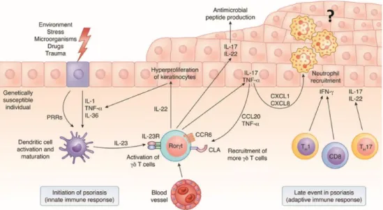
From Becher B & Pantelyushin, 2014
Figure 3. Initiation of psoriasis by γδ T cells. Several insults can directly activate DCs
through pattern recognition receptors (PRRs) or indirectly through keratinocyte stress (release of IL-1, TNF-α and IL-36). Stress-sensing DCs can produce IL-23, which activates γδ T cells to make TNF-α, IL-17 and IL-22, representing an early event in psoriasis that is mediated by the innate immune response. IL-22 induces keratinocyte proliferation, and TNF-α and IL-17 activate DCs and keratinocytes, leading to upregulation of adhesion molecules by the skin epithelium, angiogenesis and chemokine production. IL-17–induced chemokines include CXCL1 and CXCL8 and are responsible for the recruitment of neutrophils. IL-17R engagement by keratinocytes also leads to local production of CCL20, which attracts more circulating CCR6+ γδ T cells. This escalates into a self-amplifying inflammatory loop that can also be mediated by the adaptive immune system.
24
3. Materials and Methods
3.1 Mice
Sykfl/fl and Sykfl/flMrp8-cre+ mice, were previously described (Van Ziffle & Lowell 2009), p47phox−/− mice were a gift from Prof. Romani (University of Perugia) and were previously described (Jackson et al. 1995). Tcra-/- mice were a gift from Prof. Constantin (University of Verona). C57BL/6 mice were purchased from The Jackson Laboratory (Bar Harbor, ME, USA). All mice used in this study were on a C57BL/6 background and kept in a specific pathogen-free facility. All mouse experiments were carried out in accordance with guidelines prescribed by the Ethics Committee for the usage of laboratory animals for research purposes at the University of Verona and by the Italian Ministry of Health.
3.2 IMQ-induced psoriasis model
For induction of psoriasis-like skin inflammation, mice at 8–12 wk of age were shaved on the back with an electric clipper 1 d prior to treatment and received a daily topical dose of 62,5 mg of commercially available IMQ cream (5%) (Aldara Cream™, Meda AB) or control cream (vaseline) on their shaved backs for 3 or 6 consecutive days as previously described (van der Fits et al. 2009; Costa et al. 2017). On the fourth or seventh day, the animals were euthanized (Figure 4). Back skin was isolated and half was fixed in 10% formaldehyde for histopathology analysis while the other half was finely chopped and stored in RNAlater (Ambion) for quantitative real-time PCR (qRT-PCR) or digested, as described below, to achieve single-cell suspensions for flow cytometry analysis.
25
3.3 Neutrophil depletion
Mice were injected intra-peritoneally (i.p.) with 300 μg of rat anti-mouse Ly6G Ab (clone 1A8; BioXcell) or isotype control Rat IgG2a (clone 2A3; BioXCell), dissolved in 300 μl phosphate-buffered saline (PBS) every other day from day 0 to day 6 (Figure 4).
Figure 4. Neutrophil depletion in the IMQ-induced mouse model of psoriasis.
3.4 Cell preparation and flow cytometry
Skin tissue (2 cm X 2 cm) was cut from dorsal skin of the mouse. After removing subcutaneous tissue and collagen intensively with forceps, the skin was cut into small pieces and digested with 0,4 mg/ml Liberase TM (Roche Ltd.) and 0,5 mg/ml DNase I (Sigma) in RPMI 1640 medium (Sigma) for 1 hour. Single cell suspension was obtained by shredding with gentle Macs Dissociator (Miltenyi Biotec) and filtering with 70 μm and 40 μm cell strainer in series. Lymph nodes were mechanically dissociated by two frosted microscope slides and passage through a 70 μM cell strainer to yield a single-cell solution. Cells were resuspended in phosphate buffered saline containing 2 % (vol/vol) fetal calf serum, 2 mM EDTA and maintained at 4°C. For flow cytometry, 1–2×10⁶ cells were stained. Non-specific binding was blocked by pre-incubation with 0.5 µg anti-CD16/32 (2.4G2, Biolegend) and 100 µg mouse IgG (Sigma). Surface staining was performed with the following anti-mouse Abs: Ly6G(1A8), TCRαβ
26
(H57-597), CD62L (MEL-14), CD11b (M1/70), CD45 (30-F11), I-Ab (MHCII)(AF6-120.1), CD44 (IM7), TCR γ/δ (GL3) from Biolegends; Ly6C (AL-21), CD11c (HL3), CD3 (145-2C11) and GR-1 (RB6-8C5), from BD Biosciences. After final wash, cells were resuspended in staining/wash buffer containing 1 mg/ml propidium iodide (PI; Sigma-Aldrich) for viability staining according to the manufacturer‟s instructions. For intracellular cytokine staining, the cells were activated for 4 hours in phorbol 12-myristate 13-acetate (PMA; 50 ng/ml) and ionomycin (750 ng/ml) in the presence of brefeldin A (1 mg/ml). Thereafter, cells were surface-stained, washed, and then fixed and permeabilized using the eBioscience kit as previously described (Scapini et al. 2011). Intracellular staining was performed with anti-mouse IL-17A (TC11-18H10.1; eBioscience) or its relevant isotype control mAbs. Sample fluorescence was measured by a seven-color MACSQuant Analyzer (Miltenyi Biotec), while data analysis was performed by using FlowJo software Version 8.8.6 (Tree Star, Ashland, OR, USA).
3.5 Quantitative real-time PCR
Real-time reverse transcription-PCR was performed, as previously described
(Tamassia et al. 2014), using total RNA isolated from 30 mg of the skin by RNeasy Fibrous Tissue Mini Kit (QIAGEN) and utilizing the following gene-specific primer pairs (all purchased from Invitrogen) (Table 1). Data were calculated by Q-Gene software (http://www.gene) quantification.de/download.html) and expressed as mean normalized expression (MNE) units after RPL32 normalization.
3.6 Skin histology
Dorsal skin samples (3 mm) were obtained by a transversal cut of the central skin area, fixed in 10% neutral buffered formalin and embedded in paraffin blocks by using a Tissue-Tek® Tissue Embedding Console System from Diapath (Bergamo, Italy). The paraffin blocks were cut into 3 µm thick cross-section and stained with hematoxylin and eosin following the standard procedure (immersion in Mayer‟s hematoxylin: 2 minutes; immersion in eosin: 3 min) by using a Leica Microsystem Autostainer XL ST5010 (Milano, Italy). Epidermal thickness was
27
determined by measuring the average interfollicular distance under the microscope in a blinded manner. Pictures were taken using Leica DFC 300FX Digital Color Camera on a Leica DM 6000 B microscope at a 100x magnification.
3.7 Isolation of peritoneal neutrophils
Peritoneal exudates were recovered 16 hours after i.p. injection of Bio-Gel® P Polyacrylamide Beads (Bio Rad). After a passage through a 70 μM cell strainer, cell suspensions were incubated with different mAbs as described in the flow cytometry section. CD11b+Ly6G+ neutrophils were sorted using a FACS AriaTM II flow cytometer (Becton Dickinson) (> 99.0 % purity). Alternatively, neutrophils were purified using EasySep™ Mouse Neutrophil Enrichment Kit according to the manifacturer‟s instructions (>95% purity). Purified cells were suspended at proper concentration depending on the assay in RPMI 1640 medium supplemented with 10 % FBS, 1 % ultraglutammine and 1 % penicillin/streptomycin (BioWhittaker-Lonza), added to 96-well plates and cultured at 37˚C, 5 % CO2.
3.8 Proliferation and IL-17A production by γδ T cells
γδ T cells were isolated from single-cell suspensions of mouse spleen and lymph nodes from wild-type or Tcra-/- mice, using the TCRγ/δ+ T Cell Isolation Kit (Miltenyi Biotec) (> 85.5 % purity). Proliferation assay was performed in 96-well plates pre-coated (for 1 hour at 37˚C) with 10 µg ml-1 anti-CD3 mAbs [(G23-8, eBioscience). 1 x 10⁵ γδ T cells /well and 2 µg/ml anti-CD28 mAbs (B122, eBioscience) were then added to the plates (at 37˚C, 5 % CO2), in the presence of 10 ng/mL IL-1β (eBioscience) plus 100 ng/mL IL-23 (eBioscience). After 24 hours purified peritoneal neutrophils were added to the culture at the appropriate ratio. Following a 72 hour-incubation BrdU was added to the co-cultures for additional 4 hours; supernatants were then harvested for measurement of IL- 17A by using a specific ELISA kit (R&D Systems) while proliferation was determined by BrDU incorporation by ELISA (Cell proliferation ELISA, Roche), following the manufacturer‟s protocol. γδ T cell inhibition of proliferation is expressed as percentage of decrease of the absorbance value (at 450 nm: revealing the amount
28
of BrdU incorporated in proliferating γδ T cells) of each experimental conditions over the absorbance value of anti-CD3/CD28-stimulated γδ T cells.
3.9 Quantification of reactive oxygen species
Oxidative stress detection was performed using the cellROX® Deep Red Flow Cytometry Assay Kit (Life Technology). 96-well plates were pre-coated (for 1 hour at 37˚C) with 10 µg ml-1
anti-CD3 mAbs [(G23-8, eBioscience). 1 x 10⁵ γδ T cells/well and 2 µg/ml anti-CD28 mAbs (B122, eBioscience) were added into the plates (at 37˚C, 5 % CO2), in the presence of 10 ng/mL IL-1β (eBioscience) plus 100 ng/mL IL-23 (eBioscience). The day after purified peritoneal neutrophils were added to the culture at 1:1 γδ T cells-to-neutrophils ratio for 1,5 hours. Then cells were stained with the CellROX® Deep Red reagent for additional 1.5 hours. Sample fluorescence was measured by a seven-colour MACSQuant Analyzer (Miltenyi Biotec), while data analysis was performed by using FlowJo software Version 8.8.6 (Tree Star, Ashland, OR, USA).
3.10 Statistical analysis
Data were expressed as the mean ± SD and analysed using GraphPad Prism Version 5 software (GraphPad Software, Inc.). The comparison of variables was performed using two-tailed Student t- test (for comparison between two groups) or a 1-way ANOVA with Bonferroni‟s post test (used for multiple comparisons), Dunnett‟s post-test (when multiple comparisons to control group were made). P-values of less than 0.05 were considered significant and symbols indicate significant increases: */#, P <0.05; **/##, P ≤ 0.01; ***/###, P ≤ 0.001; ****/####, P ≤ 0.0001. Graphs were elaborated using GraphPad Prism Version 5 software (GraphPad Software, Inc.).
29
30
4. Results
Neutrophil depletion reduces the progression, but not the initiation, of skin inflammation and epidermal thickening in the IMQ-induced psoriasis.
To investigate the specific contribution of neutrophils to the development of IMQ-induced psoriasis, we performed neutrophil depletion by injecting anti-Ly6G (clone 1A8) Ab, or isotype control Ab, in mice treated with IMQ (or vaseline control cream), for 3 or 6 consecutive days as originally described by Van der Fits et al. (van der Fits et al. 2009). First, we confirmed that the anti-Ly6G-treatment successfully depleted neutrophils in lymph nodes and the skin of either vaseline or IMQ-treated mice after both 3 and 6 days of treatment (Figure 5).
31
Figure 5. anti-Ly6G antibody (αLy6G) efficiently depletes neutrophil infiltration in the lymph nodes and the skin of IMQ-treated mice. Dorsal skin of mice was topically
treated with IMQ containing-cream (Aldara®) or vaseline, utilized as control cream, for 3 or 6 consecutive days. To deplete neutrophils, mice were injected i.p. with anti-Ly6G antibody (αLy6G) or the isotype control (isotype Ab). (A) Draining lymph nodes were collected and analysed by flow cytometry. The total number of neutrophils (CD11b+Ly6Cint GR-1high Ly6Ghigh) is reported. (B) Total skin (2x2 cm) was digested and analysed by flow cytometry. The total number of neutrophils (CD11b+Ly6Cint GR-1high Ly6Ghigh) is reported. (C) Representative FACS plots showing the frequencies of CD11b+Ly6Cint GR-1high Ly6Ghigh neutrophils in the lymph nodes of αLy6G- or isotype Ab- treated mice after treatment with IMQ, or vaseline, for 3 or 6 consecutive days. Data are pooled from 2 separate time course experiments and are expressed as means ± SD (n = 5 mice per time point). Statistical differences of IMQ-treated vs. vaseline-treated mice (#) and IMQ-treated vs. IMQ-treated mice following neutrophil depletion (*) are reported. #P ≤ 0.05; **P ≤ 0.01; ###/***P ≤ 0.001; ####/**** P ≤ 0.0001 by 1-way ANOVA with Bonferroni‟s post-test.
32
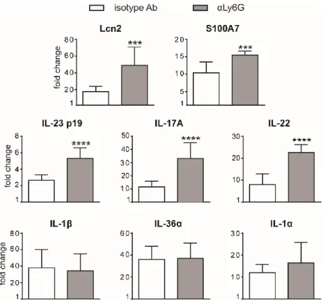
Interestingly, neutrophil depletion did not significantly affect epidermal thickening up to 3 days of IMQ treatment (Figure 6A). However, differently from what previously published (Singh et al. 2016; Sumida et al. 2014), we observed an unexpected significant increase of epidermal thickening in neutrophil-depleted mice, as compared to control mice, upon 6 days of IMQ treatment (Figure 6A, B). Consistently, the expression of skin-associated psoriatic genes by qRT-PCR, such as Lipocalin-2 (Lcn2) and S100 calcium binding protein A7/psoriasin (S100A7) was significantly higher in IMQ-treated mice receiving anti-Ly6G (clone 1A8) Ab, as compared to control isotype-treated mice (Figure 7). Strikingly, we also observed that, upon IMQ treatment, mice receiving anti-Ly6G Ab manifested a significantly increased expression of cytokines implicated in the IL-23/T17 axis, including IL-23, IL-22, IL-17, as compared to control isotype-treated mice (Figure 7). Neutrophil depletion, instead, did not significantly affect the expression of other inflammatory cytokines induced by IMQ treatment, such as IL-1, IL-36α and IL-1 (Figure 7).
Overall, these data suggest a novel potential role for neutrophils as negative modulators of disease progression and of the IL-23/T17 axis in the IMQ-induced psoriasis.
33
Figure 6. Increased epidermal thickening in neutrophil-depleted mice in response to IMQ treatment. Dorsal skin of mice was topically treated with vaseline or
IMQ-containing cream (Aldara®) for 3 or 6 consecutive days. Mice were injected with the depleting antibody αLy6G or isotype control antibody (isotype Ab). (A) The height of epidermal hyperplasia was measured in interfollicular epidermis on H&E-stained slides by light microscopic evaluation. Data are pooled from 3 separate time course experiments and are expressed as means ± SD. Statistical differences of IMQ-treated vs. vaseline-treated mice (#) and IMQ-vaseline-treated vs. IMQ-vaseline-treated mice following neutrophil depletion (*) are reported. **P ≤ 0.01; ####P ≤ 0.0001 by 1-way ANOVA with Bonferroni‟s post-test. (B) Representative H&E-staining of dorsal skin from mice injected with isotype Ab or αLy6G treated with vaseline or IMQ for 6 days. Original magnification, X100; original scale bars 40μm.
34
Figure 7. Gene-expression analysis of inflammatory molecules in the skin of IMQ-treated control or neutrophil-depleted mice. The dorsal skin of mice was topically
treated with IMQ-containing cream (Aldara®) or vaseline for 6 consecutive days. Mice were injected with the depleting antibody αLy6G or isotype control antibody (isotype Ab). Total skin RNA was extracted and reverse transcribed. mRNA expression of the indicated genes for IMQ-treated control or neutrophil-depleted mice is displayed as fold change of MNE units (after RPL32 normalisation) over vaseline-treated control. Data are pooled from 2 separate experiments and are expressed as means ± SD (n = 8-12 mice). Statistical differences of IMQ-treated vs. IMQ-treated mice following neutrophil depletion (*) are reported. ***P ≤ 0.001; ****P ≤ 0.0001 by t test.
35
Neutrophil depletion increases the expansion and infiltration of γδ T cells in lymph nodes and skin of IMQ-treated mice.
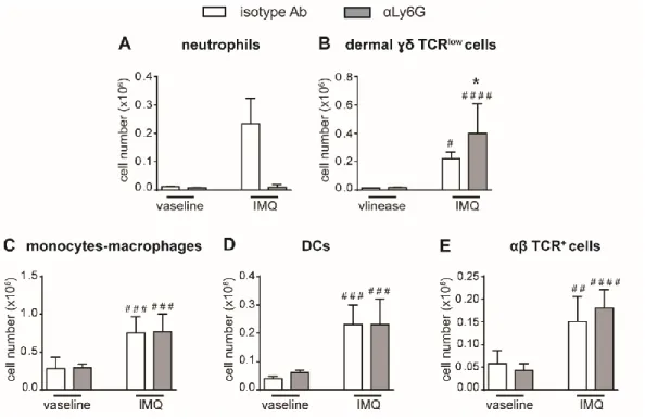
We then performed a careful characterization of the CD45+ cells infiltrating the draining lymph nodes and the skin of IMQ-treated mice receiving anti-Ly6G Ab or control isotype Ab by flow cytometry, utilizing the gating strategies previously described (Costa et al. 2017). Interestingly, after 6 days of IMQ treatment, we found strongly increased infiltration of γδ T cells in the draining lymph nodes of neutrophil-depleted mice, γδ T cells being the main pathological T cells in this mouse model of psoriasis (Cai et al. 2011; Pantelyushin et al. 2012) (Figure 8A, B-left panel). Besides the total number, also the number of CD44highCD62Llow effector γδ T cells and of IL-17-producing γδ T cells were significantly increased, indicating that not only the infiltration but also the activation of these cells was profoundly affected by neutrophil depletion (Figure 8B-right panel, C, D). No significant differences in the infiltration of αβ T cells (Figure 8E), monocytes/macrophages (Figure 8F) and DCs (Figure 8G) were instead found in the draining lymph nodes of anti-Ly6G-treated, as compared to control isotype-treated mice. Notably, a strong expansion of dermal γδ TCRlow T cells was evident also in the skin of anti-Ly6G-treated, as compared to control isotype-treated mice after 6 days of IMQ treatment (Figure 9A, B). No significant differences in the infiltration of monocytes/macrophages (Figure 9C), DCs (Figure 9D) and TCR+αβ T cells (E) were instead found in the skin of anti-Ly6G-treated, as compared to control isotype-treated mice. It is worth to point out that, under our experimental conditions, γδ T cells and neutrophils infiltrated the lymph nodes and the skin of IMQ-treated mice with similar kinetics, being the infiltration of both cell types much more consistent after 6 rather than 3 days of IMQ-treatment (Figure 5A, B and figure 10A, B).
Overall, these data point out at neutrophils as potential negative regulators of the infiltration and expansion of γδ T cells in the lymph nodes and skin of IMQ-treated mice.
36
Figure 8. Infiltration of inflammatory cells in the draining lymph nodes of IMQ-treated control or neutrophil-depleted mice. The dorsal skin of mice was topically
treated with IMQ-containing cream (Aldara®) or vaseline, utilized as control cream, for 6 consecutive days. Mice were injected with the depleting antibody αLy6G or isotype control antibody (isotype Ab). Draining lymph nodes were collected and analysed by flow cytometry. Panels report: the total number of neutrophils (Ly6Cint Ly6Ghigh GR-1high) (A), the number of total (left panel) and effector (CD44highCD62Llow, right panel)γδ TCR+ cells (B), the number of IL-17A-producing γδ TCR+ cells (C), the frequencies of IL-17A-producing γδ TCR+
cells (representative FACS plots) (D), the number of αβ TCR+ T cells (E), the number of monocytes/Mϕ (CD11bhighLy6G-CD11clow/-MHCIIlow/- cells plus CD11bhighLy6G-CD11clow/-MHCIIhigh cells) (F), and the number of DCs (CD11c+/highMHCIIhigh) (G). Data are pooled from 3 separate experiments and are expressed as means ± SD (n = 14-15 mice). Statistical differences of IMQ-treated vs. vaseline-treated mice (#) and IMQ-treated vs. IMQ-treated mice following neutrophil depletion (*) are reported. #/*P ≤ 0.05; ##P ≤ 0.01; ###/***P ≤ 0.001 by 1-way ANOVA with Bonferroni‟s post-test.
37
Figure 9. The infiltration of γδ T cells is increased in the skin of neutrophil-depleted mice treated with IMQ. The dorsal skin of mice was topically treated with
IMQ-containing cream (Aldara®) or vaseline for 6 consecutive days. Mice were injected with the depleting antibody αLy6G or the isotype control antibody (isotype Ab). Total skin (2x2 cm) was digested and analysed by flow cytometry. Panels report: the total number of neutrophils (Ly6Cint GR-1high Ly6Ghigh) (A), dermal γδ TCRlow T cells (B), monocytes/Mϕ (CD11bhighLy6G-CD11clow/-MHCIIlow/- cells plus CD11bhighLy6G-CD11clow/-MHCIIhigh cells) (C), DCs (CD11c+/highMHCIIhigh) (D) and αβ TCR+ T cells (E). Data are pooled from 2 separate experiments and are expressed as means ± SD (n = 8-10 mice). Statistical differences of IMQ-treated vs. vaseline-treated mice (#) and IMQ-treated vs. IMQ-treated mice following neutrophil depletion (*) are reported. #/*P ≤ 0.05; ##P ≤ 0.01; ###P ≤ 0.001; ####P ≤ 0.0001 by 1-way ANOVA with Bonferroni‟s post-test.
38
Figure 10. Skin infiltration of γδ T cells in the lymph nodes and skin of mice treated with IMQ for 3 and 6 days. Dorsal skin of mice was topically treated with vaseline or
IMQ-containing cream (Aldara®) for 3 or 6 consecutive days. (A) Draining lymph nodes were collected and analysed by flow cytometry. The total number of γδ TCR+
cells is reported. (B) Total skin (2x2 cm) was digested and analysed by flow cytometry. The total number of dermal γδ TCRlow
T cells is reported. Data are pooled from 2 separate time course experiments and are expressed as means ± SD (n = 5 mice). Statistical differences of treated vs. vaseline-treated mice (#) and treated mice after 3 days vs. IMQ-treated mice after 6 days (*) are reported. #/*P ≤ 0.05; **P ≤ 0.01; #### P ≤ 0.0001 by 1-way ANOVA with Bonferroni‟s post-test.
39
Neutrophils inhibit the proliferation and the production of IL-17 by γδ T cells via cell contact-dependent reactive oxygen species (ROS) production.
Previous findings have highlighted the capacity of neutrophils to both positively and negatively modulate the effector functions of γδ T cells (Davey et al. 2014; Kalyan et al. 2014a; Sabbione, María L. Gabelloni, et al. 2014; Hassane et al. 2017). Therefore we investigated the effect of neutrophils on the proliferation and the production of IL-17 by γδ T cells stimulated with anti-CD3 Abs plus anti-CD28 Abs in the presence of 100 ng/mL IL-23 and 10 ng/mL IL-1β, as previously described (Cai et al. 2011; Costa et al. 2017). As shown in Figure 11A, B, neutrophils inhibited both the proliferation and the production of IL-17 by activated γδ T cells. Given that the degree of this inhibitory effect was dependent on the ratio with T cells (Figure 11A, B), in all subsequent experiments we used the 5/1 neutrophil/T cell ratio, condition in which we obtained a strong and reproducible inhibition of γδ T cell functions by neutrophils. Similarly, to what published by Sabbione et al. (Sabbione, María L. Gabelloni, et al. 2014) with human neutrophils, we found that the addition of either catalase (a H2O2 scavenger) or of diphenyleneiodonium (DPI, a NADPH oxidase inhibitor) strongly reverted the immunosuppressive functions of mouse neutrophils on γδ T cells (Figure 11C, D), while other inhibitors, including pentoxyfilline (PTX, a degranulation inhibitor) or L-arginine [an arginase-1 (ARG1) inhibitor] were not effective (Figure 11C, D). Taken together, these data suggested that the inhibitory effects of neutrophils on γδ T cell functions in the IMQ-induced mouse model of psoriasis involves the production of ROS. In line with these observations, neutrophils isolated from p47phox-/- mice were unable to effectively inhibit γδ T cells (Figure 12A). Furthermore, by performing a flow cytometric measurement of ROS production, we also observed that the production of ROS by wild-type (WT) neutrophils, but not by p47phox-/- neutrophils, was strongly enhanced by the presence of γδ T cells in the culture (Figure 12B). Finally, in line with the fact that the inhibitory functions of different immunosuppressive neutrophil populations have been shown to occur through direct cell contact-dependent mechanisms (Choi et al. 2012; Marini et al. 2017; Pillay et al. 2012; Schmielau & Finn 2001), we found that the capacity of neutrophils to inhibit T cell proliferation
40
was significantly lower if neutrophils were separated from T cells by the use of transwells (Figure 12C).
Overall, the results obtained so far show that neutrophils inhibit γδ T cell functions via a cell contact-dependent triggered ROS production.
Figure 11. Neutrophils inhibit the proliferation and IL-17 production by γδ T cells
via reactive oxygen species (ROS) production. (A, B) γδ T cells were stimulated with
CD3/CD28, 100 ng/ml IL-23 plus 10 ng/ml IL-1β and cultured for 72h in the presence or absence of neutrophils at different ratios. (C, D) γδ T cells were stimulated with CD3/CD28, 100 ng/ml IL-23 plus 10 ng/ml IL-1β and cultured for 72h with neutrophils added at a 1 to 5 γδ T to neutrophil cell ratio, with or without inhibitors: catalase (1000 U/ml), diphenyleneiodonium (DPI) (0,1 μM), L-arginine (200 μg/ml-1), pentoxifillin (PTX) (0,5 μM). The percentages of inhibition of proliferation, as measured by BrdU incorporation (A, C), or IL-17A production (B, D) by γδ T cells, are reported. Graph values indicate means ± SD from 2 to 3 independent experiments. Statistical differences of the effect of neutrophils in the presence or absence of inhibitors are reported. *P ≤ 0.05; **P ≤ 0.01; ***P ≤ 0.001; ****P ≤ 0.0001 by 1-way ANOVA with Dunnett‟s post-test.
41
Figure 12. Neutrophil-mediated inhibition of γδ T cell proliferation requires (NADPH) oxidase-dependent ROS production and direct cell-to-cell contacts. γδ T
cells were stimulated with CD3/CD28, 100 ng/ml IL-23 plus 10 ng/ml IL-1β and cultured with neutrophils, from wild-type (WT) or p47phox-/- mice. Neutrophils were added at a 1 to 5 γδ T to neutrophil cell ratio for 72 hours (A, C) or at a 1 to 1 γδ T to neutrophil cell ratio for 3 hours (B). (A) Percentages of inhibition of γδ T cell proliferation by
neutrophils from WT or p47phox-/- mice as measured by BrdU incorporation. (B) Representative FACS histogram plots depicting the Cell-RoX MFI of CD11b+Ly6G+
neutrophils from WT or p47phox-/- mice in the presence or absence of γδ T cells, as evaluated by FACS analysis. (C) Stimulated γδ T cells were cultured with neutrophils under direct contact or transwell conditions. The graph shows the percentages of inhibition of γδ T cell proliferation, as measured by BrdU incorporation. Graph values indicate means ± SD from 2 independent experiments. *P ≤ 0.05; **P ≤ 0.01, by t test.
42
Syk signalling modulates the capacity of neutrophils to inhibit γδ T cell functions and disease progression in the IMQ-mouse model of psoriasis.
Spleen tyrosine kinase (Syk), a member of non-receptor tyrosine kinases, transmits signals in neutrophils from a variety of immunoreceptors, including Fcγ receptors (FcγRs) (Futosi et al. 2013), adhesion molecules, such as β2 integrins
(Mócsai et al. 2002) and P-Selectin glycoprotein ligand 1 (PSGL-1) (Stadtmann et al. 2013). As a consequence, Syk -/- neutrophils display impaired effector functions, including the production of ROS and the release of granule contents, in response to several inflammatory stimuli (Futosi et al. 2013).
Syk-based signalling in neutrophils alone was previously shown to be critical for appropriate host defence to Staphylococcus aureus (Van Ziffle & Lowell 2009) or the development of inflammatory arthritis (Elliott et al. 2011), suggesting the relevance of this signalling pathway in neutrophils during immune responses. Therefore, we decided to utilize mice carrying the specific deletion of Syk in neutrophils [Sykfl/flMrp8-cre+ mice (Elliott et al. 2011; Van Ziffle & Lowell 2009)], available in our laboratory, as an experimental model to test whether the specific impairment of this signalling pathway in neutrophils was sufficient to affect the interactions of these cells with T cells and IMQ-induced psoriasis. Consistently, Syk -/- neutrophils failed to produce ROS and to inhibit γδ T cells proliferation under our experimental conditions in vitro (Figure 13A, B). These data validated therefore Syk as crucial signalling molecule involved in the modulation of neutrophil capability to inhibit γδ T cell functions via a contact-dependent ROS production.
43
Figure 13. ROS-mediated inhibition of γδ T cell proliferation by neutrophils requires the activation of Syk-dependent signalling pathways. γδ T cells were
stimulated with CD3/CD28, 100 ng/ml IL-23 plus 10 ng/ml IL-1β and cultured with neutrophils from Sykfl/fl or Sykfl/flMrp8-cre+ mice at a 1 to 1 γδ T to neutrophil cell ratio for 3 hours (A) or at a 1 to 5 γδ T to neutrophil cell ratio for 72h (B). (A) Representative FACS histogram plots depicting the CellROX MFI of CD11b+Ly6G+ neutrophils from Sykfl/fl or Sykfl/flMrp8-cre+ mice in the presence or absence of γδ T cells , as evaluated by FACS analysis. (B) Percentages of inhibition of γδ T cell proliferation by neutrophils from Sykfl/fl or Sykfl/flMrp8-cre+ mice, as measured by BrdU incorporation. Graph values indicate means ± SD from 2 independent experiments. *P ≤ 0.05 by t test.
As far as IMQ-induced psoriasis experiments, we initially found that Sykfl/fl Mrp8-cre+ mice did not manifest, as compared to control Sykfl/fl mice, a significant increase of epidermal thickness, after 6 days of IMQ-treatment (Figure 14A). Interestingly, however, similarly to neutrophil-depleted mice, Sykfl/flMrp8-cre+ mice manifested, as compared to control Sykfl/fl mice, an enhanced expression of skin-associated psoriatic genes, such as S100A7 and Lcn2, as well as a specific increase in the expression of cytokines implicated in the IL-23/T17 axis, including IL-23, IL-22, IL-17, after 6 days of IMQ treatment (Figure 14B). Furthermore, also the number of total and activated γδ T cells producing IL-17, but not of other cell types, was increased in the draining lymph nodes of Sykfl/flMrp8-cre+ mice, as compared to control mice, after 6 days of IMQ treatment (Figure 15). On a similar fashion, the number of dermal γδ T cells infiltrating into the skin of IMQ-treated Sykfl/flMrp8-cre+ mice was significantly increased as compared to IMQ-treated control mice (Figure 16A, B). It is noteworthy to remark that, in line with the fact that Syk is not directly involved in controlling neutrophil migration to the