Review
ER functions of oncogenes and tumor suppressors: Modulators of
intracellular Ca
2 +
signaling
☆
Mart Bittremieux
a, Jan B. Parys
a, Paolo Pinton
b, Geert Bultynck
a,⁎
a
KU Leuven, Laboratory of Molecular and Cellular Signaling, Department of Cellular and Molecular Medicine & Leuven Kanker Instituut, BE-3000 Leuven, Belgium
bUniversity of Ferrara, Department of Morphology, Surgery and Experimental Medicine, Section of Pathology, Oncology and Experimental Biology and LTTA Center, IT-44121 Ferrara, Italy
a b s t r a c t
a r t i c l e i n f o
Article history:
Received 21 October 2015
Received in revised form 4 January 2016 Accepted 5 January 2016
Available online 6 January 2016
Intracellular Ca2+signals that arise from the endoplasmic reticulum (ER), the major intracellular Ca2+-storage
organelle, impact several mitochondrial functions and dictate cell survival and cell death processes. Furthermore, alterations in Ca2+signaling in cancer cells promote survival and establish a high tolerance towards cell stress
and damage, so that the on-going oncogenic stress does not result in the activation of cell death. Over the last years, the mechanisms underlying these oncogenic alterations in Ca2+signaling have started to emerge.
An important aspect of this is the identification of several major oncogenes, including Bcl-2, Bcl-XL, Mcl-1, PKB/Akt, and Ras, and tumor suppressors, such as p53, PTEN, PML, BRCA1, and Beclin 1, as direct and critical regulators of Ca2+-transport systems located at the ER membranes, including IP
3receptors and SERCA Ca2+
pumps. In this way, these proteins execute part of their function by controlling the ER-mitochondrial Ca2+fluxes,
favoring either survival (oncogenes) or cell death (tumor suppressors). Oncogenic mutations, gene deletions or amplifications alter the expression and/or function of these proteins, thereby changing the delicate balance between oncogenes and tumor suppressors, impacting oncogenesis and favoring malignant cell function and behavior. In this review, we provided an integrated overview of the impact of the major oncogenes and tumor suppressors, often altered in cancer cells, on Ca2+signaling from the ER Ca2+stores. This article is part of a Special
Issue entitled: Calcium and Cell Fate. Guest Editors: Jacques Haiech, Claus Heizmann, Joachim Krebs, Thierry Capiod and Olivier Mignen.
© 2016 Elsevier B.V. All rights reserved.
Keywords: Tumor suppressors Oncogenes Calcium Cancer Cell death Endoplasmic reticulum 1. Introduction
Intracellular Ca2 +signals regulate cell function and cell survival by controlling many processes. Ca2 +pumps, Ca2 +channels, Ca2 + exchangers and Ca2 +-binding proteins present at the plasma mem-brane and in different cellular compartments tightly control the cyto-solic Ca2 +concentration ([Ca2 +]
cyt)[1–3]. In resting condition, the [Ca2 +]
cytis maintained at a concentration of approximately 100 nM, whereas the extracellular [Ca2+] is about 1 mM[1,4]. In response to var-ious stimuli, the intracellular [Ca2 +] can increase to the micromolar range in the cytosol[1–3]and reach 10μM and higher in microdomains like the interface between the endoplasmic reticulum (ER) and the mitochondria[5,6]. This [Ca2+] increase can be evoked by mobilizing Ca2 +from intracellular stores, such as ER and Golgi apparatus, or by Ca2+entry from the extracellular environment[1–4]. The free [Ca2+] in the ER, the main intracellular Ca2+store, varies around 500μM[7]. Although the free [Ca2+] is around 500μM, the total [Ca2+] in the ER is more than 2 mM as a result of the action of different Ca2+buffering proteins such as calreticulin, Grp94 and BiP. These proteins bind Ca2+ with low affinity and high capacity[7]. The sarco/endoplasmic reticu-lum Ca2+-ATPase (SERCA), localized in the ER membrane, lowers the [Ca2+]
cytby pumping Ca2+from the cytosol into the ER in an adenosine Abbreviations: a.a., amino acid; ATP, adenosine triphosphate; BH domain,
Bcl-2-homology domain; BI-1, Bax inhibitor-1; BIRD-2, Bcl-2/IP3R disruptor-2; BRCA1, breast
and ovarian cancer susceptibility gene 1; [Ca2+
]cyt, cytosolic Ca2+concentration; CLL,
chronic lymphocytic leukemia; DL-BCL, diffuse large B-cell lymphoma; ER, endoplasmic reticulum; GAP, GTPase-activating protein; IICR, IP3-induced Ca2+release; IP3R, inositol
1,4,5-trisphosphate receptor; MAM, mitochondria-associated ER membrane; MCU, mito-chondrial calcium uniporter; MEF, mouse embryonicfibroblast; MOMP, mitochondrial outer membrane permeabilization; mPTP, mitochondrial permeability transition pore; mTOR, mammalian target of rapamycin; OMM, outer mitochondrial membrane; PDK1, phosphatidylinositol-dependent kinase 1; PDT, photodynamic therapy; PEST region, pro-line/glutamic acid/serine/threonine-containing region; PI3K, phosphoinositide 3-kinase; PIP2, phosphatidylinositol 4,5-bisphosphate; PIP3, phosphatidylinositol
3,4,5-trisphos-phate; PKB, protein kinase B; PKC, protein kinase C; PLC, phospholipase C; PML, promyelocytic leukemia protein; PP2A, protein phosphatase 2 A; PTEN, phosphatase and tensin homolog deleted on chromosome 10; RA2, Ras-associating domain 2; ROS, reactive oxygen species; RyR, Ryanodine receptor; SERCA, sarco/endoplasmic reticulum Ca2+
-ATPase; tBid, truncated Bid; TCR, T-cell receptor; TKO DT40 cells, triple-IP3R-knockout
DT40 cells; VDAC1, voltage-dependent anion channel 1.
☆ This article is part of a Special Issue entitled: Calcium and Cell Fate . Guest Editors: Jacques Haiech, Claus Heizmann, Joachim Krebs, Thierry Capiod and Olivier Mignen.
⁎ Corresponding author at: Laboratory of Molecular and Cellular Signaling, Department of Cellular and Molecular Medicine, KU Leuven, Campus Gasthuisberg O&N 1 Box 802, Herestraat 49, BE-3000 Leuven, Belgium.
E-mail address:[email protected](G. Bultynck).
http://dx.doi.org/10.1016/j.bbamcr.2016.01.002
0167-4889/© 2016 Elsevier B.V. All rights reserved.
Contents lists available atScienceDirect
Biochimica et Biophysica Acta
triphosphate (ATP)-dependent manner[8]. On the other hand, inositol 1,4,5-trisphosphate receptors (IP3Rs) release Ca2+from the ER store, allowing it to function as an intracellular messenger in several signal transduction pathways[9]. The IP3R, of which three isoforms exist (IP3R1, IP3R2, and IP3R3), is activated by elevated cytosolic levels of IP3, produced by phospholipase C (PLC) after stimulation of G-protein-coupled receptors or receptor tyrosine kinases by hormones, growth factors or antibodies[9–11]. IP3R activity is regulated by Ca2 +itself as well. Depending on the intracellular [Ca2 +], Ca2 +either activates (low [Ca2 +]
cyt) or inhibits (high [Ca2 +]cyt) the channel [12–14]. Furthermore, regulatory proteins[9,10,15], protein kinases and phos-phatases[16]and ATP[17–20]also modulate the Ca2+-flux properties of the IP3R. Ca2+released from the ER can be efficiently accumulated by the mitochondria, thereby directly impacting several mitochondrial functions[21–23]. Ca2+is transferred across the outer mitochondrial membrane (OMM) by voltage-dependent anion channel 1 (VDAC1), a weakly anion-selective channel that is permeable to Ca2 +as well
[24,25]. Subsequently, the mitochondrial calcium uniporter (MCU) complex removes Ca2+from the mitochondrial intermembrane space and imports Ca2+into the matrix, a process that is driven by the nega-tive mitochondrial membrane potential generated by electron transport
[26]. In the mitochondria-associated ER membranes (MAMs), which are mitochondria-associated ER subfractions involved in multiple cellular processes, including lipid synthesis, ER stress, autophagy and apoptosis, ER-mitochondrial Ca2+transport is facilitated by coupling of the IP3R to VDAC1 via glucose regulated protein 75[27,28].
Intracellular Ca2+signaling impacts many cellular processes, includ-ing mitochondrial bioenergetics, senescence, mitophagy, autophagy, and apoptosis[29–33]. First, mitochondrial bioenergetics are main-tained by constitutive low-level IP3R-mediated Ca2 +transfer from the ER to the mitochondria[29]. Ca2+stimulates mitochondrial respira-tion and ATP producrespira-tion by promoting the activities of the Ca2 + -dependent rate-limiting enzymes of the tricarboxylic acid cycle, i.e. α-ketoglutarate, isocitrate and pyruvate dehydrogenases[34]. Importantly, absence of constitutive low-level ER-mitochondrial Ca2+ signaling results in inhibition of the aforementioned dehydrogenases and can lead to activation of AMP-activated protein kinase, stimulating pro-survival mammalian target of rapamycin (mTOR)-independent auto-phagy[29,32]. Second, apoptosis is another process depending on intra-cellular Ca2+signaling. In cells suffering from varying forms of stress or damage, excessive Ca2+is released from the ER through the IP
3R and transferred to the mitochondria via VDAC1 and MCU, causing mitochon-drial Ca2+overload[35]. This in turn leads to opening of the mitochon-drial permeability transition pore (mPTP)[36], loss of mitochondrial membrane potential, permeabilization and rupture of the OMM and subsequent release of mitochondrial pro-apoptotic factors from the in-termembrane space into the cytosol, such as cytochrome c, apoptosis-inducing factor and endonuclease G. Recently, the disassembly of respiratory chain complex II due to loss of cardiolipin was proposed to be responsible for cell death triggered in response to mitochondrial Ca2+overload[37]. Third, intracellular Ca2+is also an important regula-tor of macro-autophagy, a lysosomal degradation process disposing long-lived proteins, protein aggregates, damaged organelles and intra-cellular pathogens[32,38–41]. In this way, macro-autophagy, which will be further referred to as autophagy, maintains cellular homeostasis and promotes cell survival during stress conditions, though cell death is induced if the stress is too high or persists for a longer time[38,42]. Depending on the cellular state, intracellular Ca2+ signaling either inhibits or stimulates autophagy[42]. In healthy cells, basal autophagy is suppressed by small, spontaneous IP3R-mediated Ca2+signals trans-ferred from the ER into the mitochondria that drive ATP production
[29,32]. In contrast, Ca2+signaling is enhanced in stressed cells, leading to elevated [Ca2+]
cytthat promotes autophagy by activating autophagy-stimulating proteins, such as Ca2+/calmodulin-dependent protein kinase kinase-β[42,43]. Besides the control of macro-autophagy by in-tracellular Ca2+signaling, also mitophagy, a process by which excessive
or damaged mitochondria are removed, is modulated by Ca2+uptake into the mitochondria: mitochondrial structure, membrane potential and reactive oxygen species (ROS) are not only important regulators of the Ca2+ uptake capacity of the mitochondria, but also determine whether or not to trigger mitophagy[31]. Finally, intracellular Ca2+ signaling plays an important role in cellular senescence, a protective mechanism against oncogenic events and tumorigenesis[30]. Loss of retinoblastoma and p53 pathways is usually involved in senescence, but recently it was revealed that the IP3R2 and MCU regulate this process as well[30]. Cells escaped oncogene-induced and replicative senescence by loss of IP3R2 as well as loss of MCU, since IP3R2-mediated Ca2+release and subsequent mitochondrial Ca2+accumulation through MCU lead to a decreased mitochondrial membrane potential, followed by ROS pro-duction and accumulation and eventually senescence.
During the last years, it has become clear that the remodeling of intracellular Ca2 +signaling pathways is a hall-mark of cancer cells that favors their survival and augments their cell death resistance
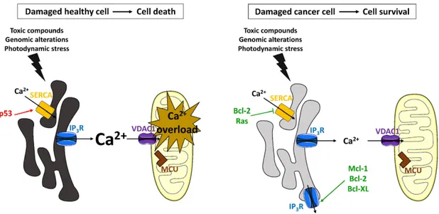
[44–47]. Many tumor cells display altered expression levels of proteins, including oncogenes and tumor suppressors that have functions at the MAMs and directly impact ER-mitochondrial Ca2+transfer (Fig. 1)
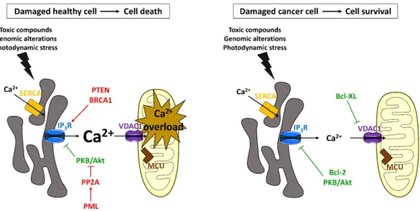
[48]. Oncogenes promote cell survival by suppressing pro-apoptotic ER-mitochondrial Ca2+signaling events, whereas tumor suppressors stimulate these ER-mitochondrial Ca2+fluxes. Hence, it is not surprising that toxic Ca2+signaling events are circumvented in cancer cells by the upregulation of oncogenes and/or the downregulation of tumor sup-pressors. The Ca2+-mediated cell death response is modulated either directly or indirectly in cancer cells. Cell death is escaped indirectly in tumor cells by a lowered ER Ca2 +content which prevents toxic, pro-apoptotic Ca2+signals emanating from the ER and avoids mitochondrial Ca2+overload (Fig. 2)[46,47]. For example, the oncogenes Mcl-1, Bcl-2 and Bcl-XL lower the ER store content by stimulating IP3Rs outside of the MAMs, thereby increasing Ca2+leak from the ER. Oncogenes Bcl-2 and Ras also decrease the ER Ca2 +levels by inhibiting the SERCA pump or by lowering SERCA2b expression levels, respectively. On the other hand, p53, which is frequently mutated in tumor cells, increases the ER store content by stimulating SERCA activity. Apoptotic cell death can also be circumvented in cancer cells by a direct modulation of toxic ER-mitochondrial Ca2 +signaling events (Fig. 3)[46,48–50]. This occurs through an upregulation of oncogenes that inhibit excessive, toxic Ca2+signals, including Bcl-2 and PKB/Akt, which both inhibit the IP3R, and Bcl-XL, which inhibits VDAC1. In contrast, tumor suppressors that promote pro-apoptotic Ca2+signaling events are downregulated or mutated in cancer cells. This includes PTEN and BRCA1, which stimu-late IP3Rs in the MAMs. The direct and indirect modulation of pro-apoptotic Ca2+transfers from the ER to the mitochondria by oncogenes and tumor suppressors will be discussed in detail in the paragraphs below. Furthermore, oncogenes that sensitize IP3Rs to low levels of IP3, such as Bcl-XL and Mcl-1, enhance pro-survival Ca2+oscillations and stimulate mitochondrial bioenergetics by enhancing ATP produc-tion, hence preserving cell proliferation and cell survival (Fig. 4)[46]. The present review aims to offer a thorough discussion of the various oncogenes and tumor suppressors that are most often altered in cancer cells and influence Ca2+signaling from the ER.
2. Oncogenes and tumor suppressors controlling intracellular Ca2+ signaling
2.1. Bcl-2
Bcl-2 is a proto-oncogene localized at the mitochondria and the ER that serves as an anti-apoptotic protein by preventing mitochondrial outer membrane permeabilization (MOMP) [51–53]. Bcl-2 is the founding member of the Bcl-2-protein family, which consists of differ-ent anti-apoptotic and pro-apoptotic members[54,55]. At the mito-chondria, Bcl-2 scaffolds and neutralizes pro-apoptotic proteins like Bax/Bak and activator BH3-only proteins such as Bim and truncated
Bid (tBid), thereby hindering the activation and oligomerization of Bax/ Bak[51,52]. Bcl-2's anti-apoptotic function is counteracted by sensitizer BH3-only proteins, like Bad, Puma and Noxa. At the ER, Bcl-2 prevents excessive Ca2+signaling that would lead to mitochondrial Ca2+ over-load and opening of the mPTP[56]. At the structural level, the anti-apoptotic Bcl-2 protein is organized in different Bcl-2-homology (BH) domains, including the BH4, BH3, BH1 and BH2 domain, and a C-terminal transmembrane domain for anchoring in membranes of intra-cellular organelles, including the mitochondria and the ER[57]. The BH3–BH1–BH2 domains together form a hydrophobic cleft, which tar-gets and scaffolds the BH3 domain of pro-apoptotic Bcl-2-family
members, such as the multidomain proteins Bax/Bak and the BH3-only proteins Bim, Bid and Bad. Bcl-2 expression is dysregulated in a va-riety of cancers, including chronic lymphocytic leukemia (CLL), diffuse large B-cell lymphoma (DL-BCL) and non-small cell lung cell carcinoma
[58–60]. In these cancers, the Bcl-2-expression levels are upregulated. Different mechanisms for Bcl-2 upregulation in cancers have been iden-tified, including (i) a chromosomal translocation t(14;18)[58,61], which places Bcl-2 under the control of an IgG promoter sequence, (ii) downregulation of endogenous miRNA targeting Bcl-2[62], and (iii) hypomethylation of the Bcl-2 gene resulting in an altered epige-netic regulation of Bcl-2[63].
Fig. 1. ER-mitochondrial Ca2+
signaling in healthy cells and in cancer cells. Oncogenes are indicated in green, whereas tumor suppressors are depicted in red. Bar-headed lines indicate an inhibitory interaction. Arrow-headed lines indicate a stimulatory interaction. The black arrows represent Ca2+fluxes. The level of shading of the ER corresponds to the ER [Ca2+], i.e. a
darker shade indicates a higher [Ca2+
] while lighter shades represent lower Ca2+
levels. Left panel: In normal, healthy cells, tumor suppressors (PTEN, BRCA1, Beclin 1, and p53) and oncogenes (Bcl-2-family members, PKB/Akt, and Ras) are in balance, thereby providing proper Ca2+
transfer from the ER to the mitochondria. This Ca2+flux ensures cell survival by
driving mitochondrial bioenergetics. SERCA pumps cytosolic Ca2+
into the ER lumen, whereas Ca2+
is released from the ER by the IP3R. Ca2+released from the ER is taken up by the
mitochondria via VDAC1, present in the outer mitochondrial membrane, and MCU, located in the inner mitochondrial membrane. Right panel: Alterations to the ER-mitochondrial Ca2+
-signaling pathway can render cancer cells resistant to cell death stimuli. Upregulation of oncogenes, such as Bcl-2, and/or downregulation of tumor suppressors, like p53, prevent the occurrence of toxic pro-apoptotic ER-mitochondrial Ca2+fluxes.
Fig. 2. Indirect modulation of the Ca2+
-mediated cell death response in damaged healthy or damaged cancer cells by tumor suppressors and oncogenes. Symbols and colors are as explained in the legend ofFig. 1. Left panel: Pro-apoptotic ER-mitochondrial Ca2+
signaling events can be modulated indirectly by altering the ER Ca2+
-store content. Tumor suppressor p53 is upregulated in healthy cells that became damaged due to various stress conditions, such as the presence of toxic compounds or DNA damage. Upregulation of p53, which stimulates SERCA activity and therefore results in an increase of the ER Ca2+content, boosts excessive Ca2+transfers from the ER to the mitochondria and subsequently leads
to mitochondrial Ca2+overload and apoptotic cell death. Right panel: Ca2+-mediated cell death can be circumvented in stressed cancer cells by a decreased ER Ca2+content, which
indirectly prevents mitochondrial Ca2+
overload. Oncogenes Bcl-2 and Ras can decrease the ER store content by inhibiting the SERCA pump or by lowering SERCA2b expression levels respectively. Bcl-XL, Mcl-1 and Bcl-2 can also sensitize IP3Rs located in parts of the ER membrane outside of the MAMs to low levels of IP3signaling, promoting basal ER Ca2+leak.
This will lower the ER Ca2+
levels and prevent cell death caused by mitochondrial Ca2+
The impact of anti-apoptotic Bcl-2 on the ER Ca2+homeostasis and dynamics is complex. Different but not mutually exclusive models have been proposed for ER-localized Bcl-2. Earlier work showed that anti-apoptotic Bcl-2 lowered the steady-state ER Ca2 +-store content (Fig. 2)[64–66], thereby protecting the mitochondria from Ca2+ over-load. Several underlying mechanisms were proposed, including Bcl-2 as a Ca2 +-leak channel[67], Bcl-2 as an IP
3R sensitizer[68,69]and Bcl-2 as a SERCA regulator[70]. (i) The original proposal that Bcl-2 could function as a putative Ca2+-pore-forming protein[67]has been excluded by a study showing that the effects of Bcl-2 in Ca2+signaling were not dependent on its putative pore-forming domain [71]. (ii) Bcl-2's function as an IP3R sensitizer was supported by evidence from Bax/Bak-knockout mouse embryonicfibroblasts (MEF cells), which display an increased ratio of Bcl-2 over Bax/Bak[68]. Bax/Bak-knockout MEF cells displayed a decreased ER Ca2+-store content that
was due to an increased sensitivity of IP3Rs towards its agonist IP3. As such, in the presence of excess Bcl-2, hypersensitive IP3Rs could become activated at basal IP3levels, thereby promoting the basal Ca2+leak from the ER through open IP3R channels and lowering the steady state ER Ca2 +levels. At the molecular level, IP
3R sensitization was due to an increased protein kinase A-dependent phosphorylation on Ser1755
[68]. Follow-up work implicated an even more complex regulation of the Bcl-2-dependent IP3R phosphorylation status, involving a role for calcineurin and protein phosphatase 1/Inhibitor-1[72–74]. Also, the effect of Bcl-2 on ER Ca2 +-store content appears to be dynamically regulated by phosphorylation. Bcl-2 phosphorylation mediated by c-Jun N-terminal protein kinase, neutralizing its anti-apoptotic properties, counteracts Bcl-2's ability to lower ER Ca2+levels[75]. Another mecha-nism for the IP3R sensitization by Bcl-2 has also been proposed. Bcl-2 was found to directly bind the C-terminal part of the IP3R channel, i.e. Fig. 3. Direct modulation of the Ca2+-mediated cell death response in stressed healthy or stressed cancer cells by tumor suppressors and oncogenes. Symbols and colors are as explained in
the legend ofFig. 1. Left panel: Apoptotic cell death is stimulated in damaged healthy cells by directly modulating IP3R-mediated Ca2+signaling events. Tumor suppressors PTEN, PML and
BRCA1 increase IP3R activity, thereby facilitating mitochondrial Ca2+overload. In contrast to PTEN and BRCA1, PML appears to act indirectly on the IP3R by facilitating the recruitment of
PP2A, which counteracts the apoptosis-suppressing effects of PKB/Akt on the IP3Rs. Right panel: Upregulated levels of oncogenes that reduce toxic ER-mitochondrial Ca2+transfers can
cause malignant cells to survive stress conditions, like stress induced by anti-cancer therapies. Bcl-2 as well as PKB/Akt suppresses excessive, pro-apoptotic Ca2+
signaling events by directly inhibiting IP3R activity. Bcl-XL inhibits VDAC1-mediated Ca2+uptake into the mitochondria, hence preventing mitochondrial Ca2+overload.
Fig. 4. Regulation of mitochondrial bioenergetics by oncogenes and tumor suppressors. Symbols and colors are as explained in the legend ofFig. 1. Left panel: Mitochondrial energy production is maintained by constitutive low-level IP3R-mediated Ca2+transfer from the ER to the mitochondria, boosting ATP production. Low-level IP3R-mediated Ca2+signaling is
stimulated by Mcl-1, Bcl-2 and Bcl-XL, which sensitize the IP3Rs present in the MAMs to basal IP3levels. Mcl-1 also increases mitochondrial Ca2+uptake by stimulating VDAC1's Ca2+
-flux properties. Right panel: Phosphorylated K-Ras4B suppresses mitochondrial Ca2+
dynamics by counteracting the sensitization of IP3Rs by Bcl-XL, thereby suppressing
a domain that is in close proximity to the Ca2+-channel pore domain (Fig. 5)[69]. Bcl-2 targeted the 6thtransmembrane domain of the IP
3R, thereby sensitizing the channel and promoting pro-survival Ca2+ oscil-lations linked to enhanced mitochondrial bioenergetics (Fig. 4). Hence, the C-terminal site in IP3R responsible for Bcl-2 binding corresponded to the site previously identified for Bcl-XL[76,77]. The affinity of the binding of Bcl-2 to the C-terminal IP3R domain has not been deter-mined. Bcl-2 overexpression in DT40 cells caused a lowering of the ER Ca2+levels in an IP
3R-dependent manner[69]. Consistent with this, Bcl-2 overexpression was more effective in protecting IP3R-expressing than IP3R-deficient DT40 cells against pro-apoptotic stimuli. (iii) Bcl-2 has been shown to directly interact with various SERCA isoforms (SERCA1 and SERCA2b), thereby limiting their ER Ca2+-uptake activity (Fig. 2)[70,78]. The binding of Bcl-2 to SERCA has also been proposed to destabilize the SERCA protein, thereby lowering its protein levels.
Besides its effect on the ER Ca2 +-store content, Bcl-2 can also directly control ER Ca2 +-release mechanisms without impacting the ER Ca2+-store content[79–81]. Bcl-2 directly interacts with the three isoforms of the IP3R channel, thereby inhibiting their function (Fig. 3). Bcl-2 overexpression in immature T cells expressing low Bcl-2 levels suppressed IP3R-mediated Ca2+release elicited by strong T-cell recep-tor (TCR) stimulation or by a cell-permeable version of IP3[80,82]. Remarkably, Bcl-2 did not suppress Ca2 +signals triggered by weak TCR stimulation. This suggests that Bcl-2 mainly suppresses excessive, pro-apoptotic Ca2+signaling events but not pro-survival Ca2+signaling events like Ca2+oscillations. The frequency of these Ca2+oscillations even appeared to be stimulated, supporting a putative sensitizing effect of Bcl-2 on IP3Rs under certain circumstances. In addition, Bcl-2 directly regulated the channel properties of the IP3R, since purified Bcl-2 could suppress IP3R single-channel activity and could interact with purified IP3R domains[80]. The Bcl-2 domain responsible for IP3R interaction and inhibition was the N-terminal BH4 domain[83]. This domain was essential and sufficient for binding IP3Rs, suppressing IP3R-mediated Ca2 +-flux properties and protecting against apoptotic stimuli. The
binding of the central IP3R domain to BH4-Bcl-2 occurred with an ap-parent affinity in the low μM range, while the IC50for the inhibition of IP3-induced Ca2 +release by BH4-Bcl-2 in permeabilized cell systems was about 30μM[83,84]. Theα-helical properties of the BH4 domain and the presence of Lys17 in the center of this domain were critical for IP3R interaction and regulation[84,85]. Bcl-2 binding to IP3R was mapped to a region of 20 amino acids (a.a. 1389–1408 in the mouse IP3R1), located in the central, modulatory domain of the channel (Fig. 5)[86]. A peptide corresponding to this Bcl-2-binding site on IP3Rs was sufficient to disrupt IP3R/Bcl-2-complex formation [86], thereby augmenting TCR-triggered IP3R-mediated Ca2 +release and apoptosis without being cytotoxic by itself[86]. A modified version of this peptide coupled to TAT and stabilized (called BIRD-2 for Bcl-2/IP3 receptor disruptor-2) triggered spontaneous, large Ca2 +-signaling events that resulted in apoptotic cell death in CLL cells[87]. Similar results were obtained in DL-BCL cells, in which the sensitivity of the cancer cells correlated with the expression level of the IP3R2, the IP3R isoform with the highest sensitivity towards its ligand IP3[88]. Further-more, DL-BCL cells that were less sensitive to BH3 mimetics were more sensitive to BIRD-2 and vice versa, suggesting a dual role for Bcl-2 in B-cell cancers at both the mitochondria and the ER[49]. Not only CLL and DL-BCL cells but also multiple myeloma, follicular lymphoma and small cell lung cancer cells appeared to be dependent on the BH4-domain biology of Bcl-2 and thus were sensitive to BIRD-2 treatment[89,90]. Moreover, in vivo injection of BIRD-2 suppressed the in vivo growth of multiple myeloma cancer cells xenografted on immunodeficient mice
[89]. Prolonged exposure of multiple myeloma cancer cells to BIRD-2 resulted in an increase in Bim levels, thereby sensitizing cancer cells towards the BH3-mimetic drugs ABT-263 and ABT-199[89]. This was further supported by evidence in small cell lung cancer cells, in which ABT-263 potentiated BIRD-2-induced cell death[90]. Moreover, BIRD-2-induced cell death could be potentiated by other Bcl-2 inhibitors that also impact Ca2+-transport systems like HA14-1[91]. BIRD-2 can trigger Ca2+-induced apoptosis through both caspase-dependent and Fig. 5. Linear representation of the IP3R isoform 1 and its regulation by tumor suppressors and oncogenes. Oncogenes are represented in green, while tumor suppressors are indicated in
red. Bar-headed lines indicate an inhibitory interaction. Arrow-headed lines indicate a stimulatory interaction. Interactions with the IP3R without functional effect are represented by oval
arrows. The IP3R1 consists of three large functional domains: a ligand-binding domain, which is composed of a suppressor domain and an IP3-binding core, a modulatory and transducing
domain, and a C-terminal channel region that contains six transmembrane domains indicated as black bars. Beclin 1 binds to the N-terminus of IP3R1 (suppressor domain), causing IP3R
sensitization that is required for driving mTOR-dependent autophagicflux. Bcl-2 inhibits IP3R activity by binding with its N-terminal BH4 domain to a.a. 1389–1408 in the mouse IP3R1
(green region of IP3R1), located in the central, modulatory domain of the channel. Bok interacts with a.a. 1895–1903 (indicated as the red region) of the IP3R. Through this interaction, Bok
protects the channel from caspase 3-dependent cleavage by shielding a caspase 3-cleavage site (DEVD) that was identified at a.a. 1888–1892 in the mouse IP3R1. Interaction of Bcl-2, Bcl-XL
and Mcl-1 with the C-terminal region of the IP3R (a.a. 2570–2749) appears to be dependent on the sixth transmembrane domain of the receptor, which results in sensitization of the
channel to basal IP3levels. Phosphorylated K-Ras4B directly binds to the IP3R (a.a. 2591–2734), and this binding is promoted in presence of Bcl-XL. Phosphorylated K-Ras4B
counteracts the sensitization of IP3R by Bcl-XL, thereby suppressing mitochondrial Ca2+dynamics. In the absence of Bcl-XL, phosphorylated K-Ras4B mildly sensitizes the IP3R to
low [IP3]. PKB/Akt suppresses IICR by phosphorylating S2681 in the C-terminal part of the IP3R. In contrast, the Akt-mediated IP3R inhibition is counteracted by PTEN,
which dephosphorylates the IP3R at S2681 via its protein phosphatase activity. Also, PML inhibits Akt-mediated phosphorylation of the IP3R by recruiting PP2A to IP3
R-PKB/Akt-protein complexes at the MAMs, where PP2A then dephosphorylates active Akt. BRCA1 binds to the C-terminal tail of IP3R (a.a. 2589–2749), thereby potentiating IP3R-mediated Ca2+
caspase-independent mechanisms, since caspase inhibitors suppressed BIRD-2-induced cell death in Bcl-2-positive lymphoid malignancies[89]
but not in small cell lung cancer cells[90]. In the latter, BIRD-2-induced apoptosis was counteracted by calpain inhibitors.
The“dual” role of Bcl-2 as a dampener of the ER Ca2+-store content or as a direct inhibitor of the IP3R not affecting the ER Ca2 +-store content has been causing controversy in thefield[92]. Yet, the varying effect of Bcl-2 on ER Ca2+could be explained by different factors. First, the reduced ER Ca2+levels upon Bcl-2 overexpression might be mediat-ed by a downstream ER Ca2+-leak pathway that is activated by Bcl-2. For instance, Bcl-2 interacts with Bax Inhibitor-1 (BI-1), an ER-located Ca2 +-leak channel belonging to the transmembrane Bax Inhibitor-1 motif-containing protein family[93–96]. Interestingly, Bcl-2 over-expression only lowered the steady state ER Ca2 +levels in BI-1-expressing cells but not in BI-1-knockout cells[97], indicating that Bcl-2 may lower the ER Ca2+-store content in a BI-1-dependent man-ner. Second, the effect of Bcl-2 on ER Ca2+might be cell type dependent. For instance, high expression levels of IP3R3 in certain cell types might favor Bcl-2's ability to lower ER Ca2 +levels as shown in the DT40 triple-IP3R knockout cell system in which Bcl-2 only lowered the ER Ca2 +content in IP
3R3-expressing cells. Third, the cellular effects of Bcl-2 have also been shown to depend on its localization and its concen-tration. For instance, transient overexpression of mitochondrially-targeted Bcl-2 or wild-type Bcl-2, but not ER-mitochondrially-targeted Bcl-2, could actually induce apoptosis in certain cells[98]. Furthermore, this effect might be dependent on its cellular concentration, since excessive over-expression has been implicated to trigger apoptosis[99]. Hence, the lowering of the ER Ca2+-store content in stably Bcl-2-overexpressing cells might be a compensatory mechanism.
Besides IP3Rs, Bcl-2 proteins also target and inhibit ryanodine recep-tors (RyRs)[100], another class of intracellular Ca2+-release channels. All RyR isoforms (RyR1, RyR2 and RyR3) interacted with Bcl-2. The binding of Bcl-2 to RyRs occurred via its central domain, consistent with the fact that the stretch of amino acids responsible for Bcl-2 bind-ing to the IP3R is highly conserved in RyRs[101]. The molecular determi-nants in Bcl-2 responsible for RyR binding were similar but not identical to the ones responsible for IP3R binding[100]. While the BH4 domain of Bcl-2 was responsible for RyR binding, the Lys17 residue of Bcl-2 ap-peared dispensable for the interaction with RyRs. Also, the BH4 domain of Bcl-2 was sufficient for inhibiting RyRs, overexpressed in HEK cells or endogenously present in rat hippocampal neurons. The binding of Bcl-2 to RyRs also appeared independent of its hydrophobic cleft, which is re-sponsible for binding the BH3 domain of pro-apoptotic Bcl-2-family members[102]. The relevance of the RyR/Bcl-2 interaction for cancer cell survival ought to be further studied. Yet, recently, elevated RyR3 ex-pression has been implicated in breast cancer risk[103]. Knocking down RyR3 in breast cancer cells impaired cell proliferation and migration. Furthermore, certain breast cancers host a signal nucleotide polymor-phism in the 3′-UTR of the RyR3 gene, which prevented binding of a reg-ulatory miRNA and caused elevated RyR3 expression in patient samples. Patients carrying the signal nucleotide polymorphism had a higher risk of developing breast cancer and its occurrence correlated to increased micro-calcification and poor progression-free survival. Also in breast cancer cells, a strong RyR2 upregulation has been described upon epi-thelial–mesenchymal transition, a process that promotes invasive prop-erties of cells and that is stimulated by hypoxia or epidermal growth factor[104]. However, the relevance of RyR/Bcl-2 complexes in breast cancer properties and their ability to undergo epithelial–mesenchymal transition remains to be further studied.
2.2. Bcl-XL
Bcl-XL is an anti-apoptotic protein related to Bcl-2. This proto-oncogene is expressed in many tissues[105]. At the mitochondria, Bcl-XL scaffolds and neutralizes pro-apoptotic Bcl-2-family members like Bak/Bax and BH3-only proteins Bim and Bid, thereby
preventing MOMP and promoting cell survival[51]. Bcl-XL is fre-quently overexpressed in cancers by which apoptosis is inhibited and cell survival is promoted. High levels of Bcl-XL have been detected in advanced and relapsed multiple myeloma, prostate, rectal, small-cell lung, gastric, pancreatic cancer, and tongue carcinomas[106,107]. Fur-thermore, constitutive activation of the epidermal growth factor recep-tor in human glioblastoma cells increases the expression of Bcl-XL, resulting in apoptosis resistance[108]. Of interest, thrombocytopenia is a major side effect of inhibiting Bcl-XL in anti-cancer therapy, since Bcl-XL is essential to maintain platelet survival by restricting Bax activ-ity[109]. Nevertheless, selective Bcl-XL inhibitors have been reported to be highly efficacious in combination with docetaxel to target a variety of solid tumors[110].
Bcl-XL can directly bind the IP3R channel and regulate its single-channel Ca2+flux properties[76]. Bcl-XL caused a potent sensitization of the IP3R, promoting its opening in response to very low [IP3] (10 nM). At the molecular level, Bcl-XL bound all three IP3R isoforms by targeting a site located outside thefirst 600 amino acids of the IP3R1. Further experiments revealed that the C-terminal part of the IP3R between a.a. 2512 and 2750 contained a Bcl-XL-binding site (Fig. 5)[76]. Bcl-XL overexpression promoted IP3R-dependent Ca2 + oscillations. Interestingly, purified tBid and Bax prevented Bcl-XL bind-ing to IP3Rs and the sensitization of IP3R channels by Bcl-XL, suggesting an involvement of Bcl-XL's hydrophobic cleft in the regulation of IP3Rs. This was underpinned by follow-up work, indicating that the C-terminal site of the IP3R contains two domains reminiscent of BH3 domains, which may recruit Bcl-2/Bcl-XL via their hydrophobic cleft[111]. In DT40 cells, the decrease of steady-state ER Ca2+levels and protection against strong B-cell receptor stimulation by overexpression of Bcl-XL required the presence of IP3Rs (Fig. 2)[76]. Consistent with this, Bcl-XL promoted spontaneous Ca2 +oscillations in wild-type but not in triple-IP3R-knockout (TKO) DT40 cells. As a consequence, Bcl-XL boosted mitochondrial bioenergetics in an IP3R-dependent manner, increasing nicotinamide adenine dinucleotide phosphate production (Fig. 4). Further work revealed that all three IP3R isoforms could enhance Bcl-XL's anti-apoptotic properties[77], consistent with the fact that Bcl-XL can bind to all three IP3R isoforms[76]and can promote Ca2+oscillations mediated by IP3R1, IP3R2 or IP3R3[77]. However, Bcl-XL only lowered the ER Ca2 +-store content of IP
3R3-, but neither of IP3R1- or IP3R2-expressing TKO-DT40 cells. This suggested that lowering of the ER Ca2 +-store content was not essential for the anti-apoptotic properties of Bcl-XL. Of note, similarly to Bcl-2, Bcl-XL too could directly bind to RyRs, which involved its BH4 domain and a Lys residue located in the BH3 domain[112]. Bcl-XL binding to RyRs suppressed Ca2+release through the channels. The presence of these complexes in cancer cells and their relevance for oncogenic properties remain unknown.
Besides IP3Rs and RyRs, Bcl-XL has been implicated in targeting Ca2+-flux pathways of the mitochondria like VDAC1, although different outcomes have been reported with Bcl-XL inhibiting or stimulating VDAC1-mediated Ca2+uptake into the mitochondria[113–115]. The inhibitory effect of Bcl-XL on VDAC1 could be attributed to its BH4 domain[116]. Of course, these effects of Bcl-XL on the rate of mitochon-drial Ca2+uptake could indirectly impact the ER Ca2+-release proper-ties. For instance, decreased mitochondrial Ca2+uptake, e.g. by Bcl-XL inhibiting VDAC1, may lead to an increase in cytosolic Ca2+signaling for a given Ca2+release from the ER. Furthermore, the inhibitory action of Bcl-XL on VDAC1 may be an additional mechanism to prevent toxic mitochondrial Ca2+overload (Fig. 3). In contrast to these studies, Bcl-XL has also been reported to enhance VDAC1-mediated mitochondrial Ca2+uptake[114]. Although we and others[113,115,116]reported an inhibitory role for Bcl-XL on VDAC1, these data could reflect the dual role of VDAC1 in both mediating survival and apoptosis signaling
[117]. As such, Bcl-XL may promote the transfer of pro-survival Ca2+ signaling while inhibiting pro-apoptotic Ca2+signaling. Alternatively, differences in the experimental methods could account for this. For
instance, many experiments performed by White et al. were based on mitochondrial Ca2+uptake experiments in permeabilized cells where all VDAC1 channels across the mitochondrial outer membrane will participate in mitochondrial Ca2 +uptake[114]. In contrast, experi-ments in other studies relied on mitochondrial Ca2+uptake measure-ments in intact cells exposed to agonists for which Ca2 +would be preferentially transferred via ER-mitochondrial contact sites[113,115, 116]. Nevertheless, since a stimulatory effect of VDAC1 by Bcl-XL has never been shown in direct measurements based on purified VDAC1 channels, the molecular properties and the relevance of the VDAC1/ Bcl-XL connection in promoting mitochondrial Ca2+transfer ought to be further scrutinized.
2.3. Mcl-1
Mcl-1 is an anti-apoptotic member of the Bcl-2-protein family and has many important cellular functions. This proto-oncogene is crucial for the development and maintenance of several cell types including lymphocytes[118,119], neurons[120]and hematopoietic stem cells
[121]. Additionally, Mcl-1 plays an important role in early embryogene-sis[122]. The Mcl-1 protein has a very short half-life and is structurally different from the other anti-apoptotic Bcl-2-family members[123]. The sequence of the putative BH4 domain of Mcl-1 substantially differs from the sequences of the BH4 domain of the Bcl-2 and Bcl-XL proteins. Furthermore, its N-terminus contains two PEST (proline/glutamic acid/serine/threonine-containing) regions, which affect the rate of turnover and contain several sites for phosphorylation[123]. Mcl-1's pro-survival functions are advantageous to many types of cancer. Overexpression of Mcl-1 has been observed in hematopoietic and in lymphoid cancers as well as in solid tumors[123]. This is supported by an analysis of somatic copy-number alterations in different cancer types, showing that the gene encoding for Mcl-1 is frequently amplified in a broad spectrum of cancer cells[124]. Mcl-1 overexpression induces resistance to chemotherapeutic agents like paclitaxel and vincristine, but also to Bcl-2 inhibitors such as ABT-737, while the sensitivity of chemoresistant cells to these compounds can be restored after Mcl-1 silencing[123,125].
One study reported the binding of Mcl-1 to IP3Rs[69]. It was shown that besides Bcl-2 and Bcl-XL, Mcl-1 too targeted the C-terminal tail of the IP3R (Fig. 5). All three Bcl-2-family members bound the IP3R domain with similar affinities. It was proposed that the last transmembrane domain of the IP3R was essential for these interactions. Mcl-1 enhanced IP3R-mediated Ca2 +release, resulting in decreased steady-state ER Ca2 +levels (Fig. 2)[69]. Store depletion in Mcl-1-expressing cells became more prevalent in the presence of low [IP3], indicating that the sensitivity of IP3-dependent Ca2 + re-lease is enhanced by Mcl-1. The Mcl-1-mediated IP3R sensitization also contributes to low-level IP3R-mediated Ca2 +signaling from the ER to the mitochondria and therefore stimulates mitochondrial bioenergetics (Fig. 4). Also, Mcl-1's anti-apoptotic properties were promoted when IP3Rs were present in the DT40 cells[69]. Yet, the protective effect of Mcl-1 overexpression in IP3R-expressing DT40 cells was less pronounced than the effects observed for Bcl-2 or Bcl-XL overexpression.
Of interest, Mcl-1 has recently been implicated to bind with high affinity to VDAC1, thereby stimulating VDAC1's Ca2 +-flux properties and thus increasing mitochondrial Ca2 +uptake, hence promoting ATP production (Fig. 4)[126]. Among all Bcl-2-family members, Mcl-1 was the strongest VDAC1-binding protein. The VDAC1/Mcl-1 complex appeared to be important for the survival of cancer cells, since the increase in mitochondrial [Ca2 +] stimulates ROS produc-tion which in turn promotes cell migraproduc-tion[126]. In addition to this, Mcl-1 has recently been implicated in the control of mitochon-drial dynamics by promoting Drp1-mediated mitochonmitochon-drialfission and preventing mitochondrial hyperpolarization, which limits mito-chondrial Ca2 +uptake[127].
2.4. Bok
Bok, Bcl-2-related ovarian killer, is a multi-BH domain-containing pro-apoptotic protein that resembles Bax/Bak. It is scaffolded by Mcl-1 and Bfl-1, but not by Bcl-2 or Bcl-XL[128]. Although afirst report suggested that Bok expression was restricted to reproductive tissues
[128], Bok appears to be expressed in many tissues besides ovary with high levels in brain, lung, spleen and stomach[129]. Since Bok-knockout mice develop normally, it was proposed that the functions of Bok largely overlap with Bax/Bak or are only critical in certain conditions of cell stress[129]. Compared to Bax/Bak, the role of Bok in apoptosis is however much less characterized. A major portion of the endogenous Bok protein resides outside the mitochondria, including the ER, Golgi and nucleus[130]. Bok overexpression in cells resulted in apoptosis that required the presence of Bax/Bak proteins[130]. Bok overexpression caused a rapid disintegration and fragmentation of the ER and Golgi independently of downstream caspase 3 activation and ap-optosis. ER/Golgi-targeting of Bok was dependent on its C-terminal transmembrane domain. Cells lacking Bok displayed a normal sensitiv-ity towards cell death stimuli like staurosporine and etoposide but displayed an increased sensitivity and abnormal ER-stress response to brefeldin A, a pro-apoptotic ER-stress inducer that disrupts ER-Golgi trafficking[130,131]. In further work, it was shown that Bok has an important function in mediating cell death upstream of Bax/Bak in response to ER stress, since Bok-deficient cells were protected against thapsigargin- or bortezomib-induced apoptosis[132]. The presence of Bok was essential for the activation of Bim, activating transcription factor 4 and CCAAT/enhancer-binding protein homologous protein, pro-apoptotic components of the unfolded protein response. The role of Bok in cancer is not fully understood, but deletions in the Bok gene were identified in high-resolution analyses of somatic copy-number alterations from more than 3000 cancer specimens as a frequent event to occur in different types of cancer[124]. However, in contrast to Bax deletion, which accelerated cancer development, Bok deletion did not accelerate the development of lymphomas in Eμ-Myc transgenic mice[129].
Interestingly, among all Bcl-2-family members, Bok displayed the strongest binding to the IP3R[133]. Bok binding to IP3Rs was isoform-specific, since IP3R1 and IP3R2, but not IP3R3, were targets for Bok. IP3Rs may serve as a sink for Bok, as most of the cellular Bok was found in complex with IP3Rs. The domains responsible for IP3R1/Bok in-teraction were on the one hand a stretch of amino acids (PSRKKAKEP) located between a.a. 1895 and 1903 of IP3R1 (Fig. 5) and on the other hand a stretch of amino acids (LGREYV) located between a.a. 34 and 39, corresponding to a part of the N-terminal BH4 domain of Bok. Furthermore, Bok-deficient cells displayed increased levels of IP3R1 and decreased IP3R2 and IP3R3 levels[133]. Yet, overall, Bok-expressing and Bok-deficient cells displayed similar IP3-induced Ca2+ release (IICR) properties. Bok also did not cause major alterations in IP3R downregulation by the ubiquitin proteasome pathway and Bok was co-degraded with IP3R by this system in conditions of persistent activation of the IP3-dependent signaling pathway[133]. However, the association of Bok with the IP3R protected these channels from proteo-lytic digestion by chymotrypsin, which has a cleavage site in the IP3R in the proximity of the Bok-binding site[133]. Bok also protected IP3Rs against cleavage by caspase 3, an apoptosis executioner caspase, since Bok-deficient cells exposed to apoptotic stimuli displayed a rapid cleavage of IP3Rs into an N-terminal 170-kDa fragment and a C-terminal 95-kDa fragment containing the channel pore. This indicates that the Bok-expression levels may critically control the caspase 3-dependent cleavage of IP3Rs. This is particularly relevant in the context of the conflicting results on whether or not IP3Rs are bona fide substrates of caspase 3. About 15 years ago, the team of Mikoshiba reported that IP3R1 was a caspase 3 substrate as IP3R1 cleavage upon apoptotic stimuli was only observed in cells expressing caspase 3
IP3R1 (a.a. 1888–1892 in mouse IP3R1) (Fig. 5). The removal of this site (by mutation) diminished Ca2+overload in DT40 cells and rendered them more resistant to apoptotic stimuli[135]. Furthermore, expression of the C-terminal channel domain, an IP3R1 cleavage product, increased the tendency of these cells to undergo apoptosis. IP3R1 cleavage by caspase 3 was further shown to play an important role in apoptotic events that take place during mouse oocyte maturation[136,137]. How-ever, other groups made observations contradicting the role of IP3R1 as a relevant caspase 3 substrate. For instance, Guillemette and co-workers showed that HeLa or Jurkat cells exposed to a variety of apoptotic stim-uli with robust caspase 3 activation did not display cleavage of IP3R1
[138]. Here, recombinant caspase 3 also failed to cleave IP3R1 in the microsomal fraction. Consistent with this, Boehning and co-workers showed that the staurosporine-induced rise in cytosolic [Ca2 +] in HeLa cells is independent of caspase 3 expression or activity[139]. Also, IP3R1 cleavage did not occur in staurosporine-treated MCF7 cells re-complemented with caspase 3. These contradicting results raise the question whether differing endogenous Bok levels may account for the varying sensitivity of IP3R1 to caspase 3 cleavage in different cell models (e.g. DT40 cells versus HeLa or MCF7/caspase 3 cells). 2.5. Beclin 1
Beclin 1 is an important regulator of autophagy, a catabolic pathway in which damaged organelles and macromolecules are degraded and recycled[140]. This protein is localized within cytoplasmic structures, such as the ER, mitochondria and the perinuclear membrane. Beclin 1 is critical to initiate autophagy since it is part of one of the main signal-initiating complexes, consisting of class III phosphatidylinositol 3-kinase/Vps34, Beclin 1 and p150 protein[141]. The Beclin 1 protein is composed of three main domains: (i) a BH3 domain located at the N-terminus (a.a. 114–123) that interacts with anti-apoptotic Bcl-2-family members, (ii) a central coiled-coil domain (a.a. 144–269) medi-ating Beclin 1 self-association and dimerization, and (iii) a C-terminal evolutionarily conserved domain (a.a. 244–337) that enables protein in-teractions and binds lipid membranes of cell organelles[140–143]. Via these domains Beclin 1 interacts with co-factors, including Vps34, PKB/Akt, Bif-1, Bcl-2 and Bcl-XL, to positively regulate autophagy and initiate autophagosome formation[140]. Beclin 1 is a known tumor suppressor, since Beclin 1 haplodeficient mice display increased tumor formation[144]. Also, in ovarian, breast and prostate cancer, the tumor suppressive gene BECN1 was mono-allelically deleted, whereas a decreased expression of Beclin 1 was observed in many types of cancer, including brain tumors and cervical cancer[140,141].
Several studies showed that IP3Rs are directly targeted by Beclin 1, which has a prominent binding site in the suppressor domain of the IP3R (a.a. 1–225) (Fig. 5)[145]. Originally, it was proposed that IP3R served as a scaffold for Beclin 1, thereby inhibiting autophagy by making it less available for its autophagy-inducing role in the Vps34 complex
[146]. This complex mediates one of the earlier steps within the autoph-agy process downstream of mTOR/unc-51-like kinase 1 complex but preceding the autophagosome formation. As such, the IP3R served as a negative regulator of autophagy. In this context, Beclin 1 did not modu-late IICR and thus IP3Rs rather served as a sink for Beclin 1, thereby limiting Beclin 1 availability for driving the autophagy process. Further work actually found an autophagy-promoting role for the IP3R/Beclin 1 complex[145]. Beclin 1 binding to the IP3R occurred in a dynamic manner during starvation-induced autophagy, leading, together with an increased ER Ca2 +-store content, to a sensitization of agonist-induced Ca2+signaling. Cells lacking Beclin 1 failed to display enhanced IP3R-mediated Ca2+signaling. This involved a direct sensitization of the IP3R by Beclin 1, since purified Beclin 1 enhanced IICR in permeabilized cells. IP3R sensitization and subsequent Ca2+signaling were essential to drive starvation-induced autophagicflux, since pharmacological IP3R inhibition and intracellular Ca2+buffering prevented starvation-induced autophagy[145]. Yet, at this point it is not clear whether
IP3Rs and Ca2+signaling are involved in Beclin 1's function as a tumor suppressor.
2.6. PKB/Akt and PTEN
The phosphoinositide 3-kinase (PI3K)/protein kinase B (PKB)/Akt signaling pathway is a tightly controlled process regulating cell survival, proliferation and cell death[147,148]. PI3K, a lipid kinase, converts phosphatidylinositol 4,5-bisphosphate (PIP2) into phosphatidylinositol 3,4,5-trisphosphate (PIP3) after it is activated by G-protein-coupled receptors and receptor tyrosine kinases[147,149]. Next, PIP3recruits phosphatidylinositol-dependent kinase 1 (PDK1) and PKB/Akt, a serine/threonine kinase, to the plasma membrane. This leads to PKB/ Akt activation through phosphorylation of T308 by PDK1 and S473 by mTORC2. Subsequently, active PKB/Akt phosphorylates its substrates in the cytoplasm and nucleus, thereby regulating several cellular func-tions, including cell growth, proliferation, survival, metabolism, protein synthesis and apoptosis[147]. There are three Akt isoforms, Akt1, Akt2 and Akt3, which are structurally highly homologous but exhibit differ-ent functions[150]. Akt1 and Akt2 are ubiquitously expressed, whereas Akt3 expression is restricted to a few tissues. Phosphatase and tensin homolog deleted on chromosome 10 (PTEN), a lipid and protein phos-phatase, is the most important negative regulator of the PI3K/Akt signal-ing pathway[149]. Via its lipid phosphatase activity, PTEN reverses the action of PI3K by dephosphorylating PIP3to PIP2, counteracting all downstream functions controlled by PKB/Akt. The protein phosphatase activity of PTEN is used to inhibit cell migration and cell cycle arrest
[149]. The tumor suppressive activity of PTEN is lost in many human tumors, such as breast, thyroid, kidney, colorectal and prostate cancer
[124,149]. Somatic mutations, gene silencing or epigenetic mechanisms cause this loss of functional PTEN in tumor cells. Hyperactivity of PKB/ Akt, caused by mutations in the catalytic subunit or amplification of Akt, has also been observed in several cancer types[150]. For example, Akt1 amplification has been detected in gastric carcinomas, whereas amplification of Akt2 occurred in ovarian, breast, colorectal and pancre-atic cancers. In several tumor types, including breast, colorectal, ovarian, lung and bladder cancer, an activating mutation on Akt1 (E17K), which results in constitutive localization of Akt at the plasma membrane and promotes growth factor-independent phosphorylation of T308 and S473, has been observed[150].
Over the past years, several studies reported the interaction of Akt kinase with IP3Rs at the ER. Joseph and co-workers showed that phos-phorylation of the IP3R by PKB/Akt occurred in vivo[151]. All three IP3R isoforms contain a consensus substrate motif for PKB/Akt kinase (RXRXX(S/T)) located in the cytosol-exposed C-terminal tail (Fig. 5). Upon Akt activation by insulin, endogenous IP3Rs present in CHO-T cells were phosphorylated[151]. A prostate cancer cell line with consti-tutively active Akt due to loss of functional PTEN (LnCAP cells) exhibited constitutive phosphorylation of endogenous IP3R1 as well. Exposure to a PI3K inhibitor removed IP3R phosphorylation in the two aforemen-tioned cell lines. In COS cells transfected with constitutively active Akt, IP3R phosphorylation was inhibited by mutating a serine amino acid residue present in the Akt substrate motif of the IP3R (S2681A and S2681E). IP3R channel function was not directly modified by Akt phosphorylation, as IICR was comparable between wild-type, non-phosphorylatable S2681A mutant and phosphomimic S2681E mutant IP3Rs expressed in COS cells[151]. Nevertheless, caspase 3 activation, induced by staurosporine, was enhanced in TKO-DT40 cells expressing the S2681A mutant compared with cells expressing wild-type IP3Rs or IP3R mutants with a mimicked effect of Akt phosphorylation (S2681E). In contrast, other studies did report reduced Ca2+release from IP3Rs phosphorylated by PKB/Akt[152,153]. It was shown that phosphoryla-tion of IP3Rs, induced by overexpressing constitutively active PKB/Akt, inhibited IICR and histamine-induced Ca2 +release, whereas the ER Ca2 +content was not decreased[152]. In this study, a greater ATP-induced Ca2+response was observed in COS cells expressing mutant
IP3RS2681Acompared to cells expressing wild-type IP3R. The former was also more sensitive to apoptotic stimuli like menadione and displayed higher mitochondrial Ca2 +rises in response to these stimuli. Also, glioblastoma cells exhibiting Akt hyperactivity due to loss of PTEN showed reduced IICR, menadione-induced mitochondrial Ca2+uptake and apoptosis compared with glioblastoma cells re-expressing PTEN
[152]. These results indicate that phosphorylation of IP3Rs by active PKB/Akt reduces Ca2 + release from the ER and subsequent Ca2 + transfer to the mitochondria, leading to the protection of cells from apoptotic stimuli (Fig. 3). Another independent study reported that agonist-induced Ca2 +release from the ER and the subsequent mito-chondrial Ca2+rise is reduced by expressing constitutively active Akt1 in HeLa cells, thereby protecting cells from apoptotic stimuli that act through mitochondrial Ca2+overload like H
2O2[153]. This study elegantly showed that Akt modulated IP3R activity, resulting in an inhi-bition of agonist-induced Ca2 +release and protection from Ca2 + -mediated apoptotic stimuli. In further work it was demonstrated that Akt reduces ER-mitochondrial Ca2+transfer and protects against apo-ptosis by specifically acting on IP3R3[154]. Activated Akt suppressed IICR and apoptosis, induced by arachidonic acid, in COS7 cells, which lack detectable IP3R1, but not in IP3R3-deficient SH-SY5Y cells. How-ever, Akt did reduce Ca2 +release and did protect against apoptotic stimuli in SH-SY5Y cells expressing IP3R3. Interestingly, it has been proposed that pro-apoptotic ER-mitochondrial Ca2+transfers preferen-tially occur via IP3R3[155,156], although a pivotal role of IP3R1 in Ca2+ -mediated apoptotic cell death was reported in other studies as well
[135,157].
Recently, tumor suppressor PTEN was found to be localized at the ER and MAMs and to modulate Ca2+transfer from the ER to mitochondria in a protein phosphatase-dependent manner[158]. PTEN silencing, which leads to increased phosphorylation and activity of Akt, impaired the release of Ca2+from the ER and lowered cytosolic and mitochondri-al Ca2+transients in response to agonists[158]. Consequently, sensitiv-ity to arachidonic acid, an apoptotic inducer causing ER Ca2+release, and subsequent mitochondrial Ca2 + overload were decreased in PTEN-silenced cells, but were both increased upon overexpression of PTEN that is targeted to the ER, indicating that ER-localized PTEN sensi-tizes cells to Ca2 +-mediated apoptotic stimuli (Fig. 3). Moreover, initiation of Ca2+-dependent apoptosis with arachidonic acid enriched PTEN localization at the ER. Co-immunoprecipitation experiments revealed that PTEN interacts with IP3R3[158]. Finally, it was determined that ER-localized PTEN directly reduces the Akt-dependent phosphory-lation state of the IP3R through its protein phosphatase activity, hence modulating Ca2+release from the ER and cellular sensitivity to Ca2+ -mediated apoptotic stimuli (Fig. 5).
2.7. Promyelocytic leukemia protein (PML)
The promyelocytic leukemia protein (PML) is an important tumor suppressor, which controls various cellular functions, including apo-ptosis, DNA-damage response, cellular proliferation and senescence
[159,160]. The PML gene was identified through its location at the breakpoint of the t(15;17) chromosomal translocation, which is often observed in acute promyelocytic leukemia[160]. As a result of this translocation, the PML gene is juxtaposed to the gene encoding the retinoic acid receptorα, which leads to the formation of fusion proteins that hinder the differentiation of hematopoietic cells. At the structural level, the N-terminal part of the PML protein is highly structured, in contrast to its C-terminus, and contains a RING domain, which is a zinc-finger that exhibits E3 ligase activity, two additional zinc-finger motifs called B-boxes and anα-helical coiled-coil domain[160,161]. These N-terminal domains are responsible for the oligomerization of PML and they mediate protein–protein interactions, allowing PML to concentrate in nuclear bodies. To exert its tumor suppressive function, PML interacts with a large number of proteins, including sumoylated PML, p53, protein phosphatase 2 A (PP2A) and PKB/Akt[160,161].
PML expression is lost or reduced in hematopoietic malignancies as well as in solid tumors, such as colon, lung, prostate and breast cancers
[160]. Interestingly, PML-knockout mice and primary cells lacking PML are protected from apoptosis triggered by a variety of stimuli[162].
A few years ago, extra-nuclear PML was discovered to be localized to the ER and MAMs and to regulate apoptosis at the ER by modulating Ca2 + release [163]. Cytosolic and mitochondrial Ca2 + responses induced by agonists or oxidative apoptotic stimuli were higher in cells expressing PML than in cells lacking PML. Consistently, the presence of PML in cells was protective against ER stress-induced apoptosis
[163]. Interestingly, the dampened Ca2+responses observed in PML-deficient cells could be fully restored by ER-targeted PML[163]. As a consequence, these cells regained sensitivity to ER stress-induced apoptosis, but remained resistant to etoposide, an apoptotic inducer that acts independently of Ca2+. These functional observations could be linked to a physical interaction between PML and IP3R3[163]. More-over, the levels of phosphorylated IP3R3 were higher in PML−/−than in PML+/+MEFs cells due to higher amounts of phosphorylated, active Akt and reduced amounts of protein phosphatase PP2A associated with IP3R3 in the former[163]. Hence, the tumor suppressor PML was found to be essential to recruit PP2A to IP3R3-Akt complexes at the MAMs (Fig. 3), where PP2A counteracts Akt activity, thereby suppressing Akt-mediated phosphorylation of the IP3R3 (Fig. 5). As a consequence, IP3R3-mediated Ca2+transfer from the ER into the mito-chondria and, eventually, apoptosis are promoted in PML-expressing cells[161,163]. Thus, similarly to PTEN, PML stimulates pro-apoptotic Ca2 +signaling events by negatively regulating the activity of Akt at the ER.
2.8. Breast and ovarian cancer susceptibility gene 1 (BRCA1)
Tumor suppressor BRCA1 is a chromatin-interacting protein that has a critical role in homologous recombination repair of double-strand DNA breaks[164]. Its main function is to preserve genomic integrity, although growing evidence indicates that BRCA1 activity is necessary for mammary epithelial differentiation as well[164]. Breast and ovarian cancer are frequently associated with impaired functioning of BRCA1, caused by BRCA1 mutations, loss of expression or BRCA1 downregula-tion. BRCA1 mutations predispose to breast and ovarian cancer, although it is still not clear why this predisposition exists since this tumor suppressor is expressed in all cells. In families with hereditary breast and ovarian cancer, the N-terminal RING domain and the C-terminal BRCT repeats of BRCA1 are frequently mutated[164]. These two domains contribute to the tumor suppressive activity of BRCA1. The N-terminal RING domain exhibits E3 ubiquitin ligase activity when heterodimerized with BARD1, another RING- and BRCT domain-containing protein[164,165]. The BRCT repeats are needed to concen-trate BRCA1 in subnuclear repair foci that appear after DNA damage. These repeats are phosphopeptide-binding domains responsible for the interaction of BRCA1 with partner proteins like the endonuclease CtIP[166], the adaptor protein Abraxas[167]and the DNA helicase BRIP1[168]. Through their interaction with BRCA1, these partner pro-teins regulate DNA repair and maintain genome stability.
Recently, BRCA1 was identified as a novel binding partner of the IP3R1[169]. A major subpopulation of the BRCA1 pool localized outside the nucleus, residing at ER membranes by binding phospholipids. Full-length IP3R/BRCA1 complexes were identified. The complex was formed through a direct interaction of the C-terminal tail of the IP3R1 with the N-terminal domain of the BRCA1 protein (a.a. 1–112, the so-called RING domain) (Fig. 5). BRCA1 overexpression caused an increase in IP3R-mediated Ca2+signaling in intact HeLa cells, particularly sensitiz-ing cells towards low [agonist][169]. This was due to a direct sensitiza-tion of IP3R channels by BRCA1 via its RING domain, which was sufficient by itself to increase IP3R single-channel activity (Fig. 3). FRET studies re-vealed that the IP3R1/BRCA1-protein complex was dynamically formed during paclitaxel-induced apoptosis[169]. Furthermore, in an ovarian
cancer cell line paclitaxel-induced cytosolic Ca2+rise and apoptotic susceptibility were critically dependent on the BRCA1-expression status. Comparison of wild-type versus TKO-DT40 cells revealed that the recruitment of BRCA1 to the ER membranes increased in an IP3 R-dependent manner during apoptosis[169]. However, also other mecha-nisms appeared to contribute to the recruitment of BRCA1 to ER membranes, likely involving the C-terminal domain of BRCA1 (the BRCT repeats), which displayed an intrinsic lipid-binding property
[169]. The BRCT repeats, but not the N-terminal RING domain, bound several ER-resident phospholipids, including phosphatidic acid and PIP2. Further studies will be needed to determine whether BRCA1's pro-apoptotic function is dependent on the presence of the IP3R, e.g. by comparing the effect of BRCA1 overexpression in wild-type versus TKO-DT40 cells.
2.9. p53
The transcription factor p53 is one of the most important tumor sup-pressor proteins in the cell. The p53 protein transcriptionally regulates its downstream target genes, preventing tumorigenesis[170]. Multiple biological processes are regulated by p53, including cell cycle arrest, apoptosis, senescence, DNA repair and energy metabolism[171]. In unstressed cells, the level of p53 is kept low through proteasomal degradation of the protein. In response to stress signals, such as DNA damage, hypoxia and oncogene activation, p53 is stabilized principally through post-translational modifications, leading to p53 activation and accumulation in the cell. Consequently, activated p53 regulates the expression of its target genes by binding to a specific degenerate DNA sequence, known as the p53-responsive element. In approximately 50% of all human tumors and in almost every type of cancer, including leukemia, lung, colorectal, cervical and ovarian cancer, p53 mutations, which are mainly missense mutations, occur[170–172]. These mutant p53 proteins either lose the tumor suppressive functions of wild-type p53 or acquire new oncogenic activities that are independent of wild-type p53, such as promoting angiogenesis, metastasis and tumor cell proliferation[170].
A recent study revealed a novel, non-transcriptional role for cytosolic p53 at the level of the ER[173,174]. Under untreated condi-tions p53 was found to localize at the ER, the MAMs and the cytosol
[173]. After p53 induction in response to stress, elicited by chemother-apeutics like adriamycin or oxidative stress like H2O2, p53 accumulated at the ER and the MAMs. Furthermore, apoptotic cell death, triggered by H202, was enhanced after adriamycin-induced p53 accumulation at the ER and the MAMs[173]. This correlated to an altered regulation of Ca2+homeostasis after p53 induction at the ER/MAM compartments. The elevated ER Ca2 +levels and agonist-induced Ca2 +release by adriamycin was critically dependent on the cellular presence of p53 and promoted apoptosis. Using pharmacological compounds inhibiting p53's transcriptional function and p53-targeted chimeras (p53-ΔNLS and ER-p53), it was excluded that the p53-dependent modulation on Ca2 +homeostasis was caused by a transcriptional role of p53[173]. Instead, p53 modulated Ca2 +homeostasis and apoptosis through an interaction of its C-terminal regulatory domain with the SERCA pump at the ER. In cells overexpressing wild-type p53, the rate of Ca2+ accu-mulation in the ER increased proportionally with the induction of p53 by adriamycin, indicating that p53 promoted SERCA activity. Interest-ingly, oncogenic p53 mutants failed to stimulate SERCA activity. The un-derlying molecular mechanism involved the lowering of the oxidation state of SERCA upon p53 activation, leading to increased Ca2+loading of the ER (Fig. 2)[173]. As a consequence, Ca2 +transfer from the ER to the mitochondria is boosted, resulting in a higher level of mitochon-drial Ca2 +overload and apoptotic cell death. The interplay between p53 and Ca2+signaling is not limited to chemotherapy but is also an inherent part of the cellular response of cancer cells exposed to photo-dynamic therapy (PDT)[175]. In MEF cells, PDT transiently increased the cytosolic and mitochondrial [Ca2 +] in a p53-dependent manner.
Consequently, p53−/−MEFs were more resistant to apoptosis triggered by PDT compared with the p53+/+clone. This indicates that p53 is required to regulate the efficacy of apoptotic Ca2+signals in vitro, sug-gesting that cancer cells can become resistant to PDT by mutation or loss of p53. These concepts were also validated by in vivo Ca2+signaling and cell death measurements in three-dimensional tumor masses[175]. PDT triggered higher cytosolic Ca2 +responses in p53+/+than in p53−/−xenografted tumors, indicating that functional p53 is needed to generate an efficient Ca2 +
response after PDT to trigger apoptotic cell death in tumor cells. Moreover, the sensitivity to PDT-triggered apoptosis was reduced in tumor masses expressing p53 by buffering intracellular Ca2+[175]. Oppositely, the PDT-induced Ca2 +response and apoptosis in p53-deficient cells could be restored by overexpressing either MCU or SERCA. Hence, both in vitro and in vivo, extra-nuclear p53 promotes pro-apoptotic Ca2+signaling that originates at the ER, there-by determining the sensitivity of tumor cells to PDT.
Furthermore, these studies also provide novel insights in the mechanisms that underlie the poor responses of cancer cells deficient in p53 to chemotherapy or PDT[176,177], linking the absence of p53 to a failure to cause pro-apoptotic Ca2+transmission from ER to mito-chondria and to kill cancer cells. Hence, boosting the efficiency of ER-mitochondrial Ca2 +transfer may therefore offer a potential strategy to enhance the responses of cancer cells to chemo- and photo-therapeutic treatments[176,177].
2.10. Ras
Ras is a small GTPase that regulates various intracellular signaling pathways, thereby controlling cell growth, differentiation and survival
[178,179]. There are three Ras isoforms, K-Ras, N-Ras and H-Ras, which are extremely homologous to one another. The Ras protein, anchored to the inner leaflet of the plasma membrane, is activated by cell surface receptors, including G-protein-coupled receptors and recep-tor tyrosine kinases, which induce guanine nucleotide exchange facrecep-tors to interchange GDP with GTP on Ras[179,180]. Ras activity is kept under control by GTPase-activating proteins (GAPs), which inactivate Ras by enabling its GTPase activity, resulting in the hydrolysis of GTP. Active Ras controls cell fate via many downstream effectors, such as Raf, PLCε and PI3Ks[179]. The oncogene Ras is mutated in 33% of human cancers, including pancreatic, colorectal and lung cancer[180]. Due to mutational activation, Ras becomes constitutively active by binding GTP in an unregulated manner instead of switching between the active and inactive states like wild-type Ras. Especially mutations of a.a. 12, 13 or 61 render Ras insensitive to GAP activity and reduce the intrinsic GTPase activity.
Oncogenic Ras has been implicated in regulating IP3Rs and Ca2+ sig-naling. Earlier work indicated that Ras could enhance agonist-induced Ca2+signaling infibroblasts and other cell types upon Ras transforma-tion[181]. This may be linked to the ability of Ras to directly interact with PLCε when GTP analogs were present[182]. PLCε appeared to be recruited to the plasma membrane in a Ras-dependent manner, resulting in an increased enzyme activity responsible for PIP2hydrolysis and the production of IP3. Ras could in vitro not induce an increase in PLCε activity, indicating that Ras may stimulate PLCε by causing its translocation to the plasma membrane. Further molecular studies revealed a prominent role for the Ras-associating domain 2 (RA2), but not RA1, within the PLCε structure for the regulation by Ras[183]. Interaction of Ras with the RA2 domain of PLCε enabled its translocation to the plasma membrane and altered its conformation from an auto-inhibited state into an active state in which PIP2is available to the cata-lytic site, resulting in PIP2hydrolysis and IP3production.
Besides targeting IP3-producing enzymes, Ras has also been impli-cated in altering IP3R-expression profiles. This was shown in a study comparing two isogenic colorectal cancer cell lines: one cell line ex-pressing oncogenic mutant K-RasG13Dand one cell line expressing wild-type K-Ras[184]. Cells expressing K-RasG13Ddisplayed a decrease