UNIVERSITY OF PIEMONTE ORIENTALE
“AMEDEO AVOGADRO”
Department of Translational Medicine PhD School in Clinical and Experimental Medicine
XXVII Cicle
Thesis for Doctoral Degree
Beta Human Papillomavirus Infection
and Skin Cancer
in Immunocompromised Host
SSD: MED/07Coordinator Tutor
Prof.essa Marisa Gariglio Prof.essa Marisa Gariglio
Candidate
I" "
SUMMARY
1
INTRODUCTION
5
Papillomaviruses: general characteristics
5
Classification and tropism
6
HPV structure, genome organization and gene products
11
HPV life cycle
18
α-HPVs and cervical carcinogenesis
23
Viral life cycle deregulation and cancer progression
24
Cervical high-risk α-HPV infection: possible outcome
27
The high-risk α-HPV oncoproteins
30
Papillomavirus vaccine and cervical screening
36
β-HPVs and skin carcinogenesis
38
Epidermodysplasia verruciformis
39
Epidemiological approaches to investigate β-HPV infection and association with
NMSC
41
Epidemiological evidence in the general population
42
Epidemiological evidence in immunocompromised individuals
44
Organ transplant recipients
45
Molecular mechanisms: the β-HPV oncoproteins
47
The “hit-and-run” model of β-HPV-associated skin carcinogenesis
51
Future prospectives
54
HPV life cycle biomarkers for the visualization of active infection patterns
54
Influence of HPV gene expression on keratinocytes differentiation in in-vivo model 58
II" "
Collection of human skin specimens
59
β-HPV DNA detection and genotyping
59
Antibodies
60
Immunofluorescent detection
62
β-HPV influence on keratinocyte differentiation in HPV8 transgenic mice
64
Genotyping of progeny
64
Skin samples
65
Whole mount protocol
65
Immunofluorescent staining on epidermal whole mount samples
66
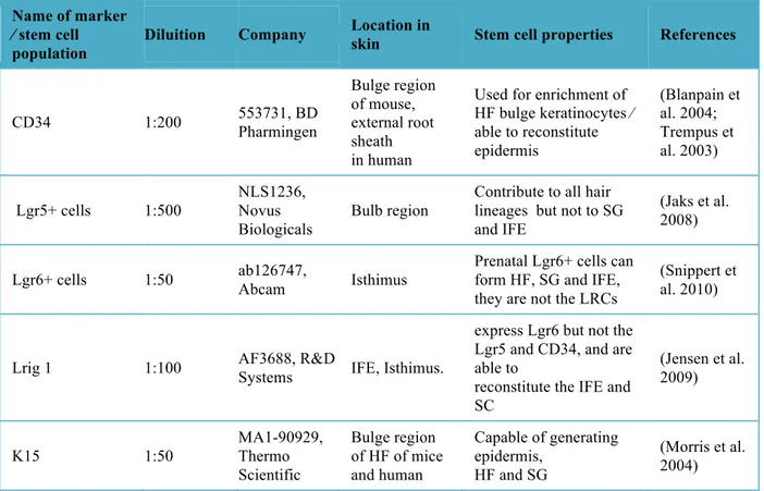
Antibodies for keratinocyte stem cell characterization in hair follicle
67
RESULTS
68
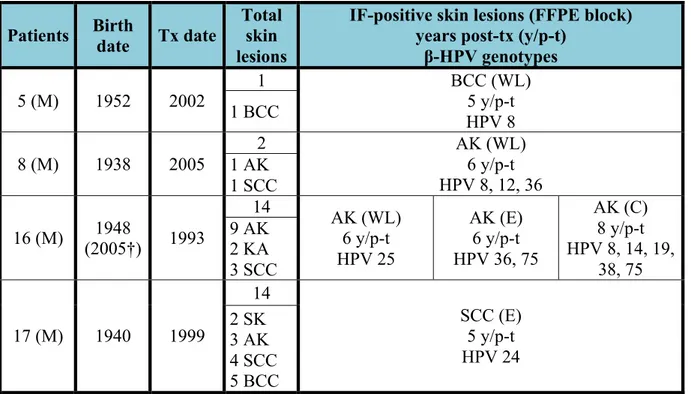
β-HPV detection in Kidney transplant recipients (KTRs)
68
Characteristics of the study cohort
68
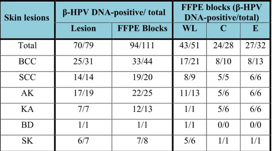
β-HPV DNA detection and genotyping in the skin lesions
70
Visualization of viral proteins in the skin lesions
71
HPV8 transgenic mice
80
Phenotype of mice expressing HPV8 early genes
80
Visualization of keratinocyte stem cell marker phenotypes in transgenic and wild
type mice
81
Conclusions
83
DISCUSSION
84
! ! ! ! ! ! ! ! ! ! ! ! ! ! ! ! !
!
!
A!Lirio,!nonno!e!maestro…!
! !1
SUMMARY
Human papillomaviruses (HPVs) are small non-enveloped viruses with a double-stranded DNA genome that infect cutaneous or mucosal squamous stratified epithelia of different body sites. They can establish aysintomatic infections that usually remain latent in healthy individuals but may also be responsible for the development of benign or neoplastic proliferative lesions, depending on the specific oncogenic properties of the different type of virus involved.
To date, more than 150 HPV types have been completely sequenced and classified into five genera based on phylogenetic analyses. These display a variety of different epithelial tropisms and life-cycle strategies, of which, the Alpha genus (α-HPVs) is the best characterized and comprises mucosa-tropic types associated with the development of genital cancer (e.g. HPV 16, 18).
HPVs belonging to the skin-tropic Beta genus (β-HPVs, e.g. HPV 5 and 8) appear to cause widespread unapparent infections without associated disease in the general population.
In patients with epidermodysplasia verruciformis (EV; an inherited primary immunodeficiency characterized by a high susceptibility to active β-HPV infection), β-HPVs replicate very efficiently and reveal their full transforming potential, inducing disseminated wart-like lesions and driving their progression to non-melanoma skin cancer (NMSC). Their involvement in skin carcinogenesis in both immunocompetent and non-EV immunocompromised patients is unclear, as both epidemiological and molecular evidences often conflict and are inconclusive. However, it is thought that, at least in conditions of impaired immune function (resulting from either primary or acquired immunodeficiency) β-HPVs might reactivate and contribute to the pathogenesis of NMSC, whose incidence is greatly increased in immunosuppressed individuals compared to the general population (Doorbar et al. 2012; Akgül et al. 2006; Aldabagh et al. 2013)
Solid organ transplantation is a treatment offered to an increasing number of patients with end-stage organ diseases; although life-saving, it is associated with an increased risk of second malignancies, presumably as result of the long-term post-transplant iatrogenic immunosuppression. Most of these (cancers such as Epstein-Barr virus-associated B-cell lymphomas, Human Herpesvirus 8-associated Kaposi’s sarcoma, and Merkel cell carcinoma of the skin, associated with Merkel cell polyomavirus) result from reactivated viruses whose oncogenic potential is suppressed by immunological reactions in healthy individuals. The most frequent type of malignancy arising in
2
organ transplant recipients (OTRs) is NMSC, and in particular the squamous cell carcinoma (SCC). The incidence of SCC in OTRs is estimated to be 60-250-fold as great as in the general population, and these post-transplant tumours are often found as multiples (commonly more then 10) and are highly aggressive.
Almost all the available studies are aimed to investigate the association between β-HPV infection and NMSC are mostly based on the detection of the presence of viral DNA in tumor tissues or positive antibody responses. Very few studies have addressed whether the viruses are actually localized in malignant cells or whether they are replicating and transcriptionally active in the context of the lesions. Confirmation of the presence of active virus cancerous lesions would greatly strengthen the evidence for an involvement of β-HPVs in the pathogenesis of NMSC.
It has been shown that detection of viral gene products and viral genome amplification by tissue staining procedures can be helpful for the visualization of active HPV infections in associated lesions, and that different expression patterns of viral lifecycle markers can be correlated to different stages of disease progression. This has been particularly noted in the well characterized α-HPV infections, and a recent study on skin tumors from EV patients has suggested that expression of the viral protein E4 and detection of viral DNA may be exploited as markers of viral activity during β-HPV associated skin cancer progression (Aldabagh et al., 2013; Pfister, 2003; Feltkamp et
al., 2008; Doorbar, 2007; Doorbar et al., 2012; Middleton et al., 2003; Borgogna et al., 2012).
To date a number of studies have attempted to clarify the mechanism that leads to HPV becoming persistent infection and to subsequent lesion formation. The general hypothesis has been that lesion formation begins with the infection of a basal stem cell (rather than a basal transiently amplifying cell) and that the longevity of the stem cells is a key factor in the formation of a persistent lesion, with these cells being a reservoir for the infection. High-risk α-HPV chronic infections, characterized by long persistence in the slow-cycling epithelial stem cells in the absence of apparent disease, is a condition sine qua non for the development of HPV-associated malignancy. Several studies have reported the ubiquitous presence of these β-HPV types on the body surface in immunosuppressed individuals and in the general population. Viral burden was greater in sun-exposed sites (e.g. forehead) than in sun-protected skin when its presence was monitored in plucked hairs and skin swabs. HPV appear to establish a reservoir of infection in the follicular stem cells of hair bulbs, which are thought to be an immune privileged site protecting them from immune clearance and therefore enabling the persistence of multiple β-HPV types.
3
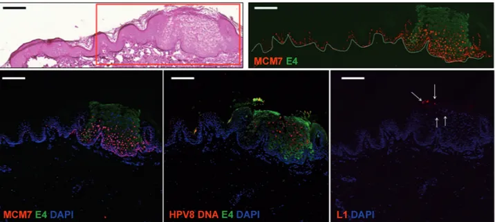
The purpose of this study was to extend observations already made in EV patients to other groups of subjects at high risk of developing NMSC. A series of skin lesions from kidney transplant recipients were systematically analyzed for the presence of βHPV infection, both at the DNA and at the protein level, looking for viral markers of transcriptional activity and replication. Using a combination of antibodies raised against the βHPV E4 and L1 proteins, it was possible to visualize the completion of the viral lifecycle in some precancerous lesions such as actinic keratosis or at the periphery of more advanced disease, in organ transplant recipient.
With the aim of investigating the influence of viral gene expression on keratinocyte differentiation, this study took advantage of a transgenic mice model expressing the complete early region of βHPV8. A specific protocol, for characterization of the keratinocyte stem cell populations has been established on epidermal whole mount samples of both wild type and transgenic mice. Keratinocyte stem cell markers CD34, K15, LGR6, LGR5 and Lrig1 were analysed in correlation with the proliferation marker Ki67.
These data presented in this study demostrates that β-HPVs are transcriptionally active and replicating at site of skin transformation and further reinforces the evidence that these viruses are involved in the process of skin carcinogenesis in patients who have undergone organ transplantation. Detection of viral markers in premalignant lesions and in the adjacent pathological epithelium of high-grade tumors supports the idea that β-HPVs may play a role in the early stages of skin transforming processes, probably enhancing the carcinogenic potential of UV damage rather than in progression and maintenance of neoplastic disease (Akgul et al., 2006; Feltkamp et
al., 2008; Pfister, 2003; Aldabagh et al., 2013). Furthermore the analysis of epidermal whole mount
samples has shown the expansion of the Lrig1+ area in the hair follicles of the transgenic mice compared with the wild type littermates. Hair follicle keratinocyte stem cell proliferation was demonstrated to be elevated within the Lrig1+ cells, compared to the other keratinocytes stem cells populations in βHPV8 transgenic mice. Intriguingly, these data suggest that the presence of the viral gene may influence normal keratinocyte differentiation, and identifies the specific keratinocyte stem cell population that is positive for the Lrig1 marker in the β-HPV8 in vivo model as a possible target. .
4
Publications:
Borgogna C*, Lanfredini S*, Peretti A, De Andrea M, Zavattaro E, Colombo E, Quaglia M, Boldorini R, Miglio U, Doorbar J, Bouwes Bavinck JN, Quint KD, de Koning MN, Landolfo S, Gariglio M.
“Improved detection reveals active β-papillomavirus infection in skin lesions from kidney transplant recipients.”
Mod Pathol. 2014; doi: 10.1038/modpathol.2013.240. [Epub ahead of print] *Equal contribution
Borgogna C, Landini MM, Lanfredini S, Doorbar J, Bouwes Bavinck JN, Quint KD, de Koning MN, Genders RE, Gariglio M.
“Characterization of skin lesions induced by skin-tropic α- and β-papillomaviruses in a patient with epidermodysplasia verruciformis.”
Br J Dermatol. 2014 Dec;171(6):1550-1554. doi: 10.1111/bjd.13156. Epub 2014 Nov 30
The data obtained from the study of the keratinocytes stem cell population in the β-HPV8 transgenic mice were achieved in the laboratory of Dr G. Patel (European Cancer Stem Cell Research Institute, Cardiff University, UK) as a visiting scientist, over a period of 7 months as part of the Ph.D program.
5
INTRODUCTION
Papillomaviruses: general characteristics
Papillomaviruses are small, unenveloped, double-stranded, circular DNA viruses exhibiting icosahedral symmetry. The papillomavirus genome ranges from 6953 bp [Chelonia mydas papillomavirus type 1 (CmPV1)] to 8607 bp [Canine papillomavirus type 1 (CPV1)] in length, and have been isolated and characterized from reptiles, birds, marsupials, and multiple other mammalian. This wide spectrum in the host species suggests an evolutionary history spanning more than 300 million years. They are species-specific and exhibit a strict tropism for the squamous stratified epithelia where they can induce cutaneous and mucosal hyperplastic lesions; some PVs have also been implicated in the development of epithelial malignancies, especially cancer of the uterine cervix and other tumors of the urogenital tract.
Human Papillomaviruses (HPV) can infect different body sites, such as the upper respiratory, oral and genital mucosae and the skin according to the specific tropism of the different viral species. They typically establish persistent infections which can either remain in an unapparent silent status or either cause clinical manifestations after a latency period of variable duration. The various types of epithelial disease that HPVs cause appear linked to their different strategies of transmission and propagation within the epithelium, and probably also to their different interactions with the immune system. HPVs can be responsible not only for benign hyperproliferative lesions (papillomas) in mucosal and cutaneous sites, but also for malignancies (especially squamous cell carcinoma, SCC). The specific association of HPV type 5 (HPV-5) with skin cancer of epidermodysplasia verruciformis (EV), a rare genetic disease, provided the first molecular evidence for the role of HPVs in human cancer.
HPVs have been implicated in cancers at several sites; in particular, the best documented HPV oncogenic activity concerns cervical cancer, on the basis of a meta-analysis of 1 million women with normal cervical cytology, around 291 million women worldwide are estimated to have human papillomavirus infection of the cervix at a given point, corresponding to an average prevalence of 10·4%, though prevalence is higher in women younger than 25 years (16·9%). Human papillomavirus types 16 and 18 account for roughly 70% of all cervical cancer.
6
Although the causal association of HPV infection with cervical carcinogenesis is epidemiologically and experimentally ascertained, its implication in the development of precancerous skin lesions that can evolve to high-grade tumors of epithelial origin – comprehensively grouped into the histological classification of non-melanoma skin cancer (NMSC) – has been much less clear to date; such uncertainties partly come from the fact that cutaneous HPVs have been shown to be ubiquitously present in the skin in the general healthy population. Nevertheless, several epidemiologic and experimental evidences are in favor of their potential oncogenic role in the insurgence of skin lesions (Knipe and Howley 2007; Doorbar 2005; Doorbar et al. 2012; Akgül, Cooke, and Storey 2006; Pfister 2003; Burk 2014; Van Doorslaer 2013)
Classification and tropism
Historically, PVs were classified together with the polyomaviruses as a single family, the Papovaviridae. This grouping arose because, although PV genomes and capsids are larger than those of polyomaviruses, the viruses share many features, including a double-stranded circular DNA genome, an icosahedral capsid composed of 72 pentamers, a nonenveloped virion, and the nucleus as the site of viral replication and virion assembly. Sequencing of PV genomes, however, indicated that, although PV share a common genetic organization, they differ from that of polyomaviruses and have no major sequence homology to polyomaviruses, and PV transcription is unidirectional, in contrast to the bidirectional transcription of polyomaviruses. Recognition of these differences, and others, have led to PV being designated as a separate family, the Papillomaviridae, by the International Committee on the Taxonomy of Viruses (Ethel-michele De Villiers et al. 2004; Bernard 2013).
After many years of research and genomic sequencing of thousands of PV isolates, PV classification was based on phylogenetic criteria following hierarchical taxonomic levels (family, genus, species, types, subtypes and variants). Two PV genomes are recognized as distinct types (genotypes) if they share less then 90% identical nucleotides in the most conserved genomic region L1 ORF, encoding the major capsid protein. Types were designated by a number, following the chronological order of their characterization. The PV taxonomy recently adopted is based on the phylogenetic relationship of the L1 ORF of animal and HPVs. Higher-order assemblages are considered as a genera (identified by Greek alphabetical prefixes) and lower-order assemblage as species (each species identified by the number of the best known type). Genera share less than 60% nucleotide identity in the L1 ORF, species within a genus share 60-70%, and types within a species
7
share 71-89%. By this approach, more than 200 PV types have been discovered to date, encompassing more than 150 HPV types and 103 animal PV types (Ethel-michele De Villiers et al. 2004; Mahy and Van Regenmortel 2008; Bernard 2013).
Historically, viral evolution has mainly been considered from a predator–prey perspective. Under this model, viral fitness (and thus its evolutionary success) is measured by the viral capacity to cause disease in its host. However, papillomaviruses cause benign, mostly unapparent, persistent infections in their hosts. In addition, papillomaviruses are highly host-restricted, and cause abortive infections in non-host species. In fact, the only exceptions to strict species specificity were described in mammalian hosts known to hybridize, thereby challenging the hosts' species definition. The observation that papillomaviruses cause benign infections unable to cross the hosts' species-barrier has led to the hypothesis of “host-linked evolution”.
The traditional (orthogenetic) definition of co-evolution states that parasites of closely related host species should be closely related themselves and cluster together in the parasite phylogenetic tree. Furthermore, dates associated with parasite divergence should coincide with the host-species divergence. Therefore, any incongruence between both trees should be considered as evidence that parasite and host did not co-evolve.
With an increase in the number of papillomavirus sequences (and their associated hosts), it became clear that papillomaviruses and their hosts did not follow an identical evolutionary path. Several violations of strict co-evolution can be observed in the phylogenetic tree in Figure 1. For example, human papillomaviruses can be found in five different genera dispersed throughout the phylogenetic tree. Also, strict co-evolution would place the non-human primate papillomaviruses basal to human papillomaviruses, not intermingled as is observed.
Evolutionary events such as cross-species infection, recombination and virus duplication (e.g. following ecological niche adaptation) have been suggested to explain the observed conflicts Because of the absence of cross-species infections, it is unlikely that horizontal gene transfer played any role in the evolution of the Papillomaviridae. In fact, a study specifically looking at the influence of horizontal gene transfer identified only a single potential cross-species transmission event. This event involved ancestors of a porcupine (EdPV1) and human (HPV41) papilloma- virus. These two viruses are the only members of a divergent genus (Nupapillomavirus).
A more recent version of the co-evolution theory was initially proposed in the early 1960s. This updated theory states that the evolution of parasites follows the evolution of host resources, not the evolution of the host species per-se. The shape of the papillomavirus phylogenetic tree could potentially be explained using this interpretation of co-evolution. Under this model, specific events
8
in the evolution of hosts (e.g. presence/absence of fur, evolution of sweat glands, etc.) created new ecological niches for papillomaviruses to adopt. Therefore, the data suggests a model in which a generalist ancestral papillomavirus diverged into four or five increasingly specialized viruses (reflected in the 4–5 major clades of the phylogenetic tree). Following these niche adaptation events, the virus evolved alongside its hosts. Throughout the co-evolutionary process, the availability of new niches would in turn drive viral radiation, followed by further co-speciation. In conclusion, the papillomavirus phylogenetic tree cannot be explained solely by co-evolution. However, initial niche sorting followed by virus–host linked speciation was a key determinant of the papillomavirus evolutionary history (Van Doorslaer 2013).
On the basis of L1 nucleotide sequences, HPVs are distributed in five of the 16 PV genera (Figure 1). The Alphapapillomavirus (α-HPV), comprises types differing by their genital, oral cutaneous tropism and by their pathogenicity (low-risk types associated with benign proliferations and high-risk types associated with invasive carcinomas). The genera Gammapapillomavirus (γ-HPV), Mupapillomavirus (µ-(γ-HPV), and Nupapillomavirus (ν-HPV) comprise types associated with cutaneous warts, whereas the types usually associated with EV belongs to the genus
Betapapillomavirus (β-HPV).
The Alpha genus is the most represented one and comprises both mucosal and cutaneous species. Infections by mucosal α-HPVs are more common than those by cutaneous α-HPVs and the majority of them are asymptomatic. Based on their oncogenic potential and association with the development of malignancies, mucosal α-HPVs are further subdivided into low-risk and high-risk groups.
The World Health Organization has defined 12 mucosal α-HPV types (HPV 16, 18, 31, 33, 35, 39, 45, 51, 52, 56, 58, 59) as being high-risk cancer-causing types; they are mainly responsible for the development of cervical cancer and can also be associated with cancers at other sites with much lower incidence (head and neck carcinomas such as oropharyngeal cancers, and cancers of the penis, anus, vagina and vulva). Nevertheless, high-risk α-HPVs do not cause cancer in the vast majority of the individuals they infect.
Low-risk α-HPVs cause benign mucosal lesions. Certain types (e.g. HPV 6 and 11) are associated with the development of respiratory papillomatosis (especially HPV 11), which is a laryngeal disease often occurring in children, and with benign external ano-genital warts (especially HPV 6); these types are sometimes occasionally found to be associated with cancers in these sites especially in individuals with immune defects, where such infections are more difficult to manage. Other
low-9
risk α-HPV types (e.g. HPV 13 and 32) are responsible for the development of oral papillomas as it occurs in the case of oral focal epithelial hyperplasia.
Cutaneous α-HPVs (e.g. HPV 2, 3, 7, 10, 27, 28, 57) are associated with the development of different types of benign skin warts arising on various sites of the hands, face, elbows and knees.
The Beta genus comprises to date 43 types, which exhibit cutaneous tropism (e.g. HPV 5, 8, 9, 14, 17, 20, 21, 23, 36, 38, 47, 49). β-HPVs are evolutionarily distinct from the Alpha genus and establish widespread unapparent asymptomatic infections that remain latent without any clinical manifestation in the general healthy population. In subjects with impaired immune function, it seems that these viruses can spread unchecked and they have been implicated in the development of NMSC. In particular, in individuals suffering from epidermodysplasia verruciformis (EV), a primary immunodeficiency associated with abnormal susceptibility to β-HPV infection, they are responsible for the development of disseminated wart-like lesions that often undergo malignant progression; there is also increasing body of evidence for their involvement in the onset of precancerous skin lesions with potential to evolve to NMSC in other immunosuppressed populations (e.g. OTRs, organ transplant recipients). Their involvement in the pathogenesis of NMSC in immunocompetent individuals is still unclear.
Gamma (e.g. HPV 4), Mu (e.g. HPV 1) and Nu (HPV 41) genera comprise a smaller number of types, exhibiting cutaneous tropism; they are associated with the development of benign palmar and plantar skin warts (Ethel-michele De Villiers et al. 2004; Akgül, Cooke, and Storey 2006; Pfister 2003; Doorbar 2007; Doorbar et al. 2012; Cubie 2013).
10
Figure 1. Papillomavirus phylogenetic tree. The DNA sequence coding for E1, E2, L1 and L2 for all 241 papillomaviruses currently on PaVE were downloaded and aligned. A partitioned gene alignment was used as the base for a maximum likelihood reconstruction of the phylogenetic treeGenera marked with an asterisk have been proposed to the ICTV, and are awaiting official recognition (http://talk. ictvonline.org/files/proposals/taxonomy_proposals_vertebrate1/m/vert01/4244.aspx). The tree is color-coded according to presence/absence of the “adaptive proteins”. Red clades lack an E6 ORF. The viruses highlighted in green do not code for an E7 protein. The purple clades code for a hydrophobic E5 protein. The Xipapillomaviruses lack an E6 (red), but contain an E5 (purple)(Van Doorslaer 2013).
11
HPV structure, genomic organization and gene products
The genomes of many of the human and animal papillomaviruses have been sequenced in their entirety, and the genomic organization of each of the PVs is similar. HPVs are characterized by small (52-55 nm diameter), nonenveloped, icosahedral capsid composed of 72 pentameric capsomeres. Their genome is in a double-stranded, covalently closed, circular DNA molecule of 7500-8000 bp. The viral DNA is associated with cellular histones to form a chromatin-like structure. Genomic organization is highly conserved among all HPV family members, with 8-9 open reading frames (ORFs) on a single transcriptionally active DNA strand encoding a larger number of gene products as a result of mRNA splicing (Figure 2). The viral genome is divided into three regions:
- The early region coding for functional proteins (E1, E2, E4, E5, E6 and E7) expressed in all the phases of the viral life cycle, which are responsible for the persistence of the viral genome in a cell, its replication and the stimulation of cell proliferation necessary to support viral replication itself;
- The late region, coding for the structural coat proteins L1 and L2 that are expressed in the final phases of the productive viral life cycle;
- The long control region (LCR), a non-coding fragment regulating the expression and replication of the viral genome (Mahy and Van Regenmortel 2008).
12
Figure 2. The genome organization of the high-risk α-type HPV16 and β-type HPV8. The HPV genome comprises a long control region (LCR) and eight genes that are necessary for different stages of the virus life cycle. These genes encode a larger number of gene products as a result of mRNA splicing. The LCR contains binding sites for cellular transcription factors (e.g., SP1, AP1, Oct1), as well as for the viral E1 and E2 proteins that control viral replication and gene expression. The best characterized HPV types have two promoter elements (P97 and P670) known as early promoter (PE) and late promoter (PL) that regulate the expression of differentially-spliced mRNAs during epithelial differentiation. Specific conserved motifs (M33 and M29) among the beta-HPVs are shown in the LCR of HPV8 (Lazarczyk et al. 2009).
13
The LCR is a significant non-coding fragment of viral DNA located between the L1 and E6 ORFs, accounting for about 10% of the entire genome. It contains an origin of replication and cis-responsive elements for regulatory transcription and replication factors of viral genetic material; in detail, there are binding sites for cellular transcription factors (e.g. SP1, AP1, Oct1), as well as for the viral E1 and E2 proteins that control viral replication and gene expression (Figure 2). All the LCR regions contain enhancers that provide the virus with specific tropism to the stratified squamous epithelial cells. Other regulatory enhancer elements are positioned within genes.
The LCR harbors one of the two well-characterized promoter elements, namely the early promoter upstream of the E6 ORF, while in many HPV types the late promoter is located within the E7 ORF (Figure 2). These promoters regulate the expression of differentially spliced mostly polycistronic mRNAs, being trans-activated in a timely and coordinated differentiation-dependent fashion in specific epithelial layers where the different phases of the viral life cycle take place. The early promoter mainly drives E6 and E7 expression, while the late promoter regulates the expression of all the other viral genes.
The mRNA species that encode E1, E2 and E4 terminate at the early polyadenylation site and for many α-HPVs include E5 as their second or third ORF; transcripts encoding L1 and L2 terminate at the late polyadenylation site. The E4 ORF is contained within the E2 ORF, with the primary E4 gene product (E1^E4) being translated from a spliced mRNA including the E1 initiation codon and adjacent sequences. Upon mRNA splicing involving a small portion within the E1 ORF and most of the E2 ORF, some high-risk α-HPV types produce the additional gene product E8^E2C. In addition, for many HPV types several other spliced transcripts have been reported, encoding variants of the early proteins such as N-terminal truncated forms of E2 and E6 (Ciesielska et al. 2012; Doorbar et al. 2012; Doorbar 2007; Tommasino 2014; Doorbar 2013).
The papillomavirus major capsid protein, L1, is a ~55 kD protein with the ability to spontaneously self-assemble into virus-like particles (VLPs). These VLPs present an exterior surface essentially indistinguishable from the native 60 nm non-enveloped papillomavirus virion. Purified recombinant L1 proteins can achieve this complex assembly reaction in the absence of any chaperones (Schiller and Lowy 2012). Assembled VLPs are potent immunogens, likely due to innate B-cell recognition of the regular icosahedrally displayed spacing of surface epitopes (Bachmann et al. 1993). These discoveries laid the foundation for the development of the current VLP-based vaccines that offer highly effective protection against infection with the cancer-causing human papillomavirus (HPV) types 16 and 18. The assembled VLP immunogens used in current
14
HPV vaccines represent the state of L1 in only one phase of the viral life cycle–namely, the rigid mature form of the virion during its transmission from one cell to another. In contrast to the mature virion state, during the process of virion assembly L1 interactions must be flexible enough to allow selective uptake of the viral genomic DNA into the virion lumen. Transmission of some papillomavirus species is thought to occur via deposition on environmental surfaces, such that the initially fragile immature virion must gain the ability remain infectious in a desiccated state for days or longer to achieve transmission (Roden, Lowy, and Schiller 1997). This high degree of stability is achieved through a maturation process in which the flexible immature virion gradually achieves a more rigid state that is stabilized by disulfide crosslinks between neighboring L1 molecules. Since L1 forms the entire exterior surface of the stabilized mature virion, it obviously must mediate initial attachment to host tissues or cells. After attachment to cells, L1 must again become pliable enough to ultimately allow release of the viral genome into a new target cell (Buck, Day, and Trus 2013).
Similar to L1 the minor capsid protein L2 is a 55KDa protein, and plays major roles in both papillomavirus assembly and the infectious process. While L1 forms the majority of the capsid and can self-assemble into empty virus-like particles (VLPs), L2 is a minor capsid component and lacks the capacity to form VLPs. However, L2 co-assembles with L1 into VLPs, enhancing their assembly. L2 also facilitates encapsidation of the ~8kbp circular and nucleosome-bound viral genome during assembly of the non-enveloped T=7 d virions in the nucleus of terminally differentiated epithelial cells, although, like L1, L2 is not detectably expressed in infected basal cells. With respect to infection, L2 is not required for particles to bind and enter in the cells.
Much of the studies and analysis about L2 are based on HPV16 L2’s primary sequence and these mapped domains because unfortunately very little information exists on its higher order structure (Wang and Roden 2013).
The E1 protein, an ATP-dependent DNA helicase, is the only enzyme encoded by the PV genome. It is tightly regulated in vivo, in particular by post-translational modifications that restrict its accumulation in the nucleus. E1 is essential for replication and amplification of the viral episome in the nucleus of infected cells. It does so by interacting with cellular DNA replication factors and assembling with E2 to the origin of replication to trigger viral DNA replication (Bergvall, Melendy, and Archambault 2013).
The E2 proteins function primarily by recruiting cellular factors to the viral genomes, which activate or repress transcriptional processes. The E2 proteins bind specifically to sequence motifs in the viral genome and can activate or repress transcription, depending on the context of these binding
15
sites and nature of the associated cellular factors. In particular, it negatively modulates the expression of E6 and E7 by down-regulating the activity of the early promoter. Furthermore, E2 recruits E1 to a specific E1-binding motif in the origin of replication and is implicated in the maintenance of the viral genome in its episomal (extrachromosomal) form. It seems to be also involved in the regulation of accurate genome partitioning during basal cell division by associating to the pericentromeric regions of mitotic chromosomes to drive the tethering of viral episomes to the cellular chromatin during mitosis (McBride 2013).
The E4 open reading frame (ORF) lies within the larger E2 ORF, and varies considerably in size between papillomavirus types. In human papillomaviruses, the primary E4 gene product is expressed from a spliced mRNA (the E1 E4 message). It is the most abundant viral gene product and is expressed before L2 and L1. The structure and function being modified, first by kinases as the infected cell progresses through the S and G2 cell cycle phases, but also by proteases as the cell exits the cell cycle and undergoes true terminal differentiation. E4 is thought to be involved, in association with E2, in viral genome amplification and suppression of cellular proliferation in the late phases of the productive life cycle; The accumulation to very high levels in the upper epithelial layers of the host seems also to be able to disrupt the cellular keratin network facilitating the extracellular release of new viral particles (Doorbar 2013).
In the HPVs genome are presents three proteins with transforming properties: the E5, E6 and E7 oncoproteins. Through combined and cooperative action, they abrogate the activity of tumor suppressor factors, promoting evasion of all cell cycle checkpoints and inducing a deregulated progression of the cell cycle; they mostly function by associating with and functionally reprogramming key components of cellular signal transduction networks. Their activity allows the maintenance of S-phase competence and a forced replicative status in the host cell, which is essential to the viral life cycle; the result is a stimulation of cell growth, survival and proliferation and a delay of terminal cell differentiation. Because of such properties, HPV oncoproteins can trigger the initiation and progression of epithelial cancers; in particular, these transforming activities are stronger in the case of high-risk α-HPVs, underlying their involvement in the development of malignancies.
E5 is the smallest oncoprotein which functionally resembles a viroporin (Venuti et al. 2011). It is an 84-aminoacids viral replication protein involved in the early stages of HPV infection. The hydrophobic E5 protein localizes to cell membranes (including Golgi, endoplasmic reticulum, nuclear membrane) where it may dimerize and trigger cell fusion (Hu et al. 2009). This phenotype
16
is associated with mitogenic signal intensification via E5-inhibited degradation of epidermal growth factor receptors (EGFR). However, E5 from dermatotropic α-viruses such as HPV6 or EV- type β-viruses such as HPV5 does not cause cell fusion, yet retains even stronger affinity for growth factor receptors (EGFR, HER2 or platelet-derived growth factor b-receptors) than does HPV16 E5. E5 also exerts immunosuppressive effects mediated via inhibition of host MHC class II complexes and/or reduced cytotoxic T cell recognition of HPV (Campo et al. 2010). These actions could be pertinent to the tumorigenicity of HPV16 in the immune-activated niche of the lymphoid tonsil, plausibly reducing immune surveillance to the lower levels of the cervix or post-transplant skin. It is also possible that the stimulatory effects of E5 on keratinocyte EGFR signaling contribute to the epidermotropism of β-HPVs, consistent with the reactivation of E5 signalling seen in some skin SCCs containing multiple HPVs (Orth 2006). Still another tumorigenic effect of E5 is to reduce expression of the epithelial adhesion molecule E-cadherin, increasing the invasiveness of infected cells while driving the epithelial–mesenchymal transition (EMT) (Boulenouar et al. 2010).
E6 and E7 are small proteins, approximately 18 and 13 kDa in size, respectively, and are localized in the nucleus. The E6 proteins are also found in the cytoplasm and some studies have suggested that E7 also has a cytoplasmic. Are multimeric proteins with potential to associate with multiple cellular partners. Such functional differences contribute to the respective transforming abilities of various HPV species and types.
About E7 protein it has been shown to bind zinc and is phosphorylated by casein kinase II (CK II). In the best-characterized HPV species, E7 binds and targets for ubiquitin-dependent proteasomal degradation the hypophosphorylated form of members of the retinoblastoma tumor-suppressor family (pRb, p107, p130). In uninfected epithelium, cell cycle entry and cell division in the basal and parabasal cell layers is controlled by growth factors that stimulate the activity of G1 cyclins including cyclinD/Cdk, which phosphorylates Rb family members and displace them from transcriptional activators of the E2F family allowing the trans-activation of genes necessary for S-phase progression. The continual stimulation of these cells physiologically allows renewal of the epithelium as surface cells exfoliate. As part the regulated stimulation of this cell cycle entry, p16ink4a is up-regulated and forms a compensatory negative feedback loop that suppresses cyclinD/Cdk activity, so preventing the over-expression of itself and other E2F-activated genes (e.g. MCM, PCNA, Ki67). E7 binds to Rb proteins and displaces them and transcriptional repressors of the E2F family from target promoters required for S-phase gene expression without the need for Rb phosphorylation and growth factor stimulation; E2F1, 2 and 3 can then occupy these vacant sites
17
and stimulate expression of the host genes necessary for DNA replication and cell cycle progression.
In certain HPV species and types, E7 is necessary for the stable maintenance of HPV episomes in epithelial cells. It can stimulate cell cycle progression and subsequent hyperproliferation, induce genomic instability, exert antiapoptotic activities, reprogram host cell metabolism and contribute to evasion of the local antiviral host immune responses to different extents by a number of other mechanisms depending on the specific oncogenic potential.
The HPV E6 proteins are approximately 150 amino acids in size and contain four Cys-X-X-Cys motifs, which form two unusually big zinc finger domains of 29-30 AA, respectively, which are important for protein stability and activity. The E6 proteins from the low risk and high risk HPV appear to have similar transcriptional activation properties.
E6 interferes with DNA damage repair, growth arrest and apoptosis. The function of E6 complements that of E7. The efficient binding of Rb proteins by E7 can lead to inhibited cell growth and apoptosis through a p53-dependent pathway; as a result, E6 proteins of many HPV types have evolved to target the tumor suppressor p53 for ubiquitin-dependent proteasomal degradation or other forms of inactivation, resulting in the abrogation of its activity that plays an essential role in protecting genomic integrity. In uninfected epithelium, p53 expression increases in response to cellular stress such as DNA damage and induces the expression of p21, which allows cell cycle arrest at G1-S checkpoint until errors in DNA replication can be repaired; if DNA damage cannot be repaired, p21 activates the expression of proapoptotic genes to prevent the replication of damaged DNA. In addition, p53 can also trigger apoptosis upon DNA damage through pathways unrelated to its transcriptional activity The consequence of E6 action is the abrogation of all these p53-dependent tumor suppressor functions in the infected cells.
In certain HPV species and types, E6 can inhibit apoptosis by interfering with p53-dependent or p53-independent apoptotic pathways through other mechanisms to different extents depending on the specific oncogenic potential.
In any case, E6 inactivates key mediators of stress-induced programmed cell death more or less strongly depending on the transforming properties of specific HPV species and types. As a consequence, cell cycle arrest and apoptosis in response to E7-mediated cell cycle entry are prevented and DNA replication followed by cellular proliferation is promoted in cells with damaged DNA because of the disruption of the DNA repair process in the host cells; this events, which are more evident in oncogenic HPVs, increase the frequency of spontaneous mutations in the cellular genome during replication, favoring the occurrence of genomic instability underlying transforming processes.
18
Another important way how E6 proteins of some oncogenic HPVs contribute to transformation is the activation of the human telomerase reverse transcriptase promoter, which controls the transcription of the catalytic telomerase subunit.
In high risk HPVs, E6 can also exert transforming activities by inducing host cell immortalization, loss of host cell polarity and anchorage-independent growth and altering host cell differentiation and metabolism; finally, E6 can facilitate viral escape of the antiviral immune surveillance mechanisms (Knipe and Howley 2007; Moody and Laimins 2010; Doorbar et al. 2012; Doorbar 2007; Akgül, Cooke, and Storey 2006; Ciesielska et al. 2012; McLaughlin-Drubin, Meyers, and Munger 2012).
HPV life cycle
Irrespective of their evolutionary origin, all papillomaviruses must complete their life cycle in the epithelial tissue that they infect (summarized in Figure 3), and produce infectious particles that are eventually secreted from the epithelial surface. Human papillomavirus infects cells in the basal layer of the epithelium, probably via microabrasions in the epithelial surface. It capitalises on the lateral extension of basal cells that accompanies wound healing to gain entry to the cell.
Infectious internalization takes several hours, after which viral DNA is released from the capsid and transported into the nucleus as free genetic material or extrachromosomal episomes. The synthesis of new virions occurs only after the infected cell has undergone mitosis and one of the infected daughter cells has differentiated.
HPV replication requires the timely and coordinated expression of the different viral gene products as the infected cell moves towards the epithelial surface; this highly regulated pattern of gene expression allows the different stages of the life cycle to be completed appropriately. For this reason, HPV life cycle takes 2-3 weeks – the time necessary for an epithelial cell to migrate from the basal to most superficial layers, mature, undergo senescence and die. Whether a productive life cycle is or is not completed depends on the nature of the epithelial site where infection occurs, as well as on the presence of external factors such as hormones and cytokines.
Due to the clear association of high-risk α-HPV types with human carcinogenesis, most of the biological studies so far have been focused on these types (Doorbar et al. 2012; Doorbar 2007; Tommasino 2014; Crosbie et al. 2013; Moody and Laimins 2010).
19
Figure 3. Productive HPV life cycle. HPVs replicate only in fully differentiating squamous epithelia. The
life cycle involves both temporal and spatial separation of viral protein expression. The virus first infects a cell in the basal layer of the epithelium where access is naturally facilitated (e.g. microtrauma, epithelial transitional zones, hair follicles). In the lower proliferative compartments of the epithelium, there is a phase of viral episome maintenance al low copy number, in which viral and cellular DNA replicate together. As long as the cell is dividing, HPVs control the expression of their viral proteins very tightly; the E6 and E7 oncogenes are thus expressed at very low levels, along with the genes coding for E1 and E2 replication factors. When the host cells stop dividing and begin to differentiate into mature epithelial cells, this provides a signal to the virus to activate all of its genes to amplify the viral genome copy number to the thousands. In the top layers of the epithelium, all of the viral genes, including those encoding the L1 and L2 proteins, are expressed, and many thousands of viral genomes are encapsidated; finally, the newly assembled infectious viral particles exit the cells in the context of epithelial surface desquamation. The time taken from infection to the generation of new virions is at least 2-3 weeks (Stanley, 2012).
Experimental models suggest that infection requires access of virus particles to the basal lamina and the interaction with heparin-sulphate proteoglycans and possibly also laminin. Structural changes in the virion capsid, which includes furin cleavage of L2, facilitate transfer to a secondary receptor on the basal keratinocyte, which is necessary for virus internalization and subsequent transfer of the viral genome to the nucleus; although the α-6 integrin and growth factor receptors have been implicated in this process, the precise nature of the entry receptor remains somewhat controversial.
Once internalized, virions undergo endosomal transport, uncoating and cellular sorting. The L2 protein-DNA complex ensures the correct nuclear entry of the viral genomes, while the L1 protein is retained in the endosome and ultimately subjected to lysosomal degradation.
20
In many cases, infection is thought to require epithelial wounding or micro-wounding to allow access of the virus to the basal lamina, and a role for the wound healing response in simulating the lateral expansion of the infected cells has been suggested. Indeed, active cell division, as would occur during wound healing, is thought to be necessary for entry of the virus genome into the nucleus, and it has been proposed that lesion formation requires the initial infection of a mitotically active cell. Given the diversity of HPV types and HPV-associated diseases, is not always possible make such broad generalizations regarding the route of infection, as multiple entry pathways have been invoked depending on the virus type under study. Some HPV species are thought to infect sites where access to the basal layer is naturally facilitated, such as the base of the hair follicle for cutaneous HPVs, or sites where columnar and stratified squamous epithelial cells meet each other, such as the cervical or anal squamo-columnar junctions for high-risk α-HPVs. For some time now, the general hypothesis has been that lesion formation begins with the infection of a basal stem cell (rather than a basal transiently amplifying cell) and that the longevity of the stem cells is a key factor in the formation of a persistent lesion, with these cells being a reservoir for the infection (Doorbar 2007; Doorbar et al. 2012).
Infection of the basal stem cell is followed by initial phase of genome amplification and then by maintenance of the viral episome al low copy number (100-200 copies per cell). In benign oral papillomas in animals, the basal copy number has been quantified using laser capture methods as 50 to 100 copies per cell, but it is likely that there will be variation from lesion to lesion and between different sites.
Due to the lacking of the enzyme machinery necessary for viral DNA replication, the viral genome amplification depends on the presence of the cellular DNA replication machinery. Thus the viral DNA replication follows cellular DNA replication as the cells progress through S-phase.
The viral replication proteins E1 and E2 are thought to be essential for this initial amplification phase, which are expressed from the late promoter and ensure viral DNA replication and transcription. It has been proposed that the use of a viral DNA helicase (i.e., E1), which is distinct from the cellular replication helicases (MCM proteins), allows viral DNA replication to be disconnected from cellular DNA replication during genome establishment and amplification; however, at this stage the viral genomes are replicated in synchrony with cellular DNA replication. E2 also regulates accurate genome partitioning during basal cell division.
The precise role of the E6 and E7 proteins in infected basal cells is uncertain, particularly for the low-risk α-HPVs and cutaneous HPVs. In these HPV types, the role of the wound healing response in driving the initial proliferation of the infected cell(s) may well be critical. In the case of the
high-21
risk α-HPV types there is a clear role of the viral E6 and E7 proteins in driving cellular proliferation in the basal and parabasal cell layers, where an expansion in lesion size is facilitated.
Viral proteins are not readily detectable prior to the onset of genome amplification during normal productive infection; the low level expression of viral proteins in the basal layer is thought to reflect, at least in part, the need of the virus to avoid detection by the host’s immune system (Doorbar 2007; Doorbar et al. 2012).
The maintenance of viral genome in the proliferating basal cells, and in cells in the lower epithelium, is a common feature of all papillomaviruses.
The late productive stage of the life cycle, starting with vegetative viral genome amplification (around 1000 copies per cell), is triggered as the infected cells differentiates and are pushed towards the epithelial surface by the division of the cell beneath. The specific events that induce the onset of genome amplification are not well understood, but depend in part on changes in the cellular environment as the infected cells move to the upper epithelial layers; of key importance is the up-regulation of the late promoter, whose activation in cells expressing E6 and E7 leads to an increase in the levels of viral proteins necessary for replication, including E1, E2, E4 and E5. After an initial increase in viral copy number, the infected ‘differentiating’ cells move from an S-like to a G2-like phase, with viral genome amplification occurring primarily in G2 after cellular DNA replication has been completed; the virus at this point fully recruits the cellular DNA replication machinery for its own replication.
A key function of the E6 and E7 proteins in most HPVs is not to promote basal cell proliferation, but rather to stimulate cell cycle re-entry in the mid-upper epithelial layers in order to allow genome amplification. These differentiating cells normally would not be replication–competent, since they do not naturally express the replicative machinery that the virus depends on; the expression of the E6 and E7 proteins in these layers allows the infected cells to aberrantly re-enter S-phase and for viral genome copy-number to rise. In high-risk α-HPVs, the stimulation of cell cycle entry occurs in the basal layer and above; for unclear reasons, high-risk oncoproteins can also promote the proliferation of the infected basal cells, which is important for the induction of neoplasia.
There is also a need for the viral replication proteins E1 and E2, which increase in abundance following the up-regulation of the late promoter.
In addition to E1 and E2, it is thought that the E4 and E5 proteins contribute indirectly to genome amplification success by modifying the cellular environment, with E5 also being involved in koilocyte formation.
22
Abundant high-risk E4 can inhibit cell cycle progression during G2-phase, and this leads to cell cycle arrest at the G2-M boundary by sequestering these proteins in the cytoplasm. It has been suggested that continued expression of E7 in a cell containing abundant E4 might lead to the maintenance of an S-phase environment, which allows accumulation of the viral genomes. The increase in E2 that is thought to accompany E4 must eventually down-regulate the viral early promoter that induces E7 production and entry into S-phase; the steady rise in E2 may act both to initiate genome amplification, and in a timely manner, to turn it off once amplification is complete. E5 is also thought to make an important contribution to genome amplification success through its ability to stabilize EGFR and to enhance EGF signaling and MAPK activity, thus allowing the maintenance of a replication-competent environment (Doorbar 2007; Doorbar et al. 2012).
The late productive stage of the life cycle takes place in the infected cells that undergo terminal differentiation in the most superficial epithelial layers. The completion of the life cycle ultimately involves the expression of the minor coat protein L2, the exit of the cell from the cell cycle and the expression of the major coat protein L1 to allow genome packaging. L1 and L2 are expresses only in cells that have already undergone genome amplification and which contain elevate concentration of E4 protein in their cytoplasm. This seems to require a change in splice site usage rather than promoter activation, leading to transcripts initiated at the late promoter that terminate at the late polyadenylation site rather than the early site, an event that is aided by high levels of E2 expression and by the presence of negative regulatory elements that destabilize late transcripts and allow the preferential synthesis of early transcripts in proliferating cells; furthermore, the pattern of codon usage within the late genes is distinct from that of the host cells, and this may further contribute to the suppression of L1 expression in the lower epithelial layers. It is thought that the accumulation of virion structural proteins to high level is retarded until the cells reach the superficial layers in order to limit detection by the host immune response.
Genome encapsidation involves the recruitment of L2 to regions of replication via E2, prior to the expression of L1 and the assembly of the icosohedral capsid in the nucleus. Virus maturation occurs in the most superficial, dying keratinocytes, leading to the production of extremely stable infectious particles. Although not precisely defined, the abundant E4 protein is thought to contribute to virion release and infectivity in the upper epithelial layers, as it organizes into amyloid-like fibres that disrupt keratin structure and compromise the normal assembly of the cornified envelope; new infectious particles are eventually shed from the epithelial surface as the infected cells exfoliate through desquamation (Doorbar et al. 2012; Doorbar 2007).
23
α-HPVs and cervical carcinogenesis
Cervical cancer is the second most common malignancy among women worldwide, with approximately 500,000 newly diagnosed cases each year and about 275,000 deaths annually. Despite its worldwide distribution, the frequency of cervical cancer varies considerably, being about ten times more common in some countries than in others. About 80% of cervical cancer occurs in developing countries, where it is frequently the most common cancer of women, accounting for as many as one quarter of female cancers. It occurs less frequently in developed countries.
The frequency of high-risk α-HPV-associated cervical cancers is of around 30-40 people per 100000 and over 99% of cervical lesions harbor viral sequences, although the proportion associated with specific high-risk α-HPV types is different in different countries and shows demographic, ethnic and socio-economic variation. Genital α-HPV infection has a peak of incidence in young individuals between 15-25 years of age following the onset of sexual activity and a progressive fall with age; over 80% of sexually active women become infected at some stage in their life with one or several high-risk and low-risk α-HPV types.
HPV 16 and 18 cause approximately 50% and 20% of the cases of cervical SCC - arising in the stratified squamous cells of the ectocervix - respectively, and both types are equally associated with around 35% of the cases of cervical adenocarcinoma – arising in the columnar glandular cells of the endocervix and more aggressive.
HPV associated pre-cancers present as intraepithelial neoplasia and are named after the site: cervical intraepithelial neoplasia (CIN), VIN and VAIN (vulval and vaginal intraepithelial neoplasia respectively), PIN (penile intraepithelial neoplasia) and AIN (anal intraepithelial neoplasia). Cervical intraepithelial neoplasia and are histologically classified for diagnostic purposes according to the grade of dysplasia: CIN1 (mild dysplasia), CIN2 (moderate dysplasia), CIN3 (in situ carcinoma); CIN1 lesions are low-grade lesions (LSIL, low-grade squamous intraepithelial lesions), while CIN2 and CIN3 lesions are high-grade lesions (HSIL, high-grade squamous intraepithelial lesions). The accurate identification of lesion grade has prognostic significance, as it has been estimated that around 20% of CIN1 will progress to CIN2, and that around 30% of CIN2 will progress to CIN3 if left untreated; CIN3 are generally considered to be the direct precursors of cervical cancer, and it has been suggested that around 40% of CIN3 lesions will progress to cervical cancer in the absence of intervention. In general, more regions with different histological grade can be found in the context of a cervical lesion; different high-risk α-HPV types are usually associated with discrete areas of disease except at junction regions (where lesions abut or are in close proximity) where more than one type may be detected.
24
Most cervical cancers develop in the transformation zone, corresponding to the squamo-columnar junction; the transitional site where the squamo-columnar glandular cells of the endocervical canal meet the stratified squamous cells of the ectocervix. The particular susceptibility of the transformation zone to cancer onset and progression may also be linked to the increased accessibility and proliferation of the basal cell layers at this metaplastic epithelial site, particularly around the time of puberty and the onset of sexual activity. In this case, we can hypothesize that the primary preferential target cells for high-risk α-HPV infection may be cells close to the squamo-columnar junction, such as the epithelial reserve cells, which lie immediately underneath the columnar epithelium of the endocervix, and eventually form the stratified epithelial layers of the transformation zone as the cervix matures. Early acquisition of high-risk α-HPV infections, also facilitated by the low levels in antigen-presenting cells in this site, can disturb the metaplastic changes occurring at this time in the transformation zone and increase the risk of cervical cancer in the future (Yang et al. 2004; Cubie 2013; Doorbar 2007; Doorbar et al. 2012; Tommasino 2014; Moody and Laimins 2010).
Viral life cycle deregulation and cancer progression
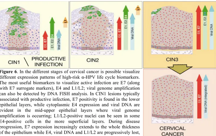
The ordered expression of viral gene products that leads to virus particle production is disrupted in HPV-associated neoplasia. In cervical disease, where most research has been done, it is generally thought that the levels of E6 and E7 expression increase from CIN1 to CIN3; this deregulation of early gene expression is accompanied by a concomitant failure to trigger late events until the cells are close to the epithelial surface, and in CIN3 genome amplification is often confined to small pockets at the most superficial layers. Changes in the timing of viral gene expression are accompanied by changes in the levels of the viral proteins, and this is likely to have a key influence in determining lesion grade.
In this scheme, CIN1 lesions typically retain the ability to complete the life cycle and release viral particles, showing a lower level of cell proliferation in the basal and parabasal layers. High-grade lesions such as CIN2 and CIN3 are characterized by the persistence of cycling cells into upper layers, and in CIN3 such cells are detectable at the epithelial surface; such lesions represent abortive infections in which viral gene expression is not properly controlled, and late events in the virus life cycle are not properly supported.
The elevation of E6 and E7 expression in high-risk α-HPV infection that leads to the CIN2+ phenotype predisposes the cell to the accumulation of genetic changes, which increasingly contribute to cancer progression. According to this hypothesis, the relatively low levels of E6 and E7 present in CIN1 do not compromise the functions of their cellular targets sufficiently to facilitate
25
cancer progression. The viral deregulation seen in CIN2/3+ is also thought to facilitate integration of the viral episome into the host cell chromosome, which can further deregulate the expression of E6 and E7.
It is not clear exactly how gene expression from the viral episome can become deregulated in early CIN. The viral gene expression may be deregulated by changes in cell signaling as can be brought about by hormonal changes, or epigenetic modifications such as viral DNA methylation, which may depend on the nature of the infected epithelial cells. The HPV16 LCR contains hormone response elements that can be stimulated by estrogen, and there is ample evidence of cooperation between estrogen and HPV in the development of cervical cancer. In CIN, it has been reported that the LCR is differentially methylated according to disease severity, which suggests that epigenetic changes may also regulate gene expression (Doorbar 2007; Doorbar et al. 2012).
In early CIN most HPV genomes persist in an episomal state, whereas in many HSIL they are found integrated into the host cell genome (Figure 4). Several studies have shown that the frequency of viral DNA integration increases with the severity of the cervical lesion, indicating that this event is implicated in the progression of the disease.
The majority of cervical cancers contain one or many HPV genome copies, integrated more or less randomly into the host cell genome in a monoclonal fashion, with the viral integration site frequently lying within the regulatory E1 or E2 genes; this event often leads to the disruption of E2 and adjacent ORFs (E4, E5, L2), and as a consequence to the loss of expression of full-length E1, E2, E4, E5 and late genes (Figure 4). The key event is the loss of E2, which is a virally-encoded transcription factor that normally regulates E6/E7 abundance by binding to and silencing the early promoter within the LCR as part of a viral regulatory mechanism.
It is thought that integration occurs in HSIL, and that once this occurs, the already deregulated expression of E6 and E7 can increase still further or else be maintained at a constitutive level. Moreover E6 and E7 transcripts expressed from integrated copies show increased stability, and this was attributed to the longer half-life of transcripts derived from integrated HPV DNA, mediated by 3’-cellular sequences of the fusion transcripts. Furthermore integration imparts a selective growth advantage over cells that harbor only episomal copies of viral DNA. Usually only a segment of the viral genome is integrated containing the LCR and truncated parts of the early and late region (Figure 4); multiple tandem repeats of such viral DNA fragments - from a single to several hundred copies – are often found integrated at random locations in the cellular genome. Upon integration the host cells contain an insignificant amount of viral DNA and proteins other than E6 and E7; the E5 protein might be a weak cofactor in the development of malignancy, mostly in early stages by
26
driving cellular proliferation cooperating with E6 and E7. This is evident in high-risk α-HPV-positive cervical cancer-derived cell lines, where viral DNA is found to be randomly integrated in the host genome leading to the disruption of several viral ORFs and the preservation of E6 and E7, which are the only actively transcribed genes.
Another significant consequence of the integration is the deregulated expression of E6/E7, which leads to persistent stimulation of S-phase entry, and the loss of the cellular p53 tumor suppressor protein. Thus promoting the emergence of a clonal population of cells with a growth advantage and an increased propensity for transformation and malignant progression; this tumorigenic pathway requires episome loss, and if viral episomes persist, E2 expression continues to block transcription of integrated E6/E7. Noteworthy, the key kinases implicated in sensing and repairing DNA damage, ATM – induced in response to double-stranded DNA breaks - and ATR – induced on the appearance of single-stranded DNA breaks – whose pathways also involve p53 in downstream signaling, which leads to the amplification of integrated HPV sequences and the flanking cellular sequences. This combination of events predisposes to the accumulation of secondary genetic changes in the host cell genome, and compromises the ability of the cell to effectively repair damaged DNA; the consequence is the induction of a condition of genomic and genetic instability which further favors progression towards malignancy.
Experimental models have shown that sustained E6/E7 expression is necessary for the maintenance of the transformed phenotype; however, this condition is necessary but not sufficient for malignant progression. The accumulation of alterations in the host cell genome, particularly in genes controlling the cell cycle, is an important and necessary event in the development of cervical cancer, and can appear over time in women who carry persistent active infections. Such mutations can occur by chance, or can arise as a result of environmental events. Tobacco metabolites, which are present in the cervical secretions of women who smoke, are thought to act as co-factors in the development of cervical cancer. Multiparity and the long-term use of oral contraceptives are also associated with increased risk, as is the presence of additional genital infections such as Chlamydia; hormonal contraception is associated with increased risk due to the effects of estrogens and progesterone, which stimulate transcription and cell proliferation inducing also E6/E7 over-expression.
Cervical cancer can arise from cells containing exclusively episomes, and for HPV 16, around 30% of cervical cancers develop in this way; around 70% of HPV16-associated cervical cancers contain integrated HPV16 sequences, while for HPV18, the viral genome is almost exclusively integrated. Predominantly “episomal” tumors appear characterized by a more favorable natural history than “integrated” tumors, correlating with a stronger immune response to L1/L2 coat