48
Case Report Veterinarni Medicina, 62, 2017 (01): 48–51
doi: 10.17221/48/2016-VETMED
Cutaneous leishmaniosis in a dog vaccinated with
LiESP/QA-21: effective or defective vaccine-related
immune surveillance? A case report
A. de Cosmo
1, D. Mazzoni
2, A. Campanati
3, G.E. Magi
1, D. Beghelli
1*
1School of Biosciences and Veterinary Medicine, University of Camerino, Camerino, Italy 2Dorica Veterinary Clinic, Ancona, Italy
3Dermatological Clinic, Polytechnic Marche University, Ancona, Italy
*Corresponding author: [email protected]
ABSTRACT: Leishmania, an intracellular protozoan parasite, is endemic, widespread and represents a public health problem in most countries of the Mediterranean basin as it is implicated in a wide spectrum of diseases both in humans and animals. Vaccination of canines remains the best control strategy to counteract the progression of active infection for canine disease in areas of the world where transmission to humans is primarily zoonotic. This case report describes the history of a four-year-old dog vaccinated against canine leishmaniosis that was presented to a private clinic for the onset of a nodular skin lesion. Besides normal haematological and biochemical analyses, the histopathological examination of the removed skin lesion revealed the presence of Leishmania amastigotes. The presence of the protozoa in the skin lesion of a vaccinated dog is discussed.
Keywords: Leishmania infantum; skin nodule; vaccine; amastigote
Case description
This report describes a case of an atypical form of canine leishmaniosis observed in a 4-year-old male Boxer dog with a history of regular vaccina-tion course against Leishmania infantum (LiESP/ QA-21, CaniLeish®, Virbac France). The complete
primary vaccination course of three injections started when the dog was 18 months old, subse-quently continuing with an annual booster vacci-nation.
The owner brought the animal to a private vet-erinary clinic (April 2015) for the appearance of a cutaneous nodule on its head (the parietal area) that had started to grow over the preceding three months. The booster vaccine dose had been ad-ministered seven months previously. Physical ex-amination revealed that the dog was in good health condition, alert, hydrated, afebrile and without other systemic signs, including local or peripheral lymphadenopathy.
The nodular skin lesion had the appearance of a “cabbage” excrescence with a “chickpea” diameter (about 1 cm); it was not ulcerated, and seemed to be the sole manifestation of the skin disease.
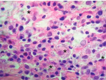
Initially, the differential diagnosis of the lesion included skin tumours (histiocytomas, mast cell tumours, cutaneous lymphoma), infectious and sterile granulomas, eosinophilic granulomas, cuta-neous cysts (Ferrer et al. 1990; Koutinas et al. 1992), cutaneous abscesses, subcutaneous helmintho-sis caused by Dirofilaria repens and mycetomas (Blavier et al. 2001). Under an appropriate sedation, the nodular lesion was removed, and histopatho-logical examination revealed follicular keratosis and a severe and chronic inflammatory infiltrate. The latter, localised particularly near the skin annexes, was composed of many histiocytes and plasma cells, a moderate number of mature lym-phocytes, a few eosinophils and rare neutrophils, and detectable leishmanial amastigote (2–3 µm in diameter) forms in the macrophages (Figure 1).
49
Veterinarni Medicina, 62, 2017 (01): 48–51 Case Report
doi: 10.17221/48/2016-VETMED
After histopathological diagnosis, serum bio-chemical analysis was performed and the total titres of anti-Leishmania antibodies were determined us-ing an indirect immunofluorescence antibody test (IFAT) (MegaFLUO® LEISH; Megacor Diagnostik
Gmbh).
The IFAT provided a negative result (titre of 1 : 40; positive test result is ≥ 50) and the complete blood count (by ADVIA 2120 V automated counter) revealed a normal haematological profile (Table 1; first blood sample = 1st BS). The serum biochemical
indicators (by Architect c8000 Clinical Chemistry Analyzer, Abbott Diagnostics, USA), reported in Table 1 (1st BS), revealed a normal pherogram
pro-file and serum protein levels as well as a moderate increase in alanine aminotransferase. For this rea-son, the dog was not treated against Leishmania, and eight months after the diagnosis (December 2015), the dog was still in good body condition at the physical examination, with blood and serum parameters within the physiological range (Table 1; second blood sample = 2nd BS). The IFAT again
provided a negative result (titre of 1 : 40).
DISCUSSION AND CONCLUSIONS
Leishmania is an intracellular protozoan para-site that is endemic, widespread and represents a public health problem in most countries of the Mediterranean basin as it is implicated in a wide spectrum of diseases both in humans and animals. Leishmaniosis currently threatens 350 million people in 98 countries, with 1.2 million new cases per year (Handler et al. 2015; Kahime et al. 2015). In regions where L. infantum is endemic, canine seropreva-lence ranges from 5% to 37% (Alvar et al. 2004), with dogs also playing an important epidemiological role as a reservoir of infection for humans (Viegas et al. 2012). After inoculation by the sand fly, the parasites disseminate towards the lymph nodes, bone marrow and viscera, such as the spleen and liver, where they replicate, causing disease. Depending on the ability of the host’s immune response to control parasite replication, the infection in dogs could range from subclinical/asymptomatic to fully developed disease (Alexandre-Pires et al. 2010). Skin lesions caused by Leishmania have been well-described but the nodular form of canine leishmaniosis is much less frequent (Blavier et al. 2001).
According to some authors (Ferrer et al. 1988; Vidor et al. 1991; Amara et al. 2000), the nodular form of leishmaniosis is more commonly diagnosed in boxers that, together with the Shepherd dogs, seem to be the breeds more predisposed to overt disease (Paltrinieri et al. 2010). Two cases of atypi-cal nodular forms, one in the interdigital space, Table 1. Serum haematological profile evaluated in a
4-year-old male dog at the first clinical examination – 1st blood sampling (BS), and after eight months – 2nd BS
Parameter 1st BS 2nd BS Range RBC (106/µl) 8.16 9.18 5.8–8.8 WBC (103/µl) 10.9 10.8 5.5–15.5 PLT (103/µl) 199 163 150–500 Urea (mmol/l) 14.81 16 6.43–19.6 Creatinine (µmol/l) 83.1 98.1 70.7–123.8 Glucose (mmol/l) 6.6 5.01 3.3–6.05 Fructosamine (µmol/l) 338 349 160–350 AST (IU/l) 29 37 15–40 ALT (IU/l) 72 83 15–55 Protein (g/l) 66.7 72.2 56–78 Albumin (g/l) 36.1 39.8 26.3–45.3 α1-globulin (g/l) 3.3 3.6 1.9–3.4 α2-globulin (g/l) 9.3 10.7 9–16.1 β1-globulin (g/l) 4.7 4.7 2.7–10.2 β2-globulin (g/l) 6.1 6.3 3.4–8.7 γ-globulin (g/l) 7.2 7.1 3–7.8 Albumin/globulin (ratio) 1.17 1.22 0.6–1.41 PLT = platelets, RBC = red blood cells, WBC = white blood cells
Figure 1. Skin, within the cytoplasm of a macrophage and between inflammatory cells numerous 2–4 µm, ovoid, protozoan amastigotes are visible (× 100 magnification)
50
Case Report Veterinarni Medicina, 62, 2017 (01): 48–51
doi: 10.17221/48/2016-VETMED
canine disease in areas of the world where trans-mission to humans is primarily zoonotic (Dye 1996; Alvar et al. 2004). This field case further supports that hypothesis. Furthermore, if the immune system directly controls the parasite’s replication making the infected dogs less infectious to same sand flies (Bongiorno et al. 2013), this in turn could further reduce the number of dogs infected by sand flies (Moreno et al. 2014).
REfERENCES
Alexandre-Pires G, Villa de Brito MT, Alguero C, Martins C, Roos Rodrigues O, Pereira da Fonseca I, Santos-Gomes G (2010): Canine leishmaniosis. Immunophenotypic pro-file of leukocytes in different compartments of sympto-matic, asymptomatic and treated dogs. Veterinary Immunology and Immunopathology 137, 275–283. Alvar J, Canavate C, Molina R, Moreno R, Nieto J (2004):
Canine leishmaniosis. Advances in Parasitology 57, 1–88. Amara A, Jemli MH, Kilani M, Ghorbel A, Aouina M (2000): A case of Leishmania nodular dermatitis in a dog (in French). Le Point Veterinaire 31, 514–516.
Blavier A, Keroack S, Denerolle P, Goy-Thollot I, Cha-banne L, Cadore JL, Bourdoiseau G (2001): Atypical forms of canine leishmaniosis. The Veterinary Journal 162, 108–120.
Bongiorno G, Paparcone R, Foglia Manzillo V, Oliva G, Cuisinier AM, Gradoni L (2013): Vaccination with LiESP/ QA-21 (CaniLeish®) reduces the intensity of infection in Phlebotomus perniciosus fed on Leishmania infantum infected dogs – A preliminary xenodiagnosis study. Vet-erinary Parasitology 197, 691–695.
Dye C (1996): The logic of visceral leishmaniasis control. American Journal of Tropical Medicine and Hygiene 55, 125–130.
Ferrer L, Rabanal R, Fondevila D, Ramos JA, Domingo M (1988): Skin lesions in canine leishmaniosis. Journal of Small Animal Practice 29, 381–388.
Ferrer L, Fondevila D, Marco A, Pumarola M (1990): Atyp-ical nodular leishmaniosis in two dogs. Veterinary Record 126, 90.
Handler MZ, Patel PA, Kapila R, Al-Qubati Y, Schwartz RA (2015): Cutaneous and mucocutaneous leishmaniasis. Clinical perspectives. Journal of the American Academy Dermatology 73, 897–908.
Kahime K, Boussaa S, Laamrani-El Idrissi A, Nhammi H, Boumezzough A (2015): Epidemiological study on acute cutaneous leishmaniasis in Morocco. Journal of Acute Disease 5, 41–45.
and the other in the right axilla were reported by Ferrer et al. (1990) in dogs that were not vaccinated. These nodules, as the one described here, were not ulcerated. Other reports of atypical nodular lesions caused by Leishmania have been reported on a dog tongue (Blavier et al. 2001; Viegas et al. 2012). All these animals, however, also showed other findings attributable to leishmaniosis (i.e. positive serology or the presence of Leishmania in the bone mar-row). Although in our presented case it was not possible to exclude dissemination to the bone mar-row through cytological or PCR tests (the owner did not consent to these further investigations), a seroconversion was neither observed at the first visit nor eight months later.
Although the nodular lesion observed in the pre-sent boxer dog could be attributable to Leishmania as the causative agent, it was not an atypical clinical manifestation of disseminated infection. Rather, it stemmed from a strong non-suppurative inflam-matory reaction to the entrance of the Leishmania protozoa at the bite site of sand flies, in an immune-competent animal.
The vaccine that is administered to dogs acts by developing long-lasting cell-mediated immune re-sponses against L. infantum, specifically with a domi-nant CD4+ Th1 influence in an overall mixed cellular
response. Among all the activated inflammatory cells, macrophages show a strong ability to reduce the levels of intracellular parasites through the induction of in-ducible nitric oxide synthase and production of nitric oxide derivatives (Moreno et al. 2014). This vaccine, recently licensed in Europe, displayed 68% to 92% efficacy in protecting animals against the appearance of clinical signs, under field conditions (Otranto and Dantas-Torres 2013; Oliva et 2014).
In this case, even if the vaccination was not able to prevent the entrance of the protozoa into the dog, it seems it allowed the animal to effectively control the infection and remain healthy. This hypothesis is supported by the absence of any clinical, serologi-cal and haematologiserologi-cal symptoms other than the observed nodular lesion, both at the moment of the physical examination and eight months later. Based on its histopathological features, the skin lesion could represent the result of a local reaction mediated by the cellular immune response against the attempt of protozoa to enter and disseminate in the dog.
Vaccination remains the best control strategy to counteract the progression of active infection for
51
Veterinarni Medicina, 62, 2017 (01): 48–51 Case Report
doi: 10.17221/48/2016-VETMED
Koutinas AF, Scott DW, Kantos V, Lekkas L (1992): Skin lesions in canine leishmaniosis (Kala-Azar): a clinical and histopathological study on 22 spontaneous cases in Greece. Veterinary Dermatology 3, 121–130.
Moreno J, Vouldoukis I, Schreiber P, Martin V, McGahie D, Gueguen S, Cuisinier AM (2014): Primary vaccination with the LiESP/QA-21 vaccine (CaniLeish®) produces a cell-mediated immune response which is still present 1 year later. Veterinary Immunology and Immunopathology 158, 199–207.
Oliva G, Nieto J, Manzillo VF, Cappiello S, Fiorentino E, Di Muccio T, Scalone A, Moreno J, Chicharro C, Carrillo E, Butaud T, Guegand L, Martin V, Cuisinier AM, McGahie D, Gueguen S, Canavate C, Gradoni L (2014): A ran-domised, double-blind, controlled efficacy trial of the LiESP/QA-21 vaccine in naive dogs exposed to two Leish-mania infantum transmission seasons. PLOS Neglected Tropical Diseases 8. Available: http://journals.plos.org/ plosntds/article?id=10.1371/journal.pntd.0003213.
Otranto D, Dantas-Torres F (2013): The prevention of ca-nine leishmaniasis and its impact on public health. Trends in Parasitology 7, 339–345.
Paltrinieri S, Solano-Gallego L, Fondati A, Lubas G, Gradoni L, Castagnaro M, Crotti A, Maroli M, Oliva G, Roura X, Zatelli A, Zini E (2010): Guidelines for diagnosis and cli-nical classification of leishmaniasis in dogs. JAVMA-Journal of the American Veterinary Medical Association 236, 1184–1191.
Viegas C, Requicha J, Albuquerque C, Sargo T, Machado J, Dias I, Pires MA, Campino L, Cardoso L (2012): Tongue nodules in canine leishmaniosis – a case report. Parasites and Vectors 5, 120.
Vidor E, Dereure J, Pratlong F, Dubreuil N, Bissuel G, Moreau Y, Rioux JA (1991): The site of inoculation of Leishmania infantum in the canine leishmaniosis. Study of a cohort in traditional Cevennes Region (in French). Pratique Medicale et Chirurgicale de l’Animale de Com-pagnie 26, 133–137.
Received: March 14, 2016 Accepted after corrections: September 30, 2016