Thorax 1992;47:833-834
Short
reports
Air
transport
and
the
fate of
pneumothorax
in
pleural
adhesions
M MHaid, P Paladini,MMaccherini,
M DiBisceglie, G Biagi, G Gotti
Abstract
Air travel is contraindicated in patients
with apneumothorax but was necessary
because of the exigenciesofwar inthree
patients. Three patients with high
velocity missile injuries to thechest and
pleural adhesionsarereported.Allhadto
be evacuated by air, without an
inter-costal drainoroxygensupplement,from
the war stricken area of Northern
Somalia (HornofAfrica) to Mogadishu.
Two patients with a partial
pneumo-thorax flew on military transport
aero-planes atanaltitude of 3000 mina
non-pressurised cabin and recoveredrapidly
afterafewdays in hospital. One patient,
transportedon asmall Cessnaaeroplane,
died after developing bilateral tension
pneumothoraces.
(Thorax 1992;47:833-834)
Pneumothorax is considered anabsolute
con-traindication for air travel, as reduction of
barometric pressure causes trapped gases to
expand.' Duringthe conflict in Somalia medical
personnel under extremely stressful
circum-stances had to evacuate three patients with
penetrating chest wounds by air without
facilitiesforprovidingameansofegressfor the
trapped pneumothorax. Two of the three
patientssurvived.
There was no past history of any important chest diseaseapartfromaninfluenza likeillness
accompanied by wheezing and dry cough.
On examination he was calm and
co-operative. The respiration rate was 20/min, pulse96/min and regular, and blood pressure
110/80mmHg. The physical signs suggested
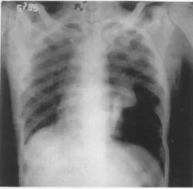
a right sided haemopneumothorax. A chest radiograph showed a partial (roughly 20%) right hydropneumothorax and upper zone
shadowing (fig 2).A needle thoracocentesiswas
performed. He was discharged on the second
dayafteradmissionand has remained well.
PATIENT3
A35 yearoldmanwithapenetrating injuryto
theleft thoraxwasadmittedto apolice casualty hospital. He had been shotbyanAK47rifle and
was transported 24 hours after injury from Erigavo(Northern Somalia)toMogadishuona
smallCessnaaeroplaneflyingat analtitude of
2150 m. The patient travelled in a cabin
exposed to external barometric pressure and arrivedatthehospital three hourslater,having become restless andseverely dyspnoeic. Little
FigureI Patient1:Radiograph showingapartial pneumothoraxontheleftside.
IstitutoPolicattedra diChirurgia Toracica
eCardiovascolare, Universit'a degliStudi diSiena, Italy PPaladini MMaccherini MDiBisceglie GBiagi GGotti Departmentof Surgery,University of Mogadishu,Somalia MM Haid
Reprintrequests to:
Dr G Gotti, via G Verdi 2, 53100Siena,Italy
Accepted11December 1991
Casereports
PATIENT1
A 21 yearoldmansufferedapenetratingwound
totheleft thorax andwasadmittedto hospital
inMogadishu after atwo and ahalf hour air
journey on amilitary transport aeroplane. He
had a history of chronic dry cough. He had
clinical signs that suggested a left sided
pneumothorax and thiswasconfirmed by chest
radiography, which showed a partial (about
50%) pneumothorax localisedto the lower left
pleuralcavity (fig 1). No treatment was neces-sary other than needle thoracocentesis.
PATIENT2
A 23year old man wasadmitted to hospital in
Mogadishu with a penetrating wound to the
right thorax after a two and a half hour air
journey on a military transport aeroplane.
Figure2 Patient 2: Radiographshowing a partial haemopneumothoraxand parietalhaematoma on the rightside.
833
on January 9, 2020 at Universitr degli Studi di Siena. Protected by copyright.
http://thorax.bmj.com/
Haid, Paladini, Maccherini, Di Bisceglie, Biagi, Gotti Figure 3 Patient 3:
Radiographshowing bilateralpneumothorax, extensivesubcutaneous emphysema, alargeleft lungcontusion, anda
bullet.
is known about the weather conditionsduring
theflight.
The patient had a history of pulmonary
tuberculosis and smoking. On examination-.he
was confused and dyspnoeic with a rapid
respiratory rate, a blood pressure of 110/60 mmHg, andapulserateof104/min. Extensive subcutaneousemphysemawaspresent.Hewas
severely hypoxaemic. A chest radiograph shortly after admission showed a right sided
pneumothorax and extensive left sided shad-owing;aright side chest tubewas inserted. A
chestradiographtwohours later showeda new
left sided pneumothorax (fig 3). A chest tube
wasinsertedonthe left side butthelung failed tore-expand. The patient's progressively
wor-sening respiratory distress, continued air leak,
and considerable haemoptysis prompted
sur-gery. A leftposterolateral thoracotomyunder
general anaesthesia was performed. The lung was freed from the chest wall and no major
lesion was found except for two pulmonary
perforations,whichwere oversewn.The bullet
was retrieved from the dorsal muscles.
Intraoperativeventilationwascontrolled with
positive end expiratory pressure. Monitoring
duringanaesthesiawaslimitedtonon-invasive measures.
Duringthepostoperative periodthepatient
continued to be restless and severely
dys-pnoeic. The lungs did not re-expand fully despite resuscitative measures and oxygen
enriched ventilation (with an ambubag via a
tracheostomy). He died ofhypoxaemia onthe
fourth day after injury. Owing to the war conditions a necropsy was not possible.
Discussion
The risk of air transport for patients with a pneumothorax is well known.' The first two patients fared well, as they had peripheral lung lesions with minimal lung contusion and the resulting air leak presumably sealed. The third patient had a central lesion, resulting in a massive leak ofair, which we assume passed into the anterior mediastinum as the media-stinal aspect of the lung is usually less adherent. Further accumulation of air from the lung laceration and from mediastinal extension of subcutaneous emphysema may have caused the air to spread into the right pleural space. Pulmonary laceration compounded possibly by pleural adhesions from old tuberculosis could have resulted in massive mediastinal and sub-cutaneous emphysema without the develop-ment of a pneumothorax on theleft.2This may explain why the left pneumothorax appeared only after the right pneumothorax had been drained.2 His exposure to high altitude during air travel seems likely to have been responsible for the expansion and dissipation of the trapped air in the mediastinum.
The patient died of hypoxaemia despite vigorous ventilation via a tracheostomy with 100% oxygen. Although the pneumothoraces and chronic pulmonary disease may have con-tributed to this picture, shunting of blood through the contused lung with no gas exchangewasprobably the mainfactor.34
Although two patients with a partial pneumothorax survived air travel, chest drain-agebefore evacuation is undoubtedly the safest option and should be carried out whenever possible.
1 StonehillRB. Air travel and thecardiopulmonarypatient.
PostgraduateMed1962;32:387-93.
2 BessonA,SaegesserF. A colour atlasofchesttraumaand
associatedinjuries.London:Wolfe, 1982:134-9,309-42.
3Wanebo H, Van Dyke J. The high velocity pulmonary
injury:relation to traumaticwetlunginjury. JThorac
CardiovascSurg 1972;64:537-50.
4 FischerRP,GeigerJP, GuernseyJM.Pulmonaryresection for severe pulmonary contusions secondary to high
velocitymissile wounds. J Trauma1974;14:293-302.
834
on January 9, 2020 at Universitr degli Studi di Siena. Protected by copyright.
http://thorax.bmj.com/