The reasons for end-to-side coaptation: how does
lateral axon sprouting work?
Introduction
The role of end-to-side (ETS) coaptation has been debat-ed for a long time since first experience by Viterbo et al. (1982).
Several studies have been published proposing the theoreti-cal bases for its clinitheoreti-cal application. To date, experimental ev-idences can be considered as well-established scientific data, but a debate remains open about clinical utilization (Dvali and Myckatyn, 2008). Since the pioneering work from Terzis and Tzafetta (2009a, b) about distal babysitting of injured facial nerve through ETS coaptation and partial neurotomy, some authors are still wondering whether an important dam-age to the donor nerve could derive from this application.
Reviewing the whole literature reveals how this technique remains a domain of some experts (Qattan and Al-Thunyan, 1998; Battiston and Lanzetta, 1999; Al-Qattan, 2001; Tung and Mackinnon, 2001; Mennen et al., 2003; Tung et al., 2003; Frey and Giovanoli, 2003a, b; Bertelli and Ghizoni, 2003; Amr and Moharram, 2005; Millesi and Schmidhammer, 2008); Magdi Sherif and Amr, 2010; Barbo-ur et al., 2012) and is mostly performed by choosing selected donors for nerve transfers to protect distal effectors in cases of proximal trunk injuries.
Millesi and Schmidhammer (2008) published an
interest-ing review and concluded that in case of high proximal inju-ries of nerve trunks in the upper extremity, more proximal nerve transfers failed to function, whereas choosing distal motor nerves gave the best results. These observations were supported by experimental evidences in the baboon (Men-nen, 1998) and were confirmed by a clinical series of brachi-al plexus injuries.
We review the literature starting from the biological bases of ETS, and discuss the usefulness of this technique, relating experimental and clinical data from other scholars and from their own experience.
ETS Coaptation: An Effective Technique
ExperimentalETS nerve repair has proven an effective tool for repairing peripheral nerves in experimental microsurgery, from both functional and morphological points of view (Bontioti and Dahlin, 2009; Papalia et al., 2016).
Three mechanisms have been proposed: a) contamination from axons regenerating from the proximal stump of the re-cipient nerve, b) true collateral sprouting from healthy fibers of the donor nerve and c) true axonal regeneration from damaged fibers of the donor nerve (“terminal sprouting”) (Bontioti and Dahlin, 2009; Papalia et al., 2016).
Abstract
Nerve fibers are attracted by sutureless end-to-side nerve coaptation into the recipient nerve. Opening a window in the epineurium enhances axon attraction and myelination. The authors analyze the features of nerve repair by end-to-side coaptation. They highlight the known mechanisms of axon sprouting and dif-ferent hypotheses of start up signals (presence or absence of an epineurial window, role of Schwann cells, signaling from the distal trunk). The clinical literature is also presented and differences between experi-mental and clinical applications are pointed out. The authors propose their point of view and perspectives deriving from recent experimental and clinical experiences.
Key Words: peripheral nerve repair; nerve coaptation; end-to-side nerve repair; side-to-side nerve repair; epineurial window; Schwann cells; nerve regeneration; nerve babysitter procedures; nerve transfer; nerve graft; glues in nerve repair
INVITED REVIEW
*Correspondence to: Michele R. Colonna, Ph.D., [email protected]. #These authors contributed equally to this study. orcid:
0000-0001-5586-4066 (Michele R. Colonna) doi: 10.4103/1673-5374.205081 Accepted: 2017-04-05
Stefano Geuna1, Igor Papalia2, #, Giulia Ronchi1, #, Francesco Stagno d’Alcontres3, Konstantinos Natsis4, Nikolaos A. Papadopulos5,
Michele R. Colonna3
1 Department of Clinical and Biological Sciences, University of Turin, Turin, Italy
2 Department of Biomedical and Dental Sciences and Morphological and Functional Sciences, University of Messina, Messina, Italy 3 Department of Human Pathology of the Adults, the Children and the Adolescents, University of Messina, Messina, Italy
4 Department of Anatomy, Faculty of Health Sciences, School of Medicine, Aristoteles University, Thessaloniki, Greece
5 Department of Plastic Surgery and Burns, Alexandroupoli University General Hospital, Democritus University of Thrace, Alexandroupoli, Greece How to cite this article: Geuna S, Papalia I, Ronchi G, Stagno d’Alcontres F, Natsis K, Papadopulos NA, Colonna MR (2017) The reasons for end-to-side coaptation: how does lateral axon sprouting work? Neural Regen Res 12(4):529-533.
Open access statement: This is an open access article distributed under the terms of the Creative Commons Attribution-NonCommercial- ShareAlike 3.0 License, which allows others to remix, tweak, and build upon the work non-commercially, as long as the author is credited and the new creations are licensed under the identical terms.
“Simple” coaptation
Dealing with “simple coaptation”, few studies have been conducted without opening any window into the nerve con-nective tissue; a simple coaptation of a cut distal stump into a donor nerve trunk, is a stimulus for nerve repair by sutures passing through the donor nerve (see below, A. 2) (Kanje et al., 2000; Papalia et al., 2016).
Our original research about sutureless (cyanoacrylate glue) ETS nerve repair opened another scenario, demonstrating that simple glue coaptation without any window was capable to attract nerve fibers from the donor to the recipient trunk; however, from a morphological point of view, regenerating axons were smaller and less myelinated (Papalia et al., 2016). Really, the presence of only a cut distal stump represents a powerful attraction for newly formed axons from a donor healthy trunk, maybe for its production of growth factors and signals, which have been shown also coming from distal effectors via retrograde transportation (Wood and Mackin-non, 2015).
How does a window opened into the connective nerve sheaths work?
Indeed, opening a window acts on Schwann cells and mo-lecular components breaking the antineurotropic continuity of the wall constituted by epi- and/or perineurium (Bontioti and Dahlin, 2009; Kovačič et al., 2012); the effect of this breakthrough is a realignment of Schwann cells creating open gates both from a cellular and an ionic/molecular point of view. Through these gates acting as positive signals for axon outgrowth, neurite cones are attracted; moreover, attractive properties from degenerated distal axons together with retrograde chemiokinetic signals from distal effectors
via the distal stump complete the job (Kovačič et al., 2012;
Wood and MacKinnon, 2015).
Different sizes of epi-perineurial windows have been stud-ied (Kovačič et al., 2012): enlarging the window from 1 to 4/5 mm produces an increased ingrowth of larger axons.
In these basic conditions, even a simple sutureless coap-tation has proven effective in directing donor fibers to the recipient trunk (Papalia et al., 2016).
Other factors playing a positive role
Sutures have a role by an additional trauma represented by their passage through the connective tissue. Some authors suggested they could act through pressure and associat-ed bleassociat-eding and inflammation (Kelly et al., 2007). Also glues have been shown to have a positive role through early inflammatory steps (Bontioti and Dahlin, 2009). As in wound healing process, where macrophages have been shown to play a critical role, early inflammation in nerve repair promotes Schwann cells activation (Papalia et al., 2016), and could be therefore defined as an “healthy”
in-flammation.
Disinhibition: let inhibitory factors shut up
Switching inhibitory factors off is a major task in nerve re-pair. To date, we know that the major inhibitory molecules are associated either with myelin or with scar tissue forma-tion. NogoA, MAG, and OMgp are present in myelin debris and work through the Nogo receptor. In nerve scar tissue, chondroitin sulphate, proteoglycans, and semaphorins, to-gether with the formation of a collagen-based membrane, act as inhibitory factors (Fawcett et al., 2012).
Time also runs against
Time has also been studied as an important factor in nerve regeneration, through both progressive increasing of inhib-itory factors and decreasing activities (a true senescence) of Schwann cells. Peripheral nerve surgeons aware that for more proximal injuries, more time is required for nerve regeneration, with a finally negative outcome due to distal effectors atrophy (Papalia et al., 2016).
Do ETS procedures produce damage to the donor nerve?
Some studies found that ETS neurorrhaphy results in donor nerve injury and regeneration of the injured nerve (Okajima and Terzis, 2000; Brenner et al., 2007). In these experimen-tal papers, it has been observed that ETS coaptation which epineurial window either alone or together with partial neurotomy produces a certain loss (“escape”) of nerve fibers from the donor nerve; this report ever restricts surgeon’s choice keeping in mind that performing ETS coaptation will damage donor nerve function (Colonna et al., 2015). Since this point, investigators have been searching for the best donor: should it be agonist/antagonist, sensory even if recip-ient is motor (Papalia et al., 2007, 2016; Colonna et al., 2015; Nghiem et al., 2015)?
Which is the mechanism of fibers spreading through an ETS repair?
There is plenty of evidence on ETS about collateral sprout-ing of the donor nerve: surgical windows through neural connective tissue open the way for cathionic passageways and activate neuronal molecular pathways (by regulating expression of IGF-1 and TWEAK-Fn14) for regeneration from the donor nerve into the recipient one (Lundborg et al., 1994; Noah et al., 1997; Zhang et al., 1999; Xiong et al., 2003; Hayashi et al., 2004; Bontioti et al., 2005; Samal et al., 2006; Kovacic et al., 2007; Zhu et al., 2008; Liu et al., 2015, 2016b).
Which is the precise site from which new fibers spread out to enter ETS coaptation site?
A number of investigators (Bajrovic et al., 2002; Matsuda et al., 2005; Akeda et al., 2006; Bontioti et al., 2006) questioned
the true origin of axons from the donor nerve: do they come from the axons closest to the coaptation site or from a more proximal site?
Morphological evidences have been reported of true regeneration from the Ranvier’s nodes just close to the co-aptation site (Zhu et al., 2008). However, in this study, only a qualitative analysis showing the sprouting of myelinated axons is reported. No further information about the amount of collateral sprouting fibers nor evidences of presence of unmyelinated fibers are described.
Other studies have proposed a more proximal origin, just from the dorsal ganglia or the spinal cord (Kim et al., 2012). To date, this question remains debated.
Fibers specificity: does it work?
Donor motor fibers have been proposed specifically for motor recipient trunks, as well as donor sensory fibers for sensory recipient trunks. As far as it concerns motor fibers, to regulate the number of spreading axons and the number of impulses, several authors have introduced partial neurec-tomy of the donor nerve (Terzis and Tzafetta, 2009a, b; Liu et al., 2015, 2016a). The use of sensory nerves as donors even in motor nerve injuries has been supported by a conspicuous report (Nghiem et al., 2015). To date, however, using sensory nerves for motor neurotization has not yet been proposed in the clinical field. But why not?
The choice for agonists in ETS nerve transfer
Even when choosing motor nerves as donors, their role as agonists of the recipient nerve has been debated; it has been concluded not to introduce conflict between agonists (Papalia et al., 2007). Millesi and Schmidhammer (2008) selected the trunk for the short head of Flexor Pollicis Brevis as donor for median thenar branch babysitting, as FPB acts as an agonist of thenarian muscles.
Double end-to-side: side-to-side. A look into clinical work and perspectives
A nerve graft can be coapted ETS as a bridge between the donor and the recipient trunk; Yüksel et al. (1999) in their experimental paper showed that babysitter procedure with side-to-side technique gave functional results superiors to ETS technique. In clinical studies, this kind of nerve fiber transfer has been proposed and used successfully in most distal nerve transfers for proximal nerve injuries (Magdi Sherif and Amr, 2010; Colonna et al., 2015, Gesslbauer et al., 2016).
Clinical: What have we learned from experimental research which can be translated to clinical practice? To date, the peripheral nerve surgeons yet face challenging problems, especially with proximal nerve injuries produced by avulsion. A very small or no regeneration follows this dramatic event, producing anatomical changes in distal ef-fectors, as well as severe functional deficits.
Even if direct or graft nerve repair between proximal and distal stump remains the gold standard, distal motor end-plate and sensory receptors hypotrophy will occur be-cause of the long distance and time to be run by regenerat-ing fibers from their proximal coaptation/graft site. Nerve fibers need to be brought distally as fast as possible and this is why we need short and quick motor nerve transfers (Bontioti and Dahlin, 2009; Colonna et al., 2015; Papalia et al., 2016).
However, we must remember that in the case of distal babysitter nerve transfers, clinical reports (MacKinnon et al., 2005; Moore et al., 2014; Sassu et al., 2015; Ray et al., 2016) agree that grading of the motor function cannot be achieved with the same precision (Isaacs, 1999) as for proximal tech-niques (Oberlin et al., 1994).
Indeed, opening a window in epiperineurium creates a
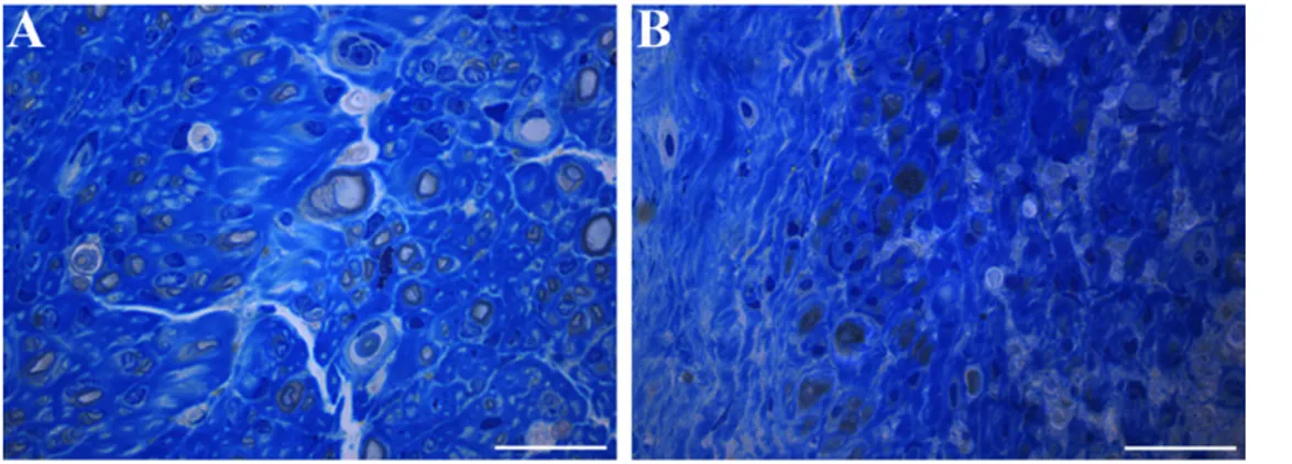
Figure 1 Representative high resolution light microscopic images of toluidine-blue-stained transverse sections of median nerve 36 weeks after end-to-side coaptation with N-butyl-2-cyanoacrylate.
(A) Coaptation with an epineurial window; (B) coaptation without an epineurial window. With an epineurial window, the regenerated fibers are more numerous. Bars: 20 µm.
sort of epineurial chamber similar to the experimental Lund-borg chamber (LundLund-borg et al., 1982). It attracts all collater-al and centrcollater-al axon sprouts towards empty endoneurcollater-al tube on all recipient nerve stump surface. It should be reserved to an experienced microsurgeon trained in microsurgical pe-ripheral nerve reconstruction.
Conclusions
New techniques and materials have been proposed as vari-ants for ETS, and we expect novel reports regarding new materials to be used as conduits and gene therapy, too. New techniques: reverse ETS
More recently, a new variant of ETS nerve coaptation has been described (Li et al., 2014), taking nerve fibers from a healthy trunk, which is severed distally and coapted ETS to the recipient injured nerve. This report seems very interest-ing as it couples the use of ETS with a donor sensory trunk to a recipient motor nerve.
Our experience
A recent study opened new perspectives for the comprehen-sion of the mechanisms underlying ETS nerve regeneration. In this work, a sutureless technique for performing ETS neurorrhaphy was developed to verify whether axon regen-eration can be induced by the biological lure coming from the damaged nerve without any trauma at the coaptation site (Papalia et al., 2016).
In adult female rats, the median nerve was glued to the ul-nar nerve epineurium using N-butyl-2-cyanoacrylate. In one experimental group a small epineurial window was opened before coaptation while in another group gluing was per-formed without any intentional damage to the ulnar nerve epineurium.
“Simple” gluing excludes trauma, inflammation and starts up signals for regeneration from sutures; nevertheless, regen-eration and axons income from the donor to the recipient nerve occurred no matter epineurium was opened or not. Both from a quantitative and qualitative points of view, ax-ons and fibers were more represented when the window was opened (Figure 1).
Thus, to date we can say that opening a window and gluing produces a valid regeneration through an ETS coaptation.
Moreover, even if epi-perineurial sheaths are not opened, a simple sutureless coaptation of a severed nerve trunk to the side of an healthy trunk is capable to attract axons from this last one.
Our group is on the way with further studies about the role of Schwann cells and attracting factors from the severed trunk, as well as stimulation with gene transfer, bioactive materials, and protecting the coaptation site (or sites when a graft is required) with wrapping.
Author contributions: All the coauthors have been working on the man-uscript, either writing single parts or revising the whole paper, and giving their suggestions. SG, IP and GR cared the morphological part and figure preparation; MRC cared the preparation and architecture of the manu-script and its final version.
Conflicts of interest: None declared.
References
Akeda K, Hirata H, Matsumoto M, Fukuda A, Tsujii M, Nagakura T, Ogawa S, Yoshida T, Uchida A (2006) Regenerating axons emerge far proximal to the coaptation site in end-to-side nerve coaptation with-out a perineurial window using a T-shaped chamber. Plast Reconstr Surg 117:1194-1203.
Al-Qattan MM, Al-Thunyan A (1998) Variables affecting axonal regener-ation following end-to-side neurorrhaphy. Br J Plast Surg 51:238-242. Al-Qattan MM (2001) Terminolateral neurorrhaphy: review of
experi-mental and clinical studies. J Reconstr Microsurg 17:99-108. Amr SM, Moharram AN (2005) Repair of brachial plexus lesions by
end-to-side side-to-side grafting neurorrhaphy: experience based on 11 cases. Microsurgery 25:126-146.
Bajrovic F, Kovacic U, Pavcnik M, Sketelj J (2002) Interneuronal sig-naling is involved in induction of collateral sprouting of nociceptive axons. Neuroscience 111:587-596.
Barbour J, Yee A, Kahn LC, Mackinnon SE (2012) Supercharged end-to-side anterior interosseous to ulnar motor nerve transfer for intrin-sic musculature reinnervation. J Hand Surg Am 37:2150-2159. Battiston B, Lanzetta M (1999) Reconstruction of high ulnar nerve
lesions by distal double median to ulnar nerve transfer. J Hand Surg Am 24:1185-1191.
Bertelli JA, Ghizoni MF (2003) Nerve repair by end-to-side coaptation or fascicular transfer: a clinical study. J Reconstr Microsurg 19:313-318. Bontioti E, Kanje M, Lundborg G, Dahlin LB (2005) End-to-side nerve
repair in the upper extremity of rat. J Peripher Nerv Syst 10:58-68. Bontioti E, Dahlin LB, Kataoka K, Kanje M (2006) End-to-side nerve
repair induces nuclear translocation of activating transcription factor 3. Scand J Plast Reconstr Surg Hand Surg 40:321-328.
Bontioti E, Dahlin LB (2009) Chapter 12: Mechanisms underlying the end-to-side nerve regeneration. Int Rev Neurobiol 87:251-268. Brenner MJ, Dvali L, Hunter DA, Myckatyn TM, Mackinnon SE (2007)
Motor neuron regeneration through end-to-side repairs is a function of donor nerve axotomy. Plast Reconstr Surg 120:215-223.
Colonna MR, Russo A, Galeano M, Delia G, Pajardi GE, d’Alcontres FS (2015) “Babysitting” procedures in proximal nerve trunk injuries: two case reports and a review. Plast Aesthet Res 2:208-212.
Dvali LT, Myckatyn TM (2008) End-to-side nerve repair: review of the literature and clinical indications. Hand Clin 24:455-460.
Fawcett JW, Schwab ME, Montani L, Brazda N, Müller HW (2012) Defeating inhibition of regeneration by scar and myelin components. Handb Clin Neurol 109:503-522.
Fortes WM, Noah EM, Liuzzi FJ, Terzis JK (1999) End-to-side neuror-rhaphy: evaluation of axonal response and upregulation of IGF-I and IGF-II in a non-injury model. J Reconstr Microsurg 15:449-457. Frey M, Giovanoli P (2003a) End-to-side neurorrhaphy of sensory
nerves. Eur J Plast Surg 26:85-88.
Frey M, Giovanoli P (2003b) End-to-side neurorrhaphy of motor nerves: reinnervation of free muscle transplants–first clinical appli-cation. Eur J Plast Surg 26:89-94.
Giovanoli P, Koller R, Meuli-Simmen C, Rab M, Haslik W, Mittlböck M, Meyer VE, Frey M (2000) Functional and morphometric evaluation of end-to-side neurorrhaphy for muscle reinnervation. Plast Recon-str Surg 106:383-392.
Hayashi A, Yanai A, Komuro Y, Nishida M, Inoue M, Seki T (2004) Collateral sprouting occurs following end-to-side neurorrhaphy. Plast Reconstr Surg 114:129-137.
Isaacs J (2017) Commentary on ‘Nerve grafts bridging the thenar branch of the median nerve to the ulnar nerve to enhance nerve re-covery: a report of three cases’. J Hand Surg Eur Vol 42:286-288. Kanje M, Arai T, Lundborg G (2000) Collateral sprouting from sensory
and motor axons into an end to side attached nerve segment. Neu-roreport 11:2455-2459.
Kelly EJ, Jacoby C, Terenghi G, Mennen U, Ljungberg C, Wiberg M (2007) End-to-side nerve coaptation: a qualitative and quantitative assessment in the primate. J Plast Reconstr Aesthet Surg 60:1-12. Kovačič U, Zele T, Tomšič M, Sketelj J, Bajrović FF (2012) Influence of
breaching the connective sheaths of the donor nerve on its myelinat-ed sensory axons and on their sprouting into the end-to-side coaptmyelinat-ed nerve in the rat. J Neurotrauma 29:2805-2815.
Li Q, Zhang P, Yin X, Han N, Kou Y, Jiang B (2014) Early sensory protection in reverse end-to-side neurorrhaphy to improve the func-tional recovery of chronically denervated muscle in rat: a pilot study. J Neurosurg 121:415-422.
Liu HF, Chen ZG, Shen HM, Zhang H, Zhang J, Lineaweaver WC, Zhang F (2015) Efficacy of the end-to-side neurorrhaphies with epineural window and partial donor neurectomy in peripheral nerve repair: an experimental study in rats. J Reconstr Microsurg 1:31-38.
Liu HF, Chen ZG, Lineaweaver WC, Zhang F (2016a) Can the babysit-ter procedure improve nerve regeneration and denervated muscle atrophy in the treatment of peripheral nerve injury? Plast Reconstr Surg 1:122-131.
Liu HF, Chen ZG, Lineaweaver WC, Friel MT, Zhang F (2016b) Molec-ular mechanism of the” babysitter” procedure for nerve regeneration and muscle preservation in peripheral nerve repair in a rat model. Ann Plast Surg doi: 10.1097/SAP.0000000000000952.
Lundborg G, Dahlin LB, Danielsen N, Hansson HA, Johannesson A, Longo FM, Varon S (1982) Nerve regeneration across an extended gap: a neurobiological view of nerve repair and the possible involve-ment of neuronotrophic factors. J Hand Surg Am 6:580-587. Lundborg G, Zhao Q, Kanje M, Danielsen N, Kerns JM (1994) Can
sensory and motor collateral sprouting be induced from intact pe-ripheral nerve by end-to-side anastomosis? J Hand Surg (Br) 19:277-282.
Mackinnon SE, Novak CB, Myckatyn TM, Tung TH (2005) Results of reinnervation of the biceps and brachialis muscles with a double fas-cicular transfer for elbow flexion. J Hand Surg Am 5:978-985. Magdi Sherif M, Amr AH (2010) Intrinsic hand muscle reinnervation
by median-ulnar end-to-side bridge nerve graft: case report. J Hand Surg Am 35:446-450.
Matsuda K, Kakibuchi M, Fukuda K, Kubo T, Madura T, Kawai K, et al (2005) End-to-side nerve grafts: experimental study in rats. J Recon-str Microsurg 21:581-591.
Mennen U (2004) End-to-side nerve suturing technique. J Hand Surg Br 29:514.
Mennen U, van der Westhuizen MJ, Eggers IM (2003) Re-innervation of M. biceps by end-to-side nerve suture. Hand Surg 8:25-31. Millesi H, Schmidhammer R (2008) Nerve fiber transfer by end-to-side
coaptation. Hand Clin 24:461-483.
Moore AM, Franco M, Tung TH (2014) Motor and sensory nerve transfers in the forearm and hand. Plast Reconstr Surg 4:721-730. Nghiem BT, Sando IC, Hu Y, Urbanchek MG, Cederna PS (2015)
Sen-sory protection to enhance functional recovery following proximal nerve injuries: current trends. Plast Aesthet Res 2:202-207.
Noah EM, Williams A, Jorgenson C, Skoulis TG, Terzis JK (1997) End-to-side neurorrhaphy: a histologic and morphometric study of axo-nal sprouting into an end-to-side nerve graft. J Reconstr Microsurg 13:99-106.
Oberlin C, Beal D, Leechavengvongs S, Salon A, Dauge MC, Sarcy JJ (1994) Nerve transfer to biceps muscle using a part of ulnar nerve for C5-C6 avulsion of the brachial plexus: anatomical study and report of four cases. J Hand Surg Am 2:232-237.
Okajima S, Terzis JK (2000) Ultrastructure of early axonal regeneration in an end-to-side neurorrhaphy model. J Reconstr Microsurg 16:313-323.
Papalia I, Cardaci A, d’Alcontres FS, Lee JM, Tos P, Geuna S (2007) Se-lection of the donor nerve for end-to-side neurorrhaphy. J Neurosurg 107:378-382.
Papalia I, Magaudda L, Righi M, Ronchi G, Viano N, Geuna S, Col-onna MR (2016) Epineurial window is more efficient in attracting axons than simple coaptation in a sutureless (cyanoacrylate-bound) model of end-to-side nerve repair in the rat upper limb: functional and morphometric evidences and review of the literature. PLoS One 11:e0148443.
Ray ZW, Chang J, Hawasli A, Wilson JT, Yang L (2016) Motor nerve transfers: a comprehensive review. Neurosurgery 78:1-26.
Samal F, Haninec P, Raska O, Dubovwy P (2006) Quantitative assess-ment of the ability of collateral sprouting of the motor and primary sensory neurons after the end-to-side neurorrhaphy of the rat mus-culocutaneous nerve with the ulnar nerve. Ann Anat 188:337-344. Sassu P, Libberecht K, Nilsson A (2015) Nerve transfers of the forearm
and hand: a review of current indications. Plast Aesthet Res 2:195-201. Terzis JK, Tzafetta K (2009a) The “babysitter” procedure: mini hypo-glossal to facial nerve transfer and cross-facial nerve grafting. Plast Reconstr Surg 123: 865-876.
Terzis JK, Tzafetta K (2009b) “Babysitter” procedure with concomitant muscle transfer in facial paralysis. Plast Reconstr Surg 124:1142-1156.
Tung TH, Mackinnon SE (2001) Flexor digitorum superficialis nerve transfer to restore pronation: two case reports and anatomic study. J Hand Surg Am 26:1065-1072.
Tung TH, Novak CB, Mackinnon SE (2003) Nerve transfers to the bi-ceps and brachialis branches to improve elbow flexion strength after brachial plexus injuries. J Neurosurg 98:313-318.
Viterbo F, Trindade JC, Hoshino K, Mazzoni Neto A (1992) Latero-ter-minal neurorrhaphy without removal of the epineural sheath. Exper-imental study in rats. Rev Paul Med 110: 267-275.
Wood MD, Mackinnon SE (2015) Pathways regulating modality-specif-ic axonal regeneration in peripheral nerve. Exp Neurol 265:171-175. Xiong G, Ling L, Nakamura R, Sugiura Y (2003) Retrograde tracing
and electrophysiological findings of collateral sprouting after end-to-side neurorrhaphy. Hand Surg 8:145-150.
Yüksel F, Karacaoglu E, Güler MM (1999) Nerve regeneration through side-to-side neurorrhaphy sites in a rat model: a new concept in pe-ripheral nerve surgery. Plast Reconstr Surg 7:2092-2099.
Zhang Z, Soucacos PN, Bo J, Beris AE (1999) Evaluation of collateral sprouting after end-to-side nerve coaptation using a fluorescent dou-ble-labeling technique. Microsurgery 9:281-286.
Zhang Z, Johnson EO, Vekris, MD, Zoubos AB, Bo J, Beris AE, Souca-cos PN (2006) Long-term evaluation of rabbit peripheral nerve re-pair with end-to-side neurorrhaphy in rabbits. Microsurgery 26:262-267.
Zhu QT, Zhu JK, Chen GY (2008) Location of collateral sprouting of donor nerve following end-to-side neurorrhaphy. Muscle Nerve 38: 1506-1509.