Edited by: Terry Francis Davies, Icahn School of Medicine at Mount Sinai, United States
Reviewed by: Michele Colaci, University of Modena and Reggio Emilia, Italy Andrea Di Domenicantonio, University of Pisa, Italy *Correspondence: Fabrizio Guarneri [email protected] Specialty section: This article was submitted to Thyroid Endocrinology, a section of the journal Frontiers in Endocrinology Received: 22 March 2017 Accepted: 09 June 2017 Published: 27 June 2017 Citation: Guarneri F, Giuffrida R, Di Bari F, Cannavò SP and Benvenga S (2017) Thyroid Autoimmunity and Lichen. Front. Endocrinol. 8:146. doi: 10.3389/fendo.2017.00146
Thyroid Autoimmunity and Lichen
Fabrizio Guarneri1*, Roberta Giuffrida1, Flavia Di Bari2, Serafinella Patrizia Cannavò1 and Salvatore Benvenga2,3,4
1 Department of Clinical and Experimental Medicine – Dermatology, University of Messina, Messina, Italy, 2 Department of Clinical and Experimental Medicine – Endocrinology, University of Messina, Messina, Italy, 3 Master Program on Childhood, Adolescent and Women’s Endocrine Health, University of Messina, Messina, Italy, 4 Interdepartmental Program of Molecular & Clinical Endocrinology and Women’s Endocrine Health, University Hospital Policlinico “G. Martino”, Messina, Italy
Lichen planus (LP) and lichen sclerosus (LS) are cutaneous-mucous diseases with uncertain epidemiology. Current data, which are likely to be underestimated, suggest a prevalence in the general population of 0.1–4% for cutaneous LP, 1.27–2.0% for oral LP, and 0.1–3.3% for LS. While etiology of lichen is still unknown, clinical and histological evidence show an (auto)immune pathogenesis. Association of lichen with autoimmune thyroid disease (AITD) has been investigated in few studies. This association appears better defined in the case of LS, while is more controversial for LP. In both situations, the frequency of the association is higher in females. We review the available literature on the correlation between the different types of lichen and AITD, and the literature on the genetic risk factors which are shared by both conditions. Such data suggest that a com-mon pathogenic mechanism could be the cause for co-occurrence of lichen and AITD, at least in some patients. Additionally, analyzing literature data and in continuity with our previous work on other autoimmune diseases, we suggest that molecular mimicry could trigger both diseases, and thus explain their co-occurrence.
Keywords: autoimmune thyroid disease, oral lichen planus, mucous lichen planus, cutaneous lichen planus, lichen sclerosus, human leukocyte antigen, infections, molecular mimicry
inTRODUCTiOn
Lichen planus (LP) is a cutaneous-mucous disease characterized by small papules and pruritus. Histologically, LP is characterized by hyperkeratosis, hypergranulosis, acanthosis with formation of colloid bodies (Civatte or Sabouraud bodies), vacuolar degeneration of the basal epidermal cell layer, “saw-tooth” appearance of the rete pegs, enlarged and deformed dermal papillae with infiltration of lymphocytes and histiocytes in a “band-like” pattern in contact with, and sometimes invading, the basal epidermal cell layer (1). Because of clinical similarity, over the years, several dermatological conditions were defined as “lichen” to eventually form the so-called lichenoid dermatoses, but with different specifications. For instance, lichen striatus is actually classified among spongiotic dermatoses, lichen amyloidosus among amyloidoses, and lichen aureus among mucinoses (1). A detailed discussion about lichenoid dermatoses is beyond the scope of this review. Only LP (cutaneous, mucosal and oral) and lichen sclerosus (LS)—also known as LS and atrophicus—for which autoimmunity has been postulated as a relevant part of the pathogenic mechanism, were reported to be associated with autoimmune thyroid disease (AITD).
Abbreviations: AITD, autoimmune thyroid disease; GD, Graves’ disease; HT, Hashimoto’s thyroiditis; LP, lichen planus; LS,
The epidemiology of LP and LS is not exactly defined. Cur-rent data, probably underestimated, suggest a prevalence in the general population of 0.1–4% for cutaneous LP (2), 1.27–2.0% for oral LP (3), and 0.1–3.3% for LS (4). Few authors inves-tigated the association between these conditions and thyroid diseases and, even fewer, the possible common etiopathogenic mechanism(s).
We, herein, review the available literature on this topic. Moreover, in continuity with our previous studies on molecular mimicry as a trigger of autoimmune diseases, we suggest molecu-lar mimicry as a potential pathogenic mechanism.
MATeRiALS AnD MeTHODS
We searched the PubMed database (https://ncbi.nlm.nih.gov/ PubMed) using the search string “thyroid AND autoimm* AND lichen.” The retrieved articles were revised, and only those dis-cussing the association between lichen and AITD were selected. The reference lists of these articles were examined to find other relevant articles, which were also revised and included in this review if appropriate.
ReSULTS
As of February 24, 2017, a PubMed search for “AITD” or “lichen” retrieved 14,864 and 15,311 articles, respectively. Conversely, a search for “thyroid AND autoimm* AND lichen,” as described in Section “Material and Methods,” yielded 40 articles of which 17 were selected as they were relevant for our review. Of these 17 articles, 15 analyzed the occurrence of AITD in patients with lichen (Table 1) and 2 analyzed the occurrence of lichen in patients with AITD (Table 2).
The first paper which suggested that LS “may be related to or caused by an autoimmune process” and highlighted the frequent association with thyroid autoimmunity was published in 1974 (5). In 26 patients (25 females and one male) with LS and 443 control subjects without autoimmune diseases, the authors evaluated the presence of antibodies to thyroglobulin (Tg), thy-roid cytoplasm, gastric parietal cells, and type I intrinsic factor. Almost half of the female patients were positive for antithyroid cytoplasm antibodies [10/25 (40%), p < 0.001 vs controls] or anti-gastric parietal cells antibodies [11/25 (44%), p < 0.001 vs controls]; other tests were negative or not different from controls. Eight patients with antithyroid cytoplasm antibodies had subclinical thyroiditis (5).
The topic was brought again to attention many years later, in a case report of a 65-year-old woman with coexisting LS, AITD, morphea, and insulin-dependent diabetes mellitus (6). Next, a comment (7) to a review article (8) provocatively defined LS “a cutaneous manifestation of thyroid disease” (7). While this state-ment appears excessive in the light of modern knowledge, it is suggestive of the increase, started in the early 1990s, of awareness and interest concerning the possible connection between lichen and AITD (9–11). Larger studies, needed to define the epidemio-logical relevance of the association, have been performed only relatively recently.
LS and AiTD
The largest, yet retrospective, study available on LS and AITD is on 532 patients with LS, predominantly adults and females (n = 500 and 396, respectively), who were visited in a German University hospital during 12 years (12). All patients were examined for cutaneous and extracutaneous autoimmune dis-eases. Tests included complete blood cell count, routine blood chemistry testing, complement components 3 and 4, C-reactive protein, and a panel of serological analyses for autoimmunity. This panel included antinuclear antibodies, extractable nuclear antibodies, rheumatoid factor, anti-cyclic citrullinated peptide antibodies, circulating immune complexes, and antibodies to thyroid peroxidase (TPO) and Tg. One or more autoimmune diseases were found in 82 patients (15.4%), with a significantly (p < 0.0001) different frequency among women (n = 75, 18.9%) and men (n = 7, 5.1%). AITD [Hashimoto’s thyroiditis (HT) or Graves’ disease (GD)] was the condition most often associ-ated with LS, with a total of 65 cases (12.2%), again prevalently among females [n = 60 (15.2%), p = 0.0002 vs males]. Overall, cases of all other autoimmune diseases accounted for 3.3% of the studied sample.
Similar results were found by others. A case–control study in women was performed (13) on a smaller population (190 with adult-onset vulvar LS, 126 with adult-onset erosive vulvar LP, 922 age-matched controls). AITD was observed in 16.3% of patients with LS and 15% of patients with erosive LP compared to 7.9% in controls (p < 0.001 for both comparisons) (13). Kazandi et al. (14) retrospectively analyzed 82 women with vulvar LS and found 15 cases of thyroid disease (18.2%). A greater prevalence (29.9%) was reported in a retrospective evaluation of 211 patients visited in a 10-year period for vulvar LS (15). The percentage was higher among patients aged less than 55 years (33.8%) than among those aged 55 years or more (27.7%).
A different picture can be observed in studies on male LS. After the aforementioned article by Kreuter et al. (12), the largest and probably most complete study was published by Kantere et al. (16). The authors randomly chose 100 patients from 771 diagnosed with LS between 1997 and 2007 and re-evaluated their clinical condition. Such re-evaluation included clinical examina-tion and laboratory tests, namely thyroid stimulating hormone (TSH), thyroxine (T4), antinuclear antibodies, and autoantibod-ies to extracellular matrix protein-1 (ECM-1), ECM-1 being the likely autoantigen of LS (17). Only five patients had mild abnormal thyroid function: low levels of T4 and raised levels of TSH (which is consistent with overt primary hypothyroidism) in two cases, normal T4 and raised TSH (which is consistent with subclinical primary hypothyroidism) in two other cases, normal T4 and decreased TSH in one (16). However, one paper (18) shows a different trend. Indeed, in a population of 60 women and 42 men, Hagedorn et al. (18) found that LS was associated with AITD in 39% of female and 12.5% of male patients.
LP (Oral, Mucous, Cutaneous) and AiTD
The association between thyroid autoimmunity and LP, in its different subtypes, is more controversial, and the scarcity of papers does not allow clear conclusions. Soy et al. (19) evalu-ated the frequency of rheumatic and autoimmune diseases in 65
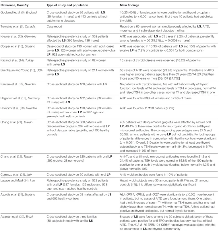
TAbLe 1 | Studies reporting clinical and/or laboratory data about subjects affected by lichen who were found positive for AITD.
Reference, Country Type of study and population Main findings
Goolamali et al. (5), England Cross-sectional study on 26 patients with LS (25 females, 1 males) and 443 controls without autoimmune diseases
10/25 (40%) of female patients were positive for antithyroid cytoplasm antibodies (p < 0.001 vs controls); 8 of these 10 patients had subclinical thyroiditis
Tremaine et al. (6), Canada Case report Report on a 65-year-old woman simultaneously affected by LS, AITD,
morphea, and insulin-dependent diabetes mellitus Kreuter et al. (12), Germany Retrospective prevalence study on 532 patients
affected by LS (396 females, 136 males)
AITD was associated with LS in 65 cases (12.2% of patients), prevalently among females [n = 60 (15.2%), p = 0.0002 vs males]
Cooper et al. (13), England Case–control study on 190 women with adult-onset vulvar LS, 126 women with adult-onset erosive vulvar LP, 922 age-matched control women
AITD was observed in 16.3% of patients with LS and 15% of patients with erosive LP vs 7.9% of controls (p < 0.001 for both comparisons) Kazandi et al. (14), Turkey Retrospective prevalence study on 82 women
with vulvar LS
15 cases of thyroid disease were observed (18.2% of patients) Birenbaum and Young (15), USA Retrospective prevalence study on 211 women with
vulvar LS
63 cases of AITD were observed (29.9% of patients). Prevalence of AITD was higher among patients aged less than 55 years [25/74 (33.8%)] than those aged 55 years or more [38/137 (27.7%)]
Kantere et al. (16), Sweden Cross-sectional study on 100 patients with LS No patients with thyroid disease, five with mild abnormality of thyroid function: low levels of T4 and raised levels of TSH in two cases, normal T4 and raised TSH in two other cases, normal T4 and decreased TSH in one Hagedorn et al. (18), Germany Cross-sectional study on 102 patients (60 females,
42 males) with LS
AITD was found in 39% of females and 12.5% of males Ebrahimi et al. (20), Sweden Cross-sectional study on 120 patients (89 females,
31 males) with mucosal LP and 87 age- and sex-matched healthy controls
AITD was found in 11/120 patients (9.2%)
Chang et al. (21), Taiwan Cross-sectional study on 500 patients with desquamative gingivitis, 287 with erosive oral LP without desquamative gingivitis, and 100 healthy controls
455 patients with desquamative gingivitis were affected by erosive oral LP; 46.4% of them were positive for anti-Tg and 45.1% for antithyroid microsomal antibodies. The corresponding percentages were 27.5 and 30.3%, among patients with erosive LP but not gingivitis. For both groups of patients, differences in comparison with healthy controls were significant (p < 0.001). Overall, 210 patients were positive for at least one thyroid autoantibody, and TSH levels were normal in 84.3%, decreased in 6.7% and increased in 9% of them
Chang et al. (22), Taiwan Cross-sectional study on 320 patients with oral LP (292 erosive, 28 non-erosive)
Anti-Tg and antithyroid microsomal antibodies were found in 21.3 and 24.4% of patients. TSH levels were normal in 85.8% of the 190 patients, positive for one or both thyroid autoantibodies, below normal in 4.2% and above normal in 10%
Carrozzo et al. (23), Italy Cross-sectional study on 50 patients with oral LP Antithyroid antibodies were found in 10% of patients Lavaee and Majd (24), Iran Retrospective prevalence study on 523 patients
with oral LP (387 females, 136 males) and 523 age- and sex-matched healthy controls
Hypothyroid subjects were 35 among patients (6.7%) and 21 among controls (4%); this difference was not statistically significant Azurdia et al. (31), England Cross-sectional study on 58 males affected by LS
and 602 healthy controls
HLA-DR11, -DR12, and -DQ7 were significantly (p ≤ 0.05) more frequent in patients, but no cases of AITD were found among them. One patient had a mild increase of serum T4 with normal TSH levels, another one had slightly lower than normal serum T4, with normal TSH. A third patient had positive antithyroid antibodies, but normal thyroid function
Aslanian et al. (32), Brazil Cross-sectional study on three families (30 subjects in total) with familial LS
8 cases of LS were found among the 30 subjects visited; seven of these patients were positive for anti-TPO antibodies, but only four had clinical AITD. The HLA-B*15-DRB1*04-DRB4* haplotype was associated with the co-occurrence of LS and thyroid autoimmunity
The type of lichen studied in each paper is written in boldface.
AITD, autoimmune thyroid disease; LP, lichen planus; LS, lichen sclerosus; T4, thyroxine; Tg, thyroglobulin; TPO, thyroid peroxidase; TSH, thyroid stimulating hormone; HLA, human leukocyte antigen.
patients (56 women and 9 men) with AITD. Oral LP was one of the less represented diseases, as it was found in only two patients (3.1%). Ebrahimi et al. (20) evaluated 120 patients with mucosal LP and 87 age- and sex-matched healthy controls for the presence of other diseases, with particular attention to autoimmune and
thyroid diseases. They found a significantly high frequency of autoimmune diseases in general (28%) and AITD in particular (9.2%) among patients. These results, together with the observa-tion that lichen was multifocal in 72% of women and 64% of men, led the authors to conclude that “LP with mucosal involvement
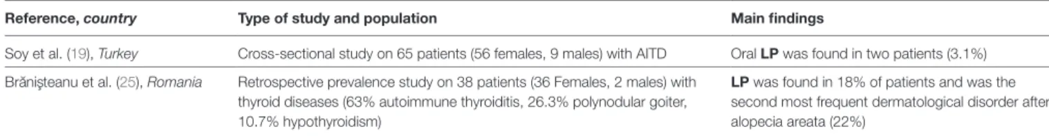
TAbLe 2 | Studies reporting clinical and/or laboratory data about subjects affected by AITD who were found positive for lichen.
Reference, country Type of study and population Main findings
Soy et al. (19), Turkey Cross-sectional study on 65 patients (56 females, 9 males) with AITD Oral LP was found in two patients (3.1%) Bra˘nis¸teanu et al. (25), Romania Retrospective prevalence study on 38 patients (36 Females, 2 males) with
thyroid diseases (63% autoimmune thyroiditis, 26.3% polynodular goiter, 10.7% hypothyroidism)
LP was found in 18% of patients and was the second most frequent dermatological disorder after alopecia areata (22%)
The type of lichen studied in each paper is written in boldface. AITD, autoimmune thyroid disease, LP, lichen planus.
should be considered and taken care of as a systemic disease” and to point out “the need for a multidisciplinary clinic to get optimal care and treatment” (20). Another paper concerning mucosal lichen, namely adult-onset erosive vulvar LP, is by Cooper et al. (13) (see above, Section “LS and AITD”). Further, interesting elements come from a recent paper (21). This paper (21) aimed to define the number of patients with desquamative gingivitis (a disease often associated with erosive oral LP) who were positive for anti-gastric parietal cells, anti-Tg, and antithyroid microso-mal antibodies. They analyzed 500 patients with desquamative gingivitis, 287 with erosive oral LP but without desquamative gingivitis, and 100 healthy controls. Upon careful reevaluation, erosive oral LP was found in 455 patients of the first group: 46.4% of them were positive for anti-Tg and 45.1% for antithyroid microsomal antibodies. The percentages were 27.5 and 30.3%, respectively, among patients of the second group. Differences from controls were significant (p < 0.001) for both groups of patients. Overall, 210 patients were positive for at least one thyroid-related autoantibody, and TSH levels were normal in 84.3%, low in 6.7%, and raised in 9% of them (21). In a previous study (22), the same group had found a prevalence of 21.3 and 24.4% for anti-Tg and antithyroid microsomal antibodies, respectively, among 320 patients with oral LP (erosive in 292 cases, non-erosive in 28). In the same study, TSH levels were normal in 85.8% of the 190 patients positive for one or both thyroid-related autoantibodies, low in 4.2% and raised in 10% (22). Other authors had reported antithyroid antibodies in 10% of 50 patients with oral LP (23).
Lavaee and Majd (24) retrospectively evaluated the frequency of hypothyroidism in 523 patients with oral LP (387 females, 136 males) and in an equal number of age- and sex-matched healthy controls. They found statistically similar proportions (6.7 and 4%, respectively).
Finally, Brănişteanu et al. published data about the association between AITD and cutaneous LP (25). The study population included 38 patients (36 females, 2 males) with thyroid diseases (63% autoimmune thyroiditis, 26.3% multinodular goiter, 10.7% hypothyroidism), who accessed the Dermatovenereology Unit of a University hospital over 2 years. LP was the second most frequent dermatological disorder observed (18%) after alopecia areata (22%).
Genetic Risk Factors: Possible Role of
Human Leukocyte Antigen (HLA)
The studies mentioned in the previous sections suggest that all subtypes of lichen, AITD, and also their association, are more frequent in females.
Given the autoimmune pathogenesis of both conditions, several authors analyzed the possibility of a link with specific alleles of the HLA genes. As well known, HLA genes generate the major histocompatibility complex (MHC) molecules, responsi-ble for presentation of (auto)antigenic peptides to the immune system and activation of the consequent specific (auto)immune reaction.
Studies on the HLA haplotype of patients with lichen are few, not very recent, and often performed on small cohorts. Porter et al. (26) reported that cutaneous LP has been associated to HLA-A3, -A5, -A28, -B16, and -Bw35, mucosal LP to HLA-A3 and -A28, oral LP to HLA-B16, -DR1, and -DRw9, erosive oral LP to HLA-DR2, -DR3, -DR9, -B27, and -Bw57, mixed oral LP to HLA-B51 [for references, see Ref. (26)]. The studies reviewed had populations ranging from 10 to 82 patients, and had been published between 1976 and 1994. For LS, the most recent review (27) suggests a strong linkage to HLA-DQ7. In a subsequent case– control study (28), an increased frequency of HLA-DRB1*12/ DQB1*03 was found in 187 patients with vulvar LS.
A comparison with AITD-associated HLA alleles (29) shows some elements in common: HLA-B16 confers increased risk for HT in Asians, HLA-DR3 is linked to GD (in Caucasians) and HT, HLA-DR9 is a risk factor for GD in Japanese and Chinese subjects and HT in Chinese patients only [for references, see Ref. (29)]. Among patients with stress-related GD, HLA-A28 is significantly more frequent (at least 3-X) in those with exacerbations of hyper-thyroidism compared with those with no exacerbations during treatment with antithyroid drugs, while HLA-DR3 is almost 3-times more frequent in the whole group of patients with stress-related GD compared with healthy controls (30).
The above data could suggest a common genetic background of susceptibility for lichen and thyroid autoimmunity. However, we found only two studies that evaluated the HLA haplotypes of patients for which the association between lichen and AITD was explicitly investigated (31, 32). Azurdia et al. (31) analyzed 58 males with LS and 602 healthy controls, and showed a sig-nificantly (p ≤ 0.05) higher frequency of HLA-DR11, -DR12, and -DQ7 in patients. In detail, the frequencies of HLA-DR11, -DR12, and -DQ7 were 22, 9, and 45% among patients and 13, 3, and 31% among controls, respectively. Abnormal thyroid function was observed in two cases: one patient had a mild increase of serum T4 with normal TSH levels, while another had slightly subnormal serum T4, and normal TSH. Positive antithyroid antibodies, but normal thyroid function, were found in a third patient (31). In the second paper, Aslanian et al. (32) examined three families, of 20, 8, and 2 members, respectively, with familial LS. Eight subjects with LS were found among the
30 visited, 7 of whom were positive for anti-TPO antibodies, but only 4 had a thyroid disease. The HLA-B*15-DRB1*04-DRB4* haplotype was associated with the co-occurrence of LS and thy-roid autoimmunity (32). HLA-DRB1*04 was almost threefold more frequent in patients with stress-related GD compared with healthy controls (30).
environmental Triggering Factors:
Association with infections and the
Molecular Mimicry Hypothesis
Like most autoimmune diseases, the environmental triggers of LS and AITD are unknown, and also unknown is whether an etiopathogenic link between the two conditions exists.
We previously reported a woman who developed both LS and HT after infection by Borrelia burgdorferi (33). In that occasion, the chronological sequence and correlation between the pathological events led us to hypothesize that molecular mimicry between bacterial antigen(s) and human autoantigens could have been the pathogenic mechanism by which borre-liosis had triggered both autoimmune diseases (33). According to the molecular mimicry hypothesis, structural similarity between microbial antigens and human autoantigens can turn a defensive immune reaction into an autoimmune reaction in genetically predisposed subjects (mainly because of specific HLA alleles). This model has been postulated, and in many cases demonstrated, as a possible explanation for the onset of autoimmunity (34–41).
Several studies on the possible role of molecular mimicry in the pathogenesis of autoimmune and allergic diseases were performed also by our group, with extensive use of bioinformat-ics tools (42–53). In detail, we searched for amino acid sequence homology between human protein autoantigens involved in spe-cific autoimmune diseases and proteins from microbes that are clinically linked to such diseases. In many cases, we also searched the homologous segments of human and microbial proteins for binding motifs of MHC molecules derived from specific HLA alleles.
Following the hypothesis formulated in our case report (33), we aimed to identify the molecules most probably involved in triggering autoimmunity after Borrelia infection (42, 45). We found that human TSH-R has four segments homologous to proteins from Borrelia and five homologous to proteins from Yersinia, another bacterial species associated with AITD. In a subsequent study (45), we extended our work to include the other known thyroid autoantigens (TPO, Tg, sodium iodide symporter) and to search human and microbial proteins for the occurrence of peptide-binding motifs of HLA-DR molecules. Eleven additional homologies were found with proteins from Borrelia (2 with Tg, 3 with TPO, 6 with sodium iodide symporter) and 15 with proteins from Yersinia (2 with Tg, 2 with TPO, 11 with sodium iodide symporter). The number of binding motifs related to the different HLA-DR alleles agreed well with literature data, which suggest that AITD is associated with HLA-DR3, -DR4, -DR5, -DR8, and -DR9.
Concerning the association between Borrelia and lichen, in 1985, Asbrink wrote that “a Borrelia infection may result in lichen
sclerosus et atrophicus-like reactions” (54), a claim that was subsequently supported by others (55–59). Although the debate remains open, a pathogenic link between borreliosis and lichen seems to exist in some cases (60). Our preliminary data (61) show that ECM-1, which is the autoantigen of LS (17), is homologous to BBG23 and methyl-accepting chemotaxis protein (mcp-3) of B. burgdorferi. All four thyroid autoantigens, ECM-1, and their corresponding homologous Borrelia proteins contain 4–32 copies of the binding motif related to HLA-DQ7, this allele conferring genetic susceptibility to both AITD (62) and LS (31).
Molecular mimicry appears as an interesting field of inves-tigation, and might explain, at least in part, associations found in epidemiological studies and/or single case reports. In our experience, it gave a plausible explanation for the association between AITD and Yersinia infection (42, 45, 50), anti-tumor vaccination with NY-ESO-1 (51), or rickettsiosis (52).
The main other infectious agents that the literature has linked to AITD are Epstein–Barr virus (63), hepatitis C virus (64), parvovirus B19 (64), human herpesvirus-6 (65), and Helicobacter pylori (66). For lichen, association was reported with Epstein– Barr virus—also known as human herpesvirus 4 (67), hepatitis C virus (67, 68), human papillomavirus (67, 69), and human her-pesvirus-7 (70), while correlation with H. pylori is controversial (71, 72).
COnCLUSiOn
The existence and nature of a connection between AITD and lichen are still unresolved issues. Echoing Braun-Falco et al. (1), the etiology of lichen is currently “a mistery.” Molecular mimicry is a likely mechanism, especially considering the advantage of providing an explanation for the occurrence of the association in patients with given HLA genotypes. However, molecular mimicry alone may not explain entirely the complex pathogenesis of the association, and other possibilities should be evaluated. Better awareness and attention to the association of lichen and AITD, and increased interdisciplinary collaboration, is desirable to define epidemiological magnitude and detailed clinical characteristics of the association, taking into account variables, such as ethnicity, socioeconomic issues, and environ-mental issues. Hopefully, better basic and clinical research will generate more effective care to patients with coexisting lichen and AITD.
AUTHOR COnTRibUTiOnS
SB and FG: substantial contributions to the conception or design of the work, drafting of the work, final approval of the version to be published, and agreement to be accountable for all aspects of the work in ensuring that questions related to the accuracy or integrity of any part of the work are appropriately investigated and resolved. SPC, FDB, and RG: acquisition/analysis/interpretation of data for the work, critical revision for important intellectual content, final approval of the version to be published, and agree-ment to be accountable for all aspects of the work in ensuring that questions related to the accuracy or integrity of any part of the work are appropriately investigated and resolved.
ReFeRenCeS
1. Braun-Falco O, Plewig G, Wolff HH, Burgdorf WHC. Dermatology. Berlin: Springer Verlag (2000).
2. Le Cleach L, Chosidow O. Clinical practice. Lichen planus. N Engl J Med (2012) 366:723–32. doi:10.1056/NEJMcp1103641
3. Thongprasom K, Carrozzo M, Furness S, Lodi G. Interventions for treat-ing oral lichen planus. Cochrane Database Syst Rev (2011) 7:CD001168. doi:10.1002/14651858.CD001168.pub2
4. van Cranenburgh OD, Nijland SB, Lindeboom R, de Korte J, de Rie MA, Ter Stege JA, et al. Patients with lichen sclerosus experience moderate satis-faction with treatment and impairment of quality of life: results of a cross- sectional study. Br J Dermatol (2017) 176:1508–15. doi:10.1111/bjd.15125 5. Goolamali SK, Barnes EW, Irvine WJ, Shuster S. Organ-specific antibodies
in patients with lichen sclerosus. Br Med J (1974) 4:78–9. doi:10.1136/ bmj.4.5936.78
6. Tremaine R, Adam JE, Orizaga M. Morphea coexisting with lichen sclerosus et atrophicus. Int J Dermatol (1990) 29:486–9. doi:10.1111/j.1365-4362.1990. tb04840.x
7. Poskitt L, Wojnarowska F. Lichen sclerosus as a cutaneous manifestation of thyroid disease. J Am Acad Dermatol (1993) 28:665. doi:10.1016/ S0190-9622(08)81795-1
8. Heymann WR. Cutaneous manifestations of thyroid disease. J Am Acad Dermatol (1992) 26:885–902. doi:10.1016/0190-9622(92)70130-8
9. Wright AJ. Lichen sclerosus thyroid disease. J Reprod Med (1998) 43:240. 10. Lewis FM. Lichen sclerosus and autoimmune disease. J Reprod Med (1998)
43:1006.
11. Lynch PJ. Lichen sclerosus and thyroid disease. J Reprod Med (1999) 44: 315–6.
12. Kreuter A, Kryvosheyeva Y, Terras S, Moritz R, Möllenhoff K, Altmeyer P, et al. Association of autoimmune diseases with lichen sclerosus in 532 male and female patients. Acta Derm Venereol (2013) 93:238–41. doi:10.2340/ 00015555-1512
13. Cooper SM, Ali I, Baldo M, Wojnarowska F. The association of lichen sclerosus and erosive lichen planus of the vulva with autoimmune disease: a case- control study. Arch Dermatol (2008) 144:1432–5. doi:10.1001/archderm.144. 11.1432
14. Kazandi M, Sahin C, Terek MC, Cirpan T, Oztekin K. Clinical evaluation of vulvar lichen sclerosus: case series. Eur J Gynaecol Oncol (2010) 31: 555–8.
15. Birenbaum DL, Young RC. High prevalence of thyroid disease in patients with lichen sclerosus. J Reprod Med (2007) 52:28–30.
16. Kantere D, Alvergren G, Gillstedt M, Pujol-Calderon F, Tunbäck P. Clinical features, complications and autoimmunity in male lichen sclerosus. Acta Derm Venereol (2017) 97:365–9. doi:10.2340/00015555-2537
17. Oyama N, Chan I, Neill SM, Hamada T, South AP, Wessagowit V, et al. Autoantibodies to extracellular matrix protein 1 in lichen sclerosus. Lancet (2003) 362:118–23. doi:10.1016/S0140-6736(03)13863-9
18. Hagedorn M, Buxmeyer B, Schmitt Y, Bauknecht T. Survey of genital lichen sclerosus in women and men. Arch Gynecol Obstet (2002) 266:86–91. doi:10.1007/s004040100209
19. Soy M, Guldiken S, Arikan E, Altun BU, Tugrul A. Frequency of rheumatic diseases in patients with autoimmune thyroid disease. Rheumatol Int (2007) 27:575–7. doi:10.1007/s00296-006-0263-8
20. Ebrahimi M, Lundqvist L, Wahlin YB, Nylander E. Mucosal lichen planus, a systemic disease requiring multidisciplinary care: a cross-sectional clinical review from a multidisciplinary perspective. J Low Genit Tract Dis (2012) 16:377–80. doi:10.1097/LGT.0b013e318247a907
21. Chang JY, Chiang CP, Wang YP, Wu YC, Chen HM, Sun A. Antigastric parietal cell and antithyroid autoantibodies in patients with desquamative gingivitis. J Oral Pathol Med (2017) 46:307–12. doi:10.1111/jop.12490
22. Chang JY, Chiang CP, Hsiao CK, Sun A. Significantly higher frequencies of presence of serum autoantibodies in Chinese patients with oral lichen planus. J Oral Pathol Med (2009) 38:48–54. doi:10.1111/j.1600-0714.2008. 00686.x
23. Carrozzo M, Gandolfo S, Lodi G, Carbone M, Garzino-Demo P, Carbonero C, et al. Oral lichen planus in patients infected or noninfected with hepatitis C virus: the role of autoimmunity. J Oral Pathol Med (1999) 28:16–9. doi:10.1111/j.1600-0714.1999.tb01988.x
24. Lavaee F, Majd M. Evaluation of the association between oral lichen planus and hypothyroidism: a retrospective comparative study. J Dent (Shiraz) (2016) 17:38–42.
25. Brănişteanu DE, Dimitriu A, Vieriu M, Boda D, Stoleriu G, Molodoi DA, et al. Cutaneous manifestations associated with thyroid disease. Rev Med Chir Soc Med Nat Iasi (2014) 118:953–8.
26. Porter SR, Kirby A, Olsen I, Barrett W. Immunologic aspects of dermal and oral lichen planus: a review. Oral Surg Oral Med Oral Pathol Oral Radiol Endod (1997) 83:358–66. doi:10.1016/S1079-2104(97)90244-4
27. Tasker GL, Wojnarowska F. Lichen sclerosus. Clin Exp Dermatol (2003) 28:128–33. doi:10.1046/j.1365-2230.2003.01211.x
28. Gao XH, Barnardo MC, Winsey S, Ahmad T, Cook J, Agudelo JD, et al. The association between HLA DR, DQ antigens, and vulval lichen sclerosus in the UK: HLA DRB1*12 and its associated DRB1*12/DQB1*0301/04/09/010 haplotype confers susceptibility to vulval lichen sclerosus, and HLA DRB1*0301/04 and its associated DRB1*0301/04/DQB1*0201/02/03 haplo-type protects from vulval lichen sclerosus. J Invest Dermatol (2005) 125:895–9. doi:10.1111/j.0022-202X.2005.23905.x
29. Guarneri F, Benvenga S. Environmental factors and genetic background that interact to cause autoimmune thyroid disease. Curr Opin Endocrinol Diabetes Obes (2007) 14:398–409. doi:10.1097/MED.0b013e3282ef1c48
30. Vita R, Lapa D, Trimarchi F, Vita G, Fallahi P, Antonelli A, et al. Certain HLA alleles are associated with stress-triggered Graves’ disease and influence its course. Endocrine (2017) 55:93–100. doi:10.1007/ s12020-016-0909-6
31. Azurdia RM, Luzzi GA, Byren I, Welsh K, Wojnarowska F, Marren P, et al. Lichen sclerosus in adult men: a study of HLA associations and suscepti-bility to autoimmune disease. Br J Dermatol (1999) 140:79–83. doi:10.1046/ j.1365-2133.1999.02611.x
32. Aslanian FM, Marques MT, Matos HJ, Pontes LF, Porto LC, Azevedo LM, et al. HLA markers in familial lichen sclerosus. J Dtsch Dermatol Ges (2006) 4:842–7. doi:10.1111/j.1610-0387.2006.06087_supp.x
33. Vaccaro M, Guarneri F, Borgia F, Cannavò SP, Benvenga S. Association of lichen sclerosus and autoimmune thyroiditis: possible role of Borrelia burgdor-feri? Thyroid (2002) 12:1147–8. doi:10.1089/105072502321085261
34. Rashid T, Ebringer A. Autoimmunity in rheumatic diseases is induced by microbial infections via crossreactivity or molecular mimicry. Autoimmune Dis (2012) 2012:539282. doi:10.1155/2012/539282
35. Balmasova IP, Sepiashvili RI. Intestine infections, inflammation and autoim-munity. Trigger and effector mechanisms of autoimmune disease development as an outcome of intestinal infections. Zh Mikrobiol Epidemiol Immunobiol (2013) 2:102–11.
36. Coppieters KT, Wiberg A, von Herrath MG. Viral infections and molec-ular mimicry in type 1 diabetes. APMIS (2012) 120:941–9. doi:10.1111/ apm.12011
37. Brodziak A, Ziółko E, Muc-Wierzgoń M, Nowakowska-Zajdel E, Kokot T, Klakla K. The role of human endogenous retroviruses in the pathogenesis of autoimmune diseases. Med Sci Monit (2012) 18:RA80–8.
38. Cruz-Tapias P, Blank M, Anaya JM, Shoenfeld Y. Infections and vaccines in the etiology of antiphospholipid syndrome. Curr Opin Rheumatol (2012) 24:389–93. doi:10.1097/BOR.0b013e32835448b8
39. Virtanen JO, Jacobson S. Viruses and multiple sclerosis. CNS Neurol Disord Drug Targets (2012) 11:528–44. doi:10.2174/187152712801661220
40. Chastain EM, Miller SD. Molecular mimicry as an inducing trigger for CNS autoimmune demyelinating disease. Immunol Rev (2012) 245:227–38. doi:10.1111/j.1600-065X.2011.01076.x
41. Cusick MF, Libbey JE, Fujinami RS. Molecular mimicry as a mechanism of autoimmune disease. Clin Rev Allergy Immunol (2012) 42:102–11. doi:10.1007/ s12016-011-8293-8
42. Benvenga S, Guarneri F, Vaccaro M, Santarpia L, Trimarchi F. Homologies between proteins of Borrelia burgdorferi and thyroid autoantigens. Thyroid (2004) 14:964–6. doi:10.1089/thy.2004.14.964
43. Guarneri F, Guarneri C, Benvenga S. Helicobacter pylori and autoimmune pancreatitis: role of carbonic anhydrase via molecular mimicry? J Cell Mol Med (2005) 9:741–4. doi:10.1111/j.1582-4934.2005.tb00506.x
44. Guarneri F, Guarneri C, Benvenga S. Identification of potentially cross- reactive peanut-lupine proteins by computer-assisted search for amino acid sequence homology. Int Arch Allergy Immunol (2005) 138:273–7. doi:10.1159/ 000088864
45. Benvenga S, Santarpia L, Trimarchi F, Guarneri F. Human thyroid autoanti-gens and proteins of Yersinia and Borrelia share amino acid sequence homo-logy that includes binding motifs to HLA-DR molecules and T-cell receptor. Thyroid (2006) 16:225–36. doi:10.1089/thy.2006.16.225
46. Guarneri F, Guarneri C, Benvenga S. Environment-induced reactivity against autoallergens: possible role of latex. J Allergy Clin Immunol (2006) 117:957–8. doi:10.1016/j.jaci.2006.02.008
47. Guarneri F, Guarneri C, Guarneri B, Benvenga S. In silico identification of potential new latex allergens. Clin Exp Allergy (2006) 36:916–9. doi:10.1111/ j.1365-2222.2006.02516.x
48. Guarneri F, Guarneri C, Benvenga S. Cross-reactivity of Anisakis simplex: possible role of Ani s 2 and Ani s 3. Int J Dermatol (2007) 46:146–50. doi:10.1111/j.1365-4632.2006.03091.x
49. Gregoric E, Gregoric JA, Guarneri F, Benvenga S. Injections of Clostridium botulinum neurotoxin A may cause thyroid complications in predisposed persons based on molecular mimicry with thyroid autoantigens. Endocrine (2011) 39:41–7. doi:10.1007/s12020-010-9410-9
50. Guarneri F, Carlotta D, Saraceno G, Trimarchi F, Benvenga S. Bioinformatics support the possible triggering of autoimmune thyroid diseases by Yersinia enterocolitica outer membrane proteins homologous to the human thyrotropin receptor. Thyroid (2011) 21:1283–4. doi:10.1089/ thy.2010.0364
51. Vita R, Guarneri F, Agah R, Benvenga S. Autoimmune thyroid disease elicited by NY-ESO-1 vaccination. Thyroid (2014) 24:390–4. doi:10.1089/ thy.2013.0170
52. Marangou A, Guarneri F, Benvenga S. Graves’ disease precipitated by rick-ettsial infection. Endocrine (2015) 50:828–9. doi:10.1007/s12020-015-0767-7 53. Guarneri F, Guarneri B. Bioinformatic analysis of HLA-linked genetic suscep-tibility to immunoallergic disease: the MotiFinder software. Ann Ital Dermatol Allergol (2010) 64:69–75.
54. Asbrink E. Erythema chronicum migrans Afzelius and acrodermatitis chron-ica atrophchron-icans. Early and late manifestations of Ixodes ricinus-borne Borrelia spirochetes. Acta Derm Venereol Suppl (Stockh) (1985) 118:1–63.
55. Aberer E, Stanek G. Histological evidence for spirochetal origin of morphea and lichen sclerosus et atrophicans. Am J Dermatopathol (1987) 9:374–9. doi:10.1097/00000372-198710000-00002
56. Aberer E, Kollegger H, Kristoferitsch W, Stanek G. Neuroborreliosis in mor-phea and lichen sclerosus et atrophicus. J Am Acad Dermatol (1988) 19:820–5. doi:10.1016/S0190-9622(88)70239-X
57. Aberer E, Klade H. Cutaneous manifestations of Lyme borreliosis. Infection (1991) 19:284–6. doi:10.1007/BF01644968
58. Schempp C, Bocklage H, Lange R, Kölmel HW, Orfanos CE, Gollnick H. Further evidence for Borrelia burgdorferi infection in morphea and lichen sclerosus et atrophicus confirmed by DNA amplification. J Invest Dermatol (1993) 100:717–20. doi:10.1111/1523-1747.ep12472369
59. Ozkan S, Atabey N, Fetil E, Erkizan V, Günes AT. Evidence for Borrelia burgdorferi in morphea and lichen sclerosus. Int J Dermatol (2000) 39:278–83. doi:10.1046/j.1365-4362.2000.00912.x
60. Vasudevan B, Chatterjee M. Lyme borreliosis and skin. Indian J Dermatol (2013) 58:167–74. doi:10.4103/0019-5154.110822
61. Guarneri F, Guarneri C, Santarpia L, Benvenga S. Borreliosi, lichen e tiroidite di Hashimoto: tre malattie, una causa sola? Atti del 79° Congresso Nazionale SIDeMaST May 26–29 2004. Brescia: SIDeMaST (2004).
62. Bogner U, Badenhoop K, Peters H, Schmieg D, Mayr WR, Usadel KH, et al. HLA-DR/DQ gene variation in nongoitrous autoimmune thyroiditis at the serological and molecular level. Autoimmunity (1992) 14:155–8. doi:10.3109/08916939209083135
63. Dittfeld A, Gwizdek K, Michalski M, Wojnicz R. A possible link between the Epstein-Barr virus infection and autoimmune thyroid disorders. Cent Eur J Immunol (2016) 41:297–301. doi:10.5114/ceji.2016.63130
64. Fallahi P, Ferrari SM, Vita R, Benvenga S, Antonelli A. The role of human parvovirus B19 and hepatitis C virus in the development of thyroid disorders. Rev Endocr Metab Disord (2016) 17(4):529–35. doi:10.1007/s11154-016- 9361-4
65. Sultanova A, Cistjakovs M, Gravelsina S, Chapenko S, Roga S, Cunskis E, et al. Association of active human herpesvirus-6 (HHV-6) infection with autoimmune thyroid gland diseases. Clin Microbiol Infect (2017) 23:.e1–50. doi:10.1016/j.cmi.2016.09.023
66. Astl J, Šterzl I. Activation of Helicobacter pylori causes either autoimmune thyroid diseases or carcinogenesis in the digestive tract. Physiol Res (2015) 64(Suppl 2):S291–301.
67. Bunker CB, Shim TN. Male genital lichen sclerosus. Indian J Dermatol (2015) 60:111–7. doi:10.4103/0019-5154.152501
68. Alaizari NA, Al-Maweri SA, Al-Shamiri HM, Tarakji B, Shugaa-Addin B. Hepatitis C virus infections in oral lichen planus: a systematic review and meta-analysis. Aust Dent J (2016) 61:282–7. doi:10.1111/adj.12382
69. Gupta S, Gupta S. Role of human papillomavirus in oral squamous cell carcinoma and oral potentially malignant disorders: a review of the literature. Indian J Dent (2015) 6:91–8. doi:10.4103/0975-962X.155877
70. Nahidi Y, Tayyebi Meibodi N, Ghazvini K, Esmaily H, Esmaeelzadeh M. Association of classic lichen planus with human herpesvirus-7 infection. Int J Dermatol (2017) 56:49–53. doi:10.1111/ijd.13416
71. Wedi B, Kapp A. Helicobacter pylori infection in skin diseases: a critical apprai sal. Am J Clin Dermatol (2002) 3:273–82. doi:10.2165/00128071- 200203080-00009
72. Hernando-Harder AC, Booken N, Goerdt S, Singer MV, Harder H. Helicobacter pylori infection and dermatologic diseases. Eur J Dermatol (2009) 19:431–44. doi:10.1684/ejd.2009.0739
Conflict of Interest Statement: The research was conducted in the absence of any
commercial or financial relationships that could be construed as a potential conflict of interest.
Copyright © 2017 Guarneri, Giuffrida, Di Bari, Cannavò and Benvenga. This is an open-access article distributed under the terms of the Creative Commons Attribution License (CC BY). The use, distribution or reproduction in other forums is permitted, provided the original author(s) or licensor are credited and that the original publication in this journal is cited, in accordance with accepted academic practice. No use, distribution or reproduction is permitted which does not comply with these terms.