ABSTRACT
Aim The aim of this prospective clinical trial was to analyze,
using the Functional Implant Prosthodontic Score (FIPS), the clinical resultsof three different abutment-implant connections (1 hexagon vs 2 conical types) single-unit restorations after one year of clinical service.
Material and methods Thirty patients were restored with
cement-retained crowns on soft tissue level implants (10 TTc Winsix, 10 TTk Winsix and 10 Aadva GC) in posterior sites and followed-up for 1 year. FIPS was applied for objective outcome assessment beside clinical and radiographic examinations. Five variables were defined for evaluation, resulting in a maximum score of 10 per implant restoration. The patients’ level of satisfaction was recorded and correlated with FIPS.
Results All implants and connected crowns revealed survival
rates of 100% without any biological or technical complications after three years of loading. The total FIPS recorded for group 1 was 44, 43 in group 2 and 42 in group 3. The mean total FIPS score was 8.6±1.1, ranging from 6 to 10. The variable “bone” revealed the highest scores (2.0; range: 2–2), as well “occlusion” (2.0; range: 2–2). Mean scores for “design” (1.7 ±0.4; range: 1–2), “mucosa” (1.6±0.5; range: 1–2), and “interproximal” (1.5±0.6; range: 1–2) were more challenging to satisfy. The patients expressed a high level of functional satisfaction (80.5±2.5; range: 65–100). No type of connection showed to be superior to the other two. No statistically significant differences were found among the three tested groups. A significant correlation was found between FIPS and the subjective patients’ perception with a coefficient of 0.80 (P < 0.0001).
Conclusions The findings of the clinical trial indicated the great
potential of both conical and hexagon connections and their good performance after 1 year of clinical service. FIPS showed to be an objective and reliable instrument to assess implant success.
prospective study on three different connections
for single-implant restorations
E. FERRARI CAgIdIACo
1,2, F. CARBonCInI
1, S. PARRInI
1, T. doldo
1, M. nAgnI
3, n. nuTI
1, M. FERRARI
1-4InTRoduCTIon
A very high survival and success rate of implants and restorations is reported (1). However, increase of patients’ expectations regarding esthetic results and longevity of the treatment, obliges to search a natural appearance as final result of the implant-prosthodontic treatment (2,3).
Clinical scores and indices have been developed to assess single-implant crowns in the esthetic zone (4-8), but mainly to evaluate success of anterior crowns placed on implants (4-8). These methodologies wanted to address clinical scores on both prosthodontic and periodontal aspects of anterior full crowns and permit dentists to perform a clinical evaluation (4-8). Unfortunately, no similar clinical evaluation system for posterior crowns on implants has been available for a long time, untill the Functional Implants Prosthodontic System (FIPS) was proposed (9). The FIPS was recently introduced in order to validate clinical crowns on implants. It must be considered that, according to the AAID (http:// www.aaid. com), the distribution in the posterior and the anterior regions of the arch of implant-supported single-unit reconstructions shows a ratio of 2:1.
Assessment of reproducibility and observer variability of FIPS was evaluated (10). Also, FIPS was used in a prospective clinical trial on Monolithic implant-supported lithium disilicate crowns in a complete digital workflow (11), showing its high potentiality to be applied in daily practice and randomized clinical trials. FIPS can help to analyze success criteria for long term biological and technical stability.
Different implant-prosthodontic connections were proposed and the two most popular connections are hexagon and conical (12-15).
FIPS can also be used to compare different connections of single-implant restorations during randomized
1 Department of Medical Biotechnologies, University of Siena, Italy 2 Department of Periodontology, Universidad Complutense, Madrid, Spain 3 Department of Dentistry, Vita-Salute University, S. Raffaele, Milan, Italy 4 Department of Restorative Dentistry, Leeds University UK
To CITe ThIS ARTICLe
Ferrari Cagidiaco e, Carboncini F, Parrini S, Doldo T, Nagni M, Nuti N, Ferrari M. Functional Implant Prosthodontic Score of a one-year prospective study on three different connections for single-implant restorations. J osseointegr 2018;10(4):130-135.
DoI10.23805 /Jo.2018.10.04.04
KeyWoRDS Functional Implant Prosthodontic Score (FIPS), hexagon connection; Conical connection; Single-implant.
clinical trials under standardized and objective criteria using clinical and radiographic outcomes and to correlate these results to the subjective perceptions of the patients (9).
The aim of this randomized clinical study was to evaluate with FIPS three types of single-implant crowns in the posterior area using two conical and one hexagon connections after 1 year of clinical service.
The null hypotheses tested were that, scoring the single-implant restorations with FIPS, there are
1. Differences between hexagon and conical connections;
2. Differences among the three tested connections.
MATERIAl And METhodS
Clinical study setting
A sample of 30 consecutive single-implant restorations in 30 patients (17 F and 13 M, mean age 52 ±6.5 years) was selected between January and April 2017 from the pool of patients accessing the Department of Prosthodontics and Dental Materials of the University of Siena, Italy. All patients required a single-implant restoration in the posterior area (upper and lower premolar and molar sites).
All procedures performed in this study involving human participants were in accordance with the ethical standards of the institutional and/or national research committee and with the 1964 Helsinki declaration and its later amendments or comparable ethical standards.
Inclusion and exclusion criteria
All patients were periodontally healthy.
Patients with the following factors were excluded from the clinical trial: 1) underage (<18 years); 2) pregnancy; 3) disabilities; 4) (profound, chronic) periodontitis; 5) heavy occlusal contacts or history of bruxism; 6) systemic disease or severe medical complications; 7) allergic history concerning methacrylates; 8) rampant caries; 9) xerostomia; 10) lack of compliance; 11) language barriers; 12) plaque index higher than 20.
Randomization and selection of the patients
After recruitment, oral hygiene instructions were given to the patients and prophylaxis was performed to establish optimal plaque control and gingival health. Clinical assessments of periodontal parameters such as probing pocket depths (PPD) Löe and Silness (16), bleeding on probing (BoP) Ainamo and Bay (17), and a full-mouth plaque index (PI), Löe and Silness (16) were performed and recorded.
All restorative procedures were performed under local anaesthesia (Articaine with 1:100.000 epinephrine) by a single experienced operator.
Intraoral X-rays were made before starting the treatment.
Randomization, allocation concealment and masking of
examiners
Each experimental subject was randomly divided in three test groups of 10 each (3× n = 10) and assigned to one of three groups according to the implant system used: • Group A: Aadva implant (GC) with conical connection; • Group B: Winsix (Biosafin) with conical connection
(TTC)
• Group C: Winsix (Biosafin) with hexagon connection (TTK)
Treatment assignment was noted in the registration and treatment assignment form that was kept by the study. Allocation concealment was performed by opaque sealed, sequentially numbered envelopes. The statistician made the allocation sequence by means of a computer-generated random list and instructed a different subject to assign a sealed envelope containing the type of IOS. The opaque envelope was opened before implant system selection and communicated to the operator. At the one-year recall blinding of the examiner was applied. All crowns were produced in a mixed traditional - impressions were made using Vinyl Polyether Silicone impression material (EXA’lence, GC), and then were poured on dental die stone (FujiRock, GC) - and digital lab workflow, the casts were scanned with a lab scanner (Aadva Lab Scan, GC), CAD/CAM-processed with individualized titanium abutments, zirconia copings (Aadva zirconia, GC), plus manually veneered zirconia suprastructures (Initial, GC). Each single titanium abutment was screwed with a maximum torque of 35 Ncm according to the manufacturer’s instructions and the final crowns were luted with temporary cement (TempBond NE, Kerr Dental, Rastatt, Germany).
Follow-up
All patients were enrolled in a dental hygiene recall program every 6 months and annual follow-up. Clinical and intraoral radiographic examinations were performed immediately after one year of loading (follow-up). The FIPS evaluation was completed by an experienced prosthodontist for all patients at baseline and 1-year follow-up. Also, patient satisfaction was evaluated with a questionnaire covering two central issues related to the implant reconstruction. Question 1 (Q1) focused on the treatment result on how the patients’ general expectations had been fulfilled. The second question (Q2) addressed specifically the patients’ satisfaction from a functional point of view. Both questions included a visual analogue scale (VAS) ranging from “unsatisfied” to “fully satisfied” (0–100). Here, the patients could separately mark their personal degree of satisfaction for Q1 and Q2.
Statistical analysis
Descriptive statistics of FIPS were calculated for medians and quantiles Q25–Q75 as well as mean scores including standard deviations (SD), minimum and maximum (min–
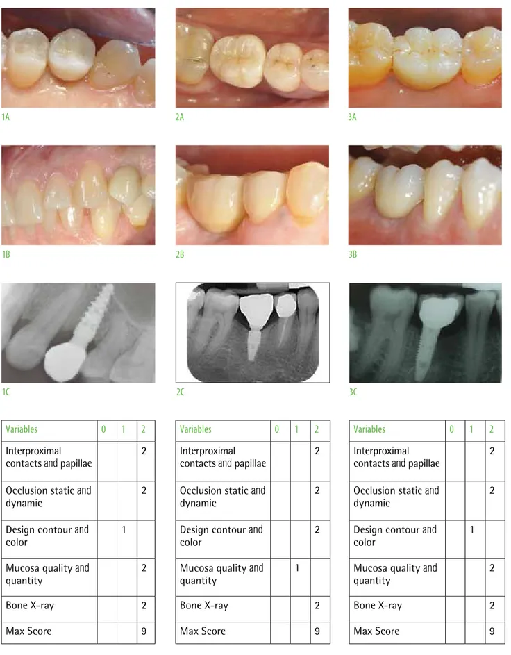
FIG. 1Group 1: restored upper premolar (FDI 24) after 1 year of loading; occlusal (A) and lateral (B) views and radiographic image (C). Application of FIPS revealed a total score of 9. 1A 1B 1C Variables 0 1 2 Interproximal
contacts and papillae 2 Occlusion static and
dynamic
2
Design contour and
color 1
Mucosa quality and
quantity 2
Bone X-ray 2 Max Score 9
2A 3A
FIG. 2Group 2: restored upper premolar (FDI 46) after 1 year of loading; occlusal (A) and lateral (B) views and radiographic image (C). Application of FIPS revealed a total score of 9.
FIG. 3Group 3: restored upper premolar (FDI 46) after one year of loading; occlusal (A)and lateral (B) views and radiographic image (C). Application of FIPS revealed a total score of 9.
2B 3B
2C 3C
Variables 0 1 2
Interproximal
contacts and papillae 2 Occlusion static and
dynamic
2
Design contour and
color 2
Mucosa quality and quantity 1 Bone X-ray 2 Max Score 9
Variables 0 1 2
Interproximal
contacts and papillae 2 Occlusion static and
dynamic
2
Design contour and
color 1
Mucosa quality and
quantity 2
Bone X-ray 2 Max Score 9
max) values. A linear regression analysis was performed for the detection of any significant correlations between the total FIPS scores and the subjective results of the patients’ VAS responses to Q1 and Q2.
A level of significance was set at P < 0.05. Statistic calculations were made with the open-source program “GraphPad Software” (http://www. graphpad.com) (Table 1, 2, 3).
RESulTS
Survival rates for all implants and connected prosthodontic reconstructions were 100%. No technical or biological complications were observed during follow-up.
Clinical examinations exhibited mean full-mouth scores for PI of 20.4±2.5 (range: 16–22) at baseline and 19.5±1.2 (range: 16–22) at 1- year follow-up, PPD of 3.6
±0.4 mm (range: 1–4) and 3.4± 0.4 mm (range: 1–4), and a mean score for BoP of 21.2-+2.5 (range: 17–24) and 19.8±1.2 (range: 16–23), respectively.
The mean total FIPS score was 8.6±1.1 (range: 6–10). In detail, all implants showed a stable level of the alveolar crest without any signs of bone loss in the radiographic analysis. Therefore, the variable “bone” demonstrated the most consistent results and highest scores with a mean value of 2±0.
Similarly mean score was recorded for the variable “occlusion” 2.0±0.
In contrast, mean scores for “design” 1.9±0.7 (range: 0–2), “mucosa” 1.8±0.4 (range: 1–2), and “interproximal” 1.7±0.4 (range: 1–2) were the most challenging to satisfy (Table 2).
Calculations of medians and quantil Q25– Q75 as well as mean total FIPS scoring for each of the five variables, including standard deviations and minimum and maximum values, are summarized in Table 3.
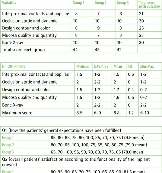
Variables Group 1 Group 2 Group 3 Total score
each outcome
Interproximal contacts and papillae 8 7 6 21 Occlusion static and dynamic 10 10 10 30 Design contour and color 8 9 8 25 Mucosa quality and quantity 8 7 8 23
Bone X-ray 10 10 10 30
Total score each group 44 43 42
TABLe 1Radiographic and clinical scores based on FIPS for each group.
TABLe 2Summarized medians and quantil Q25–Q75 as well as mean FIPS scores including standard deviations (SD) and minimum– maximum (min–max) values for each variable.
TABLe 3Both questions included a visual analogue scale (VAS) ranging from “unsatisfied” to “fully satisfied” (0–100).
N= 20 patients Mediam Q25–Q75 Mean SD Min-Max
Interproximal contacts and papillae 1.5 1-2 1.5 0.6 1-2 Occlusion static and dynamic 2 2-2 2 0 1-2 Design contour and color 1.5 1-2 1.7 0.4 0-2 Mucosa quality and quantity 1.5 1-2 1.6 0.5 0-2
Bone X-ray 2 2-2 2 0 2-2
Maximum score 8.5 6-9 8.8 1.2 6-10
Q1 (how the patients’ general expectations have been fulfilled)
Group 1 85, 80, 65, 75, 90, 100, 85, 70, 70, 75 (79.5 mean)
Group 2 80, 70, 65, 100, 100, 75, 65, 80, 80, 75 (79.0 mean)
Group 3 65, 70, 100, 95, 90, 70, 80, 70, 75, 65 (78.0 mean)
Q2 (overall patients’ satisfaction according to the functionality of the implant crowns)
Group 1 80, 95, 90, 65, 70, 75, 100, 65, 85, 90 (81.5 mean)
Group 2 75, 80, 100, 65, 65, 90, 75, 75, 95, 70 (79.0 mean)
The two questionnaires addressed the patients’ satisfaction according to the treatment outcome. Q1 focused on the fulfillment of the patients’ general expectations. Q2 asked explicitly for the overall patients’ satisfaction according to the functionality of the implant crowns.
All patients marked their level of satisfaction at or above 65% on the VAS for both questions. The mean score of Q1 was 79.0±1.5 (median: 78.8; Q25–Q75: 70– 92; range: 65–100), and 80.5±2.5 for Q2 (median: 80.3; Q25–Q75: 73–95; range: 65–100).
The linear regression analysis showed a statistically significant correlation between the total FIPS score and the VAS response of Q1 and Q2. A moderately correlation was found between FIPS and Q1 and FIPS and Q2, with a coefficient of 0.82 and 0.80 (P < 0.0001) respectively.
dISCuSSIon
The possibility to selectively assess the functional integration of single-implant restorations with an objective, reliable, and quickly applicable score was recently proposed and permits to rationalize patient’s satisfaction, to identify potential failure risks at an early stage of the treatment and to compare follow-up maintenance (9-11).
FIPS seems to be easier and more reliable to be applied than other criteria that have been proposed in the past (18-20). Some of them were mixing different parameters, such as mobility of the implant, radiolucency and substantial bone loss, bleeding and suppuration, the occurrence of technical failures, and esthetics. However, success of a single-implant restoration should ideally consider the long-term outcome of the entire implant– prosthodontic complex as a whole.
FIPS can be now generally accepted and well-established as a reliable assessment tool estimating a score by merging clinical and radiographic findings for the evaluation of implant restorations (in posterior sites) (9-11). In fact, FIPS is an outcome assessment tool that can be helpful, is easy to use, quickly and reproducibly applicable, and implies a clinical relevance for the dentist. The present trial confirmed the applicability of FIPS. This novel functional score is defined by only five variables. In contrast, esthetic indices use much more complex scoring schemes with 10 up to 15 different subcategories of assessment (4-8, 21-23). The ease applicability of FIPS is useful for long term randomized clinical trials and its use also by a wide number of clinicians.
This prospective clinical study evaluated the functional outcomes of a mixed traditional and digital workflow to fabricate single-implant crowns on three different implants after one year of loading using FIPS. It can be considered that the reaction of gingival tissue at inflammatory insults such as a placement of a single-unit restoration, can determine within one year of
clinical service some unfavorable reactions; the presence of a slight improvement of periodontal indices in combination with any bone loss can be a favorable sign of good integration of single-implant restorations. The null hypotheses tested were that, scoring the single-implant restorations with FIPS, there were differences between hexagon and conical connections and among the three tested connections; both were rejected: no differences were found between the type of abutment connections and among the three tested implant systems. That can be due to the high performances reported for both types of connections (24), the proper selection of the cases, the well performed surgical and prosthodontic procedures as well as the proper maintenance of home oral hygiene (25-26); the two null hypotheses will be reevaluated and reported along further recalls.
Although the short-term observation and the limited number of samples of each group can be considered important limitations of this study, a longer observation in a wider number of patients is planned after the three-year follow up.
The summarized analysis of the variables “inter-proximal,” “occlusion,” “design,” “mucosa,” and “bone” revealed a high mean total score of 8.6 of 10 with a relatively narrow range (SD: 1.1), indicating a precise and reliable assessment of FIPS within these groups. Although under optimal conditions, the defined variables of FIPS result in a top score of 10, all examined single- implant restorations always showed a mean score of ≥6, which can be interpreted as a successful (functional) treatment outcome.
Another important aspect was that the patients’ satisfaction was subjectively high with respect to the expected treatment outcome in general. The patients’ satisfaction reflected their expectations, confirming that their perception can correspond to the FIPS. Certainly, the method used to evaluate patients’ perception (VAS) might be improved although in this form it is very easy to be applied, repeatable and reliable (9).
ConCluSIonS
The findings of this randomized clinical trial indicated the potential of both conical and hexagon connections to perform very well after 1 year of clinical service.
FIPS showed to be an objective and reliable instrument in assessing single-implant restoration success.
REFEREnCES
1. Pjetursson Be, Asgeirsson AG, Zwahlen M, Sailer I. Improvements in implant dentistry over the last decade: comparison of survival and complication rates in older and newer publications. Int J oral Maxillofacial Implants 2014; 29(Suppl.): 308–324.
2. Papaspyridakos P, Chen CJ, Singh M, Weber hP, Gallucci Go. Success criteria in implant dentistry: a systematic review. J Dental Res 2012; 91: 242–248. 3. Sadid-Zadeh R, Kutkut A, Kim h. Prosthetic failure in implant dentistry.
Dent Clin North Am 2015; 59: 195–214.
4. Furhauser R, Florescu D, Benesch T, haas R, Mailath G, Watzek G. evaluation of soft tissue around single-tooth implant crowns: the pink esthetic score. Clin oral Implants Res 2005; 16: 639–644.
5. Meijer hJ, Stellingsma K, Meijndert L, Raghoebar GM. A new index for rating aesthetics of implant-supported single crowns and adjacent soft tissues–the implant crown aesthetic index. Clin oral Implants Res2005; 16: 645–649.
6. Belser UC, Grutter L, Vailati F, Bornstein MM, Weber hP, Buser D. outcome evaluation of early placed maxillary anterior single-tooth implants using objective esthetic criteria: a cross-sectional, retrospective study in 45 patients with a 2- to 4-year follow-up using pink and white esthetic scores. J Periodontol 2009; 80: 140–151.
7. Juodzbalys G, Wang hL. esthetic index for anterior maxillary implant-supported restorations. J Periodontology 2010; 81: 34–42.
8. Tettamanti S, Millen C, Gavric J, Buser D, Belser UC, Bragger U, Wittneben JG. esthetic evaluation of implant crowns and peri-implant soft tissue in the anterior maxilla: comparison and reproducibility of three different indices. Clin Implant Dent Related Research 2016; 18: 517–526.
9. Joda T, Ferrari M, Bragger U. A prospective clinical cohort study analyzing single-unit implant crowns after three years of loading: introduction of a novel Functional Implant Prosthodontic Score (FIPS). Clin oral Implants Res 2017 oct;28(10):1291-1295.
10. Joda T, Zarone F, Zitzmann NU, Ferrari M. The Functional Implant Prosthodontic Score (FIPS): assessment of reproducibility and observer variability Clinical oral Investig 2018 Jul;22(6):2319-2324.
11. Joda T, Ferrari M, Bragger U. Monolithic implant-supported lithium disilicate (LS2) crowns in a complete digital workflow: A prospective clinical trial with a 2-year follow-up. Clin Implant Dent Relat Res 2017; 1-7. 12. Pinheiro Feitosa PC, Barbosa de Lima AP, Silva-Concílio LR, Brandt WC,
Stability of external and internal implant connections after a fatigue test eur J Dent 2013;7: 267–271.
13. Machado LS, Bonfante eA, Anchieta RB, yamaguchi S, Coelho PG. Implant-abutment connection designs for anterior crowns: reliability and failure modes. Implant Dent 2013;22:540-5.
14. Mokhtar MA, elnagar G, Saleh M, Radwan MM. The biological complication of implant abutment materials. A systematic review and meta-analysis. J osseointegr 2018; 10 (1): 23-30 .
15. Fujiwara CA, Magro Filho o, oliveira NT, Pereira Queiroz T, Sabagg Abla M, Luiz Pardini C. Assessment of the interface between implant and abutments of five systems by scanning electronic microscopy. J osseointegr 2009; 1(2): 60-66.
16. Löe h, Silness J. Periodontal disease in pregnancy. I Prevalence and severity, Acta odont Scand 1963;21:533–551.
17. Ainamo J, Bay I. Problems and proposals for recording gingivitis and plaque. Int Dent J 1975; 25: 229–235.
18. Chen ST, Buser D. Clinical and esthetic outcomes of implants placed in post extraction sites. Int J oral Maxillofacial Implants 2009; 24(Suppl.): 186– 217.
19. Fuentealba R, Jofre J. esthetic failure in implant dentistry. Dent Clin North Am 2015; 59: 227–246.
20. Le M, Papia e, Larsson C. The clinical success of tooth- and implant-supported zirconia-based fixed dental prostheses. A systematic review. J oral Rehabilitation 2015; 42: 467– 480.
21. Gehrke P, Degidi M, Lulay-Saad Z, Dhom G. Reproducibility of the implant crown aesthetic index–rating aesthetics of single- implant crowns and adjacent soft tissues with regard to observer dental specialization. Clin Implant Dent Relat Res 2009; 11: 201– 213.
22. Vaidya S, ho yL, hao J, Lang NP, Mattheos N. evaluation of the influence exerted by different dental specialty backgrounds and measuring instrument reproducibility on esthetic aspects of maxillary implant-supported single crown. Clin oral Implants Res 2015; 26: 250–256. 23. Levine RA, Nack G. Team treatment planning for the replacement of esthetic
zone teeth with dental implants. Compendium Continuing education in Dentistry 2011; 32: 44–50.
24. Gracis S, Michalakis K, Vigolo P, Vult von Steyern P, Zwahlen M, Sailer I. Internal vs. external connections for abutments/reconstructions: a systematic review. Clin oral Implants Res 2012 oct;23 Suppl 6:202-16 25. Ciancio SG, Lauciello F, Shibly o, Vitello M, Mather M. The effect of an
antiseptic mouthrinse on implant maintenance: plaque and peri‐implant gingival tissues. J Periodontol 1995; 66, 11: 962-5.
26. Kracher CM, Smith WS. oral health maintenance dental implants. Dent Assist 2010;79:27-35.