DOTTORATO DI RICERCA
Scienze Chimiche Ciclo XX
Settore scientifico disciplinare di afferenza: CHIM / 03
Titolo della tesi:
Synthesis and characterization of implants
for bone substitutions made of
biomedical apatites containing silicon
Presentata da: Dott. Simone Sprio
Coordinatore
Dottorato
Relatore
Prof. Vincenzo Balzani
Prof. Norberto Roveri
Co-Relatori
Dott.ssa Anna Tampieri
Dott.ssa Elena Landi
Motivazione e risultati della ricerca
La presente ricerca si inquadra nell’ ambito della risoluzione dei problemi legati alla chirurgia ossea, per la cura e la sostituzione di parti di osso in seguito a fratture, lesioni gravi, malformazioni e patologie quali osteoporosi, tumori, etc…
Vi è un numero elevatissimo di casi nel mondo in cui si rende necessaria la sostituzione di porzioni di tessuto osseo e questo numero è sempre crescente per via del progressivo invecchiamento della popolazione. Questo tipo di intervento è spesso molto invasivo e traumatizzante per il paziente soprattutto nei casi in cui sono coinvolte le ossa principali degli arti o del cranio. Spesso il chirurgo deve intervenire nuovamente a distanza di mesi o di anni, con tutti i traumi legati a simili interventi chirurgici, comportando perciò una sensibile riduzione della qualità della vita del paziente.
Il problema più importante in questo ambito riguarda i materiali in uso per la sostituzione del tessuto osseo. Esiste prova che fin dai tempi più antichi l’ uomo ha utilizzato parti sostitutive in metallo mentre successivamente, in tempi più recenti, manufatti in ceramica ne hanno largamente preso il posto per via delle numerose problematiche legate all’ intro-duzione di metalli all’ interno dell’ organismo. Fin qui, la sostituzione di parti di osso era stata eseguita con il semplice scopo di mantenere la funzione strutturale della parte mancante. Tuttavia, il tessuto osseo è un’ entità molto sofisticata, sia da un punto di vista della sua composizione che della sua struttura morfologica, e questo gli conferisce proprietà uniche di adattabilità alle varie sollecitazioni meccaniche, che lo rendono al tempo stesso leggero, resistente ed elastico e soprattutto in grado di autorigenerarsi localmente in seguito a traumi di estensione limitata. Nelle ultime decadi quindi, con l’ au-mentare dell’ aspettativa di vita e dell’ accresciuto benessere, è diventato via via più rilevante il ricorso a soluzioni in grado di garantire la massima funzionalità della parte sostituita e quindi di consentire al paziente di tornare in breve tempo ad una vita attiva e soddisfacente. In molti casi, quando è possibile, si ricorre al trapianto di osso autologo, che consiste nell’ asportazione e nel successivo trapianto di piccole parti di osso dallo stesso individuo, laddove è possibile, spesso dalla cresta iliaca; questa tecnica permette solo il trapianto di piccole parti di osso e comporta tuttavia un notevole disagio poiché il paziente deve sottoporsi a due interventi chirurgici. In alternativa è anche possibile il trapianto di osso eterologo, ovvero di porzioni di osso provenienti da altro animale; in questo caso,
⎯⎯⎯⎯⎯⎯⎯⎯⎯⎯⎯⎯⎯⎯⎯⎯⎯⎯⎯⎯⎯⎯⎯⎯⎯⎯⎯⎯⎯⎯⎯⎯⎯⎯⎯⎯ ii
tuttavia, non sono pochi i casi di rigetto ed infezione. La soluzione in grado di evitare tutti questi problemi è la realizzazione di un materiale sintetico capace di riprodurre il comportamento dell’ osso naturale sia da un punto di vista chimico che strutturale e da ormai quasi cinquanta anni considerevoli sforzi sono in atto da parte della comunità scientifica per ottenere questo risultato.
Il tessuto osseo è costituito per il 70% da una sostanza minerale, l’ idrossiapatite, a base di calcio, fosforo e piccole quantità di carbonato, magnesio, silicati e altre sostanze in tracce, organizzata in una struttura porosa e complessa, tale da rendere efficiente la distribuzione degli sforzi meccanici; questa struttura minerale è permeata di cellule specializzate, gli osteociti, in grado di regolare l’ attività biomeccanica dell’ intero osso, grazie alle varie sostanze presenti nella parte minerale. Questa componente minerale nuclea su una matrice proteica a base di collagene, che conferisce all’ osso le sue uniche proprietà di elasticità e flessibilità.
Buona parte della comunità scientifica è dunque orientata alla realizzazione di materiali di sintesi biomimetici, ovvero in grado di riprodurre la composizione chimica, morfologica e strutturale dell’ osso naturale. Da un punto di vista chimico, questo consente di stimolare i processi naturali di rigenerazione del tessuto osseo e nel contempo provoca il riassorbimento dell’ impianto di sintesi da parte dell’ organismo. Da un punto di vista morfologico-strutturale, l’ ottenimento di impianti di morfologia complessa e dall’ ade-guata resistenza meccanica favorisce l’ abitazione e la proliferazione delle cellule e la formazione di nuovo tessuto osseo in grado di sostituire progressivamente l’ impalcatura di sintesi impiantata dal chirurgo. La resistenza meccanica è tanto più importante quanto più la porzione di osso sostituito ha funzioni strutturali di sostegno dell’ intero scheletro, quali le ossa lunghe delle gambe e delle braccia.
La ricerca scientifica ha finora ottenuto materiali con struttura di idrossiapatite in grado di esibire vari livelli di biomimetismo di tipo chimico, ossia materiali calcio fosfatici contenenti carbonato e/o magnesio e/o silicio in quantità adeguate per l’ attività cellulare, mostrando una più intensa attività di ricostituzione e rimodellamento del tessuto osseo in confronto con materiali di composizione chimica differente; tuttavia, una perfetta riproduzione della composizione chimica dell’ osso non è ancora stata raggiunta. Analogamente, strutture tridimensionali ceramiche con una distribuzione anisotropa dei pori e delle loro dimensioni sono state altresì ottenute, tuttavia le tecniche di
consolidamento necessarie ad ottenere tali strutture e le limitazioni tecnologiche alla possibilità di ottenere morfologie complesse simili a quelle dell’ osso hanno impedito finora di realizzare impalcature per sostituti ossei aventi proprietà analoghe a quelle del tessuto osseo naturale.
Infine, la resistenza meccanica intrinseca dei materiali bioattivi, cioè in grado di indurre una risposta biologica positiva nell’ organismo, è spesso insufficiente per sopportare gli sforzi e le sollecitazioni biomeccaniche naturali, cosicché si deve ricorrere all’ impiego di materiali ceramici inerti che non sono tuttavia in grado di essere riassorbiti dall’ or-ganismo.
Per quanto riguarda l’ aspetto chimico, il ruolo fondamentale dello ione carbonato nell’ a-patite biologica è da tempo noto, così come quello del magnesio; in tempi più recenti tuttavia gli studi si sono concentrati sugli effetti positivi del silicio sulla stimolazione del- l’ attività cellulare e su tecniche in grado di incorporarlo nella struttura dei materiali destinati ad impianto.
Nella presente ricerca il problema dell’ ottenimento di sostituti ossei biomimetici è stato affrontato da tre differenti prospettive.
In una prospettiva biochimica, si sono sperimentate ed attuate delle metodologie per ottenere polveri di idrossiapatite calcio-fosfatica contenente simultaneamente i sostituti ionici più rilevanti per la stimolazione dei processi naturali di rigenerazione ossea (carbonato, magnesio e silicio), in quantità ritenute adeguate sulla base di precedenti ricerche sul metabolismo cellulare di queste sostanze.
In prospettiva di applicazioni strutturali sono state anche sperimentate ed attuate tecnologie di consolidamento di miscele di polveri di idrossiapatite con silicati di calcio bioattivi per ottenere materiali ad elevata resistenza meccanica da utilizzare in seguito per la realizzazione di ceramici porosi.
Il problema del biomimetismo della morfologia dell’ osso è stato infine affrontato prendendo spunto dalla Natura, in particolare da strutture vegetali come legni e piante che esibiscono naturalmente strutture porose gerarchiche estremamente complesse, grazie alle quali proprietà di resistenza meccanica ed efficiente distribuzione degli sforzi sono coniugate con una elevata leggerezza. In questo modo nel presente lavoro si sono esplorate metodologie del tutto nuove per ottenere strutture ceramiche di idrossiapatite a partire da sostanze naturali, mantenendo la loro medesima morfologia.
⎯⎯⎯⎯⎯⎯⎯⎯⎯⎯⎯⎯⎯⎯⎯⎯⎯⎯⎯⎯⎯⎯⎯⎯⎯⎯⎯⎯⎯⎯⎯⎯⎯⎯⎯⎯ iv
I risultati ottenuti nel corso della presente ricerca sono stati positivi ed incoraggianti sotto tutti e tre gli aspetti investigati.
In primo luogo, da un punto di vista chimico, si sono ottenute polveri di idrossiapatite ad accresciuto biomimetismo, come dimostrato dalle varie metodologie di indagine impiegate e dal loro comportamento in ambiente fisiologico e in coltura cellulare. Il procedimento di sintesi ha permesso il controllo del tenore di ioni sostituenti nella struttura dell’ idros-siapatite, consentendo di ottenere specifiche composizioni sulla base dell’ applicazione desiderata.
In secondo luogo, materiali bioattivi con superiori proprietà meccaniche sono stati ottenuti grazie all’ impiego di silicati di calcio come materiali di rinforzo dell’ idrossiapatite. L’ impiego di tali materiali contenenti silicio ha inoltre aperto la prospettiva di ottenere materiali con accresciuto biomimetismo, oltre che dotati di accresciuta resistenza, grazie ai possibili fenomeni di migrazione di ioni silicio all’ interno della struttura dell’ idros-siapatite durante i trattamenti termici con cui vengono ottenuti i ceramici finali. Questo fenomeno, se confermato, potrà essere applicato ad una numerosa serie di materiali a base di silicati di calcio contenenti anche altri ioni influenti nel biomimetismo osseo, come il magnesio, consentendo di ottenere allo stesso tempo materiali ad accresciuto biomimetismo e resistenza meccanica.
Infine, specifici processi chimici e termici sono stati applicati ad alcune strutture lignee naturali quali abete, pino e larice, consentendo di trasformarli in idrossiapatite, mantenendo nel contempo l’ originaria morfologia porosa e gerarchicamente organizzata. Questo risultato è di assoluta novità, essendo stato applicato in passato solo per l’ ottenimento di sostanze dalla composizione più semplice, come il carburo di silicio (che può essere considerato peraltro un materiale bioinerte). Un’ idrossiapatite caratterizzata da una struttura gerarchica simile all’ osso costituisce potenzialmente la migliore soluzione per una rapida ed efficiente integrazione del nuovo tessuto osseo nell’ impianto di sintesi. Allo scopo di migliorare la riassorbibilità di simili impianti, l’ idrossiapatite deve presentare differenti sostituzioni ioniche, pertanto le ricerche future in questo campo dovranno essere orientate all’ incorporazione di ioni nelle strutture gerarchiche di idrossiapatite derivate da strutture lignee, oltre che alla determinazione delle loro proprietà meccaniche in relazione alla morfologia.
I tre aspetti del biomimetismo affrontati in questo lavoro, chimico, strutturale e morfologico, hanno delineato tre vie maestre che sono orientate a convergere nella realizzazione del sostituto osseo ideale. La ricerca futura dovrà essere rivolta particolarmente alla convergenza dei tre differenti risultati ottenuti, superando le difficoltà che questo comporta. Infatti un accresciuto biomimetismo è associato ad una ridotta stabilità chimica da parte dell’ idrossiapatite, analogamente a quanto avviene nell’ apatite naturale dell’ osso; questa instabilità si verifica soprattutto alle alte temperature, dove hanno luogo i processi di realizzazione di materiali bioattivi strutturalmente resistenti (sinterizzazione); da un punto di vista morfologico inoltre, una volta accertata la fattibilità delle trasformazioni chimiche di legni e piante in ceramici porosi a base di idrossiapatite, la ricerca dovrà essere rivolta alla selezione delle strutture legnose più adeguate in base alla loro porosità e resistenza meccanica intrinseca, e all’ adeguamento dei processi chimici e termici messi a punto nel presente lavoro allo scopo di migliorarne il biomimetismo chimico e meccanico.
⎯⎯⎯⎯⎯⎯⎯⎯⎯⎯⎯⎯⎯⎯⎯⎯⎯⎯⎯⎯⎯⎯⎯⎯⎯⎯⎯⎯⎯⎯⎯⎯⎯⎯⎯⎯ vi
L
IST OF
C
ONTENT
Page
LIST OF TABLES……….………..xi
LIST OF FIGURES………...xiii
1. GENERAL TOPICS………1
1.1 Biology of bone tissue………..1
1.2 Morphology and structure of bone tissue……….4
1.2.1 Hierarchical structure of bone………...6
1.2.2 Mechanical properties of bone………..7
1.2.3 Types of bone………8
1.3 Bone remodelling……….9
1.4 Need of the development of bone substitutes………12
1.4.1 Current solutions for bone substitutes…….………14
1.4.2 Synthetic bone substitutes………17
1.4.3 Bioceramics……….18
1.5 Calcium phosphates in biologic systems………20
1.5.1 Calcium phosphate dehydrate, Brushite (DCPD)………22
1.5.2 Tricalcium phosphate (TCP)………23
1.5.3 Amorphous calcium phosphates (ACP)………...24
1.6 Bioactivity of calcium phosphates………..24
1.7 Apatites………...26
1.7.1 Hydroxyapatite: structure and chemistry……….26
1.7.2 Biological apatites………...28
1.7.3 Effect of carbonate……….………..29
1.7.4 Effect of magnesium………30
1.7.5 Effect of silicon………30
1.8 Methods for synthesis of HA………32
⎯⎯⎯⎯⎯⎯⎯⎯⎯⎯⎯⎯⎯⎯⎯⎯⎯⎯⎯⎯⎯⎯⎯⎯⎯⎯⎯⎯⎯⎯⎯⎯⎯⎯⎯⎯ viii
1.10 Thermal behaviour of apatites………..41
1.11 Characterization of apatite biomaterials………...43
References………47
2. ANALYTICAL TECHNIQUES……….55
2.1 X-Ray Diffraction (XRD)………...55
2.2 Fourier Transform Infrared Spectroscopy (FTIR)………..61
2.3 Inductively Coupled Plasma Optical Emission Spectroscopy (ICP-OES)………..63
2.4 Scanning Electron Microscopy (SEM)………...64
2.5 Thermogravimetry-Thermoanalysis (TG-DTA)………68
2.6 Powder Analysis……….69
2.6.1 Specific Surface Area………...69
2.6.2 Electro-acoustic spectroscopy……….71
3. DEVELOPMENT OF MULTI-SUBSTITUTED
HYDROXYAPATITE……….73
3.1 Synthesis of multi-substituted hydroxyapatite powders……….73
3.2 Characterization of multi-substituted hydroxyapatite powders………..76
3.2.1 Phase analysis by XRD………79
3.2.2 Chemical analysis by ICP………83
3.2.3 Thermal analysis………..86
3.2.4 Chemical analysis by FTIR……….89
3.2.5 Powder analysis………...93
3.2.6 Solubility tests……….98
3.3 Thermal stability of multi-substituted HA powders……….105
3.4 HA powder substituted with strontium ,carbonate, silicon and magnesium………...110
3.5 In vitro tests………..114
3.5.1 Proliferation tests on Si-HA characterized by different silicon content………...114
3.6 Conclusions………..116
References………..119
4. DEVELOPMENT OF REINFORCED HA-BASED
BIOMATERIALS………...121
4.1 Need of reinforced bone substitutes……….121
4.2 Calcium silicates as biomaterials……….123
4.3 Development of composites HA / silico-carnotite (Ca5(PO4)2SiO4)………...125
4.3.1 Powder synthesis and preparation of mixtures ………..125
4.3.2 Characterization……….126
4.3.3 Cytotoxicity tests………...130
4.4 Development of composites HA / β, γ-Ca2SiO4………...131
4.4.1 Structure and phase stability in calcium disilicate system………….131
4.4.2 Synthesis of β and γ-Ca2SiO4………132
4.4.3 Preliminary sintering tests……….133
4.5 Development of dense ceramic composites by Fast Hot Pressing (FHP).……….………...134
4.5.1 Sample preparation………135
4.5.2 Sample characterization……….136
4.6 Mechanical characterization of HA and composite ceramics………..140
4.6.1 Sample preparation and mechanical testing………..140
4.6.2 Physical-chemical characterization………...143
4.6.3 Mechanical characterization………..148
4.7 Conclusions………..151
References………..154
5. HIERARCHICALLY
ORGANIZED
STRUCTURES
AS
BONE
SCAFFOLDS………...159
5.1 Introduction………..159
⎯⎯⎯⎯⎯⎯⎯⎯⎯⎯⎯⎯⎯⎯⎯⎯⎯⎯⎯⎯⎯⎯⎯⎯⎯⎯⎯⎯⎯⎯⎯⎯⎯⎯⎯⎯ x
5.3 Ceramization of native vegetable structures……….162
5.4 The European Project TEM-PLANT………170
5.5 Characterization of the transformed woods………..170
5.6 Conclusions………..179
References………..181
FINAL CONCLUSIONS AND FUTURE PERSPECTIVES………183
L
IST OF
T
ABLES
Page
Table 1.I. General properties of different bone grafts………..17
Table 1.II. Present uses of bioceramics………18
Table 1.III. Classification of Bioceramics on the basis of tissue attachment………...19
Table 1.IV. Phosphate minerals present in human tissues..……….22
Table 1.V. Atomic composition of human hard tissues………...29
Table 3.I. Amount of reagents employed in the synthesis of multi-substituted HA powders………...75
Table 3.II. Chemical analysis of the multi-substituted HA powders………..…….84
Table 3.III. Weight fraction of the various elements in the multi-substituted HA powders………..85
Table 3.IV. Percent weight loss in the range 100-400 °C………89
Table 3.V. Particle characteristics in multi-substituted HA……….94
Table 3.VI. Release kinetic data of multi-substituted HA powders in SBF at 37 °C………..104
Table 3.VII. Phase transformation in Si-CHA powders during sintering……….107
Table 3-VIII. Phase transformation in Si-MHA powders during sintering………...109
Table 3.IX. Chemical composition of Sr-Si-MCHA powder……….114
Table 3.X. Weight fraction of the various elements in Sr-Si-MCHA powder…………...114
Table 4.I. Main mechanical properties of HA compared to cortical bone……….122
Table 4.II. Mechanical properties of different bioactive calcium silicates in comparison with cortical bone………124
Table 4.III. Comparison between different FHP treatments performed on HA powder………...137
Table 4.IV. Samples to be mechanically tested obtained by FHP……….140
Table 4.V. Results of flexural strength tests performed on HA and composite………….148
Table 4.VI. Results of nanoindentation tests on HA and composite………..149
Table 4.VII. Mechanical behaviour of HA and composite materials under macroscopic investigation………...149
⎯⎯⎯⎯⎯⎯⎯⎯⎯⎯⎯⎯⎯⎯⎯⎯⎯⎯⎯⎯⎯⎯⎯⎯⎯⎯⎯⎯⎯⎯⎯⎯⎯⎯⎯⎯ xii
L
IST OF
F
IGURES
Page
Figure 1.1. Schematic drawing of an osteoblast………...……..2
Figure 1.2. Schematic drawing of the cross section of an osteocyte………..3
Figure 1.3. Schematic drawing of an osteoclast……….4
Figure 1.4. Schematic drawing of the bone structure……….5
Figure 1.5. Hierarchical structure of bone………..6
Figure 1.6. Young's modulus of trabecular bone as a function of density of bone…………8
Figure 1.7. Different types of bone………9
Figure 1.8. Bone remodelling: effect of reduction (from A to B) and of intensification of strain (from B to A) on bone trabecules………...10
Figure 1.9. Bone resorption………..10
Figure 1.10. Bone deposition………...11
Figure 1.11. Schematic diagram of the Davy and Hart model for bone remodelling……..12
Figure 1.12. Effect of age on strength and density of bone………..13
Figure 1.13. Comparison of the different chemical activities of different ceramic-based biomaterials……….…20
Figure 1.14. Examples of bioceramic implants for different applications……….…..21
Figure 1.15. Crystal structure of Hydroxyapatite………..…...26
Figure 1.16. Structure of monoclinic hydroxyapatite………..……27
Figure 1.17. View of the two possible arrangement of CO3 moieties around the PO4 tetrahedron………37
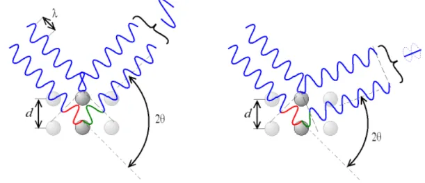
Figure 2.1. Constructive and destructive interference in Rayleigh scattering of X rays………...…..…56
Figure 2.2. The 14 possible Bravais lattices………58
Figure 2.3. Some examples of Miller planes………59
Figure 2.4. Diffractometer adopting the Bragg-Brentano geometry………61
Figure 2.5. Possible vibrations detected by infrared spectroscopy………..62
Figure 2.6. Scheme of equipment for infrared spectroscopy………...62
Figure 2.7. Signals produced by the interaction of the electron beam with the sample………...64
⎯⎯⎯⎯⎯⎯⎯⎯⎯⎯⎯⎯⎯⎯⎯⎯⎯⎯⎯⎯⎯⎯⎯⎯⎯⎯⎯⎯⎯⎯⎯⎯⎯⎯⎯⎯ xiv
Figure 2.8. Scheme of a Scanning Electron Microscope……….65
Figure 2.9. Interaction of the electron beam with the electronic cloud………68
Figure 2.10. Schematic of double layer in a liquid at contact with a solid………..71
Figure 2.11. Equipment for electro-acoustic spectroscopy………..72
Figure 3.1. Equipment for the synthesis of HA powder………...74
Figure 3.2. Effect of osteporosis on the bone trabeculae (right)………..76
Figure 3.3. Equipment for solubility tests on multi-substituted HA granulates…………...79
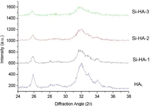
Figure 3.4. Comparison between XRD spectra of stoichiometric HA (HAL) and Si-HA with different content of silicon……….80
Figure 3.5. Comparison between XRD spectra of CHA and Si-CHA with different carbonate content………...81
Figure 3.6. Comparison between the XRD spectra of multi-substituted HA powders containing magnesium, silicon and carbonate………82
Figure 3.7. Crystallization of amorphous Si-CHA-1 (a) and Si-MCHA-1 (b) after treatment at 700 °C………..83
Figure 3.8. Thermogravimetric analysis of multi-substituted HA powders……….87
Figure 3.9. TG-DTA curves of Si-CHA-1 and Si-MCHA-1 powders……….88
Figure 3.10. TG-DSC of pure CaCO3 powder……….88
Figure 3.11. Comparison between TG spectra of Si-CHA-1 and CaCO3………89
Figure 3.12. FTIR spectra of multi-substituted HA powders………...90
Figure 3.13. Effect of silicon on hydroxyl bands in Si-HA powders………...91
Figure 3.14. Water adsorption bands in Si-CHA-1 and Si-MCHA -1 powders…………...92
Figure 3.15. Typical morphology of a silicon-substituted HA……….93
Figure 3.16. Self-agglomeration of HA powders……….94
Figure 3.17. Particle size distribution of multi-substituted HA powders.: a) differential; b) cumulative………..96
Figure 3.18. Electrical conductivity of multi-substituted HA powders………...97
Figure 3.19. ζ-potential vs. pH of multi-substituted HA powders………...98
Figure 3.20. Typical morphology of a granulated HA……….99
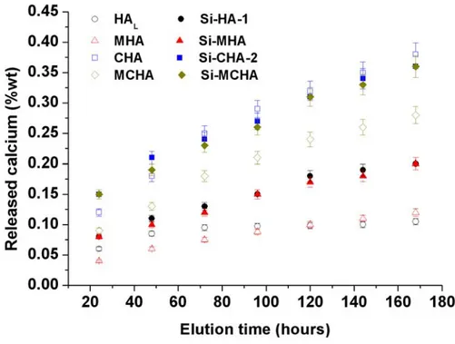
Figure 3.21. Cumulative release of calcium by multi-substituted HA powders…………100
Figure 3.22. Cumulative release of magnesium by multi-substituted HA powders……..101
Figure 3.24. Effect of silicon on the formation of α-TCP after sintering………...106
Figure 3.25. DSC curves of Si-HA powders………..106
Figure 3.26. DSC curves of Si-CHA powders………...108
Figure 3.27. Effect of silicon on MHA powder……….109
Figure 3.28. Effect of silicon on MCHA powder………..110
Figure 3.29. XRD spectrum of Sr-Si-MCHA powder………...111
Figure 3.30. Thermal behaviour of Sr-Si-MCHA powder……….112
Figure 3.31. XRD spectrum of Sr-Si-MCHA fired at 700 °C………112
Figure 3.32. Formation of β-TCP in Sr-Si-MCHA powder………...113
Figure 4.1. XRD spectrum of the calcined gel, precursor of silico-carnotite phase……..126
Figure 4.2. XRD spectrum of the calcined gel after the thermal treatment of transformation in silico-carnotite phase……….127
Figure 4.3. Decomposition of silico-carnotite after firing at different temperatures…….128
Figure 4.4. XRD spectra of mixtures HA-silicocarnotite after sintering at 1250 °C…….129
Figure 4.5. Microstructure of HA pure and doped with different amounts of silico-carnotite: a) 5%; b) 10%, c) 20%; d) 50%...130
Figure 4.6. Polymorphic transformations of Ca2SiO453………131
Figure 4.7. XRD spectrum of as-synthesized β-Ca2SiO4………...132
Figure 4.8. XRD spectrum of as-synthesized γ-Ca2SiO4………...133
Figure 4.9. Phase composition of mixture HA-γ-Ca2SiO4 after sintering in air at different temperatures……….134
Figure 4.10. Scheme of the equipment for FHP……….135
Figure 4.11. Sample preparation for FHP experiments………..136
Figure 4.12. XRD spectra of stoichiometric HA heat treated by FHP………...137
Figure 4.13. Delamination occurring during FHP of HA under excessive applied pressure………138
Figure 4.14. Different grain growth in external (a) and internal part (b) of a HA sample heated at 1500 °C (100 °C/min)……….139
Figure 4.15. Scheme of the flexural strength test performed on sample bars………141
Figure 4.16. Geometry of a Berkovich indenter tip………...142
Figure 4.17. Example of a load-displacement curve during loading and unloading……..142 Figure 4.18. Comparison between densification curves of HA
⎯⎯⎯⎯⎯⎯⎯⎯⎯⎯⎯⎯⎯⎯⎯⎯⎯⎯⎯⎯⎯⎯⎯⎯⎯⎯⎯⎯⎯⎯⎯⎯⎯⎯⎯⎯ xvi
at different heating rates………...143 Figure 4.19. XRD spectrum of γ-Ca2SiO4 treated at 1500 °C by FHP………..144 Figure 4.20. Morphology of as-obtained γ-Ca2SiO4………..145 Figure 4.21. Comparison between XRD of HA and HA / Ca2SiO4 composite…………..145 Figure 4.22. Microstructures of HA obtained by FHP at different temperatures.
a): 1500 °C; b) 1400 °C; c) 1300 °C………146 Figure 4.23. Microstructures of composites obtained by FHP
at different temperatures. a): 1500 °C; b) 1400 °C; c) 1300 °C…………...147 Figure 4.24. EDS analysis performed on several grains of
HA-β-Ca2SiO4 composite sintered at 1500 °C by FHP………147 Figure 5.1. Hierarchical structure of wood……….161 Figure 5.2. Comparison between mechanical performances of different classes
of materials………...162 Figure 5.3. Reaction pathway to obtain hydroxyapatite scaffolds starting
by native wood………...164 Figure 5.4. Typical thermal cycle in carburization process………...166 Figure 5.5. Sample arrangement in calcium infiltration. A: liquid phase;
B: gaseous phase……….168 Figure 5.6. Morphology of pyrolyzed larch-wood……….171 Figure 5.7. XRD spectrum of pyrolyzed wood………..171 Figure 5.8. XRD spectrum of transforming CaC2 wood-like structure,
when exposed to air……….172 Figure 5.9. Formation of calcium carbide into the pores of carbon preform……….173 Figure 5.10. Extensive formation of CaC2 particles into the pore of carbon preform…...173 Figure 5.11. Incomplete carburization in liquid-phase infiltration………174 Figure 5.12. SEM micrographs of pyrolysed wood before and after
the transformation in CaC2.………..174 Figure 5.13. Calcium carbide obtained by initial molar ratio Ca/C >> 1 and
Ca/C = 1………175 Figure 5.14. Comparison between XRD analyses of two different
thermal cycles to obtain CaO………...176 Figure 5.15. SEM micrographs of calcium oxide porous template………176
Figure 5.16. XRD analysis after carbonation process by autoclave
(400°C, 22bar PCO2, 24 hours)………...177
Figure 5.17. Carbonation percentage vs time of reaction in kiln (700°C)……….177 Figure 5.18. SEM micrograph calcium carbonate structure………...178 Figure 5.19. XRD analysis after hydrothermal treatment of calcium carbonate
⎯⎯⎯⎯⎯⎯⎯⎯⎯⎯⎯⎯⎯⎯⎯⎯⎯⎯⎯⎯⎯⎯⎯⎯⎯⎯⎯⎯⎯⎯⎯⎯⎯⎯⎯⎯ xviii
C
HAPTER
1
G
ENERAL
T
OPICS
1.1 Biology of bone tissue
Bones are rigid organs that form part of the endoskeleton of vertebrates. They
function to move, support, and protect the various organs of the body, produce red and white blood cells and store minerals. Because bones come in a variety of shapes and have a complex internal and external structure, they are lightweight, yet strong and hard, in addition to fulfilling their many other functions. One of the types of tissues that makes up bone is the mineralized osseous tissue, also called bone tissue, that gives it rigidity and honeycomb-like three-dimensional internal structure. Other types of tissue found in bones include marrow, endosteum and periosteum, nerves, blood vessels and cartilage. There are 206 bones in the adult body and about 300 bones in the infant body.
Bone tissue consists of cells embedded in a fibrous, organic matrix, the osteoid, which is primarily constituted by Type I collagen (90%) and 10% amorphous ground substance (primarily glycosaminoglycans and glycoproteins). Osteoid comprises approximately 50% of bone by volume and 25% by weight.
Osteoblasts (from the Greek words for "bone” and "germ" or embryonic) are
mono-nucleate cells, descending from osteoprogenitor cells, that are responsible for the secretion of osteoid, and subsequent bone formation through the mineralization of the osteoid matrix.
General Topics
⎯⎯⎯⎯⎯⎯⎯⎯⎯⎯⎯⎯⎯⎯⎯⎯⎯⎯⎯⎯⎯⎯⎯⎯⎯⎯⎯⎯⎯⎯⎯⎯⎯⎯⎯⎯
Figure 1.1. Schematic drawing of an osteoblast.
Osteoblasts are located on the surface of osteoid seams and also manufacture hormones, such as prostaglandins, to act on the bone itself (see Fig. 1.1). They robustly produce alkaline phosphatase, an enzyme that has a role in the mineralisation of bone, as well as many matrix proteins. Osteoblasts are the immature bone cells. When osteoblasts are trapped in the bone matrix which they themselves produced they become star-shaped cells named osteocytes, the most abundant cell found in bone.
Osteocytes are mature bone cells, networked to each other via long processes that occupy
tiny canals called canaliculi, which are used for exchange of nutrients and waste (see Fig. 1.2). The space that an osteocyte occupies is called a lacuna (Latin for a pit). Although osteocytes have reduced synthetic activity and, like osteoblasts are not capable of mitotic division, they are actively involved in the maintenance of bony matrix, through various mechanosensory mechanisms regulating the bone's response to stress.
Bone is a dynamic tissue that is constantly being reshaped by osteoblasts, cells which build bone, and osteoclasts, which resorb bone.
Figure 1.2. Schematic drawing of the cross section of an osteocyte.
Osteoclast (from the Greek words for "bone" and "broken") are multi-nucleated cells
responsible for the resorption of bone through the removal of the bone's mineralized matrix (see Fig. 1.3). The removal process begins with the attachment of the osteoclast to the osteon (predominant structures found in compact bone, see later); the osteoclast then induces an infolding of its cell membrane and secretes collagenase and other enzymes important in the resorption process, such as tartrate resistant acid phosphatase, secreted against the mineral substrate. High levels of calcium, magnesium, phosphate and products of collagen will be released into the extracellular fluid as the osteoclasts tunnel into the mineralized bone, also resulting in a transfer of calcium from bone fluid to the blood. During childhood, bone formation exceeds resorption, but as the aging process occurs, resorption exceeds formation.
The characteristic rigidity and strength of bone derives from the presence of mineral salts, that permeate the organic matrix, formed by the osteoid mineralization, due to the secretion of vesicles containing alkaline phosphatase, by the osteoblasts. This cleaves the phosphate groups and acts as the foci for calcium and phosphate deposition. The vesicles then rupture and act as a centre for crystals to grow on. The mineral phase comprises approximately 50% of bone by volume and 75% by weight. The principal constituents of bone mineral are calcium phosphate, mainly carbonated hydroxyapatite, amorphous calcium phosphate and calcium carbonate, with lesser quantities of sodium, magnesium, silicon and fluoride.
General Topics
⎯⎯⎯⎯⎯⎯⎯⎯⎯⎯⎯⎯⎯⎯⎯⎯⎯⎯⎯⎯⎯⎯⎯⎯⎯⎯⎯⎯⎯⎯⎯⎯⎯⎯⎯⎯
Figure 1.3. Schematic drawing of an osteoclast.
1.2 Morphology and structure of bone tissue
Fig. 1.4 illustrates the structure of bone. Whole bones are composed of two types of bony tissue: cortical and trabecular.
Cortical bone, also known as compact bone, is dense and forms the surface of bones,
contributing 80% of the weight of a human skeleton. It is extremely hard, formed of multiple stacked layers with few gaps. Cortical bone comprises the diaphysis of long bones and the thin shell that surrounds the metaphyses.
Cortical haversian bone consists of quasi-cylindrically shaped element called osteons or
haversian system. The individual haversian system themselves are composed of
concentric lamellae about 3 to 7 µm thick. These thin lamellae, in turn, are constructed from wrapped collagen fibres impregnated at regularly spaced sites with hydroxyapatite and other mineral crystals about 20 to 40 nm long.
Osteons are typically about 200 µm in diameter, the same thickness as the laminae in laminar bone, and about 10 to 20 mm long. The thickness is the same because bloody supply for the haversian system is a central lumen containing a blood vessel, and thus every point in the haversian system is no more than 100 µm from the bloody supply, as was the case with laminar bone. Haversian bone is organized to accommodate small arteries, arterioles, capillaries, and venues of the microcirculation system.
Figure 1.4. Schematic drawing of the bone structure.
The osteons of haversian bone and the laminae of laminar bone are basically just different geometric configurations of the same material. In both geometric configurations no point in the tissue is more than 100 µm away from the bloody supply.
Trabecular bone (also known as cancellous, or spongy) is a type of osseous tissue with a
low density and strength but very high surface area, that fills the inner cavity of long bones, as a three-dimensional, interconnected network of trabecular rods and plates. A network of rods produces low-density, open cells, whereas a network of plates can result in higher density, nearly closed cells. The external layer of cancellous bone contains red bone marrow where the production of blood cellular components (known as hematopoiesis) takes place. Cancellous bone is also where most of the arteries and veins of bone organs are found.
The classification of bone tissue as cortical or trabecular is based on relative density, the ratio of specimen density to that of fully dense cortical bone (usually assumed to have a density of 1.8 g⋅cm-3). The relative density of trabecular bone varies from 0.05 to about 0.7, corresponding to porosities that ranges from about 30 to more than 90%. The relative density of cortical bone ranges from about 0.7 to about 0.95. Obviously, the distinction between low-density cortical bone and high-density cortical bone is somewhat arbitrary. Bone is first deposited as woven bone, in a disorganized structure with a high proportion of osteocytes in young and in healing injuries. Woven bone is weaker, with a small number of randomly oriented collagen fibres, but forms quickly. It is replaced by lamellar bone,
General Topics
⎯⎯⎯⎯⎯⎯⎯⎯⎯⎯⎯⎯⎯⎯⎯⎯⎯⎯⎯⎯⎯⎯⎯⎯⎯⎯⎯⎯⎯⎯⎯⎯⎯⎯⎯⎯ which is highly organized in concentric arranged laminae with a low proportion of osteocytes. Lamellar bone is stronger and filled with many collagen fibres parallel to other fibres in the same layer. The fibres run in opposite directions in alternating layers, much like plywood, assisting in the bone's ability to resist torsion forces. After a break, woven bone quickly forms and is gradually replaced by slow-growing lamellar bone on pre-existing calcified hyaline cartilage through a process known as bony substitution.
The thickness of the lamina is about 200 µm. Between each lamina and the next there is a net-like system of load vessels. Occasional large radial vessels through lamina connect the net.
1.2.1 Hierarchical structure of bone
The whole architecture of bone is very complex: starting from the smallest constituting elements to the largest scales, the bone is not only organized in an anisotropic manner, but the arrangement of its constituting elements is hierarchical; this means that its structural units are organized at increasing size levels, and this feature confers unique properties to the whole bone structure.
Figure 1.5. Hierarchical structure of bone.
In Fig. 1.5 the hierarchical structure of bone is illustrated, starting from the lowest level, where nanometer-sized crystals of carbonate apatite are embedded in and surround the fibrous protein collagen. At the second lowest level, these mineralized fibres lie bundled together and are attached to each other. At the next level, these fibres come together to
form lamellae at a width of ~2 µm. The lamellae have various patterns; a very common one is a secondary osteon, in which concentric lamellae form cylindrical structures, ~200 µm in diameter, surrounding a central blood vessel. Compact bone, solid to the naked eye, is modified in places to form trabecular bone, which consists of many struts; with the spaces between the struts filled with marrow. As above stated, the struts are not just randomly arranged, but are related to the direction of mechanical loads on the bone. Mechanical and biological properties of the bone depend on the interaction taking place across all levels of organization. A deeper discussion on hierarchy in bone tissue and on the activity devoted to reproduce it by taking advantage of the hierarchic natural structure of woods and plants will be provided in Chapter 5.
1.2.2 Mechanical properties of bone
The different structures of cortical bone and trabecular bone result in different mechanical properties. Bone mechanical properties are highly variable according to species, age, anatomical site, liquid content, etc.
Cortical bone is an anisotropic material, meaning that its mechanical properties vary according to the direction of load.
Cortical bone is often considered an orthotropic material. Orthotropic materials are a class of anisotropic materials characterized by three different Young's moduli E1, E2, E3 according to the direction of load, three shear moduli G12, G13, G23 and six Poisson's ratios ν12, ν13, ν23, ν21, ν31, ν32.
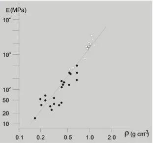
The mechanical characterization of trabecular bone is even more difficult. The mechanical properties of trabecular bone as a whole are due to the mechanical characteristics of single trabecules and to its highly porous structure. Fig. 1.6 shows the dependence of the Young's modulus of trabecular bone from bone density.
General Topics
⎯⎯⎯⎯⎯⎯⎯⎯⎯⎯⎯⎯⎯⎯⎯⎯⎯⎯⎯⎯⎯⎯⎯⎯⎯⎯⎯⎯⎯⎯⎯⎯⎯⎯⎯⎯
Figure 1.6. Young's modulus of trabecular bone as a function of density of bone.
1.2.3 Types of bone
Bone characterized by different functions in the body are strongly different in shape, so that they exhibit different structures hierarchically organized and, correspondingly, different mechanical properties. Several examples are shown in Fig. 1.7: long bones are found in our extremities (femur, tibia, ulna, radius, humerus, etc..) and provide stability against bending and buckling; they are characterized by a tubular shaft and articular surface at each end. Short bones, which include vertebrae, the head of femur, all of the metacarpals and phalanges in the hands, the metatarsals and phalanges in the feet, and the clavicle (collarbone), provide stability against compression (along the vertical axis, in the case of vertebra). They also have a tubular shaft and articular surfaces but smaller than long bones. Plate-like bones (such as the skull, the scapula, the ribs, and the sternum) are thin and have broad surfaces; their main function is to protect vital organs. Irregular bones are also found in the body; they are irregular in size and shape and are usually quite compact. They include the bones in the vertebral column, the carpal bones in the hands, tarsal bones in the feet, and the patella (kneecap).
Figure 1.7. Different types of bone.
1.3 Bone remodelling
Bone adapts and remodels in response to the stress applied. Wolff's law1 states that bones develop a structure most suited to resist the forces acting upon them, adapting both the internal architecture and the external conformation to the change in external loading conditions. This change follows precise mathematical laws.
When a change in loading pattern occurs stress and strain fields in the bone change accordingly. Bone tissue seems to be able to detect the change in strain on a local bases and then adapts accordingly. The internal architecture is adapted in terms of change in density and in disposition of trabecules and osteons and the external conformation in terms of shape and dimensions. When strain is intensified new bone is formed. On a microscopic scale bone density is raised and on a macroscopic scale the bone external dimensions are incremented. When strain is lowered bone resorption takes place. On a microscopic scale bone density is lowered and on a macroscopic scale the bone external dimensions are reduced (Fig. 1.8).
General Topics
⎯⎯⎯⎯⎯⎯⎯⎯⎯⎯⎯⎯⎯⎯⎯⎯⎯⎯⎯⎯⎯⎯⎯⎯⎯⎯⎯⎯⎯⎯⎯⎯⎯⎯⎯⎯
Figure 1.8. Bone remodelling: effect of reduction (from A to B) and of intensification of strain (from B to A) on bone trabecules.
When the change in strain is due to a change in direction of load on a microscopic scale the structure of trabecules and osteons is rearranged and on a macroscopic scale a change in bone shape may occur.
Remodelling is carried out by the cellular component of bone. When resorption takes place osteoclasts reabsorb collagen and mineral phase (Fig. 1.9A) which are then taken into the circulatory system (Fig. 1.9B). During deposition osteoblasts group on the deposition surface and build the collagen network of bone (Fig. 1.10A). Mineralization takes place afterwards (Fig. 1.10B).
Figure 1.9. Bone resorption.
Figure 1.10. Bone deposition.
Bone resorption and bone deposition processes are always active in bone. An equilibrium strain state exists in correspondence to which the two activities are perfectly balanced. When strain intensity is higher than the equilibrium strain deposition activity is more intense than resorption activity and net deposition occurs. When strain intensity is lower than the equilibrium strain deposition activity is less intense than resorption activity and net resorption occurs. Dynamical equilibrium between resorption and deposition is again achieved when the equilibrium strain state is newly established.
The cell-biology based model of Davy and Hart expresses functional dependence of bone remodelling on the strain field, based on cell activity (Fig. 1.11). The load applied to the bone together with geometric and material properties determine the local strain. Strain is detected by a transducer which generates the strain remodelling potential (SRP). This signal is modulated by genetic, hormonal and metabolic factors, generating the remodelling potential which regulates the recruitment rate and the activity of osteoblasts and osteoclasts, stimulating bone formation and bone resorption. The balance between bone deposition and bone resorption determines the net bone remodelling. remodelling modifies bone geometric and material properties through a feedback loop.
General Topics
⎯⎯⎯⎯⎯⎯⎯⎯⎯⎯⎯⎯⎯⎯⎯⎯⎯⎯⎯⎯⎯⎯⎯⎯⎯⎯⎯⎯⎯⎯⎯⎯⎯⎯⎯⎯
Figure 1.11. Schematic diagram of the Davy and Hart model for bone remodelling.
1.4 Need of the development of bone substitutes
Bone disease is a serious health condition that directly impacts on the quality of life of sufferers, particularly among the aged. In most cases, the treatment of bone defects requires a bone graft, and sometimes in extensive amount2-7.
Nowadays, more than 2 million bone grafting procedures are happening annually worldwide, with a turn-over of the order of billions of dollars per annum, in order to repair bone defects in orthopaedics, neurosurgery and dentistry8-9
, which include the treatment of posttraumatic skeletal complications, such as delayed unions, non-unions, mal-unions10. Bone grafting may be also required for the healing of spinal fusions, filling defects following removal of bone tumours and several congenital diseases.
A major contributor to the need for “spare parts” for the body is the progressive deterioration of tissue with age. Bone is especially vulnerable to fracture in older people because of a loss of bone density and strength with age. Fig. 1.12 summarizes the effect of time on bone strength and density from the age of 30 years onward.
Figure 1.12. Effect of age on strength and density of bone.
The effect is especially severe in women because of hormonal changes associated with menopause. Bone density decreases because bone-growing cells (osteoblasts) become progressively less productive in making new bone and repairing micro-fractures. The lower density greatly deteriorates the strength of the cancellous bone, in the ends of long bones and in vertebrae. An unfortunate consequence is that many old people fracture their hips or have collapsed vertebrae and spinal problems.
In addition, bone composition and properties can vary considerably within a population and are difficult to control. Thus remodelling of the implant can be very different from one person to another and from one implant to another.
The replacement of a bone or a part of must be distinguished from organ grafts (e.g. kidney, heart). In organ grafts, the cells are generally kept alive and the reestablishment of blood supply restores the functions of the whole organ. In bone tissues, however, the blood supply cannot be efficiently restored and the tissue is condemned to die, be reabsorbed, and replaced. In most cases the replacement of bone fragments by foreign bone tissue leads to poor junction, rehabitation, and remodelling problems.
As the average age of the population increases and the patients receiving such implants become younger, the above-mentioned numbers are likely to increase. For these reasons, the field of biomedical materials has grown rapidly over the past 40 years, offering
General Topics
⎯⎯⎯⎯⎯⎯⎯⎯⎯⎯⎯⎯⎯⎯⎯⎯⎯⎯⎯⎯⎯⎯⎯⎯⎯⎯⎯⎯⎯⎯⎯⎯⎯⎯⎯⎯ solutions to repair defects, correct deformities, replace damaged tissue and provide therapy, with a continuously increasing market value. This has contributed to the increase in the average lifetime of individuals in developed countries.
Although current biomaterials provide an effective immediate solution for many patients, concerns have arisen over their short- and long-term clinical success. In the short-term concerns have arisen due to the low bio-reactivity with existing bone. Such properties have highlighted clinical implications for the time required for patient rehabilitation11. In longer-term studies, biomaterials degrade with time and this may have adverse effects on interfacial apposition to the implant and its mechanical stability12-14. Moreover, it is reported that nearly 20% of the hip replacement surgeries performed in past years were to ‘‘revise’’ the original implants. As a consequence, considerable research interest has focused on investigating mechanisms that contribute to implant-prosthesis failure and on developing new biomaterials with extended lifetimes. One novel design approach towards improved implant performance has involved the use of biomimetic approaches, whereby nano- and micro-scale features and/or chemistry of the implant may be designed to mimic those of living bone tissue.
1.4.1 Current solutions for bone substitutes
Since a very ancient time, diseased bone portions were substituted with parts in metal; in the last century ceramic bodies substituted metal more and more, owing to the problems related to the metal dissolution in the human body and the release of toxic debris. Then, the substitution of bone intended mainly to restore the structural functionality of the missing part, but it was not able to completely restore all its properties; the very complex composition and morphology of the bone tissue are responsible of its unique ability to continuously adapt to the ever changing mechanical solicitations, so to be simultaneously light, tough and elastic and able to locally regenerate after traumas of limited extension. Thus, with the increasing of the expectance of life and well-being, the development of bone substitutes able to restore the whole functionality of the original tissue was of increasing importance in the last decades; beyond the commercial aspects, the problem has a highly social relevance as the patients can regain a more active and satisfactory life and sooner than in the past.
An ideal bone substitute should be biomimetic, i.e. able to perfectly mimic in vivo the behaviour of the natural bone. For this reason, not only biocompatibility is required, but
features of bioactivity, osteoinductivity, osteoconductivity and bio-resorbability are also strongly needed.
Biocompatibility is the ability of a material to perform with an appropriate host response
in a specific application, without having any toxic or injurious effects on biological systems15.
The scope of this definition is very wide and many subgroups of applications can be found to make more narrow definitions of biocompatibility. Anyway, for a long-term implantable medical device, like a bone substitute, the biocompatibility refers to the ability of the device to perform its intended function, with the desired degree of incorporation in the host, without eliciting any undesirable local or systemic effects in that host.
Bioactivity is the ability of the implant to bond to bone tissue. The process of bone
bonding is the result of multiple, parallel and sequential reactions at the material-tissue interface. These interactions are related to either physicochemical phenomena that occur in the presence or absence of cells, or are related to reactions affected by cellular activity. The events occurring at the bone-implant interface can be summarized as follows:
(1) dissolution from the ceramic16,17; (2) precipitation from solution onto the ceramic18,20; (3) ion exchange and structural rearrangement at the ceramic-tissue interface16,18,19,21,22; (4) inter-diffusion from the surface boundary layer into the ceramic23; (5) solution-mediated effects on cellular activity21,24,25; (6) deposition of either the mineral phase (a), or the organic phase (b), without integration into the ceramic surface18,19,22; (7) deposition with integration into the ceramic19,21; (8) chemotaxis to the ceramic surface21; (9) cell attachment and proliferation24-27; (10) cell differentiation21; and (11) extracellular matrix formation27,28.
Osteoinductivity is the ability of a material to induce cell differentiation oriented to the
synthesis of a bone matrix, able to mineralize in bone tissue.
Osteoconductivity is a passive property of the implant consisting in the ability of making
easy the formation and diffusion of the new bone both through its chemical (presence of ions and substances able to enhance the cell activity for osteogenesis, the process of new bone development) and morphological (hierarchically organized porous structure able to host the growing bone tissue and the vascular system) features.
Bioresorbability is the ability of the implant to be dissolved by the in vivo processes of
General Topics
⎯⎯⎯⎯⎯⎯⎯⎯⎯⎯⎯⎯⎯⎯⎯⎯⎯⎯⎯⎯⎯⎯⎯⎯⎯⎯⎯⎯⎯⎯⎯⎯⎯⎯⎯⎯ When the characteristics of bioactivity, osteoinductivity and osteoconductivity are merged together, the implant is rapidly integrated into the growing bone tissue (osteointegration) and a direct structural and functional connection between ordered living bone and the implant surface is so established29.
Several methods of reconstructing bone defects are available namely using autograft, allograft, demineralised bone matrix (DBM), calcium phosphate (in particular hydroxyapatite and tricalcium phosphate), autologous bone marrow aspirates, bone morphogenetic proteins, and several other related growth factors (VEGF, PDGF, etc.). The gold standard of bone-grafting is harvesting autologous cortical and cancellous bone (autograft), which provides optimal osteoinductive, osteoconductive, and osteogenic properties30. Iliac crest is the most frequently chosen donor site as it provides easy access to good quality and quantity cancellous autograft. Harvesting autologous bone from the iliac crest has, however, several downsides as it lengthens the overall surgical procedure and is usually complicated by residual pain and cosmetic disadvantages31,32.
Furthermore, it may fail in clinical practice as most of the cellular (osteogenic) elements do not survive transplant33. Other limitations include elderly or paediatric patients and patients with malignant disease34. In addition autograft harvesting is associated with a 8.5-20% of complications including haematoma formation, blood loss, nerve injury, hernia formation, infection, arterial injury, urethral injury, fracture, pelvic instability, cosmetic defects, tumour transplantation, and sometimes chronic pain at the donor site31,35. Another disadvantage is the elevated levels of resorption during healing36.
Allograft and xenografts, depending whether the bone portion is explanted by other
humans or animals, are regarded as the surgeon’s second option. Its use has increased 15-fold the past decade and accounts for about one-third of bone grafts performed in the United States37. The current increasing availability of allograft tissue has made it possible to manufacture customised types, such as dowels, strips, and chips33. Allograft bone has more limitations in the essential bone graft characteristics described earlier and yields more variable clinical results. In addition, allografts carry the risk of transferring viral diseases. The processing of allograft tissue lowers this risk but, that can significantly weaken the biologic and mechanical properties initially present in the bone tissue38,39. An overview of the properties of allograft and autograft bone is summarized in Table 1.I.
Table 1.I. General properties of different bone grafts.
Bone graft Strength Osteoconduction Osteoinduction Osteogenesis
Autograft Cancellous --- +++ +++ +++ Cortical +++ ++ ++ ++ Allograft Cancellous Frozen --- ++ + --- Freeze-Dry --- ++ + --- Cortical Frozen +++ + --- --- Freeze-Dry + + --- ---
1.4.2 Synthetic bone substitutes
Considerable interest has developed in creating osteoconductive matrices using non-biologic materials. Synthetic products have the advantage to offer controlled composition and properties that can be modified and tailored on the characteristics of the specific patient and optimized for the replacement of specific portions of bone. They can be easily stored, shaped, and sterilized, and do not need the heavy organization of bone organ banks40.
Degradable polymers, bioactive glasses, and various metals have been studied. Polylactic and Polyglycolic acid polymers have been used extensively as suture materials, and biodegradable fracture fixation implants. These materials have the advantage of being assembled in various forms and can be integrated with growth factors, drugs, and other compounds to create multiphase delivery systems. They provide a porous architecture for the ingrowth of new bone and then fully degrade.
A variety of porous metal surfaces and coatings have been used as surfaces for bone ingrowth intended to fix prosthetic joint replacement components to bone. These include sintered cobalt-chrome beads, titanium alloy fibre metals, and plasma-sprayed surfaces. New metallurgy techniques are creating metallic matrices of much greater porosity. Tantalum can be fabricated as metallic foam-like structure with interconnecting pores, which allows exceptionally rapid and complete ingrowth. Hydroxyapatite coating of metal surfaces enhances ingrowth and direct bonding of bone to porous surface41.
General Topics
⎯⎯⎯⎯⎯⎯⎯⎯⎯⎯⎯⎯⎯⎯⎯⎯⎯⎯⎯⎯⎯⎯⎯⎯⎯⎯⎯⎯⎯⎯⎯⎯⎯⎯⎯⎯ Essentially, these coatings can be used on implants with relatively simple surface geometry and use excessive high temperatures. This means that it is difficult to coat implants with complex surface geometry (e.g. porous surface) and that no biologically active agents can be added to the coating during the spraying process.
1.4.3 Bioceramics
Since half century, specially designed ceramic materials, called bioceramics, were adopted for the repair, reconstruction, and replacement of diseased and damaged parts of the body, usually the hard tissues of the musculo-skeletal system, such as bones, joints, or teeth6
(see Table 1.II).
The great challenge facing the use of ceramics in the body is to replace old, deteriorating bone with a material that can function the remaining years of the patients’ life (> 20 years). This demanding requirement of survivability is under conditions of use that are especially harsh to ceramic materials: corrosive saline solutions at 37 °C under variable, multiaxial, cyclical mechanical loads.
Table 1.II. Present uses of bioceramics.
Application Material
Orthopaedic load-bearing applications Al2O3
Coatings for chemical bonding (orthopaedic, dental and maxillary prosthetics)
HA, surface-active glasses and glass-ceramics Dental implants Al2O3, HA, surface-active glasses
Alveolar ridge augmentations Al2O3, HA, HA-autogenous bone composite,
H-PLA composite, surface-active glasses Otolaryngological applications Al2O3, HA, glasses and glass-ceramics
Artificial tendons and ligaments PLA-carbon fiber composites Coatings for tissue ingrowth (cardiovascular,
orthopaedic, dental, and maxillofacial prosthetics)
Al2O3
Temporary bone space fillers Trisodium phosphate, calcium and phosphate salts Periodontal pocket obliteration HA, HA-PLA composites, trisodium phosphates,
calcium and phosphate salts, glasses Maxillofacial reconstruction Al2O3, HA, HA-PLA composites, glasses
Percutaneous access devices Bioactive glass-ceramics
Orthopaedic fixation devices PLA-carbon fiber, PLA-CaP-base glass fibers Survivability of a bioceramic requires formation of a stable interface with living host tissue. The mechanism of tissue attachment is directly related to the type of tissue response at the implant interface (Table 1.III). No material implanted in living tissues is inert; all materials elicit a response from living tissue. The four types of response allow different
means of achieving attachment of prostheses to the musculo-skeletal system and can be summarized as follows:
If the material is toxic, the surrounding tissue dies
If the material is non-toxic and biologically inactive (almost inert), a fibrous tissue of variable thickness forms.
If the material is non-toxic and biologically active (bioactive), an interfacial bond forms. If the material is non-toxic and dissolves, the surrounding tissue replaces it (bioresorbable).
Table 1.III. Classification of Bioceramics on the basis of tissue attachment.
Type of attachment Example
• Dense, non porous, nearly inert ceramics attached by bone growth into surface irregularities by cementing the device into the tissues, or by press fitting into a defect (termed morphological fixation).
• Al2O3, Alumina (single crystal and polycrystalline)
• For porous inert implants bone ingrowth occurs, which mechanically attaches the bone to the material (termed biological fixation).
• Porous polycrystalline Al2O3
• Hydroxyapatite-coated porous metals. • Dense, non porous, surface-reactive ceramics,
glasses, and glass-ceramics attach directly by chemical bonding with the bone (termed bioactive fixation).
• Bioactive glasses • Bioactive glass-ceramics • Hydroxyapatite
• Dense, non porous or porous resorbable ceramics
are designed to be slowly replaced by bone. • Calcium sulfate (plaster of Paris) • Tricalcium phosphate • Calcium phosphate salts
The reactivity of the surrounding bone tissue to the implant is closely correlated with the rate of formation of an interfacial bond of implants with bone. The relative level of reactivity influences the thickness of the interfacial zone of layer between the material and tissue. The failure of implant materials generally originates from the biomaterial-tissue interface. When biomaterials are almost inert and the interface is not chemically or biologically bonded, there is relative movement, and progressive development of a non-adherent fibrous capsule. Movement at the biomaterial-tissue interface eventually leads to deterioration in function of the implant or of the tissue at the interface or of both. The thickness of the non-adherent capsule varies greatly, depending upon both the material and the extent of the relative motion. It is crucial that inert implants are implanted with a very tight mechanical fit so to maintain a little value of the fibrous tissue thickness, otherwise it can become several hundred micrometers thick, leading to the loosening of the implant and
General Topics
⎯⎯⎯⎯⎯⎯⎯⎯⎯⎯⎯⎯⎯⎯⎯⎯⎯⎯⎯⎯⎯⎯⎯⎯⎯⎯⎯⎯⎯⎯⎯⎯⎯⎯⎯⎯ clinical failure, with fracture of the implant or of the bone adjacent to it. Moreover, the bone at interface with an inert material is very often structurally weak, because of disease, localized death of bone or stress shielding, due to the higher elastic modulus of the implant which prevents the bone from being loaded properly.
Fig. 1,13 shows a comparison of the different chemical activities of different ceramic-based biomaterials.
Figure 1.13. Comparison of the different chemical activities of different ceramic-based biomaterials.
1.5 Calcium phosphates in biologic systems
The wide application in the last decades of calcium phosphates as biomaterials is mainly due to the biological properties of these materials, whose composition is very close to the one of bone mineral, which represents about 70% of the mass of dry bone tissue6,7,42. The first study dates back to the 1920, reporting an accelerated bone healing in surgically created defects in rabbits43. Anyway, specific interest in calcium phosphates for biomedical applications, and specifically in apatites, raised up in the 1960s and initial studies principally involved the synthesis and analysis of hydroxylapatites in an attempt to better
understand the physico-chemical and biological behaviour of natural apatites, which constitute the mineral part of human bones and teeth42,44,45.
Following these studies, calcium phosphate-based bioceramics have been in use in medicine and dentistry for the last 40 years. Different phases of calcium phosphate ceramics are selected depending upon whether a resorbable or bioactive material is desired. These materials constituted a wide variety of biomaterials like coatings of metal orthopaedic (hip and knee joints) and dental implants (Fig. 1.14), cements, injectable cements, composite materials, and drug carriers (antibiotics, anticancerous drugs, growth factors), three-dimensional dense and porous scaffolds for bone reconstruction or replacement, applied especially in small bones and middle ear bones, powder granulates for the repair of bone defect in maxillofacial surgery, alveolar ridge augmentation, otolaryngology.
Figure 1.14. Examples of bioceramic implants for different applications.
The stable phases of calcium phosphate ceramics depend considerably upon temperature and the presence of water, either during processing or in the use environment. At body temperature, only two calcium phosphates are stable in contact with aqueous media, such as body fluids: at pH < 4.2, the stable phase is CaHPO4·2H2O (dicalcium phosphate,
General Topics
⎯⎯⎯⎯⎯⎯⎯⎯⎯⎯⎯⎯⎯⎯⎯⎯⎯⎯⎯⎯⎯⎯⎯⎯⎯⎯⎯⎯⎯⎯⎯⎯⎯⎯⎯⎯ brushite, DCPD), whereas, at pH > 4.2, the stable phase is Ca10(PO4)6(OH)2 (Hydroxyapatite, HA). At higher temperatures, other phases, such as Ca3(PO4)2 (β-tricalcium phosphate, TCP), and Ca4(PO4)2O (tetracalcium phosphate, TeCP) are present. The unhydrated, high temperature calcium phosphate phases interact with water, or body fluids, at 37 °C to form HA. Thus, the solubility of a TCP surface approaches the solubility of HA and decreases the pH of the solution, which further increases the solubility of TCP and enhances resorption.
Other calcium phosphates have been identified with or without association with apatite; they include: Ca8H2(PO4)6⋅5H2O (octacalcium phosphate, OCP); Ca2P2O7 (calcium pyrophosphate dehydrate in mono- and triclinic forms, CPP); amorphous calcium phosphate, ACP.
Apatites in normal calcified tissues of teeth and bone have been postulated to form either directly or indirectly by way of precursor calcium phosphates such as ACP, DCPD, OCP or TCP. The general occurrence and co-existence of phosphate minerals in human tissues are summarized in Table 1.IV.
Table 1.IV. Phosphate minerals present in human tissues.
Phosphate minerals Chemical formula Occurrences Apatite or “apatitic” calcium
phosphates
(Ca, NA, Z)10 (PO4, CO3,
Y)6 (OH, X)2 Enamel, dentine, bone Whitlockite, β-TCP (Ca, Mg)9(PO4)6
Salivary stones, dental calculi, tubercular deposits, pulmonary calcifications, calcified cartilage
Octacalcium phosphate Ca8H2(PO4)6⋅5H2O Dental and urinary calculi Brushite, DCPD, calcium
phosphate dehydrate CaHPO4⋅2H2O
Dental calculi, concretions in old bones, chondrocalcinosis
Calcium pyrophosphate dehydrate, CPPD, mono and
triclinic Ca2P2O7⋅2H2O Pseudo-gout deposits, in synovium fluid Amorphous calcium phosphate (containing Mg2 and/or P2O74-) Variable composition Non-visceral calcifications associated with uremia; aortic valves
1.5.1 Calcium phosphate dehydrate, Brushite (DCPD)
The formation of DCPD in vivo is observed in dental calculi and in other pathological calcifications. It can forms either by direct precipitation or by dissolution of apatite and reprecipitation of the more stable DCPD crystals under acid conditions. The
presence of trace elements (e.g., F-, P2O74-, Sr2+ and Mg2+) is associated with different morphologies of DCPD crystals. DCPD can co-exist with other calcium phosphates, like OCP and apatite, depending on pH.
1.5.2 Tricalcium phosphate (TCP)
Tricalcium phosphate exists under two crystallographic forms: β and TCP. The α-form is unstable at low temperature and is obtained by quenching the β-α-form heated above 1125 °C (transition temperature). The β-form may however be stabilized by several ionic impurities, such as Mg2+ ions, frequently associated with Ca salts. α-tricalcium phosphate is more soluble and more reactive than β-TCP, and it can be rapidly hydrolyzed into apatite in aqueous media. Both varieties can be used in Ca-P cement preparations.
The β-form makes an excellent resorbable biomaterial. However, it is not found in biological systems and it cannot be obtained by precipitation.
Biological TCP is always partially magnesium-substituted, giving rise to whitlockite (Ca, Mg)9 (PO4, HPO4)6. It is not detected as constituents of normal tissue calcifications but their presence in several pathological tissue calcifications, in abnormally calcified cartilage and in human dental carious lesions has been reported46,47.
β-TCP is more soluble than apatite and can be hydrolyzed into apatitic phases; whitlockite on the contrary seems less soluble and has never been shown to convert into apatite48. The presence of Mg in β-TCP is thus an important parameter affecting its biological properties, especially its ability to be resorbed.
β-TCP can be obtained by thermal decomposition of a non-stoichiometric apatite with a Ca/P ratio of 1.5 or by solid state reaction at high temperatures (900 °C). The industrial product may sometimes be blended precipitated powders with a correct overall composition but grain heterogeneity. Prolonged heating at 1000 °C is generally sufficient to insure homogenization of the chemical composition.
Although the presence of other trace ions (Mn2+, Fe2+, Co2+, Ni2+) also promotes the formation of β-TCP at the expense of apatite49, the influence of Mg2+ ion is the most significant, so that the Mg/Ca ratio in biological and synthetic systems is the major determining factor in the formation of the whitlockite phase at the expense of apatite46,50. However, in presence of CO32- or F- ions, the effect of Mg2+ presence is reduced and the formation of apatite phase is favoured.