Access this article online
Quick Response Code:
Website:
www.jcasonline.com
DOI:
10.4103/JCAS.JCAS_24_18
Gentle Is Better: The Original “Gentle Technique” for Fat
Placement in Breast Lipofilling
Pietro Gentile1,2, Barbara De Angelis1, Verdiana Di Pietro1, Vittoria Amorosi1, Maria G. Scioli3, Augusto Orlandi3, Valerio Cervelli1
1Department of Plastic and Reconstructive Surgery, University of Rome “Tor Vergata,” Rome, Italy, 2Plastic and Reconstructive Surgery, Catholic University, “Our Lady
of Good Counsel,” Tirane, Albania, 3Anatomical Pathology, University of Rome “Tor Vergata,” Rome, Italy
Abstract
Context: Breast lipofilling usually involves three different stages (harvesting, processing, and placement), and in each of these phases,
adipocyte cells can be damaged. Our technique of fat placement is quite different from the others as we focus our attention on the last stage of fat graft procedure, which could explain the better results in graft survival. Aims: Our method is focused on eliminating any unnecessary manipulation of the graft so as to optimize graft retention and clinical outcomes: Controlled movement and slow rate of fat injection are the cornerstone of our technique and guarantee a nontraumatic fat transfer and a greater survival rate of adipocytes.
Settings and Design: This was a retrospective cohort study. Materials and Methods: Of 120 patients (average age 41,5 years) affected by
breast soft tissue defects, 60 were managed with the lipofilling procedure using fat graft injected by “Gentle technique.” To establish the effects of the injection’s procedure, we compared the results obtained in fat graft maintenance with a control group made up of 60 patients, treated with fat graft injection according to Coleman procedure. Statistical Analysis Used: Values are expressed as mean plus standard error and analyzed using Student’s t test. Results: In patients treated with Gentle technique, we observed a 60.5% + 12.5% maintenance of contour restoring and three-dimensional volume after 1 year (P < 0.0001 vs. control group); we compared the results obtained with only 39% + 4.4% of the control group treated with fat graft injected according to Coleman. Conclusions: Controlled 26 movement and slow rate of fat injection are the cornerstone of our technique and guarantee a nontraumatic fat transfer and a greater 27 survival rate of adipocytes.
Keywords: Breast fat injection, breast lipofilling, fat graft, techniques of fat injection
I
ntroductIonSince its original description in 1893 by Neuber that fat can be used to fill a depressed facial scar,[1] fat grafting has become
very popular in the field of Plastic and Reconstructive Surgery.[2] Fat is in fact an ideal soft tissue filler as it is
completely biocompatible, abundantly available, and easily harvested and processed, and therefore, it is widely used for treating cosmetic, traumatic, and reconstructive deficits in the face, abdomen, trunk, and thigh.[3-6] Despite the appeal
of fat and its widespread adoption, many shadows remain around fat stability and graft survival: The amount of fat retention is far to be predictable and reliable considering that as much as 40–60% of fat graft could be lost.[7-12]
In 1993, Carpaneda and Ribeiro[13] reported a necrosis rate
of 60% in 3.5 mm diameter cylindric fat grafts when observed 2 months after transplantation. Necrosis involved especially
the central zone whereas the viability of adipose cell was better in the periferic area. In 1994, they showed how graft viability was largely influenced by the technique of placement especially concerning thickness and geometric shape: Better results were obtained with fat deposits no thicker than 3.0 mm and a viability of 40% could be reached.[14]
In the wake of these results in 1995, Coleman[15] published
his experience with fat grafting using a technique that tried to find a compromise between the necessity of fulling a loss of substance and the attempt to guarantee fat viability in Address for correspondence: Prof. Pietro Gentile MD, PhD,
Department of Plastic and Reconstructive Surgery, University of Rome “Tor Vergata,” Via Casilina 1049, 00169, Rome, Italy. E-mail: [email protected] This is an open access journal, and articles are distributed under the terms of the Creative Commons Attribution-NonCommercial-ShareAlike 4.0 License, which allows others to remix, tweak, and build upon the work non-commercially, as long as appropriate credit is given and the new creations are licensed under the identical terms.
For reprints contact: [email protected]
How to cite this article: Gentile P, De Angelis B, Di Pietro V, Amorosi V, Scioli MG, Orlandi A, et al. Gentle is better: The original “gentle technique” for fat placement in breast lipofilling. J Cutan Aesthet Surg 2018;11:120-6.
Gentile, et al.: Gentle technique for breast augmentation
Journal of Cutaneous and Aesthetic Surgery ¦ Volume 11 ¦ Issue 3 ¦ July-September 2018 121 the recipient bed. In line with Peer’s theory, he postulated
that fat necrosis happened when the graft was placed in a manner that did not ensure nutrition and oxygen from surroundings. In addition, if the graft deposits dimension was too big, adipocyte cells compete with each other for survival. So he placed the graft in multiple tunnels in a single block to ensure a better contact with vascularized bed and within each tunnel, released it in small aliquot to increase the viability of central adipocyte cell and also to ameliorate fat particle integration and tactile feelings under the skin. The Coleman technique of injecting small aliquot in multiple tunnels spread rapidly worldwide, but even if it represented a landmark technique in breast lipofilling, in recent years, surgeons have developed their own methods for graft placement according to their “artistic” judgment and patient needs. In particular, the Coleman technique is based on very quick fat graft injection.
In this article, we present the “Gentle technique” for fat placement, an original method, based on a gentle and slow fat injection that we have perfected over the years to maximize graft retention.[16-19] Our fat reabsorption rate
is significantly lower compared with the rate reported by other studies.[20-22] So to explain these findings, we focus
on the technique for graft placement that we have named the “Gentle” technique to underline the importance of a nontraumatic procedure to maximize the integrity of grafted fat.
M
aterIalsandM
ethodsThis is a retrospective review of 120 patients (average age 41,5 years) who underwent breast remodelling with fat graft injection (January 2008–January 2018). The “Gentle technique,” based on a gentle and slow fat injection, was performed in 60 patients (21 patients affected by breast soft tissue defects, 13 patients affected by unilateral breast hypoplasia, 14 patients affected by outcomes of breast reconstruction, and 12 patients after prosthesis removal). To establish the effects of this technique in terms of fat graft maintenance, we compared the results obtained with a control group made up of 60 patients (8 patients affected by unilateral breast hypoplasia, 20 patients affected by breast soft tissue defect, 20 patients affected by outcomes
of breast reconstruction, and 12 patients after prosthesis removal) treated with fat graft injection according to Coleman procedure [Table 1].
Clinical evaluation of fat graft maintenance
Three methods for the evaluation of outcomes were used: (1) evaluation by surgeons; (2) magnetic resonance imaging (MRI), mammography, and ultrasound; and (3) patient self-evaluation.
Before and after each procedure, we performed a careful anamnesis and a clinical examination and took photographs to document improvement or the disappearance of defects. Before the first lipofilling session, mammography, ultrasound, and MRI were performed to rule out signs of tumor recurrence and to have an initial point of comparison to identify new lesions. In addition, in the more complex cases, such as the case with absence of pectoralis muscle and Poland syndrome, a high resolution computed-tomography scan with three-dimensional imaging was performed. Post-operative follow-up took place at 2, 7, 15, 21, and 36 weeks and then annually. Mean follow-up was 60 months.
MRI showed that transplanted fat tissue survived and formed a significantly thick fatty layer not only subcutaneously on and around the mammary glands but also between the mammary glands and the pectoralis muscles.
Exclusion criteria
The exclusion criteria were divided into two types: systemic and local. The systemic criteria included platelet disorders, thrombocytopenia, antiplatelet drugs, bone marrow aplasia, uncompensated diabetes, sepsis, and cancer. The local criteria included cancer loss of substance. We did not consider tobacco use or genetic disorders as exclusion criteria.
Surgical technique The “Gentle technique”
Fat tissue was harvested from the abdominal region using some specific cannulas.
Fat tissue was centrifuged according to the Coleman techniques with modifications. In particular, the fat graft (80–340 ml) was harvested in a closed system Table 1: Patients data and fat graft assessment
Gentle technique Coleman technique
Number of patients, no° 60 60
Age at surgery, yr 41,5 (min 20, max 63) 41,5 (min 19, max 64)
Mean weight loss, kg 61,25 (min 22, max 91) 58,6 (min 22, max 91)
BMI at surgery, kg/m2 28 (min 22, max 34,16) 28 (min 22, max 34,16)
Maximum BMI, kg/m2 50,73 (min 41,77, max 63,29) 49,45 (41,77 max 63,29)
Fat maintenance percentage 60,5% + 12,5% at 1 yr 39% + 4.4% at 1 yr
Fat graft injected volume for each breast 50–150 ml (average, 93.54 ml) 50–150 ml (average, 93.54 ml) Fat graft injected volume for each breast 187 ml (range, 110–250 ml) per patient. 187 ml (range, 110–250 ml) per patient
with 20 ml Luer-Lok syringes and collected in a bag, and was subjected to an automatic filtration (120 ìm filter). Maintaining asepsis, we collected to the bag 10 ml Luer-Lok syringes and harvesting the filtered fat previous collected. We took the plungers off the syringes; after closing them with a cap, we positioned them flat in the sterile centrifuge. The syringes were processed for 3 min at 3,000 rpm according to the Coleman technique. The filtered and centrifuged body fat was put in 1 ml syringes and aseptically reinserted using the specific microcannulas for implanting.
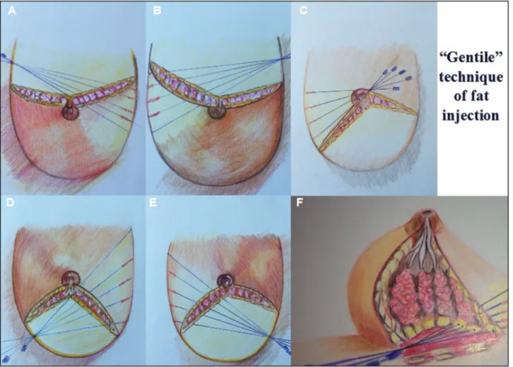
Fat obtained was implanted in multiple tunnels with slow and controlled movements through different entrances: three in the inframammary fold [Figure 1D–F], two in supero and inferoexternal quadrant [Figure 1A and D], two in super and inferointernal quadrant [Figure 1B and E], and four periareolar [Figure 1C]. The inframammary ones are roughly located where the breast groove joins respectively the parasternal, the
midclavicular, and the front axillary line [Figure 1D–F]: a 1.5 mm cannula connected to a 5 ml syringe is pushed through the access deep to the suprafascial level, and the tunnel so created is drawn with a dermographic sterile pen or methylene blue to point out the site of injection. The fat was then released with a retrograde technique while withdrawing the cannula and placed along linear deposits in the suprafascial, retroglandular e intraglandular space. The operation was repeated with other four syringes turning the injection directional axis of 10°–15° medially to have five tunnels arranged radially around the skin access [Figure 1D–F].
Other tunnels were created in the spaces between the beams previously drawn so that the fat was homogeneously distributed [Video]. The whole procedure was repeated for each inframammary access. We usually spend ten 5 ml syringes for every inframammary hole (about 50 cc of fat) and so a total of thirty 5 ml syringes (about 150 cc of fat) per side.
Figure 1: Gentle technique of fat injection. (A) Point of cannula access in superoexternal quadrant located at 292.5° of the breast. (B) Point of cannula
access in superointernal quadrant located at 67.5° of the breast. (C) Point of cannula access located at 45°–135° and 225°–315° on the areolar circumference. (D) Point of cannula access in inferoexternal quadrant located at 247.15° of the breast. (E) Point of cannula access in inferointernal quadrant located at 112.5° of the breast. (F) Three-dimensional painting of fat injection into the breast through three inframammary fold access located at 135°, 180°, and 225° of the breast
Gentile, et al.: Gentle technique for breast augmentation
Journal of Cutaneous and Aesthetic Surgery ¦ Volume 11 ¦ Issue 3 ¦ July-September 2018 123
Then the graft was injected through periareolar entrances located at 45°–135° and 225°–315° on the areolar circumference. If required, we place an additional third access at 12 o’clock. The fat was injected using 1–2 cc syringes through three or four tunnels arranged radially around the skin access and released with a retrograde technique while withdrawing the cannula along linear deposits within and over the mammary gland. We generally use an inferior number of tunnel for these access then the number we used for the inframammary ones because this graft is not used to fully the breast but just to refine breast contour and skin irregularities. The quantity of fat injected through these accesses is about 50–70 cc, according to the breast needs.
The entire procedure was repeated on the other side, and in the end, the patient was checked for any asymmetry and deformities that are meticulously corrected. Once the fat tissue was implanted, the access incisions were closed using 5-0 nylon stitches.
In all patients, the procedure was carried out in two sessions. According to the patient’s needs, in each of the two sessions, 50–150 ml (average, 93.54 ml) was injected, for a total of 187 ml (range, 110–250 ml) per patient.
The “Coleman technique”
Fat tissue (80–340 ml) was harvested from the abdominal region using some specific cannulas. Maintaining asepsis, we took the plungers off the syringes; after closing them with a cap, we positioned them flat in the sterile centrifuge. The syringes were processed for 3 min at 3,000 rpm. This procedure can obtain purified fat tissue, preserving the integrity of the adipocytes but separating the fluid fat
portion from the serousbloody part. The purified body fat was put in 1 ml syringes and aseptically reinserted using the specific microcannulas for implanting.
Fat was implanted quickly in multiple tunnels to ensure a better contact with vascularized bed, and within each tunnel, it was released in small aliquot to increase the viability of central adipocyte cells and also to ameliorate fat particle integration and tactile feelings under the skin. In all patients, the procedure was carried out in two sessions. According to the patient’s needs, in each of the two sessions, 50–150 ml (average, 93.54 ml) was injected, for a total of 187 ml (range, 110–250 ml) per patient.
Statistical analysis
Values were expressed as mean plus standard error and analyzed using Student’s t test. Differences were considered statistically significant for P < 0.05.
R
esultsIn patients treated with the Gentle technique, we observed a 60.5% + 12.5% maintenance of contour restoring and three-dimensional volume after 1 year (P < .0001 vs. control group); we compared the results obtained with only 39% + 4.4% of the control group treated with the Coleman technique.
Transplanted fat tissue reabsorbing was analyzed with instrumental imaging (MRI-ultrasound-mammography). There were no complications in any patient, and the results were lasting in all cases (the mean follow-up period was 60 months). All the patients were satisfied with the resulting texture, softness, and contour, and MRI
confirmed maintenance of the restoration. The images were acquired on axial and sagittal planes. Based on MRI scans acquired, volumetric assessments of the breasts were also calculated, taking as edges the anterior axillary line, anterior margin of the pectoral muscle, medio-sternal line, skin, and nipple.
D
iscussionAutologous fat grafting is widely used in breast surgery for a variety of indications including the correction of volume, shape, and contour deformities[23-29]; treatment of irradiated breast tissue; and priming of the irradiated field in breast reconstruction[30-32]: In the majority of cases, lipofilling is used as an adjunct to standard breast augmentation procedure even if the use of fat alone has been recently demonstrated.[33] Despite the appeal of graft lipofilling technique, many problems still remain concerning fat survival, and presently, there is no standardized method for fat grafting procedure. This procedure usually involves three different stages (harvesting, processing, and placement), and in each of these phases, adipocyte cells can be damaged. Our technique of fat placement is quite different from the others as we focus our attention on the last stage of fat graft procedure, which could explain the better results in graft survival. The most common procedure adopted by plastic surgeons in breast lipofilling is the Coleman technique[34]: According to this one, fat is injected using a blunt Coleman infiltration cannula connected to a 1 ml syringe and placed with a retrograde technique while withdrawing. Small aliquots of fat less than 0.1 ml were distributed equally in recipient bed to guarantee a better revascularization of the graft; in fact, injecting a single bolus of a large volume of fat is to be avoided as it leads to fat necrosis and poor outcomes due to the lack of sufficient contact with vascularized host tissue.[35] This technique rapidly spread worldwide and became the landmark technique of transplantation because it produces better results in graft survival.[36] However, while this method can be successfully used for small areas like the face, it can be inconvenient when treating large areas such as breast or buttocks because it requires a very long time and results in loss of fat placement accuracy. Our solution is to place the graft among linear deposits in a radial pattern, which permits shortening the surgical time and ensures graft viability as our results demonstrate. Chung et al.[37] have reported how the Coleman technique for fat injection could be harmful for adipocyte cell: They compared fat retention rate using Coleman procedure with the use of a hand-held device that permits disposal of preset 400 µl aliquots with every pull of the trigger. Performing an MMT viability assay, they demonstrated that there was a significant higher cellular viability of the fat injected with the device than with the Coleman technique, and similar results were reported for adipocyte
proliferation rate and fat volume retention. This study supported the important influence of mechanical trauma in graft survival and the necessity of using a “fat friendly” technique to reduce fat reabsorption rate and adipocyte necrosis. According to these findings, the “Gentle” technique pretends to be literally gentle and the fat is released with slow and controlled movements with a constant plunger pressure: A slow rate of fat injection allows us to achieve better results than a fast one as it causes a lower shear stress on adipocyte cell.[38] In fact, whereas fat tissue is widely resistant to both negative and positive pressure and static pressure can damage fat only at extreme levels,[38,39] shear stress, which is exerted on the fat as it is pushed through tubing such as a syringe barrel or cannula, has been shown to be very harmful for adipocyte cells and a flow rate of 0.5–1 cc/s has to be recommended to optimize fat viability.[38] Atashroo et al. have found fat storage rate to be significantly higher when the fat is released at a constant rate that minimizes fat trauma than when it is intermittently placed in the bed recipient.[40] A constant plunger pressure decreases fat viscosity and lets the graft flow out from the syringes more easily. Wider cannulas (2.5 mm) are also preferred because of reduced fat reabsorption compared with smaller cannulas (1.6 or 2 mm)[41]; needle size does not influence fat viability at least using 14, 16, 20 needles gauge.[42] We think that using wider diameter cannulas decreases mechanical trauma on adipocyte cell and better respects physical properties of fat.
Pattern of graft distribution is also to be thought in order to have better results. Different ways of graft placing have been tried in these years but a standardized layout still does not exist. As we have previously reported, the first revolution in graft disposition was made by Coleman: Instead of placing fat in a single block as Peer previously had done, Coleman disposed it in small aliquots into consistent numbers of subcutaneous tunnels.[34] He reported a significant deterioration of recipient site fullness in a period between 8 and 12 years and attributes much of this success to placement technique and surgeon experience. Jeong[43] recently reported the use of screw-type syringes to dispose fat like several logs that are laid on top of others oriented in the opposite direction. We prefer to dispose the graft in a radial pattern to avoid an excessive stratification of linear deposits that could decrease vascularization ratio and consequently graft viability especially in the central rows. A radial pattern permits a homogeneous distribution of large volume of fat (200-220 cc) with minimal risk of adipocyte cell necrosis due to the wide contact surface between graft and vascularized host tissue.
Yoshimura et al.[44] use 150 mm long 18-gauge needles that are inserted from either one or two points on the areola margin or one or two points at the inframammary fold in variable directions and planes to achieve a diffuse
Gentile, et al.: Gentle technique for breast augmentation
Journal of Cutaneous and Aesthetic Surgery ¦ Volume 11 ¦ Issue 3 ¦ July-September 2018 125
distribution. The grafts were injected as small aliquots or as a thin string into the fatty layers on, around, and under the mammary glands and also into the pectoral muscle. They reported a breast circumference augmentation of 4 to 8 cm at 6 months and 2–3 bra-size increase corresponding to fat volume retention of 100 to 200 ml, substantially less than the fat survival rate we report. Considering the surgical technique they perform, we find that using a double access inframammary and periareolar, we can reach a more three-dimensional distribution of the graft and a better breast re-contouring as patient satisfaction demonstrates. Moreover, we avoid intrapectoral graft injection and prefer to place the entire volume over the muscle fascia to enhance fat graft survival. In fact, Karacaoglu[45] reported a higher retention rate when fat is placed in supramuscular layer (81.95% ± 4.4%) than in the submuscular one (37.31% ± 5.77%) confirming that even the selection of recipient site could influence ultimate fat graft survival.
c
onclusionEven if different fat injection techniques have been tried in the last years to maximize fat stability while improving breast contouring, volume, and shape, a standard one still does not exist. We have showed in this article that every single aspect of graft placement (cannula size, flow rate injection, fat purification, layout of injection, and layer recipient bed) can influence fat stability, and therefore, a reference technique of transplantation has to be obtained to make different study results comparable. In these years, we have tried to improve our technique to define a procedure that ensures maximal fat survival. Our method is focused to eliminate any unnecessary manipulation of the graft so as to optimize graft retention and clinical outcomes: Controlled movement and slow rate of fat injection are the cornerstone of our technique and guarantee a nontraumatic fat transfer and a greater survival rate of adipocytes. Linear placing and radial pattern should be preferred when large amounts of fat have to be released while preserving graft vascularisation. Although our reported experience is from a single centre, the “Gentle” technique seems to be a desirable technique for fat injection as it improves fat viability and long-term patient satisfaction.
Almost all the patients were satisfied with their enlarged soft breast with natural control.
Declaration of patient consent
The authors certify that they have obtained all appropriate patient consent forms. In the form the patient(s) has/have given his/her/their consent for his/her/their images and other clinical information to be reported in the journal. The patients understand that their names and initials will not be published and due efforts will be made to conceal their identity, but anonymity cannot be guaranteed.
Financial support and sponsorship
Nil.
Conflicts of interest
There are no conflicts of interest.
EVIDENCE BASED MEDICINE: Level III
R
efeRences1. Neuber F. Fat transplantation. Chir Kongr Verhandl Dsch Gesellch Chir 1893;20:66
2. Coleman WP 3rd. The history of liposuction and fat transplantation in America. Dermatol Clin 1999;17:723–7.
3. Cárdenas-Camarena L, Arenas-Quintana R, Robles-Cervantes JA. Buttocks fat grafting: 14 years of evolution and experience. Plast Reconstr Surg 2011;128:545-55.
4. Brongo S, Nicoletti GF, La Padula S, Mele CM, DʼAndrea F. Use of lipofilling for the treatment of severe burn outcomes. Plast Reconstr Surg 2012;130:374e-6e.
5. Cervelli V, Gentile P, De Angelis B, Calabrese C, Di Stefani A, Scioli MG, et al. Application of enhanced stromal vascular fraction and fat grafting mixed with PRP in post-traumatic lower extremity ulcers. Stem Cell Res 2011;6:103-11.
6. Gentile P, Orlandi A, Scioli MG, Di Pasquali C, Bocchini I, Curcio CB, et al. A comparative translational study: the combined use of enhanced stromal vascular fraction and platelet-rich plasma improves fat grafting maintenance in breast reconstruction. Stem Cells Transl Med 2012;1:341-51.
7. Delay E, Streit L, Toussoun G, La Marca S, Ho Quoc C. Lipomodelling: an important advance in breast surgery. Acta Chir Plast 2013;55:34-43.
8. Niechajev I, Sevćuk O. Long-term results of fat transplantation: clinical and histologic studies. Plast Reconstr Surg 1994;94:496–506.
9. Zocchi ML, Zuliani F. Bicompartmental breast lipostructuring. Aesthetic Plast Surg 2008;32:313–28.
10. Rieck B, Schlaak S. Measurement in vivo of the survival rate in autologous adipocyte transplantation. Plast Reconstr Surg 2003;111:2315-23.
11. Peer LA. Cell survival theory versus replacement theory. Plast Reconstr Surg (1946) 1955;16:161-8.
12. Del Vecchio D, Rohrich RJ. A classification of clinical fat grafting: different problems, different solutions. Plast Reconstr Surg 2012;130:511-22.
13. Carpaneda CA, Ribeiro MT. Study of the histologic alterations and viability of the adipose graft in humans. Aesthetic Plast Surg 1993;17:43-7.
14. Carpaneda CA, Ribeiro MT. Percentage of graft viability versus injected volume in adipose autotransplants. Aesthetic Plast Surg 1994;18:17-9.
15. Coleman SR. Long-term survival of fat transplants: controlled demonstrations. Aesthetic Plast Surg 1995;19:421-5.
16. Bielli A, Scioli MG, Gentile P, Agostinelli S, Tarquini C, Cervelli V, et al. Adult adipose-derived stem cells and breast cancer: a controversial relationship. Springerplus 2014;3:345.
17. Scioli MG, Bielli A, Gentile P, Mazzaglia D, Cervelli V, Orlandi A. The biomolecular basis of adipogenic differentiation of adipose-derived stem cells. Int J Mol Sci 2014;15:6517-26.
18. Araco A, Gravante G, Araco F, Gentile P, Castrì F, Delogu D, et al. Breast asymmetries: a brief review and our experience. Aesthetic Plast Surg 2006;30:309-19.
19. Fiaschetti V, Pistolese CA, Fornari M, Liberto V, Cama V, Gentile P, et al. Magnetic resonance imaging and ultrasound evaluation after breast autologous fat grafting combined with platelet-rich plasma. Plast Reconstr Surg 2013;132:498e-509e.
20. Kaufman MR, Bradley JP, Dickinson B, Heller JB, Wasson K, O’Hara C, et al. Autologous fat transfer national consensus survey: trends in techniques for harvest, preparation, and
application, and perception of short- and long-term results. Plast Reconstr Surg 2007;119:323-31.
21. Gir P, Brown SA, Oni G, Kashefi N, Mojallal A, Rohrich RJ. Fat grafting: evidence-based review on autologous fat harvesting, processing, reinjection, and storage. Plast Reconstr Surg 2012;130:249-58.
22. Smith P, Adams WP Jr, Lipschitz AH, Chau B, Sorokin E, Rohrich RJ, et al. Autologous human fat grafting: effect of harvesting and preparation techniques on adipocyte graft survival. Plast Reconstr Surg 2006;117:1836-44.
23. Spear SL, Wilson HB, Lockwood MD. Fat injection to correct contour deformities in the reconstructed breast. Plast Reconstr Surg 2005;116:1300-5.
24. Illouz YG, Sterodimas A. Autologous fat transplantation to the breast: a personal technique with 25 years of experience. Aesthetic Plast Surg 2009;33:706-15.
25. Kanchwala SK, Glatt BS, Conant EF, Bucky LP. Autologous fat grafting to the reconstructed breast: the management of acquired contour deformities. Plast Reconstr Surg 2009;124:409-18.
26. de Blacam C, Momoh AO, Colakoglu S, Tobias AM, Lee BT. Evaluation of clinical outcomes and aesthetic results after autologous fat grafting for contour deformities of the reconstructed breast. Plast Reconstr Surg 2011;128:411e-8e.
27. Losken A, Pinell XA, Sikoro K, Yezhelyev MV, Anderson E, Carlson GW. Autologous fat grafting in secondary breast reconstruction. Ann Plast Surg 2011;66:518-22.
28. Del Vecchio DA. “SIEF”—simultaneous implant exchange with fat: A new option in revision breast implant surgery. Plast Reconstr Surg 2012;130:1187-96.
29. Cigna E, Ribuffo D, Sorvillo V, Atzeni M, Piperno A, Calò PG, et al. Secondary lipofilling after breast reconstruction with implants. Eur Rev Med Pharmacol Sci 2012;16:1729-34.
30. Rigotti G, Marchi A, Galiè M, Baroni G, Benati D, Krampera M, et al. Clinical treatment of radiotherapy tissue damage by lipoaspirate transplant: a healing process mediated by adipose-derived adult stem cells. Plast Reconstr Surg 2007;119:1409-22; discussion 1423-4. 31. Salgarello M, Visconti G, Barone-Adesi L. Fat grafting and breast
reconstruction with implant: another option for irradiated breast cancer patients. Plast Reconstr Surg 2012;129:317-29.
32. Sarfati I, Ihrai T, Kaufman G, Nos C, Clough KB. Adipose-tissue grafting to the post-mastectomy irradiated chest wall: preparing the
ground for implant reconstruction. J Plast Reconstr Aesthet Surg 2011;64:1161-6.
33. Khouri RK, Rigotti G, Khouri RK Jr, Thomas, TM. Total breast reconstruction with autologous fat transfer: review of a seven-year multicenter experience. Plast Reconstr Surg 2014;134:84-5
34. Coleman SR. Structural fat grafts: the ideal filler? Clin Plast Surg 2001;28:111-9.
35. Choi M, Small K, Levovitz C, Lee C, Fadl A, Karp NS. The volumetric analysis of fat graft survival in breast reconstruction. Plast Reconstr Surg 2013;131:185-91.
36. Coleman SR. Facial recontouring with lipostructure. Clin Plast Surg 1997;24:347-67.
37. Chung MT, Paik KJ, Atashroo DA, Hyun JS, McArdle A, Senarath-Yapa K, et al. Studies in fat grafting: part I. Effects of injection technique on in vitro fat viability and in vivo volume retention. Plast Reconstr Surg 2014;134:29-38.
38. Lee JH, Kirkham JC, McCormack MC, Nicholls AM, Randolph MA, Austen WG Jr. The effect of pressure and shear on autologous fat grafting. Plast Reconstr Surg 2013;131:1125-36.
39. Shiffman MA, Mirrafati S. Fat transfer techniques: the effect of harvest and transfer methods on adipocyte viability and review of the literature. Dermatol Surg 2001;27:819-26.
40. Atashroo D, Raphel J, Chung MT, Paik KJ, Parisi-Amon A, McArdle A, et al. Studies in fat grafting: part II. Effects of injection mechanics on material properties of fat. Plast Reconstr Surg 2014;134:39-46. 41. Ozsoy Z, Kul Z, Bilir A. The role of cannula diameter in improved
adipocyte viability: a quantitative analysis. Aesthet Surg J 2006;26:287-9.
42. Erdim M, Tezel E, Numanoglu A, Sav A. The effects of the size of liposuction cannula on adipocyte survival and the optimum temperature for fat graft storage: an experimental study. J Plast Reconstr Aesthet Surg 2009;62:1210-4.
43. Jeong JH. Recent advancements in autologous fat grafting. Arch Aesthetic Plast Surg 2014;20:3-7.
44. Yoshimura K, Sato K, Aoi N, Kurita M, Hirohi T, Harii K. Cell-assisted lipotransfer for cosmetic breast augmentation: supportive use of adipose-derived stem/stromal cells. Aesthetic Plast Surg 2008;32:48-55; discussion 56-7.
45. Karacaoglu E, Kizilkaya E, Cermik H, Zienowicz R. The role of recipient sites in fat-graft survival: experimental study. Ann Plast Surg 2005;55:63-8; discussion 68.