ALMA MATER STUDIORUM - UNIVERSITÀ DI
BOLOGNA
School of Science
Department of Industrial Chemistry “Toso Montanari”
Corso di Laurea Magistrale / Master
Advanced Spectroscopy in Chemistry
Classe LM-71 - Scienze e Tecnologia della Chimica Industriale
F
UNCTIONAL
DNA
NANOSTRUCTURES FOR IN VITRO
BIOSENSING IN LIVE CELLS
Experimental Master Thesis
CANDIDATE : TUTOR:
Isidro Badillo Dott. Giampaolo Zuccheri
First Session
- 1 -
Nature is the source of all true knowledge. She has her own logic, her own laws, she has no effect without cause nor invention without necessity.
- 2 -
Preface
This Master thesis is the result, in principle, of my curiosity for going into the Nanoworld to know, discover and research, how DNA nanotechnology is setting the bases for an upcoming revolution in the biomedical scene and many other scientific fields. I believe this is just the beginning of one of the more promising scientific disciplines in human sciences.
In the following pages I present the results of my master thesis internship, performed at the department of Nanobiotechnology under the supervision of Dr. Giampaolo Zuccheri. This thesis concludes my two years of the Master of Science Advanced Spectroscopy in Chemistry as part of the Erasmus Mundus program.
This research is presented in five main parts that includes from the design, synthesis, characterization and functional evaluation of two self-assembled multifunctional DNA nanobiosensors. Each structure was devised in an attempt to perform a specific area of DNA self-assembly that we find promising for the future development of functional DNA sensor nanotechnology.
- 4 -
Abstract
DNA is a fascinating biomolecule that is well known for its genetic role in living systems. The emerging area of DNA nanotechnology provides an alternative view that exploits unparallel self-assembly ability of DNA molecules for material use of DNA. The pioneering work by Professor Seeman has showed that the self-assembly of synthetic oligonucleotides can be used in order to make nanostructures of arbitrary shape and complexity; such nanostructures can also be endowed with interesting functional properties. After nearly three decades of research efforts, the meaning of DNA nanotechnology has evolved from toylike beautiful nanoscale structures to really useful building blocks for a variety of applications. In this thesis, I report the design, synthesis and characterization of multifunctional fluorescencent nanobiosensors by DNA self-assembling . Each structure was designed and implemented to be introduced in live cells in order to give information on their functioning in real-time.
Although many reports exist on the results of DNA self-assembling systems, still few of them focus on the in vitro study about the function of such DNA nanostructures in live cells. Due to this, there are still a limited knowledge about the functionality in real time of such designs. To address an aspect of this issue, we designed, synthesized and characterized two main DNA nanostructures with biosensor functionality. In the design effort, we have used computational tools to design a graphic model of two new DNA motifs and to obtain the specific sequences to all the ssDNA molecules in our models, to provide their functionality.
By thermal self-assembly techniques we have successfully synthesized the structure and corroborate their formation by PAGE technique. In addition with this last technique, we have established the conditions to characterize their structural conformation change when they perform their sensor response. The sensing behavior was also accomplished by fluorescence spectroscopy techniques; FRET evaluation and fluorescence microscopy imaging. Providing the evidence about their adequate sensing performance outside and inside the cells detected in real-time.
As a preliminary evaluation we have tried to show the evidence about the localized functionality of our structures in different cancer cell lines with the ability to work as a therapy target sensor. However, still a better understanding in the biomedical correlation process between DNA nanostructures and live systems is required.
- 6 -
Contents
Summary
10
PART I Elements of Nanoscience and
DNA anotechnology
18
1 Introduction to the Nanoworld
19
1.1 Nanoscience and Nanotechnology Definitions
19
1.1.1
Brief History of Nanotechnology22
1.2 The Creation of Nanoscale Objects
24
1.2.1
Top-Down24
1.2.2
Bottom-Up25
1.3 The Nano-Bio Interface
27
1.4 Nanobiotechnology
27
1.4.1
Towards Molecular Nanobiotechnology29
2 DNA Nanotechnology
31
2.1 Brief History of DNA Nanotechnology
31
2.2 Fundamental DNA Properties
32
2.3 DNA Nucleobase Interactions
37
2.3.1
Sticky Ends37
3 Hybridization Techniques of Creating Nanostructures
40
3.1 Holliday Junctions
42
- 7 -
3.2.1
Funtionality of DNA Polyhedra52
3.3 Future Directions
53
References Part I
54
PART II Strategy of Implementations
58
4 Structural Modelling
59
4.1 The pH Dependent Biosensor
60
4.1.1
The idea60
4.1.2
The model61
4.2 The Nucleic-Acid Sensitive Nanostructure
68
4.2.1
The idea68
4.2.2
The model69
5 The Sequence Design
71
5.1
NANEV72
5.2
NUPACK72
References Part II
76
PART III Method for Synthesis and Characterizations
78
6 Experimental Synthesis
79
6.1 Annealing DNA Strands to Self-Assemble
DNA nanostructures
79
- 8 -
6.2 Non-denaturing PAGE for the Characterization of
Self-Assembled DNA Nanostructures 80
7 Characterization of Nanostructures
81
7.1 Spectroscopic and Optical Microscopy Characterizations 81
7.1.1
Fluorescence Spectroscopy81
7.1.1.1
Quenching or FRET Interactions81
7.2 Fluorescence Microscopy
84
7.2.1
Imaging Rationale and Image Acquisition84
7.2.2
Semiautomatic Analysis of Fluorescence Images 858 Cell culture and nanostructure delivery to cells
87
References Part III
89
PART IV Results and Discussions
90
9 The Nucleic-Acid Sensitive DNA Nanostructure
92
9.1 Characterization of the Self-Assembly DNA Nanostructure 92
9.2 Probing the Binding of Targets Sequences to
TT:BAX Structure
95
9.3 The Functionality of the Nucleic-acid DNA Tetrahedron
as a Sensor
99
10 The pH Dependent DNA Tetrahedron
106
10.1 Self-Assembly and Characterizations of Nanostructures 106
- 9 -
10.3 Probing the Sense Response to pH Changes
111
11 Cell Uptake
117
11.1 The DNA nanostructures internalization by the cells 117
11.2 The internal localization inside the cells
119
12 Re-design of the Nanostructures with the Cy3/Cy5
Fluorophore pairs
121
12.1 PAGE gel characterizations
122
12.2 Fluorescence spectroscopy characterizations
124
12.3 Fluorescence microscopy characterization. The cell uptake 125
12.4 The micrograph semi-automatic analysis
131
12.5 TT:BAX15 and Staurosporine for cellular
apoptotic evaluation
131
References Part IV
133
PART V Conclusions and Appendixes
135
13 Conclusions and outlook
136
13.1 Future Perspectives
139
14 Appendixes
140
15.1 Microscope Filter Characterization
140
- 10 -
Summary
Introduction
The seminal work of Nadrian Seeman has showed that the self-assembly of synthetic oligonucleotides can be used in order to make nanostructures of arbitrary shape and complexity. As oligonucleotides can be chemically derivatized in many ways, such nanostructures can also be endowed with interesting functional properties. This field will certainly lead to a wealth of future applications in sensing and other fields.
The goal of the research performed during this Master’s internship is to make multifunctional nanobiosensors by DNA self-assembly. These are intended to be introduced in live cells in order to give information on their functioning in real-time.
Strategy of implementation
We chose to design and realize DNA self-assembled biosensors as modifications of the original DNA tetrahedron design proposed by Andrew Turberfield and coworkers. These are simple and efficiently assembled nanostructures that can be obtained with as few as 4 different oligonucleotides. The tetrahedrons can be made functional in many ways by performing the assembly of chemically-derivatized oligonucleotides.
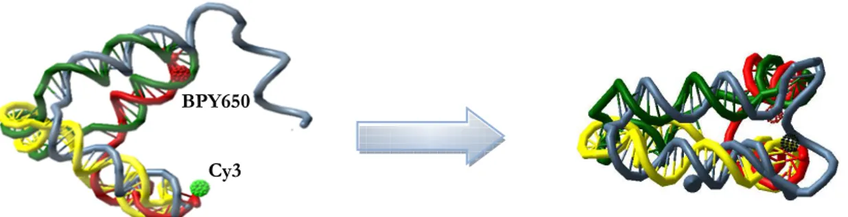
We postulated the design of ‘open’ DNA tetrahedral that can close to a compact tetrahedron (triangular pyramid) upon a chemical binding event. This large conformational transition can be coupled with a change in the fluorescence properties (FRET) of fluorophores decorating some of the component oligonucleotides, and thus it can be detected externally.
We propose the design of at least two classes of such nanostructures:
- A pH-sensitive nanostructure, where the conformational ‘closing’ transition is driven by trhe formation of an intramolecular CT-motif triple helix. See Figure 1. - A nucleic-acids sensitive nanostructure, where the conformational ‘closing’ transition is driven by the contemporary binding of a target nucleic acid to two ssDNA tails of the structure. See Figure 2.
- 11 -
Figure 1: structural model and functioning scheme for the pH sensitive nanostructure (left: high pH
state, right, low pH state). FRET between fluorophores decorating the structure vertices is more efficient if the closed conformation.
Figure 2: structural model and functioning scheme for the nucleic-acids sensitive nanostructure
(left: unbound open state, right, bound closed state). FRET between fluorophores decorating the structure vertices is more efficient if the closed conformation.
Results
We designed and made DNA nanostructures and showed they are function. Then, we treated live cells with them and demonstrated uptake in live human cancer cells.
Design of nanostructures
We successfully designed DNA nanostructures by taking advantage of available software tools and achieved two different nanostructures coded in 4 oligonucleotides each:
- A pH sensitive tetrahedral nanostructure (see Figure 1) that contains a double-stranded target and a single-double-stranded triplex-forming oligonucleotide section that
Cy3 BPY650
- 12 -
will assemble in a triple-helix upon reduction of the solution pH. The triplex sequence was obtained previously in the lab and displays a transition point between pH 6 and 7 at 37°C in physiological conditions, so it is apt for measuring changes in the intracellular pH in mammalian cells.
- A nucleic-acids sensitive tetrahedral nanostructure (see Figure 2) that contains two single stranded tails that are complementary to two near portions of the BAX transcript RNA. This is the RNA product of a gene that is involved in cell apoptosis and thus it can be triggered by apoptosis-inducing signals.
Two fluorophores are localized at two vertices of the nanostructure where their mutual distance will change the most upon conformational transition (see Figures 1 and 2, fluorophores are represented as spheres). We used Cy3 as the donor dye and Bodipy650 or Cy5 as the acceptor dye.
Characterization of nanostructures
We characterized the assembly of the nanostructures by PAGE, so that we can follow the assembly step-wise and test the thermodynamic stability of the structures in physiological conditions.
Assembly characterization
As an example of successful assembly, Figure 3 below shows the stepwise assembly of two variations of the BAX nucleic-acid sensing nanostructure. PAGE shows that assembly proceeds with high yield. Functional tetrahedrons migrate as single bands and are stable in physiological conditions. Occasionally, the gels show small stoichiometry impairments, but the tetrahedron is always the principal product.
- 13 -
Figure 3. Annealing of oligonucleotide dimers (lanes 1-6), trimers (lanes 9-11) and full tetrahedrons
for two variations (lanes 12 and 13) of the nucleic- acids sensor DNA tetrahedron.
Functional characterization
The functional characterization of the dynamic behavior of nanostructures is achieved by PAGE, by changing the conditions (running pH for the pH biosensor and titration with target oligonucleotide for nucleic-acids biosensor) and by spectrofluorimetry.
Nucleic acids biosensor
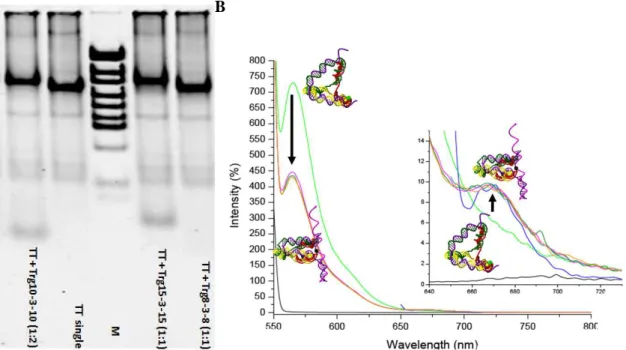
We demonstrated that the nucleic-acids sensitive tetrahedron is functional. PAGE shows that tetrahedron bind the target and it then migrates as a single band (verifying recognition and association) and that the nanostructure band gets shifted to higher mobility than the unbound state (as it is more compact in shape) as expected. Figure 4A.
Tetrahedrons are decorated with donor and acceptor fluorophores that can do energy transfer (FRET) on closing of the structure upon recognition. Upon titration with target nucleic acids of appropriate sequence and sufficient length, the structure undergoes a conformational transition towards a compact, closed structure where FRET is more efficient. Fluorescence spectroscopy shows a significant quenching of the donor dye, and correspondent increase in the acceptor sensitized emission of fluorescence. Figure 4B.
- 14 -
A B
Figure 4. A) PAGE showing the shift in mobility of the tetrahedron band (TT) upon binding with
specific DNA targets of appropriate sequence and length (20-nt –Trg10-3-10 - or 30-nt - Trg15-3-15 - recognition sequence, in these conditions). Shorter recognition sequences are not stably bound (Trg8-3-8). B) Spectrofluorimetric characterization of the conformational transition upon target binding (in the direction of arrows) as evidenced by both the quenching of donor fluorophore emission and the sensitized emission of the acceptor fluorophore (inset).
pH-biosensor
By exploiting the same strategy for functional characterization, we performed PAGE and spectrofluorimetry also in order to characterize the dynamic behavior of the conformational transition of the pH sensitive nanostructure.
In order to better characterize the behavior, we also prepared control pH-insensitive nanostructures: an open DNA tetrahedron that cannot close, and a compact tetrahedron that stays close irrespective of the solution pH. All the 3 specimen (negative ‘open’ control, positive ‘closed’ control and biosensor tetrahedron) are run in PAGE in different pH buffers. At pH 5, the pH-sensitive tetrahedron runs as fast as the positive control, ‘closed’ nanostructure. At pH 8, the pH sensing tetrahedron runs as fast as the negative-control, ‘open’ tetrahedron. Figure 5A.
The pH-driven conformational transition can also be checked via spectrofluorimetry, using the same strategy described above. pH-independent donor (Cy3) and acceptor fluorophores (Bodipy650 or Cy5) are brought in proximity by the closing of the tetrahedron at low pH, leading to donor fluorescence quenching and
- 15 -
correlated acceptor sensitized emission. The fluorescence behavior of the control positive and negative nanostructures are not changed with solution pH. Figure 5B. Our measurements show that the behavior of nanostructures is reversible and robust, as they can undergo repetitive transitions by cycling the pH.
Cell uptake
Confirming previous evidence reported by our laboratory and others, we confirmed that this class of nanostructures can spontaneously be uptaken by live cells even without the aid of transfecting agents. We tested different cell lines, including HeLa and glioblastoma cells. We have demonstrated uptake by characterizing the cellular fluorescence by epifluorescence microscopy. Nanostructures are also present as a diffuse signal in the cytoplasm, but they are manly evidenced as a localized signal in lysosomes and possibly mitrochondria.
A B
Figure 5. A) PAGE showing the mobility of the pH-sensitive tetrahedron band at acidic pH as a
comparison with pH-insensitive control nanostructures. Gel is run at 37°C in physiological conditions. B) Plot of the fluorescence signal due to the emission of the donor (Cy3) dye decorating the nanostructure during cycling of the solution pH. Fluorescence of the pH-sensitive tetrahedral nanostructure gets quenched when the structure is closed by triple helix formation and the donor fluorophore is near the acceptor. Measurements are performed at 37°C in physiological conditions. In te n si ty ( 5 6 5 n m )
- 16 -
pH-biosensor
We characterized the state of the pH-sensitive tetrahedron inside live cultured cells. By characterizing the localization of the FRET signal in the fluorescence microscope, in comparison with the direct excitation signal of the donor and acceptor dye, we evidenced that the higher FRET comes from point-like organelles recognizable as lysosomes. As it is known that these should have their contents at low pH (≤5) and thus we conclude that the nanostructure is successfully reporting on intracellular pH variations, see Figure 6.
Nucleic acids biosensor
We also proved that our nucleic-acids sensitive nanostructure gets internalized by live gliobastoma cells. These also get localized in the cytoplasm, but manly in lysosomes and possibly mitrochondria.
We made and used nanostructures that were designed to target an RNA transcript involved in the apoptotic pathway. We did a number of attempts but so far we have not been able to show the emergence of the target RNA after induction of apoptosis in the glioblastoma cells.
Figure 6. Fluorescence image of a human cultured glioblastoma cell after uptake of the
pH-sensitive DNA nanostructure. Here is the display of the overlay of the fluorescence signal of the donor dye represented in red with the signal from the FRET channel, represented in green. Localization of the green signal (or perfect overlay of signals that shows as yellow) reports on the localization of high-FRET states in point-like organelles identifiable as lysosomes.
- 17 -
Conclusions
We have successfully designed and realized functional dynamic DNA nanostructures that can respond to the change in pH or to the presence of specific nucleic acids by changing their conformation in an externally measurable fashion. We have also shown that these nanostructures can be internalized by live human cells in culture and could report on their internal state.
Outlook
Additional experiments and possibly more statistically relevant citofluorimetry experiments will be needed to quantitatively assess the functionality of the nanostructures as intracellular biosensors, especially for the case of the nucleic acid biosensor.
An important factor in the deployment of such biosensor will be the intracellular localization upon uptake. Current evidence seem to show that this could depend on the cell line, on the cell state and possibly also on the type and number of fluorophores that are used to detect the presence and signaling state of the internalized nanostructures. Further studies are needed in order to have a better control on cellular localization.
- 18 -
PART I
Elements of Nanoscience and DNA Nanotechnology
- 19 -
Elements of Nanoscience and DNA Nanotechnology
1. Introduction to the Nanoworld
1.1
Nanoscience and Nanotechnology Definitions
Nowadays the terms nanotechnology, nanoparticles, and nanomaterials are familiar to a broad audience of readers since the term “nano” has revolucionazied the way of thinking about the object size. Nanobiotechnology is a multidisciplinary field that covers a vast and diverse array of technologies coming from engineering, physics, chemistry, and biology. It is the combination of these fields that has led to the birth of a new generation of materials and methods of making them. The scope of applications is enormous and every day are discovered new areas of our daily lives where they can find use.
From a general perspective, the prefix nano is derived from the Greek word “νάνος” (nanos) meaning “dwarf”, the quantity of 10–9 (one billionth) of a measuring unit. Therefore, the nanoscience is defined as the research field where objects and those phenomena originated by nanoscale objects converge in a range size of 1 to 100 nm. Thus, Nanoscience can be viewed as an highly interdisciplinary that seeks to integrate mature nanoscale technology.
Once nanoscience was defined, it can be possible to define “nanotechnology” in general way as the field of research and fabrication that is on a scale of 1 to 100 nm. In a broad sense, nanotechnology involves the “creation and application of nanomaterials, devices and technical systems whose functions are determined by the presence of nanostructural fragments” [1].
- 20 -
In the way to understand the proper term of nanotechnology, it is helpful to delimit the nanoworld in the context of basic science. Chemistry is the study of atoms and molecules, a real matter of dimensions generally less than one nanometer, while condensed matter physics deals with solids of essentially infinite arrays of bound atoms or molecules of dimensions greater than 100 nm. A significant gap exists between these regimes, which corresponds to the previously defined nanosize region, in other words this gap corresponds to particles of 1 to 100 nm, or about 10 to 106 atoms or molecules per particle [2].
In this nanoscale domain neither quantum chemistry nor classical laws of physics hold. For example, in materials where strong chemical bonding is present, delocalization of valence electrons can be extensive, and the range of delocalization can vary with the size of the system. This effect, coupled with structural changes depending on size, can generate different chemical and physical properties.
Another way to interpret the nanoscale meaning, is to put into visual perspective the atomic, nano, micro and macro domains and compare with other usual objects in our world as it is shown in Figure 1. This illustration helps to compare in meters the size of a DNA strand, a bacterium and a regular cell.
The existing definitions of nanotechnology also emerged from the modern achievements and inventions in the area of visualization, manipulation, and analysis of nanometersized structures as well as the controllable creation of new functional materials with unique properties and nanosized devices that provided technological breakthroughs in various industrial fields.
Over time and with the advancement in this field, the definitions of Nanotechnology have become much more robust to integrate almost all the elements that describe better its meaning and understanding. Some of such particular definitions that can be found in literature are:
From the report of the Royal Society & the Royal Academy of Engineering of 2004 [3]. “Nanotechnology is the design, characterization, production and application of structures, devices and systems by controlling shape and size at nanometer scale.”
From the National Nanotechnology Initiative [4]. “Nanotechnology is science, engineering, and technology conducted at the nanoscale, which is about 1 to 100 nanometers. Nanoscience and nanotechnology are the study and application of extremely small things and can be used across all the other science fields, such as chemistry, biology, physics, materials science, and engineering”
- 21 -
The Concept of the Development of Research in the Area of Nanotechnology in Russia until 2010 (approved by the government of the Russian Federation on 18 October 2004) used the following definition: “Nanotechnology is the combination of methods and techniques that provide the possibility of controllable creation and modification of objects that include components with the size of less than 100 nm which have unique properties and can be integrated with complete functioning large-scale systems.”
- 22 -
It is obvious that all of the definitions are not perfect and can be subject to criticism, and the rapid development of various branches of nanotechnology will correct and improve them.
1.1.1 Brief History of Nanotechnology
Even though nanotechnology is considered as a relatively new field, it does not mean that technologists and scientists had not been working intuitively in this area before. The ancient Romes empirically used gold nanoparticles to color goblets and other glass products [5]. (This technology was used to create the famous Roman ruby goblets). It is also appropriate to remember that in 1857, M. Faraday was working with a red colloid solution that contained gold nanoparticles with a size of 20 nm.
The primary documented concept of Nanotechnology was presented on December 29, 1959, when Richard Feynman presented his famous lecture entitled “There’s Plenty of Room at the Bottom” at the annual meeting of the American Physical Society at the California Institute of Technology [6]. In this talk, Feynman stated for the first time several ground-breaking concepts, most of which are still relevant today. Even though his speech was completely theoretical, he described how the laws of physics do not limit our ability to manipulate single atoms and molecules. Back then, manipulating single atoms or molecules was not possible because they were far too small for available tools in that time. Nevertheless, he correctly predicted that the time for the atomically precise manipulation of matter would inevitably arrive. Today, that lecture is considered to be the first landmark of science at the nanolevel.
Later, the term nanotechnology was first used by Prof. Norio Taniguchi in 1974 at a conference in Tokyo with his paper “On the Basic Concept of 'Nano-Technology” defining it as “the processing of, separation, consolidation, and deformation of materials by one atom or one molecule” [7].
On the other hand, the progress in nanotechnology was based on inventions made at the end of the twentieth century. The first of these inventions belongs to Nobel laureates, physicists Gerd Binnig and Heinrich Rohrer from IBM research laboratory, who, in 1981, designed the tunnel scanning microscope that made it possible to see single atoms [8]. The second discovery was made in 1986, when the atoms were not only observed, but also manipulated for the first time with the help of a tunnel microscope enhanced by G. Binnig. The Nobel lecture in 1986 [9] described both the principle of operation of the tunnel microscope and the way of manipulating atoms. On the same
- 23 -
year, Dr. Kim Eric Drexler published a book entitled “Engines of Creation: the Coming Era of Nanotechnology” [10]. This book speculatively explored the concepts proposed by Feynman bringing them to a wide public attention. The concepts used by Drexler about nanotechnology are often referred as “molecular nanotechnology”, that is, the engineering of functional machines at the molecular scale designed and built atom-by-atom.
Summarizing, the principal scheme of integral parts of nanotechnology formed by the end of the twentieth century is illustrated in Figure 2. Remarks on this scheme, can be stated that engineering (technical) nanotechnology has started being focused toward solution of the following problems [11].
• Creation of solid materials and surfaces with controllable molecular structures •Designing new types of chemical compounds with controllable properties (nanoconstructions)
• Creating nanosized self-organizing or self-replicating structures
• Fabrication of devices for various purposes (components of nanoelectronics, nanooptics, nanoenergetics, etc.)
• Integration of nanosized devices with electric systems
Atoms
Nanoparticles Layers
Nanostructures
Figure 2. The principal scheme of the basic part of nanotechnology formed to the end of the twentieth
century and the main research fields.
Assembly
Synthesis
1. Dispersions 2. Nanostructured 3. Functional and smart 4. Composite and coatings materials nanostructures materials
- 24 -
In this perspective, the goal in nanotechnology may be described as the ability to assemble molecules into useful objects hierarchically integrated along several length scales and then, after use, disassemble objects into molecules. Nature already accomplishes this in living systems and in the environment.
1.2 The Creation of Nanoscale Objects
The first 30 years of the nanosciences were dedicated mainly to studying and fabricating materials at the nano scale range. In those studies, much effort was focused on shortening the dimension of fabricated materials. It was also a time when the two basic fabrication approaches were defined: “bottom-up” and “top-down” based on the type of action that is needed to go from the starting materials to the desired product. A general view and comparison of these two techniques are shown in Figure 3.
1.2.1 Top-Down
The Top-Down approach represents the type of nanofabrication first imagined by Feynman that search to create nanoscale structures or devices by using larger, externally-
Figure 3. Technological approaches to the creation of nanomaterials: Top-down and bottom-up.
Initial material Initial material
- 25 -
controlled tools to direct their assembly. This technique is referred to the molding,
carving, and fabricating of small materials and components by reducing the size of the initial substance or placement of required atoms on the surface of some material in a certain configuration, this is possible by the use of larger objects like mechanical tools and lasers.
Examples of Top-Down nanofabrication include all types of lithography (photo-, electron beam, dip-pen, soft-, nanoimprint-) and etching tools. In all these techniques, the generation of nanoscale features relies on the externally controlled relative movement of a tool and the substrate. The advantage of the top-down technique is that it enables to control the manufacture of smaller, more complex objects, as illustrated by micro and nanoelectronics, for example in the silicon chip fabrication.
1.2.2 Bottom-Up
On the other hand, the bottom-up approach, searches for the means and tools to build nanomaterials (devices) by combining smaller components from desired elements, such as single molecules and atoms, that are integrated into spatial structures as a result of a physicochemical process; generally, those elements are held together by covalent forces.
Theoretically, it can be exemplified by molecular assemblers, where nanomachines are programmed to build a structure one atom or molecule at a time; the strategy is to put the desired elements in a context where they can freely find each other and establish those interactions, without or with very limited external control. In this way, the result of the assembly is ultimately dependent on exactly how sub-component can bind to each other. Since no direct action is taken to drive the components to the position assigned to them, the arrangement will be spontaneously; this process is often called “self-assembly”.
In the context of nanoscience and nanotechnology, the self-assembling components are obviously nano-sized objects, for examples molecules or colloidal particles. Bottom-up approaches based on self-assembly could in principle be able to produce the same structures accessible to top-down methods, with the advantage to be in a highly parallel fashion and much cheaper. In addition, with the bottom-up design, the covalent bonds holding a single molecule together are far stronger than the weak interactions that hold more than one molecule together in the top-down. Thus, this
- 26 -
approach enables to control the manufacture of atoms and molecules and holds far more practical and applicative future potential.
It is necessary to remark that in any variant of the considered technologies described above, the forming nanomaterials will obtain new properties that the initial objects do not possess.
It is clear that the creation of practically significant structures at the nanometer scale is one of the fundamental challenges of science and technology in this century. This means that commercial demand for the production of miniature devices will determine the need to develop building blocks based on concept and principles that are used in nature to create nanosized systems or biosystems. At the same time, the building blocks must have a size that fills the gap between the submicrometric sizes reached by the classical top-down technology and the sizes that can be achieved using the classical bottom-up technology.
Analysis of the fundamental backgrounds of physical chemistry, biochemistry, and synthetic chemistry of biological molecules, considering the requirements for the two aforementioned approaches to obtain nanoobjects leads to the conclusion that, due to their geometric size, two groups of compounds are the most suitable for the creation of nanosized structures [12]:
1 Biopolymers (such as nucleic acids and proteins) 2 Nanoparticles (nanoclusters) of various origin
The formation of complex spatial objects, using the bottom-up approach, with regulated properties (nanostructures, nanobiomaterials, nanoconstructions) using nucleic acid molecules and their complexes as construction blocks is called nucleic acid based nanodesign or structural nucleic acid nanotechnology [13-15]. At this time, there are two different strategies of nanodesign:
1. Creation of nanostructures using single-stranded polynucleotide (DNA) molecules containing deliberately selected sequences of nucleobases.
2. Creation of nanoconstructions using spatially fixed linear double-stranded DNA molecules (or their complexes)
- 27 -
1.3 The Nano-Bio Interface
Biosystems are governed by complicated nanoscale processes and structures that have been optimized over millions of years. Biologists have been operating for many years at the molecular level, ranging from nanometers (DNA and proteins) to micrometers (cells, bacteria). A typical protein like hemoglobin has a diameter of about 5 nm, the DNA’s double helix is about 2 nm wide, and a mitochondrion spans a few hundred nanometers as compared in Figure 1. Therefore, the study of any subcellular entity can be considered “nanobiology.” Moreover, the living cell along with its hundreds of nanomachines is considered, today, to be the ultimate nanoscale fabrication system.
Considering that the groundwork of each and every biological system are nanosized molecular building blocks and machinery that cooperate to produce living entities and these elements have been integrated for nanotechnologists in the combination of two disciplines (nano and biotechnology) to give birth of a new science, nanobiotechnology. Nanotechnology provides the tools and technology platforms for the investigation and transformation of biological systems, and biology offers suitable models and bio-assembled components to nanotechnology. The difference between “nanobiology” to “nanobiotechnology” resides in the technology part of the term. Objects that are “man-made” correspond to the technology section of nanobiotechnology. Nanobiotechnology will lead to the design of entirely new classes of micro- and nanofabricated devices and machines, determining the inspiration for which will be based on bio-structured machines, the use of biomolecules as building blocks, or the use of biosystems as the fabrication machinery.
1.4 Nanobiotechnology
The nanoconstruction technology approaches are based on top-down for the construction of nonbiological systems and bottom-up for the molecular level as biological systems. Those constructions are done via a collection of molecular tool kits of atomic resolution that are used to fabricate micro- and macrostructure architectures. Nanobiotechnology, can be described in many ways: one is the incorporation of nanoscale machines into biological organisms for the ultimate purpose of improving the organism’s quality of life. To date, there are few methods for synthesizing nanodevices that have the potential to be
- 28 -
used in an organism without risk of being rejected as antigens; another way is the use of biological “tool kits” to construct from nano- to microstructures. However, the broad perspective to define “Nanobiotechnology” is probably the one that will include both and will be defined as: the engineering, construction, and manipulation of entities in the 1- to 100-nm range using biologically based approaches or for the benefit of biological systems. The biological approaches can be either an inspired way of imitate biological structures or the actual use of biological building blocks and building tools to assemble nanostructures.
Nanotechnology and nanobiotechnology could be differentiated by the technique used for the construction of nanostructures and nanodevices. In nanotechnology, the dominant approach is top-down, whereas in nanobiotechnology is bottom-up. The bottom-up approach to nanofabrication has the ambition to learn how to use biological strategies for its ends. Unfortunately, we are currently unable to design from scratch complex information-containing polymers with the same efficiency displayed by nature, not to mention synthesizing them. So one slightly different approach is to use not only the basic strategy, but also the actual molecules found in biological systems, or specific portions of them.
An interesting example of the bottom-up approach is the pioneering work of Seeman and coworkers by the use of the structural properties of DNA to produce target materials with predictable three-dimensional (3D) [16-19]. He uses DNA motifs with specific, structurally well defined, cohesive interactions involving hydrogen bonding or covalent interactions (“sticky ends”) to produce target materials with predictable 2D and 3D structures. The elements for the construction of these structures will be described in more detail in the next sections. These efforts have generated a large number of individual species, including polyhedral catenanes, such as a cube and a truncated octahedron, a variety of single-stranded knots, and Borromean rings. The combination of these constructions with other chemical components is expected to contribute to the development of nanoelectronics, nanorobotics, and smart materials. Therefore, the organizational capabilities of structural DNA nanotechnology are just beginning to be explored, and the field is ultimately expected to be able to organize a variety of species in the material world.
Actually, nanobiotechnology already has an impact on healthcare. Research on biosystems at the nanoscale has created one of the most dynamic science and technology domains at the confluence of physical sciences, molecular engineering, biology,
- 29 -
Figure 4. Relationship of Biotechnology and Nanotechnology to Nanomedicine and related technologies.
biotechnology, and medicine [20]. The confluence of nanoscience and biology are contributed to unified new particular fields of study that are even now more frequently to find in the literature such as: Nanomedicine, Nanobiomedicine, Nanobiopharmaceutical, Nanobiosystem science and engineering, Nanotherapy. The relationship of nanotechnology to biotechnology and related technologies is depicted graphically in
Figure 4.
1.4.1 Towards Molecular Nanobiotechnology
It is clear that Nanotechnology provides investigation tools and technology platforms for biomedicine. Examples include current work in the subcellular environment, investigating and transforming nanobiosystems (for example, the nervous system) rather than individual nanocomponents, and developing new nanobiosensor platforms. Investigative methods of nanotechnology have been implemented in uncovering fundamental biological processes, including self-assembling, subcellular processes, and system biology (for example, the biology of the neural system).
In those days, key improvements have been made in measurements at the molecular and subcellular levels and in understanding the cell as a highly organized
CELL/GENE THERAPY NANOBIOTECHNOLOGY NANOMEDICINE BIOTECHNOLOGY NANOTECHNOLOGY LIFE SCIENCES genomics/proteomics DRUG DISCOVERY
DRUG DELIVERY NANOARRAYS
BIOMARKERS MOLECULAR DIAGNOSTICS NANOBIO- PHARMACEUTICALS PERSONALIZED MEDICINE
- 30 -
molecular mechanism based on its abilities of information utilization, self-organization, self-repair, and self-replication [21]. Single molecule measurements are providing an improvement on the dynamic and mechanistic properties of molecular biomachines, both in vivo and in vitro, allowing direct investigation of molecular motors, enzyme reactions, protein dynamics, DNA transcription, and cell signaling. Chemical composition has been measured within a cell in vivo.
Another aspect is the transition from understanding and control of a single nanostructure to nanosystems. Scientists are beginning to recognize the interactions of subcellular components and the molecular origins of diseases, influencing areas of medical diagnostics, treatments, and human tissue replacements. Also, spatial and temporal interactions of cells including intracellular forces have been measured for the development of particular techniques. For example, atomic force microscopy has been used to measure intermolecular binding strength of a pair of molecules in a physiological solution, providing quantitative evidence of their cohesive function [22]. In this perspective, flows and forces around cells have been quantitatively determined, and mechanics of biomolecules are better understood. It is accepted that cell architecture and macro behavior are determined by small-scale intercellular interactions.
Some other trends include the ability to detect molecular phenomena and build sensors and systems of sensors that have high degrees of accuracy and cover large domains. For example, fluorescent semiconductor nanoparticles or quantum dots can be used in imaging as markers for biological processes because they photobleach much more slowly than dye molecules and their emission wavelengths can be finely tuned. Also, key challenges are the encapsulation of nanoparticles with biocompatible layers and avoiding nonspecific adsorption. Thus, nanoscience investigative tools help us understand self-organization, supramolecular chemistry and assembly dynamics, and self-assembly of nanoscopic, mesoscopic, and even macroscopic components of living systems [23, 24]
Thereby, emerging areas include developing realistic molecular modeling for “soft” matter, obtaining nonensemble-averaged information at the nanoscale domain, understanding energy supply and conversion to cells (photons and lasers), and regeneration mechanisms. Because the first level of organization of all living systems is at the nanoscale, it is expected that nanotechnology will have a strong consequences in almost all aspects of bio-domains.
- 31 -
2 DNA Nanotechnology
2.1 Brief History of DNA Nanotechnology
The beginning of DNA nanotechnology started in 1953, when James Watson and Francis Crick proposed a double-helical model of the DNA molecule to describe the arrangement of the complementary base sequences (A-T and G-C) in the DNA polymer backbone. Nadrian Seeman and coworkers proposed innovative ideas of exploiting the unique DNA molecular recognition properties for nanoconstruction in the early 1980s [15, 16]. In their initial work, they constructed artificial nucleic acid architectures using synthetic DNA branched junction motifs containing three and four arms, which has been considered as the foundation work of DNA nanotechnology [14, 25, 26]
The area of DNA nanotechnology grew quickly in the 1990s, partially because of the industrial availability of chemically synthesized DNA molecules with arbitrary sequences. Actually, it is possible to order almost all components needed for DNA nanotechnology, including modified DNA bases. On the other hand, catalyst for the rapid development of DNA nanotechnology came from the invention and commercialization of particular characterization tools, such as atomic force microscopy (AFM). AFM, as a member of scanning probe microscopy (SPM), can readily probe almost any samples deposited at flat surfaces and perform measurements and visualizations in three dimensions of a given sample. AFM, together with more recently developed techniques such as cyro-EM and high resolution fluorescent microscopy, provides a powerful set of toolbox for in-depth characterization of self-assembled DNA nanostructures. With these technological advances, numerous sophisticated DNA nanostructures rapidly appeared, greatly accelerate the growth of this area.
In 1994, Adleman reported a DNA-based “wet-lab” solution for solving a computational problem[27], which can be considered as one of the first applications of DNA nanostructures in biological computing. There was an area full of nice structures without any applications. However, nearly a decade ago, researchers started to rationally control variation of DNA nanostructures with external triggers, which extended the area of DNA nanotechnology from structure to function. One of the most attractive directions is to convert static DNA nanostructures to dynamic, this means the creation of functional
- 32 -
DNA “nanomachines” or DNA “nanodevices.” An elegant example of DNA nanomachines is “DNA tweezers” reported by Turberfield and Simmel in 2000 [28].
Also, the progress of DNA nanotechnology was benefited from the rapid development of another emerging area, functional nucleic acids (FNAs) (aptamers and DNAzymes). Aptamers are artificially in vitro selected single-stranded DNA or RNA with antibody-like high affinity and specificity [29]. Ribozymes or DNA zymes are artificial selected nucleic acid with specific enzyme catalytic activities. The introduction of FNA into DNA nanostructure developed versatile applications of DNA nanotechnology, leading to the construction of functional DNA nanostructures for biosensing, nanoplasmonics, and nanorobotics.
One of the milestone works in DNA nanotechnology was done by Rothermund in 2006 by the introduction of the DNA “origami” technique [30]. This great strategy was inspired by the ancient Asia paper-cutting art origami. In brief, DNA origami involves raster-filling a designed shape using a long single-stranded scaffold oligonucleotide and hundreds of short helper strands. After temperature annealing, the scaffold is kept in place by these staple strands. “Scaffolded DNA origami” has several important advantages as compared to previously used simple self-assembled “tile” strategies. The particular strategy construction techniques will be described in the next sections.
2.2 Fundamental DNA Properties
Before describing the properties of DNA nanostructures, it is necessary to examine the fundamental properties of these molecules that determine the possibility of their application in nanotechnology.
Chromosomes, as part of the cell nucleus of all living systems, include DNA molecules; the genetic code is encrypted in their structures. Watson and Crick proposed a double-helical model to described the DNA molecule (Figure 5). In the model they encountered that a DNA molecule is a linear polymer consisting of two sugar-phosphate chains that contain nucleotides—nitrogen bases: adenine (A), guanine (G), cytosine (C), and thymine (T) bound to sugar residues. Two opposite sugar-phosphate chains twist upon each other and form a linear double spiral (double helix). Each chain contains complementary base pairs, such as A-T (with two hydrogen bonds between them) and G-C (with three hydrogen bonds between them) (Fig. 5b). The complementarity concept
- 33 -
Figure 5 – (a) Scheme of a double-stranded DNA molecule consisting of two hydrogen bonded opposite
polynucleotide chains that run in opposite directions and are twisted around each other. Hydrogen bonds hold complementary base pairs together - adenine (green) with thymine (red) and guanine (black) with cytosine (orange), a size of a helical turn (3.4 nm) and the diameter of the helical structure (2 nm) are marked; (b) An untwisted view of the two chains with complementary base pairs that form specific Watson-Crick hydrogen bonds; (c) Structure of a portion of a single strand of DNA. It is a polymeric chain, consisting of a sugar-phosphate backbone with bases attached to the sugar residues. In a single chain, the structure places no restrictions on the sequence in which the bases can occur.
involves the formation of hydrogen bonds between A and T, as well as between G and C, and a precise steric conformity between the surfaces of nitrogen base pairs, and, consequently, the oligonucleotide chains that include these nitrogen bases. In addition to the hydrogen bonds, the double-stranded DNA structure is maintained by hydrophobic interactions between the bases. The hydrophobic nature of the bases means that a single-stranded structure, in which the bases are exposed to the aqueous environment, is unstable. Pairing of the bases allow them to be removed from interaction with the surrounding water.
In contrast to the hydrogen bonding, hydrophobic interactions are relatively non-specific, this means, nucleic acid strands will tend to stick together even in the absence of specific base pairing, although the specific interactions make the association stronger. Negatively charged groups of phosphoric acid residues provide the electrostatic repulsion between chains in a DNA molecule and give the molecule the properties of an acid soluble in standard water–salt solutions (Fig. 5c).
The simplicity of the Watson–Crick model has provided a great influence on other areas of science, from law to medicine. This discovery has answered the question
- 34 -
of how genetic information is stored and how it is interpreted and processed in a DNA molecule. Some important facts in the dimension range of a DNA double helix as a nanometric object can be summarized from the (Fig. 5a). The diameter of a molecule is about 2 nm, and the distance between pairs is 0.34 nm. Every helical turn consists from 10 to 10.5 base pairs per turn, so the total size of one turn is about 3.5 nm.
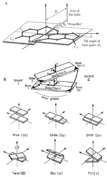
The spatial structure of a double-stranded DNA molecule depends on the base sequence: The changes in the relative orientation of mean planes of the base pair is due to the differences in spatial organization caused by the presence of different pairs in the chain. The mutual orientation of base pairs and their orientation to the axis of a linear DNA molecule are expressed in such terms as base slope angle, base pair helical twist angle, and inclination angle of base pairs (Figure 6).
Figure 6. The parameters that determine the orientation of individual nitrogen bases (A) and base
pairs (B) in respect to the axis of a linear DNA molecule.
- 35 -
These angles (Fig. 6) determine both the local bending of a DNA chain and the total curvature of a DNA molecule. The shape taken by an individual DNA molecule can be analyzed in terms of superposition of thermal fluctuations of the structure and the inner low-energy structure typical of the sequence of nucleobases [31]. Hydrophobic interactions between consecutive bases on the same strand contribute to this winding of the helix, as the bases are brought closer together, enabling a more effective exclusion of water from interaction with the hydrophobic bases. In solution, a DNA molecule exists in a canonical B form, as a rule, although, depending on the base sequence and properties of the solvent, there may be a wide range of DNA structural forms such as the A and Z form.
The structural characteristic of DNA can be studied first, by considering, the specific properties of nitrogen base sequence in a DNA chain. One of this sequences is the called paliandromic sequence or paliandrome repeat, where paliandrome can be understood as a word, phrase or sentence that reads the same from the left to the right and from the right to the left. For a DNA molecule, this term means that both DNA chains contain sequences connected by a twofold symmetry (Figure 7). In order to superimpose one repeat on the other, it must be rotated 180° around the horizontal axis and then again about the vertical axis. A mirror repeat of nitrogen bases has a symmetric sequence on each strand. Superimposing one repeat on the other requires only a single 180° rotation about the vertical axis.
Palindromic sequences of DNA can come from alternative structures where bases form pairs in one chain. When only one DNA chain is included in the process, a hairpin structure is formed (Fig. 7a). However, when both of the DNA chains take part in the process, the structure is called a cruciform (Fig. 7b).
From previous descriptions allows one to make some statements: DNA nitrogen bases have peculiar properties. First, their spatial structure provides the possibility of forming complementary pairs; second, the efficient recognition is only typical of complementary base pairs; and, finally, a stable structure formed of complementary base pairs does not depend much on exterior conditions.
- 36 -
Figure 7. Nitrogen base sequences in DNA strand (repeats) that determine the possible existence of
various local structural forms. (a) Formation of a hairpin; (b) formation of a cruciform DNA structure.
An important consequence of the previous base properties is that the formation of complementary Watson–Crick base pairs requires an intact base pair structure. Any displacement of electron or steric structure of base pairs may reduce their ability to form Watson–Crick pairs, and, consequently, different spatial forms of DNA molecules with significantly different properties correspond to different base structures.
From previous descriptions allows one to make some statements: DNA nitrogen bases have peculiar properties. First, their spatial structure provides the possibility of forming complementary pairs; second, the efficient recognition is only typical of
- 37 -
complementary base pairs; and, finally, a stable structure formed of complementary base pairs does not depend much on exterior conditions. An important consequence of the previous base properties is that the formation of complementary Watson–Crick base pairs requires an intact base pair structure. Any displacement of electron or steric structure of base pairs may reduce their ability to form Watson–Crick pairs, and, consequently, different spatial forms of DNA molecules with significantly different properties correspond to different base structures.
2.3 DNA Nuleobases Interactions
2.3.1 Sticky Ends
Double-stranded DNA molecules may have single-stranded fragments at their ends (extensions or “sticky ends”) that can form complementary pairs with other unknown single-stranded DNA fragments (Figure 8 (A)). The most important characteristic of the sticky ends is that they provide the specificity of the interaction between end nucleotide sequences of single-stranded DNAs [17]. A particular distinction of such interaction is
Figure 8. “Sticky ended” cohesion and ligation. A
B
- 38 -
that when the sticky ends overlap, specific hydrogen bonds are formed between single-stranded DNA base pairs. Figure 8 (B) represents that under proper conditions, the sticky ends of two DNA molecules recognize and bind to each other specifically by hydrogen bonding to form a single molecular complex. (Predictable affinity and structure are two key features to sticky-ended cohesion that make it important for the application in DNA nanotechnology). The additional treatment of the preformed structure that contains two sugar-phosphate chain breaks by proper enzymes (ligases) promoting the formation of a rigid, helical, a double-stranded DNA complex (B-form DNA) with complementary base pairs (Fig. 8(C)). Since the base sequences in the molecules may be known, the example proves that in the case of DNA, intermolecular interactions can be predicted and programmed.
In addition, a more complicated process, namely, the formation of a double-stranded DNA molecule from two single-double-stranded molecules with complementary base pairs, is known (Figure 9 (A)). Here, each interacting unit (oligomer) is an array of four primary nitrogen bases that selectively bind other oligomers in only two possible pairs. In consequence, a strong and specific double-stranded complex is formed between two oligomers. Sometimes a regular structure is obtained, but several incorrect complexes could also be formed (Fig. 9 (B)). Single mismatches can be relatively stable. The process of creating a double-stranded molecule from two joining single-stranded complementary DNA chains is called hybridization [32].
The DNA hybridization is a thermodynamically regulated and reversible process. While a double-stranded DNA molecule is separated into two single-stranded chains at the increase in temperature, two complementary single-stranded molecules are joined at the decrease in temperature. Another condition that has a considerable meaning for DNA nanotechnology can be emphasized as: A single-stranded DNA chain construction corresponds to the structure of flexible-chain polymers with most accidental orientation of neighboring nitrogen bases, while the double-stranded DNA molecule is formed as a result of hybridization of rigid molecules, with fixed location of nitrogen bases, respect to the DNA molecule’s axis.
In the process of hybridization, two parameters play an important role: the affinity between nitrogen base pairs and the specificity of the interaction between them. For an effective self-assembly, the hybridization of single-stranded DNA molecules must be as high as possible. This means that the formation of base pairs must take place at a maximum efficiency, while the percentage of wrong base pairs has to be minimal. For
- 39 -
Figure 9. Formation of a double-stranded DNA molecule as a result of the complementary recognition
mechanism of nucleation-zipping; A) regular structure, B) irregular structure.
example, the stability of undesirable hybrids measured by the change in the free energy at the formation of complementary pairs has to be minimized, while the stability of the desirable pairs must be maximal. This means that nitrogen bases in the initial DNA chains must be precisely chosen (synthesized), and the experimental conditions for the hybridization must be selected to provide both the affinity and the specificity of the hybridization. The optimal option is to use deliberately designed oligonucleotides that contain sequences of nitrogen bases with a specified structure.
From the previous assessments, it can be concluded that the possibility of using DNA molecules as building blocks, in the case of the hybridization technique of DNA nanostructure formation, is determined by a number of unique properties of these molecules, such as:
1. Nitrogen bases can form specific (complementary) pairs; the complementarity of bases is the fundament in DNA structural nanotechnology.
2. Flexible single-stranded fragments of nucleic acid molecules with a specified base sequence (the sticky ends) hybridize with complementary fragments and from rigid double stranded molecules whose local structure corresponds to the B form. The hybridization is the second fundament in DNA nanotechnology.
3. The predictability of the location of total atoms in the structure of a helical double-stranded molecule relative to its linear axis, as well as the properties of
- 40 -
rigid linear DNA molecules and the nature of intermolecular interactions under different conditions, is the third fundament in DNA nanotechnology.
4. The synthesis of DNA molecules of different sizes and chemical composition in reasonable quantities with the use of modern biochemical methods is the fourth fundament in DNA nanotechnology.
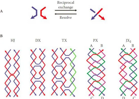
3 Hybridization Techniques of Creating Nanostructures
A fundamental drawback during the creation of DNA nanostructures by the bottom-up approach is that linear DNA molecules cannot form extended two- or three-dimensional (2-D or 3-D) structures with desired requirements under standard solvent properties [33]. This means that the formation of an extended spatial nanostructure with hydrogen bonds between neighboring elements requires the introduction of junction points into the initial DNA structure that would work as the angles in the created construction.
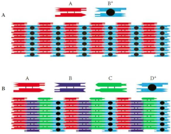
From this context, representing DNA as a branched structure attracts some interest. As shown in Figure 10, there is the hypothetical example of the connection of a four-arm branched DNA molecule (cruciform structure) with sticky ends, (the four sticky ends are labeled as A, B and their complementary fragments as A 'and B'). Due to parallelism in the assembly process of four-arm DNA molecules with sticky ends, the hybridization of four such molecules result in formation of a quadrilateral structure (Fig. 10(B)). The formed structure also possesses sticky ends (open valences) on the outside. This means that at the necessary concentration of the four-arm branched DNA molecules in the solution (on the left), this motif could be assembled into a 2-D periodic array (on the right). Under the action of DNA ligase, the splits in sugar-phosphate chains at the sites of sticky ends joining in this structure can be “linked” (eliminated), and rigid double-stranded DNA (B form) ribs, connecting flexible branching points in periodic array, can be formed. The created structure (on the right) has a nanometric size. As the backbone in the square may be from 5 to 20 nm long, this kind of structure can be called a DNA nanostructure. As a result of the self-assembly process, beside the square, an infinite 2-D lattice can be formed. This supposition is based on the presence of sticky ends on the external edges of the structure, which makes possible, the further assembly of a 2-D structure.
- 41 -
Figure 10. Assembly of a four-arm branched motif and sticky ends (A) to form a two-dimensional lattice
(B)
Is it probed that the success of the assembly depends on both the stiffness of the DNA segments that form the edges of the square and the stability of junction points on the corners [33]. If any of these components is flexible, the square will not be the desired product, and the formation of a regular structure is highly unlikely. Even if DNA molecules are considered to be flexible, they have a persistent length of about 50 nm at normal solvent properties, which means that locally a double-stranded DNA molecule is much more rigid. Thereby, short DNA molecules with a length equal to two to three helical turns (6–10 nm) can be considered as rigid building blocks.
The objective of the hybridization technique of DNA nanostructure creation is to search for and the synthesis of nitrogen base sequences that provide the formation of the desirable product and make it possible to avoid the formation of intermediate products of the assembly that interact with the final product. In other words, one can analyze the thermodynamic parameters of all of the possible sequences and choose a sequence and hybridization conditions that lead only to the desirable product. To achieve this parameter, there were developed some computer programs, based on the approach called the “minimization of sequence symmetry”, that makes possible to choose the nitrogen base sequences needed for the creation of nanoobjects. Some of these programs are SEQUIN; proposed by Seeman [34]; NANEV from the Turberfield group and some other programs can be found online [35].