1
To my parents,
and my beloved husband
2
ABSTRACTS
Citrus myrtifolia Raf. (chinotto) is a citrus fruit which originates from a mutation of Citrus aurantium (sour orange). This fruit has a small size and looks like a ping-pong. In Italy, The plant is cultivated in Calabria, Sicily and Liguria. The fruit of chinotto is widely used in food industry. Concentrated chinotto juice is a key ingredient in the production of soft drinks and liqueurs, whereas the whole fruit is used in the confectionery industry. However, despite its industrial applications, chinotto is among the least studied citrus fruit, with only a few reports present in the literature.
Here, we studied bioactive compounds contained in all parts of chinotto fruit. We also investigated the changes of those bioactive compounds during maturation and different area cultivation. First, the oil content from the seeds were analyzed, in order to assess the fatty acid profiles. After that, the methanolic extracts and DMF extracts of the defatted seeds and pulps were analyzed for their total phenolic contents (TPC) and antioxidant capacities. The results showed that all those extracts had the ability to scavenge both DPPH and ABTS radicals. TPC value is highly dependent on the level of fruit maturity. Moreover, we also isolated the pectin and β-glucan from chinotto seeds and pulps, and characterized them using FT-IR, in order to provide the valuable information about the new alternative sources of pectin and β-glucan.
Essential oils were extracted from the peels. The effects of maturation on the composition in volatile compounds of those essential oils were evaluated. After that, the potential antioxidant and anti-inflammatory properties of these fractions were tested. Our finding showed that essential oil obtained from semiripe chinotto peels effectively modulates inflammation in vitro and could, therefore, represent a potential attractive source of bioactive compounds for food, cosmetic, or pharmaceutical applications.
Key words: Chinotto, fatty acid profile, antioxidant, total phenolic content, pectin, β-glucan, essential oil, anti-inflamatory
3
ACKNOWLEDGEMENTS
This thesis has been written as a part of my three year PhD at Department of Pharmacy, Health and Nutritional Sciences in the University of Calabria, Italy, between May 2013 and February 2016.
It is a pleasure to thank the many people who made this thesis possible. Firstly, I would like to express my special appreciation and thanks to my supervisor Dr. Alessia Fazio, she has been a tremendous mentor for me. I would like to thank you for encouraging my research and for allowing me to grow as a research scientist. I could not have imagined having a better supervisor for my PhD study. Her invaluable help of constructive comments and suggestions throughout the thesis works have contributed to the success of my PhD program.
My sincere thanks also goes to Dr. Pierluigi Plastina for his advice and friendly assistance with various problems during my PhD. I greatly appreciate his time in explaining many things before I faced my final exam.
I cordially thank Prof. Galileo Violini and Dott. Gianpiero Barbuto, for their support and help towards my scholarship affairs. Next, I would like to thank Prof. Bartolo Gabriele, coordinator of OMPI curriculum and Prof. Roberto Bartolino, director of ‘Bernadino Telesio’ Doctoral school of science and technology, UNICAL.
My sincere appreciation also extends to all my colleagues in laboratory of food chemistry who have provided assistance at various occasions. Unfortunately, it is not possible to list all of them in this limited space.
I would also like to thank all my friends in Centro Residenziale, especially Lisa, Yuges, Dhanya, Narjes, Rihab, Wafa, Zahira, Rahmat,Youssef and others for their help, kindness and moral support during my study. Thanks for the friendship and memories.
Last but definitely not the leastI would like to thank my family members. Words cannot express how grateful I am to my mother and father, for all of the sacrifices that you’ve made on my behalf. Your prayer for me was what sustained me thus far. I would also like to thank my beloved husband, Rhesa Adriansyah. Thank you for supporting me for everything, and especially I can’t thank you enough for encouraging me throughout this experience. You are the apple of
4 my eye. To my mother-in-law, father-in-law, my brother, sisters-in-law and brother-in-law who have been so supportive for my overseas studies. Thank you and I love you all so much!
Praise to God for His help and guidance that I finally able to complete this dissertation.
5
CONTENTS
Abstracts i
Acknowledgements ii
Contents iv
List of Tables vii
List of Figures viii
CHAPTER 1 INTRODUCTION 1
1.1 Citrus myrtifolia Raf. 2
1.2 History of chinotto 3
1.3 Habitat of chinotto plants 4
1.4 Bioactive compound in Chinotto 4
1.4.1 Naringin 5
1.4.2 Neohesperidin 6
1.5 Fatty acid 7
1.5.1 Saturated fatty acid 8
1.5.2 Unsaturated fatty acid 10
1.6 Pectin 12
1.7 β-glucans 14
1.8 Essential oil 16
1.9 Aim of the study 18
CHAPTER 2 RESULTS AND DISCUSSIONS 19
2.1 Chinotto seeds 20
2.1.1 Oil Contents 20
2.1.2 Fatty acid composition of the oils 20
2.1.3 Biophenol extraction 23
2.1.4 Antioxidant activity 24
2.1.4.1 DPPH assay 24
6
2.1.4.2 ABTS assay 26
2.1.5 Total phenolic content (TPC) of seed extracts 28
2.1.6 Pectins 29
2.1.6.1 Characterization of pectins 29
2.1.7 β-glucans 32
2.1.7.1 Characterization of β-glucans by Infrared Transmission
Spectroscopy 32
2.1.7.2 Scanning electron microscopy of β-glucan from the seeds
of chinotto 34 2.2 Pulps of chinotto 35 2.2.1 Biophenol extraction 35 2.2.2 Antioxidant activity 35 2.2.2.1 DPPH assay 35 2.2.2.2 ABTS assay 37
2.2.3 Total phenolic content (TPC) of pulp extracts 39
2.2.4 Naringin and Neohesperidin contents 40
2.2.5 Determination of pectin in pulps 41
2.2.5.1 Characterization of pectins 42
2.2.6 β-glucans 43
2.2.6.1 Scanning electron microscopy of β-glucan from the pulps
of chinotto 44
2.3 Volatile compounds, antioxidant and anti-inflammatory properties of the essential oil from Chinotto peels
45
CHAPTER 3 EXPERIMENTAL PROCEDURES 51
3.1 Instruments 52
3.2 Reagents and Standards 52
3.3 Plant materials 52
3.4 Oil content of seeds 53
3.5 Fatty acid composition 53
3.5.1 Direct transesterification 53
7
3.5.2 HRGC-FID analysis 54
3.6 Extraction of polyphenols 54
3.6.1 Defatted seed residues 54
3.6.2 Pulp powder 55
3.7 Determination of antioxidant activity 55
3.7.1 DPPH assay 55
3.7.2 ABTS Assay 56
3.8 Determination of total phenolic content (TPC) 58
3.9 HPLC Analysis 58
3.10 Pectin extraction under different pH 58
3.10.1 Quantifying the degree of methoxylation and yield of pectins 59
3.11 Isolation of β-glucans 59
3.12 Infrared spectroscopy 60
3.13 Analysis of Scanning electron microscopy 61
3.14 Cell culture 61
3.15 Effects of CEOs on nitric oxide release and on gene expression 62 3.16 RNA purification and quantitative reverse transcription real-time PCR 62
3.17 Statistical analysis 62
CONCLUSIONS 64
REFERENCES 66
APPENDIX 74
8
List of Tables
2.1 Oil contents of the seeds 20
2.2 Fatty acid composition of chinotto seeds 21
2.3 Biophenol extracts from seeds 23
2.4 DPPH scavenging ability of the chinotto seed extracts 25 2.5 ABTS scavenging ability of the chinotto seed extracts 27
2.6 Total Phenolic Content of seed extracts 28
2.7 Pectin yield obtained from chinotto seeds 29
2.8 Degree of methoxylation of pectins in chinotto seeds 32 2.9 The amount of β-glucans contained in chinotto seeds 32
2.10 Biophenol extracts from pulps 35
2.11 DPPH scavenging ability of the chinotto pulp extracts 36 2.12 ABTS scavenging ability of the chinotto pulp extracts 38
2.13 Total Phenolic Content of pulp extracts 39
2.14 Naringin and Neohesperidin contents in pulp of chinotto 41
2.15 Pectin yield obtained from chinotto pulps 41
2.16 Degree of methoxylation of pectins in chinotto pulps 43 2.17 The amount of β-glucans contained in chinotto pulps 44
2.18 Volatile components in CEOs 47
2.19 Radical scavenging activity of CEOs 47
3.1 The Information of sample code 53
9
List of Figures
1.1 Chinotto fruits 2
1.2 Chemical structure of naringin 5
1.3 Chemical structure of Neohesperidin 6
1.4 Structure of Palmitic acid 8
1.5 Stucture of myristic acid 8
1.6 Stucture of stearic acid 9
1.7 Structure of behenic acid 9
1.8 Structure of Arachidic acid 10
1.9 Structure of Oleic acid 10
1.10 Structure of linoleic acid 11
1.11 Structure of α-linolenic acid 11
1.12 Structure of Palmitoleic acid 12
1.13 Chemical structure of pectin 13
1.14 Chemical structure of β-glucan 15
2.1 DPPH• radical scavenging activity of chinotto seed extracts 26
2.2 ABTS scavenging activity of chinotto seed extracts 28
2.3 Infrared spectrum of pectins extracted from chinotto seeds at different pH 31
2.4 Infrared spectrum of commercial β-glucans 33
2.5 Infrared spectrum of extracted β-glucan from chinotto seed 33 2.6 Scanning electron micrographs of extracted β-glucan from seeds of chinotto 34 2.7 DPPH• radical scavenging activity of chinotto pulp extracts 37
2.8 ABTS scavenging activity of chinotto pulp extracts 39
2.9 Infrared spectrum of pectins extracted from chinotto pulps at different pH 43 2.10 Infrared spectrum of β-glucan extracted from chinotto pulps 45 2.11 Scanning electron micrographs of extracted β-glucan from pulp of chinotto 45
2.12 Effect of CEOs on NO production 48
2.13 Results of XTT analysis 49
2.14 Inhibition of gene expression up-regulation 50
10
3.1 Chemical structure of DPPH 56
3.2 Chemical structure of ABTS 57
1
CHAPTER 1
2 1.1 Citrus myrtifolia Raf.
Citrus myrtifolia Raf., commonly known as chinotto or myrtle-leaved orange, is a species belonging to the Rutaceae family, subfamily Aurantioideae and genus Citrus, which originates from a mutation of C. Aurantium (sour orange) [1]. Native of southern China, its origin has not been exactly ascertained. The plant was cultivated for centuries in France and Italy. In Italy the production of chinotto fruits are concentrated in the southern regions, for its mild climate and the type of terrain, with Sicily in the first row, followed by Calabria and the Ligurian Coast (Savona). This plant is a woody plant varies from shrub to small tree one meter high, with rounded crown and compact branches with leaves of dark green color, small and similar in size to those of the myrtle; hence the Latin name Myrtifolia. The flowers are small, white and very fragrant. The fruits are in clusters, a deep orange color and the weight no more than 50-60 grams, with the size as a ping-pong. Immature fruits are green oblate spheroids with a diameter of about 2-3 cm, whereas ripe fruits are orange oblate spheroids with a diameter of about 5-6 cm. The flesh is bitter and sour and divided into 8–10 segments. The peel is tightly adherent to the pulp [2] .
Fig.1.1 Chinotto fruits
Although in many countries it is grown only for ornamental purposes, its sour-tasting fruits have a significant impact on the food industry. The smaller green ones (2-3 cm diameter) are used in the candy industry or in the preparation of jam, whereas the juice of ripe fruits is an essential flavor component of syrups, soft drinks and aperitifs and, above all else, the primary ingredient of the ‘Chinotto’ Italian soft drink. In light of the growing distribution of this popular soft drink, which is also beginning to appear in several other countries (America, Australia), it is surprising
3 that chinotto fruit is among the least studied citrus fruit, with only a few reports present in the literature [3-5].
1.2 History of Chinotto
Chinotto (Citrus Myrtifolia Raf.) is a nativeplant from southern China. In the sixteenth century, the Portuguese came to Goa, India, where the tree cultivated. The Indians considered the plant was not local but native to the areas of South-East Asia and China.
The interest was due to the fact that this fruit was extracted as an important perfume essence and also as a source of vitamin for the sailor because of the presence of many antioxidants made it suitable for the storage on the sailing ships on long journeys. It was imported to Europe because those purposes mentioned. People in Turkey, Syria, and the Black Sea area used chinotto also as a perfume essence. After that, people started to consume squeezed chinotto juice with a lot of sugar as a beverage or as an ingredient of candy.
In the eighteenth century, the cultivation of chinotto had spread to Mediterranean. It was difficult to plant chinotto tree in that area,800 cultivations were greatly reduced because of the difficulty of cultivation (slow growth, poor harvests in case of drought, plant death in case of frost, the discovery of their durable for sea traveller. Currently, the cultivation of chinotto has completely disappeared from the Iberian Peninsula, from Provence, from North Africa and the East from Turkey and Syria. However, in France the trees of chinotto exist only in the area of Nice (which until 1860 was part of Italy).
According to history document, it seems that in Italy chinotto was imported by a navigator of Savona in 1500. At that time chinotto was used to make candy or jelly. Over the centuries it spreads across the western coast a real "industry chinotto" that knows its greatest success in the last century. At the end of the Second World War the cultivation of chinotto was present only in Liguria (from Ventimiglia to Nervi) and Sicily. The fruits "turned" ("turning" is the operation that allows the removal of the thin layer of peel), "boiled", "tanned" and placed in brine were sold in wooden tubs: departed from the port of Savona to Marseille for the confectionery sector. Nowadays, the industriesof chinotto are growing only in the province of Savona (Albenga) with a high cultivation in the area from Pietra Ligure to Finale Ligure. In Sicily it is found only in the area of Taormina. At present the Liguria together with Georgia remains growing chinotto with the seeds which are imported from Asia.
4 1.3 Habitat of Chinotto plants
Chinotto, like all other citrus fruits belongs to the family of Rutaceae, suitable to be planted in subtropical climates and particularly sensitive to the extremely cold. These plants are very resistant to excess water and various diseases. It is very suitable for growing in pots. It is grown mainly in the warmer regions of Italy for its mild climate and the type of soil in both conducive to plant growth (Liguria, Calabria and Sicily).
1.4 Bioactive compound in Chinotto
It is known how citrus fruits are rich in different phytochemicals, which all contribute to determine a high nutraceutical potential: several studies have shown that these compounds possessbiological properties such as antioxidant, inflammatory andanalgesic effects, anti-viral and anti-bacterial activities, up to antithrombotic, neuroprotective and anti-tumoral properties [6].
Several studies reported that almost all parts of chinotto fruit contain many bioactive compounds that may provide health benefits to human body. Chinotto raw materials used for beverage industrial purposes is a good source of phytochemicals, mainly vitamin C and flavonoids. These bioactive compounds in whole fruit of chinotto are in fact higher than in the juice [7]. Barreca et al. identified that chinotto contained significant amount of flavonoids and furocoumarins in their peel, carpel membranes, leaves and seeds. The main phenolic components of chinotto fruits belong to the flavanone class, including narigin and neohesperidin as the major flavonoid components. The remaining flavanones contained in small quantity, such as neoeriocitrin, melitidin, brutieridin, eriocitrin, and narirutin. The other compounds that also present in chinotto in lower amount than flavanone groups, namely rhoifolin,vicenin-2, lucenin-2,polymethoxyflavone, bergapten, epoxybergamott, neodiosmin, sinensetin, tetramethoxyfavone, nobiletin, heptamethxyflavone and tengeretin [8,9,10].
Nowadays,all those aspects above are considered to be highly valuable for the commercial valorization of chinotto as a citrus with high potential as nutraceutical source. However, this study only focused on naringin and neohesperidin contents in chinotto pulp and their changes during maturation because based on the previous studies naringin and neohesperidin were the major compounds in chinotto.
5 1.4.1 Naringin
Naringin (with the molecular formula C27H32O14 and a molecular weight of 580.4 g/mol) is a
flavanone-7-O-glycoside between the flavanone naringenin and the disaccharide neohesperidose. Two rhamnose units are attached to its aglycon portion, naringenin, at the 7-carbon position. Naringin contents in various citrus species such as Chinotto fruit, and it presents in high amount in the whole part of chinotto and also in their leaves [8]. Naringin is responsible for the fruit's bitter taste.
Fig.1.2 Chemical structure of naringin
Naringin are strong antioxidants and have an abilty to scavenge free radicals and prevent lipid peroxidation. Both superoxide and hydroxyl radicals are scavenged by this flavonoids in vitro [11,12].Naringin is moderately soluble in water. The gut microflora breaks down naringin to its aglycon naringenin in the intestine; it is then absorbed from the gut [13]. Although the average daily human intake of naringin or flavonoids is not known, the total intake of polyphenols was suggested as ~1 g/d [14]. Chun et al. [15] estimated flavonoid intake by combining the USDA flavonoids database and 24-h dietary recall in NHANES 1999–2002 data. The daily mean intake of flavonoids was found from tea (157 mg), citrus fruit juices (8 mg), wine (4 mg), and citrus fruits (3 mg).
In metabolic syndrome, obesity, and related cardiovascular complications, naringin influences AMPK-, PPARα–, and CPT-1–mediated fat utilization and preserves mitochondrial function. Moreover, naringin also prevents the TNF-α–mediated inflammatory process and tissue damage in liver and vasculature. Naringin supplementation lowered elevated plasma lipid concentrations in high-fat-diet–fed rats and decreased plasma lipids and cholesterol in high-cholesterol-diet–fed rats. The cholesterol-lowering effect of naringin was observed in LDL receptor (LDLR) knockout mice. Hepatic 3-hydroxy- 3-methyl CoA (HMG-CoA) reductase activity was significantly reduced in the naringin-supplemented (0.02 g/100 g) group, whereas cholesterol
6 acyl transferase (ACAT) activity was unaffected in Ldlr knockout mice [16]. In a clinical trial, naringin supplementation (400 mg/capsul/d) reduced plasma total- and LDL-cholesterol concentrations [11]. Moreover, naringin significantly increased the production of NO metabolites in urine and improved the acetylcholinemediated endothelium function using thoracic aortic ring preparations by NO production [17]. Effect ofNaringin on Cardiac Toxicity and Hypertrophy is reducing lipid peroxidation, improved antioxidant enzymes, and decreased inflammatory cell and fibrosis in hearts of isoproterenol-treated rats [18]. Other study showed that naringin (30 mg/kg) and vitamin C (50 mg/kg) cotreatment ameliorated streptozotocininduced diabetes in rats by improving insulin concentration and prevented oxidative stress. It also improved insulin concentration and pancreatic architecture in db/db mice at a supplementation dose of 0.2 g/kg of diet [19].
1.4.2 Neohesperidin
Neohesperidin is a flavonoid which belong to flavanone glycoside group . Its aglycone form is called hesperetin [20]. Citrus fruits are rich sources of neohesperidin; the peels of oranges, lemons, and grapefruit contain bitter taste of neohesperidin. alkaline hydrogenation of neohesperidin can produce Neohesperidin dihydrochalcone (NHDC) compound, which is an intensive sweetener.
Fig.1.3 Chemical structure of Neohesperidin
NHDC showed remarkable radical scavenging activity against stable radical and reactive oxygen species (ROS). NHDC was the most potent inhibitor of H2O2 and HOCl. NHDC showed HOCl
scavenging activity of 93.5% and H2O2 scavenging property of 73.5% which was more than
those of all the tested compounds including ascorbic acid and BHT. Moreover, NHDC could inhibit protein degradation, plasmid DNA strand cleavage and HIT-T15, HUVEC cell death from HOCl attack while mannitol, BHT, and ascorbic acid could not protect them effectively. Hence,
7 NHDC is a potent antioxidant, especially it is evaluated as a novel HOCl scavenger. The intake of NHDC-containing food might contribute positive in allergic patients. There are many advantages on NHDC for drug development because of its characteristics of intense sweetener, low toxicity, and potent antioxidant activity [21]
1.5 Fatty Acid
Fatty acid is a carboxylic acid with a long aliphatic tail (chain), which is either saturated or unsaturated. Most naturally occurring fatty acids have a chain of an even number of carbon atoms, from 12 to 28 [22]. Fatty acids are usually derived from triglycerides or phospholipids. When they are not attached to other molecules, they are known as "free" fatty acids.Fatty acid chains differ by length, often categorized as short to very long:
1. Short-chain fatty acids (SCFA) are fatty acids with aliphatic tails of fewer than six carbons (e.g. butyric acid).
2. Medium-chain fatty acids (MCFA) are fatty acids with aliphatic tails of 6–12 carbons, which can form medium-chain triglycerides
3. Long-chain fatty acids (LCFA) are fatty acids with aliphatic tails 13 to 21 carbons.
4. Very long chain fatty acids (VLCFA) are fatty acids with aliphatic tails longer than 22 carbons [23, 24].
Fatty acids are usually produced by the hydrolysis of triglycerides, with the removal of glycerol. Phospholipids represent another source. Some fatty acids are produced synthetically by hydrocarboxylation of alkenes.
Fatty acids that are required by the human body but cannot be made in sufficient quantity from other substrates, and therefore must be obtained from food, are called essential fatty acids. There are two series of essential fatty acids: one has a double bond three carbon atoms removed from the methyl end; the other has a double bond six carbon atoms removed from the methyl end. Humans lack the ability to introduce double bonds in fatty acids beyond carbons 9 and 10, as counted from the carboxylic acid side [25]. Two essential fatty acids are linoleic acid (LA) and alpha-linolenic acid (ALA). They are widely distributed in plant oils. The human body has a limited ability to convert ALA into the longer-chain n-3 fatty acids eicosapentaenoic acid (EPA) and docosahexaenoic acid (DHA), which can also be obtained from fish.
8 Fatty acid consist of 2 types, first fatty acids that have carbon–carbon double bonds are known as unsaturated and second the fatty acids without double bonds are known as saturated. They differ in length as well.
1.5.1 Saturated Fatty Acid
Saturated fatty acids have no double bonds, Unbranched, straight-chain molecules. Thus, saturated fatty acids are saturated with hydrogen (since double bonds reduce the number of hydrogens on each carbon). Because saturated fatty acids have only single bonds, each carbon atom within the chain has 2 hydrogen atoms (except for the omega carbon at the end that has 3 hydrogens). The short-chain, low molecular weight fatty acids (<14:0) are triglyceride constituents only in fat and oil of milk, coconut and palmseed. In the free form or esterified with low molecular weight alcohols, they occur in nature only in small amounts, particularly in plant foods and in foods processed with the aid of microorganisms, in which they are aroma substances [26]. Fatty acids belong to saturated fatty acid are behenic acid (C:22), myristic acid (C:14), palmitic acid (C:16), stearic acid (C18:0), arachidic acid (C:20), etc.
Palmitic acid (hexadecanoic acid) (Fig. 1.4) is one of the most common fatty acids found in food. Palmitic acid contained in palm oil, ruminant milk fat, butter, cheese, fruit coat fats, seedfats and in fatty meat [27].
Fig. 1.4 Structure of Palmitic acid
Myristic acid (acid tetradecanoic) (Fig. 1.5), whose name comes from Myristica fragrans, a tropical tree of nutmeg seeds, which have a very high concentrations, up to 70-80% of the lipid fraction. Myristic acid also present in tropical oils, especially as palm and coconut, and animal fats (cheese and meat). The myristic acid is used to produce soaps and cosmetics, since its salts (sodium and potassium) have foaming properties. An ester, isopropyl myristrate is used in topical preparations for promoting the cutaneous absorption.
9 Stearic acid (octadecanoic acid) (Fig. 1.6) is not responsible for heart disease because the body is rapidly desaturated to oleic acid with the intervention of Stearoyl-CoA desaturase [28]. Stearic acid can be found in foods high fat foods, especially animal source food, and also in vegetables, especially in cocoa butter and shea butter.
Fig. 1.6 Stucture of stearic acid
Behenic acid (acid docosanoic) (Fig. 1.7) is a carboxylic acid, the saturated fatty acid with formula C21H43COOH. In appearance, it consists of white to cream color crystals or powder with
a melting point of 80 °C and boiling point of 306 °C. Behenic acid is extracted from the seeds of the Ben-oil tree (Moringa oleifera). It is so named from the Persianmonth Bahman, when the roots of this tree were harvested. Behenic acid is also present in some other oils and oil-bearing plants, including rapeseed (canola) and peanut oil and skins.
Commercially, behenic acid is often used to give hair conditioners and moisturizers their smoothing properties. It is also used in lubricating oils, and as a solvent evaporation retarder in paint removers. Its amide is used as an anti-foaming agent in detergents, floor polishes and dripless candles. Reduction of behenic acid yields behenyl alcohol [29].
Fig. 1.7 Structure of behenic acid
Arachidic acid (fig 1.8), also called eicosanoic acid, is the saturated fatty acid with a 20-carbon chain. It is as a minor constituent of peanut oil (1.1%–1.7%), corn oil (3%) and cocoa butter (1%). Its name derives from the Latin arachi, peanut. It can be formed by the hydrogenation of arachidonic acid.Reduction of arachidic acid yields arachidyl alcohol.Arachidic acid is used for the production of detergents, photographic materials and lubricants [30].
10
Fig. 1.8 Structure of Arachidic acid
1.5.2 Unsaturated Fatty Acid
The unsaturated fatty acids, which dominate lipids, have one or more double bonds between carbon atoms. (Pairs of carbon atoms connected by double bonds can be saturated by adding hydrogen atoms to them, converting the double bonds to single bonds. Therefore, the double bonds are called unsaturated). In most natural fats the double bonds of unsaturated fatty acids occur in the cis configuration. In milk fat a considerable proportion is in the trans configuration. These trans bonds result from microbial action in the rumen where polyunsaturated fatty acids of the feed are partially hydrogenated [27, 31].
Oleic acid (C18: 1) (Fig. 1.9) is a monounsaturated fatty acid belonging to the family of the ω-9. The oleic acid contain in olive oil with the percentages around 60-80%, it presents in esterified form (triglycerides).
Fig. 1.9 Structure of Oleic acid
Linoleic acid is an essential fatty acid with 18 carbon atoms. It is a key precursor of some endogenous bioregulators such as prostaglandins, which play a very important role in inflammatory processes, and thromboxanes, involved in blood clotting.
Linoleic acid can be linked to the lowering of total cholesterol, by acting on sensitization to lipoprotein receptor of the liver, with the disadvantage (absent for the ω-9) to slightly reduce the high density lipoproteins (HDL or "good cholesterol"). For some specialists, the non-selective cholesterol lowering action of linoleic acid can be defined negligible, since complications related to hypercholesterolemia relating especially to the alteration of the relationship between the
11 lipoproteins (LDL / HDL). In percentage terms, the linoleic acid lowers LDL much more than those of HDL, which in general will tend to stabilize.
Linoleic acid (Fig. 1.10) is contained mainly in sunflower seeds, wheat germ, in sesame, walnuts, soybean, corn, and olive oil.
Fig. 1.10 Structure of linoleic acid
α-Linolenic acid (ALA) (Fig. 1.11) is a carboxylic acid with an 18-carbon chain and three cis double bonds. The first double bond is located at the third carbon from the methyl end of the fatty acid chain. Thus, α-linolenic acid is a kind of omega-3 fatty acid which is found in plants and an isomer of gamma-linolenic acid (GLA), a polyunsaturated n−6 (omega-6) fatty acid. It is found in flaxseed oil, and in canola, soy, and walnut oils. These vegetable oils should be obtained by cold pressing, possibly with the addition of vitamin E, used strictly for raw salad dressings, and stored in dark glass containers, to protect from light and heat sources. α-linolenic acid is similar to the omega-3 fatty acids that are in fish oil, called eicosapentaenoic acid (EPA) and docosahexaenoic acid (DHA). Human body can change α-linolenic acid acid into EPA and DHA. However, some researchers suggest that less than 1% of ALA is converted to physiologically effective levels of EPA and DHA.
The main functions of α-linolenic acid acid are antiplatelet, anti-thrombotic and vasoprotective. Moreover, omega-3 fatty acids, especially EPA and DHA, have been shown to reduce inflammation and may help prevent chronic diseases, such as heart disease and arthritis. They may also be important for brain health and development, as well as normal growth and development. Fish oil containing EPA and DHA may help treat heart disease, prevent heart attack and stroke, and slightly reduce high blood pressure [27,31,32].
12 Palmitoleic acid (fig 1.12), or (9Z)-hexadec-9-enoic acid, is an omega-7 monounsaturated fatty acid with the formula CH3(CH2)5CH=CH(CH2)7COOH that is a common constituent of
the glycerides of human adipose tissue. It is present in all tissues but, in general, found in higher concentrations in the liver. It is biosynthesized from palmitic acid by the action of the enzyme delta-9 desaturase. A beneficial fatty acid, it has been shown to increase insulin sensitivity by suppressing inflammation, as well as inhibit the destruction of insulin-secreting pancreatic beta cells [33].
Palmitoleic acid can be abbreviated as 16:1∆9. Dietary sources of palmitoleic acid include a variety of animal oils, vegetable oils, and marine oils. Macadamia oil (Macadamia integrifolia) and sea buckthorn oil (Hippophae rhamnoides) are botanical sources with high concentrations, containing 17% and 19% min to 29% max of palmitoleic acid, respectively [34].
Fig. 1.12 Structure of Palmitoleic acid
1.6 Pectin
Pectin is a polysaccharide mixture with a complicated structure containing at least 65% of galacturonic acid (GalA). Three structural elements are involved in the make-up of a pectin molecule: a homogalacturonan consisting of (1 → 4) linked α-D-GalA, a galacturonan with differently arranged side chains (building blocks: apiose, fucose, arabinose, xylose), and a rhamnogalacturonan with a backbone consisting of the disaccharide units [→ 4)-α-D-GalA-(1 → 2)-α-L-Rha-(1 →] and with its rhamnose residues linked by arabinan and galactan chains [26].
13 O CO2H HO O O HO O CO2H HO O HO O CO2Me HO O HO O CO2H HO O HO O CO2H HO O HO
Fig. 1.13 Chemical structure of pectin
Pectin occurs commonly in most of the plant tissues as a cementing substance in the middle lamella and as a thickening on the cell wall, thereby providing vegetable tissue with consistency and mechanical resistance. However, during postharvest ripening of fruits, pectins typically undergo solubilization and depolymerisation which causes softening of the fruit texture. The number of sources that may be used for the commercial manufacture of pectins is very limited [35]. The main sources of commercially acceptable pectins are from peels of citrus fruits and from apple pomace. It is 20–40% of the dry matter content in citrus fruit peel and 10–20% in apple pomace [36,37]. Alternative sources include sugarbeet waste from sugar manufacturing, sunflower heads (seeds used for edible oil), and mango waste [38].
Pectin is a part of natural human diet, as the literature reports, the daily intake of pectin from fruit and vegetables can be estimated to be around 5 g (the consumption of approximately 500 g fruit and vegetable per day is estimated). Pectin acts as a soluble dietary fiber [39]. Consumption of pectin has been shown to reduce blood cholesterol levels. The mechanism appears to be increase of viscosity in the intestinal tract, leading to reduce absorption of cholesterol from bile or food [39, 40]. In the large intestine and colon, microorganisms degrade pectin and liberate short-chain fatty acids that have favourable influence on health (also known as prebiotic effect). Pectin has been used potentially as a carrier for drug delivery to the gastrointestinal tract, such as matrix tablets, gel beads, film-coated dose form [40].
14 In food industry, pectin is used as a gelling agent in a wide range of fruit-based products, such as jams, marmalades, jellies, fruit preparations for yoghurts and desserts and fruit filling for bakery products. Pectin can be used to improve the mouth-feel and the pulp stability in juice based drinks and as a stabiliser in soured milk beverages, yoghurts and ice creams. Pectin also reduces syneresis in jams and marmalades and increases the gel strength of low calorie jams [26,41,42]. The percentage of carboxyl groups esterified with methanol is the degree of esterification (DE) or degree of methylation (DM). Degree of methylation in pectin is an important factor characterizing pectin chains. DM of pectin molecules can influence gel formation in food industry as thickening agents [37]. There can be a wide range of DMs dependent on species, tissue, and ripening [31,43, 44]. It has been observed that the DM of pectins changes during fruit ripening [45]. The degree of methyl-esterification (DM) has a strong impact on the functional properties of pectins and these are categorized as high-ester or low-ester with DM > 50% and DM < 50%, respectively [46]. High methoxyl pectin can form a gel under acidic conditions in the presence of high sugar concentrations or a similar co-solute at pH < 3.5, whereas low methoxyl pectin forms gels only in the presence of a polyvalent salt, usually the calcium ion (Ca2+) at a very wide range of pH (between 2.6 and 7.0) [47].
1.7 β-glucans
β-glucans are indigestible polysaccharides occurring naturally in various organic sources such as corn grains, yeasts, bacteria, algae, fungi and higher plants such as fruit, corn grains, oats, barley, rye. β-glucans from different sources have different linkage types, branching manners and MW [26]. They are important components of the fibres containing unbranched polysaccharides consisting of β-d-glucopyranose units linked through (1→4) and (1→3) glycosidic bonds in cereals, (1→6) glycosidic bonds in fungal sources, and (1→3) and (1→ 6) glycosidic bonds in baker’s yeast [48,49].
15
Fig. 1.14 Chemical structure of β-glucan
The structure has an impact on the water solubility of β-glucans. Extensive research has been done into the structure and properties of water solubleβ-glucans in contrast to water-insolubleβ-glucans. Generally, no sharp distinction exists between the soluble and insoluble fractions and the ratio is highly dependent on the extraction conditions of the soluble fibre. Glucans are usually concentrated in the internal aleurone and subaleurone endosperm cells walls [50].
The health functions of β-glucan have attracted much attention in recent years. Besides being a source of dietary fiber, it is linked with certain biomedical effects such as host defense potentiator (HDP), antitumor, anti-infective, immunostimulator, reduce total cholesterol and low-density lipoprotein (LDL) levels of hypercholesterolemia and has immunomodulatory activities. When taken orally in foods, β-glucans reduce postprandial serum glucose levels and the insulin response, that is, they moderate the glycemic response, in both normal and diabetic human subjects [31]. The main attribute of β-glucans that makes them beneficial is the fact that they can form very high viscose solutions, therefore, increase intraluminal viscosity. Further biological effects ofβ-glucans, which may be prospectively utilised in the clinical practice, can reside in the stimulative action on the haematopoiesis of the bone marrow, and also in the radioactive and antimutagenic effects (scavengers of free radicals) [48].
In food Industries, β-glucans had an important technological role in processed foods, where they can be used for the elaboration of products with high dietary fiber content as non-caloric thickening and stabilizing agents, as an aid in the production of cheese and ice-cream, as a fat substitute in dairyproducts and as a gel-forming component [51].
16 1.8 Essential oil
An essential oil is defined internationally as the product obtained by hydrodistillation, steamdistillation or dry distillation or by a suitable mechanical process without heating (for Citrus fruits) of a plant or some parts of it [52]. They are aromatic oily liquids, volatile, characterized by a strong odour, rarely coloured, and generally with a lower density than that of water. They can be synthesized by all plant organs (flowers, buds, seeds, leaves, twigs, bark, herbs, wood, fruits and root) and therefore extracted from these parts, where they are stored in secretory cells, cavities, canals, epidermic cells or glandular trichomes [53]. Essential oils only represent a small fraction of plant’s composition; nevertheless they confer the characteristics by which aromatic plants are used in the food, cosmetic and pharmaceutical industries.
The aroma of each oil results from the combination of the aromas of all components, and even minor oil constituents may have major organoleptic roles to play [54]. In addition to the extraction techniques reported above there are other ones that may be used for extracting the volatile fraction, nevertheless this cannot be called an “essential oil” in those cases. Suchtechniques include: vacuum distillation, solvent extraction combined off-line with distillation, simultaneous distillation-extraction (SDE), supercritical fluid extraction (SFE), and microwaveassisted extraction and hydrodistillation (MAE and MA-HD), static (S-HS), dynamic (D-HS) and high concentration capacity headspace (HCC-HS) sampling [52]. These authors in a synthetic way explain how all of these techniques operate.
Essential oils have a complex composition, containing from a dozen to several hundredcomponents. The great majority of components identified in essential oils includes terpenes (oxygenated or not), with monoterpenes and sesquiterpenes prevailing. Nevertheless, allyl- and propenylphenols (phenylpropanoids) are also important components of some essential oils [55].
Capillary gas chromatography is the technique of choice for the analysis of essential oils due to thevolatility and polarity of essential oil components, combining two different-polarity stationary phases. Identification of oil components is generally performed by chromatographic data (Kováts indices, linear retention indices, relative retention time, retention time locking) and/or by spectral data, mainly by mass spectrometry (GC-MS).
17 In Nature, essential oils play an important role in the attraction of insects to promote the dispersion of pollens and seeds or to repel other ones. In addition, essential oils may also act as antibacterials, antivirals, antifungals, insecticides, herbicides, or have feeding deterrent effects against herbivores by reducing their appetite for such plants. Essential oils have also an important role in allelopathic communication between plants [56]. The detection of some of these biological properties needed for the survival of plants has also been the base for searching similar properties for the combat of several microorganisms responsible for some infectious diseases in humans and animals. This search intends to respond to the increasing resistance of pathogenic microbes to antibiotics.
The essential oils were also reported have ability to combat bacteria from the respiratory tract, anti-Helicobacter pylori, anti-Mycoplasma pneumoniae; essential oils against DNA virus: HSV1 (herpes simplex virus), HSV-2, NDV (Newcastle disease); or RNA virus: SARS-Cov (severe acute respiratory syndrome-associated coronavirus), and Junin virus. In addition, essential oils have also revealed to be effective on the inhibition of growth and reduction in numbers of the more serious foodborne pathogens such as Salmonella spp., E.coliO157:H7 and Listeria monocytogenes [57].
The antioxidant activity of essential oils is another biological property of great interest because they may preserve foods from the toxic effects of oxidants [58]. Moreover, essential oils being also able of scavenging free radicals may play an important role in some disease prevention such as braindysfunction, cancer, heart disease and immune system decline. Increasing evidence has suggested that these diseases may result from cellular damage caused by free radicals [59]. There is also evidence that some essential oils possess anti-inflammatory activity. For example, chamomile essential oil has been used for centuries as an anti-inflammatory and also for alleviating the symptoms associated with eczema, dermatitis and other pronounced irritation [60]. However, there are other examples of essential oils (eucalyptus, rosemary, lavender, millefolia) along with other plants (pine, clove andmyrrh) that have been used as mixed formulations as anti-inflammatory agents [61].
The anti-inflammatory activity of essential oils may be attributed not only to their antioxidant activities but also to their interactions with signalling cascades involving cytokines and regulatory transcription factors, and on the expression of pro-inflammatory genes [62,63].
18 1.9 Aim of the study
The aims of this project were to :
1. Analyze oil content in the seed of chinotto, in order to assess the profile of fatty acid quantitatively and qualitatively during maturation;
2. Determine antioxidant activities and total phenolic content in MeOH and DMF extracts from the pulps and seeds of chinotto;
3. Compare antioxidant activities and total phenolic content between pulp and seed of Chinotto and analyze their changes during maturation;
4. Analyze the naringin and neohesperidin, as the major flavonoids in fruit of chinotto, contained in pulp during maturation and from different area of cultivation. This study will allow us to know in which stages of the maturation their levels are highest;
5. Isolate the pectin and β-glucan extracted from pulps and seedsof chinotto from different areas of cultivation at various stages of maturation, and characterize them by FT-IR analyses, as a tool to elucidate the structural aspects of the isolated materials. The results of this study will provide the valuable information about the new alternative sources of pectin and β-glucan which is derived from parts of chinotto fruit, such as seeds and pulps;
6. Evaluate the effects of maturation on the composition in volatile compounds of the essential oil from Chinotto peels, and to verify the potential antioxidant and anti-inflammatory properties of these fractions.
19
CHAPTER 2
20
2.1 Chinotto seeds
2.1.1 Oil contents
The oil contents of the chinotto seeds at different stage of ripening and area of cultivation are reported as a mean value of three samples (Table 2.1) and are expressed on a dry seed weight flour (5 g). As shown in Table 2.1, the oil increased during maturation both in Calabria and Sicily. At ripe stage the percentage of oil from Sicily seeds (54.8 %) was slightly higher than that contained in Calabria seeds (52.9%).Overripe chinotto from Calabria had the highest content of oil (56.8%), otherwise the lowest content of oil belong to unripe from Calabria (47.7 %).
Table 2.1 Oil contents of the seedsa
Sample Degree of maturation Area of cultivation Weight of Oil (g) w/w (%) SC02 Unripe Calabria 23.9 ± 0,5 47.7 SC03 Semiripe Calabria 24.8 ± 0,7 49.6 SC04 Ripe Calabria 26.4 ± 1,1 52.9 SC05 Semiripe Sicily 23.3 ± 0,9 46.5 SC06 Ripe Sicily 27.4 ± 1,3 54.8 SC07 Overripe Calabria 28.4 ± 1,5 56.8 a
Values are mean ± SD of three samples of each species, analysed individually in triplicate.
2.1.2 Fatty acid composition of the oils
The fatty acid compositions of the oils are determined by gas chromatography using methyl tridecanoate as internal standard and are reported in Table 2.2 There are no qualitative differences in the fatty acid profiles of chinotto seed from all areas of cultivation at all ripening stages; the individual fatty acids contents vary according to the climatic conditions and the ripening period. The most represented fatty acids in seed oils of chinotto, both in Calabria and Sicilia seeds, were linoleic, oleic and palmitic acids. The highest amount of linoleic and oleic acids belong to SC05 (6.3–8.5 mg/goil and 3.8-4.5 mg/goil, respectively). Moreover, SC06 had
the highest content of palmitic acid (3.53-3.96 mg/goil). At ripe stage, myristic, palmitoleic and
behenic acid in Sicily seeds were lower than Calabria seeds; however, in Sicily seeds the content of other fatty acids, such as palmitic, stearic, oleic, linoleic, linolenic, and arachidic acids, was
21 higher than those in Calabria seeds. At semiripe stage, most ofthe oils from Calabria chinotto seeds contained fatty acids in greater quantities than that from Sicily seeds, except palmitoleic, linoleic and oleic acid. Behenic acid was the least fatty acid in all seed oils of chinotto.
The ripening of chinotto fruit caused the changes in the amount some fatty acids. Palmitic, palmitoleic and behenic acids increased during maturation in Calabria seed oils.Stearic, oleic, linoleic, and linolenic acids decreased during maturation in Calabria seed oils.There were no quantity alteration during maturation in myristic and arachidic acids in Calabria seed oils; their amounts were stable at 0.12 and 0.17 mg/goil, respectively. In Sicily seeds, the amounts of all
fatty acids increased during ripening, except palmitoleic, oleic and linoleic acid whose amount decreased.
Table 2.2Fatty acid composition of chinotto seeds(mg/goil)
a
Fatty Acids Sample 1 Sample 2 Sample 3
SC02 Myristic Acid (C14) 0.12 ± 0.01 0.11 ± 0.002 0.12 ± 0.03 Palmitic Acid (C16) 2.68 ± 0.7 2.48 ± 0.8 2.38 ± 0.2 Stearic Acid (C18:0) 0.39 ± 0.03 0.45 ± 0.10 0.25 ± 0.14 Oleic Acid (C18:1) 3.55 ± 0.2 3.63 ± 0.5 3.16 ± 0.04 Linoleic Acid (C18:2) 3.75 ± 0.01 4.43 ± 0.8 3.13 ± 0.18 Linolenic Acid (C18:3) 1.06 ± 0.01 1.13 ± 0.2 0.83 ± 0.08 Palmitoleic Acid (C16:1) 0.14± 0.03 0.14 ± 0.6 0.16 ± 0.14 Arachidic Acid (C20) 0.16 ± 0.06 0.18 ± 0.06 0.17 ± 0.01 Behenic Acid (C22) 0.02 ±0.005 0.01 ± 0.003 0.01 ±0.002 SC03 Myristic Acid (C14) 0.12 ± 0.003 0.12 ± 0.006 0.12 ± 0.004 Palmitic Acid (C16) 2.96 ± 0.05 2.03 ± 0.19 2.32 ± 0.10 Stearic Acid (C18:0) 0.43 ± 0.01 0.42 ± 0.02 0.35 ± 0.01 Oleic Acid (C18:1) 2.9 ± 0.04 2.97 ± 0.10 2.35 ± 0.08 Linoleic Acid (C18:2) 4.08 ± 0.06 3.47 ± 0.18 3.28 ± 0.1 Linolenic Acid (C18:3) 0.94 ± 0,01 0.84 ± 0,01 0.75 ± 0.02
22 Palmitoleic Acid (C16:1) 0.05 ± 0.001 0.07 ± 0.002 0.06 ± 0.009 Arachidic Acid (C20) 0.18 ± 0.001 0.16 ± 0.004 0.18 ± 0.001 Behenic Acid (C22) 0.03 ± 0.003 0.02 ± 0.003 0.03 ± 0.002 SC04 Myristic Acid (C14) 0.12 ± 0.003 0.12 ± 0.001 0.12 ± 0.005 Palmitic Acid (C16) 2.7 ± 0.2 3.3 ± 0.02 1.7 ± 0.16 Stearic Acid (C18:0) 0.04 ± 0.23 0.07 ± 0.009 0.05 ± 0.12 Oleic Acid (C18:1) 2.8 ± 0.03 3.5 ± 0.09 2.4 ± 0.01 Linoleic Acid (C18:2) 3.3 ± 0.09 4.02 ± 0.02 3.51 ± 0.15 Linolenic Acid (C18:3) 0.11 ± 0.07 0.13 ± 0.004 0.15 ± 0.05 Palmitoleic Acid (C16:1) 0.07 ± 0.007 0.07 ± 0.002 0.06 ± 0.002 Arachidic Acid (C20) 0.17 ± 0.006 0.17 ± 0.001 0.17 ± 0.001 Behenic Acid (C22) 0.03 ± 0.005 0.03 ± 0.0001 0.03 ± 0.001 SC05 Myristic Acid (C14) 0.10 ± 0.003 0.11 ± 0.14 0.10 ± 0.02 Palmitic Acid (C16) 2.4 ± 0.05 2.4 ± 0.11 2.6 ± 0.08 Stearic Acid (C18:0) 0.05 ± 0.01 0.03 ± 0.02 0.08 ± 0.02 Oleic Acid (C18:1) 3.8 ± 0.01 4.2 ± 0.17 4.5± 0.02 Linoleic Acid (C18:2) 6.3 ± 0.03 7.3 ± 0.03 8.5 ± 0.10 Linolenic Acid (C18:3) 0.13 ± 0.01 0.18 ± 0.1 0.18 ± 0.02 Palmitoleic Acid (C16:1) 0.15 ± 0.02 0.16 ±0.001 0.19 ± 0.06 Arachidic Acid (C20) 0.16 ± 0.04 0.16 ± 0.03 0.16 ± 0.05 Behenic Acid (C22) 0.01 ± 0.001 0.01 ± 0.002 0.01± 0.001 SC06 Myristic Acid (C14) 0.11 ± 0.03 0.11 ± 0.03 0.11 ± 0.03 Palmitic Acid (C16) 3.53 ± 0.08 3.96 ± 0.38 3.67 ± 0.1 Stearic Acid (C18:0) 0.45 ± 0.02 0.67 ± 0.07 0.63 ± 0.02 Oleic Acid (C18:1) 3.24 ± 0.06 3.41 ± 0.31 3.19 ± 0.007 Linoleic Acid (C18:2) 5.21 ±0.7 5.28 ± 0.5 4.97 ± 0.1 Linolenic Acid (C18:3) 0.75 ± 0.03 1.13 ± 0.12 1.07 ± 0.03
23 Palmitoleic Acid (C16:1) 0.05 ± 0.001 0.03 ± 0.005 0.04± 0.001 Arachidic Acid (C20) 0.19 ± 0.01 0.20 ± 0.06 0.20 ± 0.02 Behenic Acid (C22) 0.02 ± 0.009 0.02 ± 0.001 0.02 ± 0.001 SC07 Myristic Acid (C14) 0.12 ± 0.005 0.12 ± 0.003 0.12 ± 0.001 Palmitic Acid (C16) 2.65 ± 0.03 2.88 ± 0.44 2.76 ± 0.55 Stearic Acid (C18:0) 0.22 ± 0.06 0.20 ± 0.03 0.21 ± 0.04 Oleic Acid (C18:1) 3.2 ± 0.005 2.5 ± 0.006 2.9 ± 0.03 Linoleic Acid (C18:2) 3.88 ± 0.08 3.74 ± 1.01 3.18 ± 0.9 Linolenic Acid (C18:3) 0.84 ± 0.09 0.91 ± 0.12 0.69 ± 0.05 Palmitoleic Acid (C16:1) 1.80 ± 0.02 1.96 ± 0.32 1.54 ± 0.2 Arachidic Acid (C20) 0.17 ± 0.05 0.16 ± 0.005 0.17 ± 0.07 Behenic Acid (C22) 0.03 ± 0.002 0.03 ± 0.002 0.03 ± 0.005 a
Values are mean ± SD of three samples of each species, analysed individually intriplicate. Each value is expressed as mg/ g of extracted oil
2.1.3 Biophenol extraction
Biophenol compounds were extracted using two different solvents: MeOH and DMF. The extract yields were reported in Table 2.3 and are expressed on a dry defatted seed weight flour (1 g).
Table 2.3 Biophenol extracts from seeds
Sample Degree of maturation Area of cultivation MeOH (mg/g ) DMF (mg/g) SC02 Unripe Calabria 70 11 SC03 Semiripe Calabria 200 80 SC04 Ripe Calabria 210 90 SC05 Semiripe Sicily 210 120 SC06 Ripe Sicily 210 96 SC07 Overripe Calabria 190 14
24 The amounts of MeOH extracts were higner than those of DMF extracts at all stages of maturation. MeOH extracts increased during maturation in Calabria seeds, but they did not change in Sicily seeds. Ripe Calabria (SC04) seeds provide the highest amount in MeOH extract. The largest amounts of biophenols from methanol extract were found in ripe seeds from Calabria (SC04), and semi ripe (SC05) and ripe seeds from Sicily (SC06) (210 mg/g defatted seed powder).
In DMF extracts, the yield increased in Calabria seeds during maturation but it gradually decreased when the seeds reached overripe stage. In Sicily seeds, DMF extracts decreased during maturation; semiripe Sicily (SC05) seeds seeds provide the highest amount in DMF extract.
2.1.4 Antioxidant activity
The DMF and methanolic seed extracts were examined and compared for their free radical scavenging activities using DPPH and ABTS assays, whose combination is particularly useful for the screening of plant extract antioxidant capacity [64].
2.1.4.1 DPPHassay
The free-radical scavenging activity of the extracts was evaluatedusing the DPPH method. 2,2’-Diphenyl-1-picrylhydrazyl (DPPH) is a stable free radical, and when it reacts with a radicalscavenger, its maximum absorbance at 517 nm fades rapidly. The antioxidant effect is proportional to the disappearance of DPPH in test samples. All the extracts showed a significant antioxidant ability (Table 2.4). The results highlighted that scavenging capacity of the MeOH and DMF extracts on DPPH radicals increased with the concentration. However, the percentage of radical scavenging activity in methanolic extracts were higher than DMF extracts at all concentrations, except for SC02 and SC03 at lower concentration (21.6% and 11.6 % respectively at 0.03 μg/mL), and SC04 at 0.17 and 0.08 μg/mL (69.6 and 47.7%). Both methanolic and DMF extracts of overripe chinotto seeds from Calabria (SC07), showed the highest value of antioxidant ability at lower concentration (35.6% and 14.8%, respectively). It is worth noting that methanolic extract of semi-ripe chinotto (SC05) had the highest percentage of radical scavenging activityat higher concentration (93.1% at 0.33 μg/mL),.
25
Table 2.4DPPH scavenging ability of the chinotto seedextracts DPPH Scavenging Activity (%) SC02 SC03 SC04 SC05 SC06 SC07 MeOH Extract 0.33 μg/mL 81.6 ± 0.9 80.5 ± 2 90.4 ± 3.3 93.1 ± 2 86.1 ± 2.8 92.1 ± 1.4 0.17 μg/mL 65.9 ± 5.6 65.1 ± 6.2 66.7 ± 5.3 70.2 ± 7.5 77.2 ± 3.2 81.4 ± 0.9 0.08 μg/mL 38.3 ± 8.4 38.7 ± 4.4 40.1 ± 1.3 51.4 ± 3.9 41.6 ± 0.3 44.9 ±1.5 0.03 μg/mL 9.7 ± 3.3 5.6 ± 3.7 25.9 ± 8.7 32.8 ± 4 21.7 ± 4.9 35.6 ± 6.3 (μmol TE/gextract) 0.33 μg/mL 58.8 ± 0.6 59.2 ± 1.3 65.9 ± 2.1 66.3 ± 1.4 62.6 ± 1.8 65.9 ± 1.1 DMF Extract 0.33 μg/mL 78.2 ± 0.1 80.1 ± 2.3 79.3 ± 1.8 70.5 ± 0.3 87.1 ± 6.6 72.3 ± 2.9 0.17 μg/mL 72.7 ± 1.4 54.0 ± 5.7 69.6 ± 1.7 45.5 ± 1.3 50.7 ± 2.8 45.8 ± 0.8 0.08 μg/mL 34.7 ± 2.5 24.2 ± 1.5 47.7 ± 1.7 31.8 ± 0.6 35.1 ± 2.4 24.9 ± 1.7 0.03 μg/mL 21.6 ± 1.7 11.6 ± 4.9 24.8 ± 4.1 2.5 ± 2 8.8 ± 5.1 14.8 ± 1.2 (μmol TE/gextract) 0.33 μg/mL 55.3 ± 0.1 58.9 ± 1.4 57.6 ± 0.7 51.3 ± 0.2 63.6 ± 3.9 52.6 ± 1.9
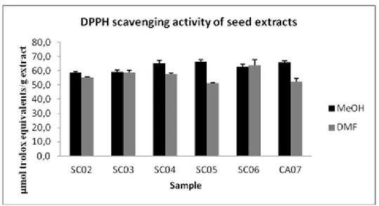
The antioxidant activity was also reported in μmol of Trolox equivalents per gram of extract (μmol TE/gextract). As shown in Figure 2.1, at the higher concentrationused (0.33 μg/mL),
methanolic extract of semi-ripe chinotto (SC05) exhibited a free radical scavenging of 66.28 μmol TE/gextract.
26
Fig. 2.1 DPPH• radical scavenging activity of chinotto seed extracts. The results are
means (±SD) of three separate experiments.The antioxidant activity was reported by μmoles of Trolox equivalents per gram ofdry extract (μmol TE/g extract).
2.1.4.2 ABTS assay
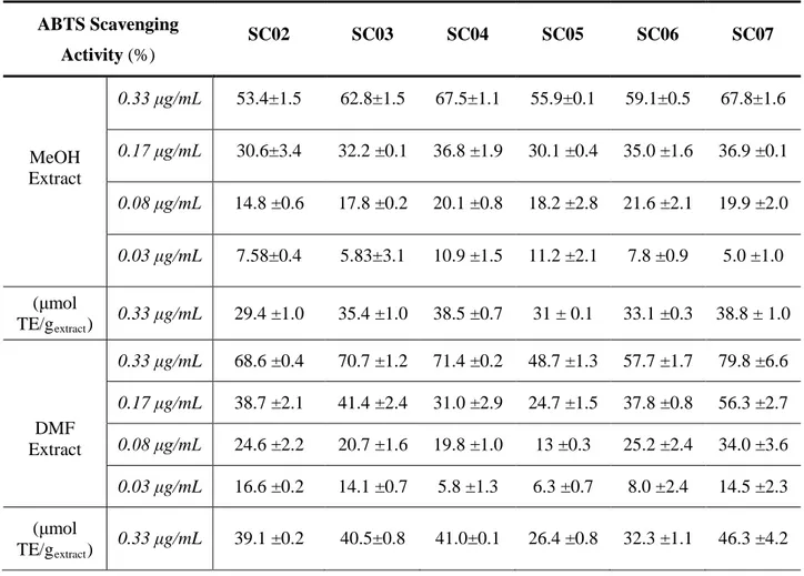
ABTS assay was also applied to measure the antioxidant activity in seed extracts. Data from this experiment were expressed as percentage of antioxidant activity in seed extracts, and were presented as means ± standard deviation (Table 2.5). Antioxidant capacity detected by ABTS assay was lower compared to that by DPPH assay; that extracts from seed had antioxidant ability inferior to 80%. Nevertheless, the results showed that antioxidant capacity by ABTS assay wasstrongly positively correlated to that by DPPH assay. Radical scavenging capacity of methanolic and DMF extracts of chinotto seedson ABTS radical cation increased with the concentration. At the highest concentration (0.33μg/mL), the percentage of radical scavenging activity of DMF extracts from Calabria seeds were higher than those of MeOH extracts. However, the MeOH extracts from Sicily seeds (SC05 and SC06) had a higher values than their DMF extracts at highest concentration (55.9% and 59.1% respectively at 0.33μg/mL). Both methanolic and DMF extracts of overripe chinotto seeds from Calabria (SC07), showed the highest value of antioxidant ability at 0.33μg/mL (67.8% and 79.8%, respectively). At lowest concentration (0.03 μg/mL), DMF extract of unripe seeds (SC02) had the highest percentage of radical scavenging activity (16.6%).
27
Table 2.5ABTS scavenging ability of the chinotto seedextracts ABTS Scavenging Activity (%) SC02 SC03 SC04 SC05 SC06 SC07 MeOH Extract 0.33 μg/mL 53.4±1.5 62.8±1.5 67.5±1.1 55.9±0.1 59.1±0.5 67.8±1.6 0.17 μg/mL 30.6±3.4 32.2 ±0.1 36.8 ±1.9 30.1 ±0.4 35.0 ±1.6 36.9 ±0.1 0.08 μg/mL 14.8 ±0.6 17.8 ±0.2 20.1 ±0.8 18.2 ±2.8 21.6 ±2.1 19.9 ±2.0 0.03 μg/mL 7.58±0.4 5.83±3.1 10.9 ±1.5 11.2 ±2.1 7.8 ±0.9 5.0 ±1.0 (μmol TE/gextract) 0.33 μg/mL 29.4 ±1.0 35.4 ±1.0 38.5 ±0.7 31 ± 0.1 33.1 ±0.3 38.8 ± 1.0 DMF Extract 0.33 μg/mL 68.6 ±0.4 70.7 ±1.2 71.4 ±0.2 48.7 ±1.3 57.7 ±1.7 79.8 ±6.6 0.17 μg/mL 38.7 ±2.1 41.4 ±2.4 31.0 ±2.9 24.7 ±1.5 37.8 ±0.8 56.3 ±2.7 0.08 μg/mL 24.6 ±2.2 20.7 ±1.6 19.8 ±1.0 13 ±0.3 25.2 ±2.4 34.0 ±3.6 0.03 μg/mL 16.6 ±0.2 14.1 ±0.7 5.8 ±1.3 6.3 ±0.7 8.0 ±2.4 14.5 ±2.3 (μmol TE/gextract) 0.33 μg/mL 39.1 ±0.2 40.5±0.8 41.0±0.1 26.4 ±0.8 32.3 ±1.1 46.3 ±4.2
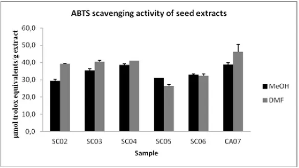
The antioxidant capacities were also expressed as micromol Trolox equivalents per gram of extract (TEAC). As shown in Figure 2.2, at the higher concentration used (0.33 μg/mL), methanolic and DMF extracts of overripe chinotto (SC07) exhibited a free radical scavenging of 38.8 and 46.3 µmol TE/gextract, respectively.
28
Fig.2.2 ABTS scavenging activity of chinotto seed extracts. The results are means
(±SD) of three separate experiments.The antioxidant activity was reported by μmoles of Trolox equivalents per gram ofdry extract (μmol TE/g extract).
2.1.5 Total phenolic content (TPC) of seed extracts
The total phenolic content of each fraction was determined using the Folin-Ciocalteu method with some modification [65] and the results obtained were expressed as mg of gallic acid equivalents per gram of extract (Table 2.6).
Table 2.6 Total Phenolic Content of seed extracts
Sample MeOH extract
(mg GAE/gextract) DMF extract (mg GAE/gextract) SC02 13.5 ± 0.9 1.5 ± 0.1 SC03 44.4 ± 1.1 52.4 ± 0.9 SC04 44.4 ± 0.7 32.3 ± 1.6 SC05 72.5 ± 2.6 80.6 ± 1.4 SC06 24.3 ± 0.7 2.8 ± 0.6 SC07 59.1 ± 2.2 12.2 ± 0.9
The changes of total phenolic content (TPC) of MeOH extracts showed different trends during maturation depending on the regions of origin; it was reduced during ripening for the extracts from Sicily seeds with highest content reached at semi-ripe stage SC05 (72.5mg GAE/gextract)
29 while it increased during ripening for the extracts from Calabria seeds to reach 59.1 mg GAE/g at mature stage (SC07). The total amount of phenolic compounds detected in the seed extracts were directly related to the values of radical scavenging activity.As the methanolic extract, DMF extracts of SC05 also had a highest value (80.6 mg GAE/gextract). These results indicated that
total phenolic content in seed residue extracts was strongly correlated with antioxidant activity, suggesting that phenolic compounds contribute to their antioxidant capacities.
2.1.6 Pectins
The pectins were extracted under 3 different pH conditions, in order to monitor the effect of extraction pH on the pectin yield and their degree of methoxylation. The results showed the differences in yields and degree of methoxylation during maturation under different pH values. The percentage yields of pectin from chinotto seeds increased during ripening in all fractions (Table 2.7). Ripe and overripe seeds from Calabria chinotto provided more pectins A (4.59% and 5.96 %, respectively) than ripe seeds from Sicily (3.83%).
Table 2.7 Pectin yield obtained from chinotto seeds
Fractions Yield (%) SC02 SC03 SC04 SC05 SC06 SC07 A 2.6 3.3 4.6 3.4 3.8 5.7 A- 11.4 11.8 12.4 21.1 40.6 12.5 A+ 15.8 17.1 18.7 34.8 57.1 21.3 2.1.6.1 Characterization of pectins
The degree of methoxylation (DM) of the pectin is the important factors in determining the texture of the gel. Fruits with high DM can be used for the production of jams and marmalades. Generally, the ingredients required for the formation of the gel may be present in the fruit itself: 65% of sugar, acidity around 3.0 and higher DM (>50%). Different pH values of extraction effected the change of degree of methoxylation.Conditions of extreme acidity (pH 1.0) caused a hydrolysis of the methyl esters and a de-polymerization of pectin chain (Fraction A-, [66]). The same situation also occurred in extreme alkaline (pH 12.0) conditions, which caused saponification of methyl esters groups and the production of different sized pectin chains by
β-30 elimination (Fraction A+, [67]). Both treatments can cause a decrease in the DM of pectins.The variation in degree of methoxylation was related with the pH. Fractions A, A- e A+ of chinotto seeds were characterized by FT-IR spectroscopy (Figure 2.3 (a), (b), (c), respectively): the band at 1631 cm-1 (peak number 4 in fig 2.3) corresponds to the symmetrical stretching vibration of free carboxyl group, while the peak at 1741 cm-1 (peak number 3 in fig. 2.3) was assigned to methyl ester group. Each pectin fraction obtained at different pH showed the same qualitative profile at all stages of ripening. After the analysis of each spectrum through identification of the two peaks, The DM was obtained using the equation [Abs 1741 cm-1/ (Abs 1631 cm-1 + Abs 1741 cm-1)] [68].
31
b. Infrared spectrum of fraction A
-c. Infrared spectrum of fraction A+
Fig. 2.3 Infrared spectrum of pectins extracted from chinotto seeds at different pH
The spectra presented in Fig. 2.3 confirmed that the variation in the degree of methoxylation was associated with the pH (Table 2.8). DM in pectin from seeds increased during the maturation (table 2.8), in the case of fraction A, both in seed from Calabria and Sicily. The highest DM was obtained for ripe seeds from Sicily (SC06, 72.7%). DM decreased during maturation in fractions A- (Calabria and Sicily) and A+ (Sicily), but increased in fraction A+ Calabria seeds.
32
Table 2.8 Degree of methoxylation of pectins in chinotto seeds
Fraction Degree of Methoxylation (%) SC02 SC03 SC04 SC05 SC06 SC07 A 40.3 ± 1.2 44.7 ± 2.2 50.3 ± 1.2 58.5 ± 3.0 72.7 ± 1.9 55.3±1.3 A- 37.3 ± 1.8 33.7 ± 3.1 47.7 ± 2.3 45.2 ±1.3 42.2 ±2.3 31.7 ± 3.7 A+ 32.7 ± 1.2 30.5 ± 1.1 42.9 ±1.7 45.4 ± 1.7 42.2 ± 3.6 45.8 ± 2.4 2.1.7 β-glucans
The content of β-glucan in Chinotto seeds were reported in Table 2.9. The amount of β-glucans decreased during maturation; seeds of unripe Calabria chinotto (SC02) contained the highest amounts of β-glucans (6.80%), this value higher than β-glucan content in barley flour with only 4.75 % [69]. The smallest content belong to SC07 (1.39%). Seeds from Calabria had a lower amount of β-glucans than those from Sicilia at the same degree of maturation.
Table 2.9 The amount of β-glucans contained in chinotto seeds
Sample Degree of Maturation β-glucans (%) SC02 Unripe Calabria 6.8 SC03 Semiripe Calabria 1.7 SC04 Ripe Calabria 1.5 SC05 Semiripe Sicily 2.1 SC06 Ripe Sicily 1.9 SC07 Overripe Calabria 1.4
2.1.7.1 Characterization of β-glucans by Infrared Transmission Spectroscopy
Infrared spectroscopy allows the measurement of molecular vibrations of covalent bonds. The IR region 4000-400 cm-1 provides information on the fundamental vibrations. The infrared spectra
33 from commercial and extracted β-glucans were shown in Figures 2.4 and 2.5, respectively. The FT-IR spectra of all seeds have the same profile; there were no changes during maturation and from different area of cultivation.
Fig. 2.4 Infrared spectrum of commercial β-glucans
Fig. 2.5 Infrared spectrum of extracted β-glucanfrom chinotto seed
In the region of 4000–3000 cm-1, the extracted and commercial β-glucans spectra showed a wide band with maximum absorption (minimum transmittance) at 3416 cm-1 and 3398 cm-1, respectively. This can be attributed to normal vibration modes of asymmetric and symmetric stretching of OH groups because polysaccharides contain a significant number of OH groups, which exhibit an absorption band above 3000 cm-1. The absorption peaks occurring at 2924 cm-1
34 (extracted) and 2926 cm-1 (commercial) in the region of 3000–2840 cm-1could be attributed to the relative values of the vibrational modes of asymmetric and symmetric stretches of CH groups [70]. The strong absorption at 1639 cm-1 for extracted β-glucans and 1640 cm-1 for commercial β-glucans were due to the stretching of CN groups and NH groups of the proteins indicating the presence of amide linkages and the presence of protein in the sample. The region 1285–242 cm-1, which showed peaks with maximum absorption at 1073 cm-1 (extracted) and 1047 cm
-1
(commercial), corresponds to COC and CO bonds of a ring of D-glucose [71], which are network vibrations in which all of the atoms of the macromolecular chain vibrate in phase and normal modes resulting from coupling of the CC and CO stretches. Therefore, the peaks at 1073 cm-1 for the extracted sample and at 1047 cm-1 for the commercial one indicate the presence of glycosidic bonds and cyclic structures of monosaccharides.
Carbohydrates can be recognized by peaks at wave numbers of 1040 cm-1 (CO bond of the alcohol group), 2940 cm-1 (CH stretch) and 3400 cm-1 (OH stretch) [70]. It is also important to note that the spectra showed absorption peaks at 993 cm-1 for the extracted sample and 887 cm-1 for the commercial sample, which is indicative of a β-glycosidic anomeric bonds.
2.1.7.2 Scanning electron microscopy of β-glucan from the seeds of chinotto
The morphology of extracted β-glucans at all stages of maturation was similar. A panoramic view of the samples (Fig.2.6a) revealed that extracted β-glucans had an irregular and small particle size distribution. A few clusters of rounded structure and small loose particles with geometric shapes.A higher magnification of a cluster of the extracted sample (Fig. 2.6b) showed some porous appearances and rough surfaces.
A. 50x B. 500x
35
2.2 Pulps of Chinotto
2.2.1 Biophenol extraction
Biophenol compounds in pulp were also extracted using two different solvents: MeOH and DMF. The extract yields were reported in Table 2.10 and are expressed on a dry pulp weight flour (1 g).
Table 2.10 Biophenol extracts from pulps
Sample Degree of maturation Area of cultivation MeOH (mg/g ) DMF (mg/g) PC02 Unripe Calabria 420.1 417.7 PC03 Semiripe Calabria 422.7 535.5 PC04 Ripe Calabria 493.2 530.3 PC05 Semiripe Sicily 467.5 481.64 PC06 Ripe Sicily 488.7 575.6 PC07 Overripe Calabria 528.9 550.1
The amount of MeOH extracts from pulps increased during maturation both in Calabria and Sicily. In DMF extracts, the amount of extracts was increased during maturation and it occurred in Sicily and Calabria pulps. The amounts ofDMF extracts were higher than those of MeOH extracts in all stages of maturation except unripe Calabria (PC02). The lowest amount in MeOH extracts was provided from the pulp of unripe Calabria chinotto (420.1 mg/g) and the lowest amount in DMF extracts was unripe Calabria (PC02) (417.7 mg/g).
2.2.2 Antioxidant activity
The DMF and methanolic extracts from pulp were examined and compared for their free radical scavenging activities using DPPH and ABTS assays.
2.2.2.1 DPPH assay
The methanolic and DMF seed extracts of chinotto pulps were examined and compared for their free radical scavenging activities against DPPH (Table 2.11). All pulpextracts exhibited an antioxidant ability. MeOH extracts had antioxidant activity higher than DMF extract, except
36 those extracted from semiripe Calabria pulp (PC03) at 0.17μg/mL and 0.08μg/mL, and overripe Calabria pulp at 0.33 μg/mL and 0.03 μg/mL.At the lowest concentration (0.03 μg/mL), unripe Calabria (PC02) still had high activity than the others (15%). DMF extract of the pulpfrom ripe Calabria chinotto (PC07) had the highest radical scavenging ability at the highest concentration (81.7% at 0.33μg/mL).
Table 2.11 DPPH scavenging ability of the chinotto pulp extracts DPPH Scavenging Activity (%) PC02 PC03 PC04 PC05 PC06 PC07 MeOH Extract 0.33 μg/mL 76.2 ± 4.2 76.1± 3.3 79.5 ± 3.9 78.8 ± 0.2 80.9 ± 3.6 78.4 ± 2.9 0.17 μg/mL 56 ± 3.3 49.3 ± 2.1 63.3±5.2 56.7 ± 5.8 58.9 ± 2.1 53.1 ± 4.9 0.08 μg/mL 22.9 ± 3.7 25.9 ± 7.2 33.7 ± 4.1 42.3 ± 3.2 37.7 ± 9.3 37.3 ± 1.1 0.03 μg/mL 15 ± 3 9.2 ± 0.4 14.5 ± 0.6 12.3 ± 0.6 8.4 ± 7.3 9.4 ± 7.2 (μmol TE/gextract) 0.33 μg/mL 56.03 ± 2.7 56.1 ± 2.1 56.3 ± 2.7 55.8 ± 0.2 59.1 ± 2.3 56.6 ± 1.2 DMF Extract 0.33 μg/mL 73.4 ±0.3 74.5 ± 1.5 79 ± 0.8 75.8 ± 1.6 78.3 ± 1 81.7 ± 3.5 0.17 μg/mL 52 ± 1.9 56.8 ± 5.9 54.9 ± 2.1 55.9 ± 6.4 55.3 ± 3.4 51.9 ± 0.3 0.08 μg/mL 22.8 ± 5.2 35.1 ± 0.8 31±2 34.5 ± 8.2 27.4 ± 0.8 31± 3.7 0.03 μg/mL 11.3 ± 2.9 7.3 ± 3.6 4.2 ± 3.3 1.4 ± 0.6 2.3±1.8 12.8 ± 0.8 (μmol TE/gextract) 0.33 μg/mL 54.2 ± 0.2 55.2 ± 0.9 56.6 ± 1.6 55.1 ± 1.0 56.2 ± 0.7 58.9 ± 2.4
The antioxidant capacities from pulp extracts were also expressed as micromol Trolox equivalents per gram of extract (TEAC). As shown in Figure 2.7 the scavenging capacity of the DMF extracts on DPPH radicals increased with the concentration while the antioxidant capacityof the methanolic extracts increased greatly with the concentration in Sicily chinotto pulps and slightly in Calabria chinotto pulps. The highest antioxidant activity belonged to methanolic extracts of pulp from ripe Sicily Chinotto PC06 (59.6 ± 1.2 µmol TE/g extract).