Research Article
Can Clinical and Surgical Parameters Be Combined to Predict
How Long It Will Take a Tibia Fracture to Heal? A Prospective
Multicentre Observational Study: The FRACTING Study
Leo Massari
,
1Francesco Benazzo,
2Francesco Falez,
3Ruggero Cadossi,
4Dario Perugia,
5Luca Pietrogrande
,
6Domenico Costantino Aloj,
7Antonio Capone,
8Michele D’Arienzo,
9Matteo Cadossi,
10Vincenzo Lorusso,
1Gaetano Caruso,
1Matteo Ghiara,
2Luigi Ciolli,
3Filippo La Cava,
3Marco Guidi
,
5Filippo Castoldi,
7Giuseppe Marongiu,
8Alessandra La Gattuta,
9Dario Dell’Omo,
11Michelangelo Scaglione,
11Sandro Giannini,
10Mattia Fortina
,
12Alberto Riva,
12Pier Luigi De Palma,
13Antonio Pompilio Gigante,
13Biagio Moretti,
14Giuseppe Solarino,
14Francesco Lijoi,
15Giovanni Giordano,
15Pier Giorgio Londini,
16Danilo Castellano,
16Giuseppe Sessa,
17Luciano Costarella,
17Antonio Barile,
18Mariano Borrelli,
18Attilio Rota,
19Raffaele Fontana,
19Alberto Momoli,
20Andrea Micaglio,
20Guido Bassi,
21Rossano Stefano Cornacchia,
22Claudio Castelli,
23Michele Giudici,
23Mauro Monesi,
24Luigi Branca Vergano,
24Pietro Maniscalco
,
25M’Putu Bulabula,
25Vincenzo Zottola,
26Auro Caraffa,
27Pierluigi Antinolfi,
27Fabio Catani,
28Claudio Severino,
28Enrico Castaman,
29Carmelo Scialabba,
29Venceslao Tovaglia,
30Pietro Corsi,
30Paolo Friemel,
31Marco Ranellucci,
31Vincenzo Caiaffa,
32Giovanni Maraglino,
33Roberto Rossi
,
34Antonio Pastrone,
34Patrizio Caldora,
35Claudio Cusumano,
35Pier Bruno Squarzina,
36Ugo Baschieri,
36Ettore Demattè,
37Stefano Gherardi,
37Carlo De Roberto,
38Alberto Belluati,
39Antonio Giannini,
39Ciro Villani,
40Pietro Persiani
,
40Silvio Demitri,
41Bruno Di Maggio,
42Guglielmo Abate,
42Francesca De Terlizzi,
4and Stefania Setti
41Orthopaedic and Traumatology Department, “S. Anna” Hospital, University of Ferrara, Ferrara, Italy
2Orthopaedic and Traumatology Department, IRCCS Foundation “San Matteo” Hospital, University of Pavia, Pavia, Italy 3Orthopaedic and Traumatology Department, “Santo Spirito in Sassia” Hospital, Rome, Italy
4Research and Development, IGEA Clinical Biophysics, Carpi, Modena, Italy 5Orthopaedic and Traumatology Department, “Sant’Andrea” Hospital, Rome, Italy
6Health Sciences Department, Operative Unit of Orthopaedics and Traumatology, “San Paolo” Hospital, University of Milan, Milan, Italy
7Orthopaedic, Traumatology and Rehabilitation Department, II Orthopaedics Clinic, CTO Hospital, Torino, Italy 8Orthopaedic Department, University of Cagliari, Cagliari, Italy
9Orthopaedic and Traumatology Department, “Paolo Giaccone” Hospital, University of Palermo, Palermo, Italy 10Department of Orthopaedic Surgery, Rizzoli Orthopaedic Institute, University of Bologna, Bologna, Italy
11Translational Research on New Surgical and Medical Technologies Department, Orthopaedics and Traumatology II∘,
University of Pisa, Pisa, Italy
12Orthopaedics and Traumatology Clinic, “S. M. alle Scotte” Hospital, University of Siena, Siena, Italy
13Clinical and Molecular Science Department, Faculty of Medicine, Polytechnic University of Marche, Ancona, Italy 14Basic Medical Science, Neurosciences and Sensory Organs Department, University of Bari, Bari, Italy
15Orthopaedic and Trauma Department, “Morgagni-Pierantoni” Hospital, Forl`ı, Italy 16Orthopaedic and Traumatology Department, “Misericordia” Hospital ASL 9, Grosseto, Italy Volume 2018, Article ID 1809091, 7 pages
17Surgery Department, “Vittorio Emanuele” Hospital, University of Catania, Catania, Italy
18Orthopaedic and Trauma Department, “San Michele” Nursing Home Hospital, Maddaloni, Caserta, Italy 19Orthopaedic and Traumatology Department, “Sandro Pertini” Hospital, ASL RMB, Rome, Italy
20Orthopaedic and Traumatology Department, “San Bortolo” Hospital, Vicenza, Italy 21Orthopaedic and Traumatology Department, A.O. Pavia Voghera Hospital, Pavia, Italy 22Orthopaedic and Traumatology Unit, Ariano Irpino Hospital, Avellino, Italy
23Orthopaedics and Trauma Department, “Papa Giovanni XXIII” Hospital, Bergamo, Italy 24Orthopaedic and Traumatology Department, “M. Bufalini” Hospital, Cesena, Italy
25Orthopaedic and Traumatology Department, “Guglielmo da Saliceto” Hospital, Piacenza, Italy 26Traumatology and Reconstructive Surgery Functional Department, “S. Anna” Hospital, Como, Italy 27Orthopaedics and Traumatology Clinic, “S. M. Misericordia” Hospital, University of Perugia, Perugia, Italy 28Orthopaedic Surgery Department, Policlinico di Modena, University of Modena and Reggio Emilia, Modena, Italy 29Orthopaedic and Traumatology Department, Montecchio Maggiore Hospital, Vicenza, Italy
30Orthopaedic and Traumatology Department, CTO Hospital ASL RM “C”, Rome, Italy 31Orthopaedic and Traumatology Department, Regione Veneto Azienda ULSS 18, Rovigo, Italy 32Orthopaedics and Traumatology Department, “Di Venere” Hospital, Bari, Italy
33Orthopaedics and Traumatology Department, “SS. Annunziata” Hospital, Taranto, Italy
34Orthopaedic and Traumatology SCDU Department, “Mauriziano Umberto I” Hospital, University of Torino, Torino, Italy 35Orthopaedic and Traumatology Surgery Department, “San Donato” Hospital, Arezzo, Italy
36Orthopaedics Department, NOCSAE Hospital, Modena, Italy
37Orthopaedics and Traumatology Department, “Santa Chiara” Hospital, Trento, Italy 38Orthopaedics Unit, “Santa Maria di Loreto Mare” Hospital, Loreto Mare, Napoli, Italy 39Specialized Surgery Department, “S. Maria delle Croci” Hospital, Ravenna, Italy 40Orthopaedic Department, Sapienza University of Rome, Rome, Italy
41Orthopaedic and Trauma Department, “Santa Maria della Misericordia” Hospital, AOUD Udine, Udine, Italy 42Orthopaedics and Traumatology Unit, Piedimonte Matese Hospital, Caserta, Italy
Correspondence should be addressed to Leo Massari; [email protected]
Received 6 December 2017; Accepted 22 February 2018; Published 30 April 2018 Academic Editor: Peter Angele
Copyright © 2018 Leo Massari et al. This is an open access article distributed under the Creative Commons Attribution License, which permits unrestricted use, distribution, and reproduction in any medium, provided the original work is properly cited.
Background. Healing of tibia fractures occurs over a wide time range of months, with a number of risk factors contributing
to prolonged healing. In this prospective, multicentre, observational study, we investigated the capability of FRACTING (tibia FRACTure prediction healING days) score, calculated soon after tibia fracture treatment, to predict healing time. Methods. The study included 363 patients. Information on patient health, fracture morphology, and surgical treatment adopted were combined to calculate the FRACTING score. Fractures were considered healed when the patient was able to fully weight-bear without pain.
Results. 319 fractures (88%) healed within 12 months from treatment. Forty-four fractures healed after 12 months or underwent
a second surgery. FRACTING score positively correlated with days to healing:𝑟 = 0.63 (𝑝 < 0.0001). Average score value was 7.3± 2.5; ROC analysis showed strong reliability of the score in separating patients healing before versus after 6 months: AUC = 0.823. Conclusions. This study shows that the FRACTING score can be employed both to predict months needed for fracture healing and to identify immediately after treatment patients at risk of prolonged healing. In patients with high score values, new pharmacological and nonpharmacological treatments to enhance osteogenesis could be tested selectively, which may finally result in reduced disability time and health cost savings.
1. Introduction
Over the past 50 years, orthopaedic surgery has defined, for the different skeletal sites and different fracture morpholo-gies, guidelines to ensure a suitable mechanical environment to allow healing [1]. The treatment of tibia fractures has become almost solely surgical, using nails, plates, and screws and external fixation, to set the mechanical conditions (sta-bility, contact, and alignment of the fracture fragments) for bone repair [2–4]. Following surgical treatment, patients have
obtained significant benefits; limb function recovery is more rapid, and joint stiffness or local osteoporosis is rare.
Bone healing results from the activity of different cell populations at the fracture site [5, 6]; nowadays, orthopaedic research aims to stimulate bone callus formation by pharma-cological [7], cellular [8], and biophysical means [9] in order to speed up fracture healing [10].
The tibia is the region with the highest incidence of fractures resulting from trauma [3]. The healing of a tibia fracture can occur over a very wide time range, from a
minimum of 2 months to a maximum of 6 months in most patients. Nevertheless, in a significant percentage of patients, healing may take place well beyond 6 months after the trauma or may require one or more surgical procedures, with significant associated health costs [11–13].
Although general and local conditions that may adversely affect fracture healing have been identified [14–18], the ability to early recognise fractures at risk of developing a nonunion is still left to the surgeon’s experience.
In a previous retrospective study, we assessed clinical records of patients treated for tibia fractures to collect information on trauma characteristics, fracture treatment, patient’s general conditions, and finally the time required for fracture healing. The data thus collected were analysed by logistic regression to identify those parameters that influ-enced time to fracture healing, and then they were combined in a score whose values increased as time to healing increased [19].
We conducted this prospective multicentre observational study to (i) investigate, in a large cohort of patients, if the score, calculated immediately after the treatment, could reliably predict the time to healing of a tibia fracture and (ii) determine the ability of the score to identify fractures at risk of nonunion, that is, healing after more than 6 months.
2. Materials and Methods
This prospective observational study mirrors the clinical practice for tibia fracture treatment throughout the country, “real world data.” On this assumption, neither indication was given on how to treat the fracture nor the review on treatment appropriateness was performed.
From January 2010 to September 2012, patients who had suffered a tibia fracture were recruited in 41 Italian orthopaedic traumatology centres. Patient treatment was left to the choice of the trauma surgeon based on experience. All patients provided written, informed consent for the handling of personal data. The study was approved by the ethical committee of the coordinating centre: University of Ferrara, Italy.
Inclusion criteria were as follows: patients with posttrau-matic fractures type 41-A and B, 42-A-B and C, 43-A and B according to AO classification [1]; fracture treatment within 3 days from trauma; and patient age> 18 years.
Exclusion criteria were as follows: fractures involving the tibia plateau and malleolar fractures, patients with autoim-mune diseases or neoplasia, and patients who could not return to the treating centre for follow-up visits.
We selected a patient-centred end point to determine fracture healing: fully weight-bearing without pain. Within 12 months from trauma, the date at which the fracture healed was used to calculate days and months elapsed since treatment (“healing time”). We chose to follow patient for 12 months after treatment as a small percentage of fractures may slowly heal 6 months after the trauma [20–22].
Criteria for failure to heal included fractures not healed within 12 months from trauma and fractures that required surgical procedures not foreseen in the initial treatment plan.
2.1. Database. For each patient, surgical and clinical data were collected in dedicated software and used to calculate the score: FRACTING (FRACTure healING). Drop-down menu was used for descriptive variables. Required fields ensured complete and consistent data collection. The score was calculated adding all values shown in Table 1.
As ancillary information, on the day of healing, surgeons were asked to record on the database the presence of bone cal-lus on tibia cortices in orthogonal radiographs; nevertheless no centralised review of the X-rays was performed.
2.2. Statistical Analysis. We conducted a power analysis to establish the number of patients required to demonstrate the correlation of the score with time to healing with a confidence interval of 0.10. Considering the correlation𝑟 = 0.69 observed in the retrospective study, we calculated a sample size of 301 patients.
In the descriptive analysis for continuous variables, mean values and standard deviations are reported. ANOVA analysis with post hoc Bonferroni test has been applied for com-parison between multiple groups. The association between continuous variables was calculated by linear regression analysis and Pearson linear correlation coefficient. In order to determine the ability of the score to identify those who would not heal within 6 months posttrauma, contingency tests and receiver operating characteristic (ROC) analysis were used; specificity, sensitivity and positive predictive values were calculated. Data were analysed using SPSS 21.0 software [IBM, New York, USA].
3. Results
3.1. Study Cohort. 519 patients were screened, 38 did not meet the inclusion criteria, and 67 did not accept to be enrolled. Finally, 414 patients with tibia fracture entered into the database. Fifty-one patients (12%) did not return for follow-up visits. Overall 363 patients completed the study (Table 2) (Figure 1).
Twenty-one percent of fractures were open; in 3%, loss of bone tissue occurred. In 75% of fractures, both tibia and fibula were fractured. According to AO classification, 6% of fractures were type 41, 72% were type 42, and 22% were type 43. Table 3 reports the treatment performed in open or closed fractures.
Out of 363 patients, 319 (88%) healed within 12 months; 268 (74%) healed within 6 months and 51 (14%) between 6 and 12 months. Forty-four fractures (12%) were considered failure as they required either further surgery or more than 12 months to heal. Figure 2 shows the percentage of fractures healed at each month.
Fracture healing was achieved on average in 130± 54 days for all patients.
At healing, the presence of callus was reported for 311 fractures: in at least three cortices in 81% of patients, in two cortices in 15% of patients, and in one cortex only in 4% of patients.
At long-term follow-up (6 months from healing), 93% of fractures were reevaluated and their healing was confirmed;
Table 1: Parameters used for FRACTING score calculation.
Parameter Values for score calculation
Age increase 18–45 1 46–60 2 >60 3 Malnutrition Yes 1 Diabetes Yes 1 Smoking Yes 1
Use of NSAIDs Yes 1
Fracture exposure severity
Closed 1
Open grade 1 2
Open skin< 5 cm 3
Open skin> 5 cm 4
Location: metaphysis or epiphysis Yes 1
Synthesis device Nail 1 Plate 2 External fixation 3 Unstable Yes 1 Misalignment> 5∘ Yes 1
Bone graft Yes 1
Plate + diastasis Yes 0.5
Angular stability plate Yes 0.5
Plate + plaster cast Yes −0.5
Fracture of tibia alone Yes 1
Loss of bone substance Yes 1
Bone diastasis,>2 mm Yes 1
Length of surgery,>120 minutes Yes 1
Blood haemoglobin before treatment< 10 g/dl Yes 1
Blood haemoglobin after treatment< 10 g/dl Yes 1
NSAIDs: nonsteroidal anti-inflammatory drugs.
Table 2: Patients’ characteristics.
Male/female 257/106
Age (yrs) 48± 17
Weight (Kg) 74± 13
Height (cm) 171± 8
Table 3: Treatment performed in open or closed fractures.
No. of fractures treated Open Closed
External fixation 76 40 36
Nail 163 25 138
Plate & screws 124 12 112
Total 363 77 (21%) 286 (79%)
2 patients reported that the fracture, initially judged to be healed, had over time been treated again.
3.2. Healing Time and FRACTING Score. The values of the score ranged from 3 to 18, with a mean value of7.3 ± 2.5, median 7.
The correlation of the score with healing time expressed in days is significant:𝑟 = 0.63; 𝑝 < 0.0001 (Figure 3).
In traumatology practice, the patient follow-up interval after treatment is usually 30 days. Therefore, we grouped the fracture healing into five time intervals:≤3, 4, 5, 6, and >6 months. The average score values by months after treatment are reported in Table 4.
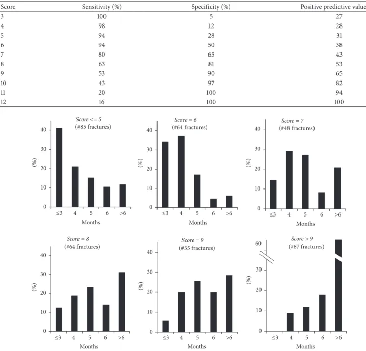
In further analysis we evaluated for different score values the percentage of fractures healed at different time intervals from trauma (Figure 4).
Among the 363 fractures, 12% of fractures with score values≤ 7 took more than 6 months to heal versus 43% of fractures with score values> 7 (𝑝 < 0.0001). We performed the ROC analysis to evaluate the ability of the score to predict fracture healing in more than 6 months (nonunion); the area under the curve (AUC) was 0.823± 0.033 (𝑝 < 0.0001). Data for the sensitivity, specificity, and positive predictive value for individual score values are shown in Table 5.
4. Discussion
To estimate healing time of a fracture immediately after its treatment is difficult, it is based on individual surgeon’s
519 screened patients 38 didn’t meet the
inclusions criteria 67 didn’t accept to be enrolled 414 included patients 51 lost to follow-up 363 evaluable patients
Figure 1: Flow diagram in which the eligible, screened, and included patients are illustrated.
0.6 4.7 17.9 25.4 21.6 13.8 8.2 4.4 1.3 0.9 0.6 0.6 0 5 10 15 20 25 30 1 2 3 4 5 6 7 8 9 10 11 12 H ea le d (%)
Time since trauma (months)
Figure 2: Percentage of fractures healed each month after treatment.
0 2 4 6 8 10 12 14 16 18 FRA C TIN G s co re 50 100 150 200 250 300 350 400 0
Healing time (days)
R = 0.63 p < 0.0001
Figure 3: Linear correlation of the score value with fracture healing time in days.
experience, and it is made even more difficult as to date there is no accepted gold standard to determine the healing of a fracture [23, 24]. Several clinical studies have considered healing on both (i) radiographic criteria: presence of bone callus in at least three cortices on radiographs performed in the two projections, and (ii) clinical criteria: absence of tenderness at the fracture site, the absence of pain on application of pressure to the fracture site and during full weight-bearing [21, 22, 25].
In this observational study, we tested the ability of the FRACTING score to estimate immediately after fracture treatment how long it will take to heal. Here, fracture healing has been based exclusively on clinical criteria: full weight-bearing without pain. This patient-centred end point is relevant in clinical practice as it corresponds to the return to work and daily activities. As confirmation of reliability of the criteria for healing adopted, at 6-month follow-up, in 2 patients only, further treatment was required.
The FRACTING score is positively correlated with the healing time in days (𝑟 = 0.63; 𝑝 < 0.0001). Furthermore, ANOVA test shows a significant association among score values and healing time in months (Table 4).
Within each score value (Figure 4), we observed fractures healing at different time periods, thus leaving a range of uncertainty that can be explained by individual biology, patient’s behaviour, and adherence to the orthopaedic sur-geon indications until healing. Nevertheless, it is noteworthy that while for scores ≤5, 12% of fractures healed after 6 months from trauma, for scores>9, the percentage increased to 61% (𝑝 < 0.0001).
The ROC analysis shows good reliability of the FRACT-ING score to assess the risk of nonunion (AUC = 0.823). In clinical practice, an effective threshold might be selected for a score value of 8 that shows a sensitivity of 63% with specificity of 81%, and a positive predictive value of 53% that shows that the fracture heals in more than 6 months (Table 3).
To our knowledge this is the first attempt to prospectively validate a score to predict fracture healing time. The results of this study cannot be extended to skeletal segments other than the tibia. However, our work suggests that the same approach can be adopted to develop specific scores for fractures located in different bones.
The major strength stems from the population studied that represents real world data. FRACTING score is associ-ated with fracture healing time and able to accurately identify fractures at risk of nonunion.
Limitations include the exclusive use of clinical criteria for definition of fracture healing, although only 2 patients experienced fracture retreatment at follow-up.
5. Conclusions
FRACTING score might be used for selecting patients in whom the efficacy of therapeutic interventions to enhance fracture healing is assessed, such as cell therapy, growth factors, drugs, or physical stimuli. Furthermore, patients with high scores may benefit from customised treatment protocols by planning closer surveillance and specific rehabilitation
Table 4: Average score values in different fracture healing months.
Healing months ≤3 4 5 6 >6
No. of fractures 74 81 69 44 51
Avg score (st.dev.) 4.97 (2.00) 6.33 (2.14) 6.86 (2.20) 7.42 (2.56) 8.71 (1.84)
ANOVA:𝑝 < 0.0001
Post hoc analysis among scores:𝑝 value
Healing months ≤3 4 5 6 >6 ≤3 1 4 0.0379 1 5 0.0007 0.6341 1 6 0.0001 0.0401 0.5711 1 >6 0.0001 0.0001 0.0001 0.0038 1
Table 5: Sensitivity, specificity, and predictive value of the score to identify fracture healing in more than 6 months.
Score Sensitivity (%) Specificity (%) Positive predictive value (%)
3 100 5 27 4 98 12 28 5 94 28 31 6 94 50 38 7 80 65 43 8 63 81 53 9 53 90 65 10 43 97 82 11 20 100 94 12 16 100 100 Score = 6 (#64 fractures) 0 10 20 30 (%) Score <= 5
(#85 fractures) (#48 fractures)Score = 7
≤3 4 5 6 >6 Score = 8 (#64 fractures) Months ≤3 4 5 6 >6 Months ≤3 4 5 6 >6 Months ≤3 4 5 6 >6 Months ≤3 4 5 6 >6 Months ≤3 4 5 6 >6 Months Score = 9 (#35 fractures) Score > 9 (#67 fractures) 60 0 10 20 30 40 (%) 0 10 20 30 40 (%) 0 10 20 30 40 (%) 0 10 20 30 40 (%) 0 10 20 30 40 (%)
that might limit the occurrence of nonunions, thus leading to significant cost savings [12, 13].
Disclosure
Level of evidence is prognostic, investigating the effect of FRACTING score on the outcome of a disease, Level I.
Conflicts of Interest
The authors declare that they have no conflicts of interest.
Acknowledgments
This study was funded in part by IGEA.
References
[1] M. E. M¨uller, P. Koch, S. Nazarian, and J. Schatzker, The
Com-prehensive Classification of Fractures of Long Bones, Springer,
Berlin, Germany, 1990.
[2] C. S. Keller, “The principles of the treatment of tibial shaft frac-tures. A review of 10,146 cases from the literature,” Orthopedics, vol. 6, no. 8, pp. 993–1006, 1983.
[3] M. Bhandari, G. H. Guyatt, M. F. Swiontkowski et al., “Surgeons’ preferences for the operative treatment of fractures of the tibial shaft: An international survey,” The Journal of Bone & Joint
Surgery, vol. 83, no. 11, pp. 1746–1752, 2001.
[4] L. E. Claes, C. A. Heigele, C. Neidlinger-Wilke et al., “Effects of mechanical factors on the fracture healing process,” Clinical
Orthopaedics and Related Research, no. 355, pp. S132–S147, 1998.
[5] R. Marsell and T. A. Einhorn, “The biology of fracture healing,”
Injury, vol. 42, no. 6, pp. 551–555, 2011.
[6] K. D. Hankenson, G. Zimmerman, and R. Marcucio, “Biological perspectives of delayed fracture healing,” Injury, vol. 45, no. 2, pp. S8–S15, 2014.
[7] D. M. Fisher, J. M.-L. Wong, C. Crowley, and W. S. Khan, “Preclinical and clinical studies on the use of growth factors for bone repair: A systematic review,” Current Stem Cell Research &
Therapy, vol. 8, no. 3, pp. 260–268, 2013.
[8] K. L. Sinclair, R. Mafi, P. Mafi, and W. Khan, “Mesenchymal stem cells and growth factors used for bone formation, fracture healing and non-unions- A systematic review,” Current Stem
Cell Research & Therapy, 2016.
[9] R. K. Aaron and M. E. Bolander, Physical Regulation of Skeletal
Repair. American Academy of Orthopaedic Surgeons Research Symposia, Chicago, 2005.
[10] W. M. Novicoff, A. Manaswi, M. V. Hogan, S. M. Brubaker, W. M. Mihalko, and K. J. Saleh, “Critical analysis of the evidence for current technologies in bone-healing and repair,” The Journal of
Bone & Joint Surgery, vol. 90, no. 1, pp. 85–91, 2008.
[11] E. Antonova, T. K. Le, R. Burge, and J. Mershon, “Tibia shaft fractures: costly burden of nonunions,” BMC Musculoskeletal
Disorders, vol. 14, article 42, 2013.
[12] L. A. Mills and A. H. R. W. Simpson, “The relative incidence of fracture non-union in the Scottish population (5.17 million): A 5-year epidemiological study,” BMJ Open, vol. 3, no. 2, Article ID 002276, 2013.
[13] Z. Dahabreh, G. M. Calori, N. K. Kanakaris, V. S. Nikolaou, and P. V. Giannoudis, “A cost analysis of treatment of tibial fracture
nonunion by bone grafting or bone morphogenetic protein-7,”
International Orthopaedics, vol. 33, no. 5, pp. 1407–1414, 2009.
[14] M. S. Gaston and A. H. R. W. Simpson, “Inhibition of fracture healing,” Journal of Bone and Joint Surgery Series: B, vol. 89, no. 12, pp. 1553–1560, 2007.
[15] L. Audig´e, D. Griffin, M. Bhandari, J. Kellam, and T. P. R¨uedi, “Path analysis of factors for delayed healing and nonunion in 416 operatively treated tibial shaft fractures,”
Clinical Orthopaedics and Related Research, no. 438, pp. 221–232,
2005.
[16] G. M. Calori, W. Albisetti, A. Agus, S. Iori, and L. Tagliabue, “Risk factors contributing to fracture non-unions,” in Injury, vol. 38, pp. S11–S18, 2007.
[17] M. Bhandari, P. Tornetta III, S. Sprague et al., “Predictors of reoperation following operative management of fractures of the tibial shaft,” Journal of Orthopaedic Trauma, vol. 17, no. 5, pp. 353–361, 2003.
[18] K. Fong, V. Truong, C. J. Foote et al., “Predictors of nonunion and reoperation in patients with fractures of the tibia: An observational study,” BMC Musculoskeletal Disorders, vol. 14, article no. 103, 2013.
[19] L. Massari, F. Falez, V. Lorusso et al., “Can a combination of different risk factors be correlated with leg fracture healing time?” Journal of Orthopaedics and Traumatology, vol. 14, no. 1, pp. 51–57, 2013.
[20] P. Joveniaux, X. Ohl, A. Harisboure et al., “Distal tibia fractures: Management and complications of 101 cases,” International
Orthopaedics, vol. 34, no. 4, pp. 583–588, 2010.
[21] H. T. Aro, S. Govender, A. D. Patel et al., “Recombinant human bone morphogenetic protein-2: a randomized trial in open tibial fractures treated with reamed nail fixation,” The Journal
of Bone & Joint Surgery, vol. 93, no. 9, pp. 801–808, 2011.
[22] T. Lyon, W. Scheele, M. Bhandari et al., “Efficacy and safety of recombinant human bone morphogenetic protein-2/calcium phosphate matrix for closed tibial diaphyseal fracture a double-blind randomized controlled phase-ii/iii trial,” The Journal of
Bone & Joint Surgery, vol. 95, no. 23, pp. 2088–2096, 2013.
[23] B. G. Dijkman, J. W. Busse, S. D. Walter, and M. Bhandari, “The impact of clinical data on the evaluation of tibial fracture healing,” Trials, vol. 12, article no. 237, 2011.
[24] D. B. Whelan, M. Bhandari, D. Stephen et al., “Development of the radiographic union score for tibial fractures for the assess-ment of tibial fracture healing after intramedullary fixation,” The
Journal of Trauma and Acute Care Surgery, vol. 68, no. 3, pp.
629–632, 2010.
[25] S. Govender, C. Csimma, H. K. Genant et al., “Recombinant human bone morphogenetic protein-2 for treatment of open tibial fractures a prospective, controlled, randomized study of four hundred and fifty patients,” The Journal of Bone & Joint