PhD course in Biochemistry
XXXI Cycle (2015-2018)
Structural and biochemical characterization of
ribosome small subunit-dependent GTPase A
(RsgA) from Pseudomonas aeruginosa
Docente guida PhD Student
Prof. Carlo Travaglini-Allocatelli Serena Rocchio
Tutor
Dr. Adele Di Matteo
PhD coordinator
Table of contents
1. Introduction ... 1
1.1 Bacterial ribosome ... 1
1.1.1 Bacterial ribosome assembly ... 3
1.1.2 30S subunit maturation ... 5
1.2 GTPases and ribosome biogenesis ... 8
1.2.1 General features of GTPases ... 10
1.2.2 Circulary permutated GTPases ... 11
1.2.3 HAS family of GTPases and mechanism of GTP hydrolysis ... 12
1.2.4 Role of GTPases in ribosomal assembly ... 14
1.3 Ribosome small subutnit-dependent GTPase A (RsgA) ... 15
1.3.1 Structural features ... 15
1.3.2 RsgA and the 30S subunit ... 16
1.3.3 GTPase activity ... 20
1.3.4 Role of RsgA in 30S subunit maturation ... 21
1.4 Targeting ribosomal assembly as novel antibacterial strategy ... 23
1.5 Pseudomonas aeruginosa ... 26
2. Aim of the thesis ... 29
3. Materials and methods ... 31
3.1 Protein Expression and Purification ... 31
3.2 Protein crystallization and structure solution ... 32
3.3 Preparation of nucleotide-free PaRsgA ... 33
3.4 CD and fluorescence spectroscopy ... 33
3.5 Binding kinetic measurements ... 34
3.6 GTPase activity ... 35
4. Results ... 39
4.1 PaRsgA expression and purification ... 39
4.2 PaRsgA structural characterization ... 42
4.2.1 PaRsgA crystallization, data collection and structure determination 42 4.2.2 Overall structure ... 44
ii
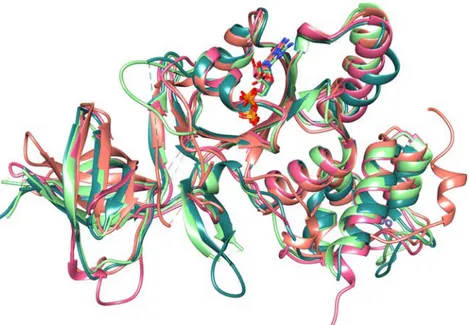
4.2.3 Structural comparison with PaRsgA orthologues ... 48
4.3 Preparation of the nucleotide-free form of PaRsgA ... 52
4.4. Biophysical characterization of PaRsgA ... 53
4.4.1 Far-UV CD spectroscopy ... 53
4.4.2 Urea-induced denaturation ... 54
4.5 Nucleotide binding kinetics ... 55
4.6 Enzyme activity ... 58
4.6.1 Intrinsic GTPase activity ... 58
4.6.2 Role of monovalent cations in the GTPase activity of PaRsgA ... 62
2.6.3 Possible determinant features for GTP hydrolysis mechanism of PaRsgA ... 65
5. Discussion ... 67
6. Conclusion and future prospects ... 75
References ... i Appendix ... xii
1. Introduction
1.1 Bacterial ribosome
Ribosomes are essential macromolecular machines that translate the genetic information into functional proteins. They are particles of more than 2.3 MDa with a diameter of about 20 nm, composed of 65% ribosomal RNA (rRNA) and 35% ribosomal proteins (r-proteins). The bacterial ribosome sediments, during ultracentrifugation, as a 70S particle composed of a small subunit (30S) and a large subunit (50S) (Ramakrishnan et al., 2002). Each subunit is therefore defined by a sedimentation coefficient, which reflect its relative mass, structure and composition differences (Connolly et al., 2008). The small subunit contains 21 ribosomal proteins and a 16S ribosomal RNA (rRNA), whereas the large subunit is made up of 34 proteins and two rRNAs: the 23S and 5S (Shajani et al., 2011) (Figure 1.1).
The 30S subunit binds mRNA during translation initiation and is mainly responsible for the mRNA decoding function. The 50S subunit contains the peptidyl transferase center and catalyzes peptide-bond formation. The 30S subunit is composed of three domains: the body (5’ domain), the platform region (central domain), and the head (3’ domain), whereas the 50S subunit principal features are the central protuberance, the L1 arm (L1 stalk) and the L7–L12 region (L7-12 stalk) (Figure 1.2). Ribosome has three functional sites designated as A (aminoacyl), which accepts the incoming aminoacylated tRNA; P (peptidyl), which holds the tRNA with the nascent peptide chain; and E (exit), which holds the deacylated tRNA before it leaves the ribosome (Ramakrishnan et al., 2002).
50S subunit 70S ribosome 30S subunit
L1 stalk L7-L12 stalk central protuberance body platform head PTC DC
Figure 1.1 E. coli 70S ribosome. The small subunit (30S) is shown on the left, with the 16S rRNA in blue and the small subunit proteins (S-proteins) in orange. The large subunit (50S) is shown on the right, with the 23S rRNA in red, the 5S rRNA in pink and the large subunit proteins (L-proteins) in green.
[Figure adapted from Shajani et al., 2011]
Structural information on the 70S ribosome (Yusupov et al., 2001; Noeske et al., 2015), the 30S (Wimberly et al., 2000; Schluenzen et al., 2000) and the 50S subunits (Ban et al., 2000) have contributed to shed light on many aspects of ribosome architecture and function (Ramakrishnan et al., 2001). Together, available structures provide a great deal of information about protein-RNA interactions in each subunit, as well as the details of the interaction of the ribosome with ligands such as initiation and elongation factors, mRNA, tRNA and antibacterial drugs (Noller et al., 2005; Carter et al., 2000; Ramakrishnan, 2001 et al.; Steiz et al., 2003). Despite the acquired knowledge on the ribosome structure and function, ribogenesis (i.e. the processes leading to a functional ribosome in cell) is far to be completely understood even if dozens of proteins involved in ribosome maturation have been identified and many genetic, biochemical and structural data have been accumulated up to now (Goto et al., 2013). To reach a complete picture of how the ribosome get its functional state in vivo is of profound interest both from a biological perspective as well as from a pharmacological point of view, since the ribosome represents an important drug target.
1.1.1 Bacterial ribosome assembly
Ribosome biogenesis is a central cellular program that accounts for a
Figure 1.2 Bacerial ribosome. The functional 70S in the middle, the 50S subunit on the left (rRNA in yellow and r-proteins in magenta) and 30S on the right (rRNA in green and r-proteins in cyan). Features in the 50S subunit include the central protuberance, L1 arm (L1 stalk) and L7–L12 region (L7-12 stalk) and the peptidyl transferase center (PTC). In the 30S subunit, these include the head, body and platform as well as the decoding center (DC).
significant fraction of the energy budget for rapidly growing bacteria, and is an essential process in all living cells. Due to the complexity of this process, understanding how different components of the bacterial ribosome come together and organize themselves remains a daunting challenge.
The in vitro assembly of the small and large subunits from individual components was achieved over 40 years ago (Nierhaus et al., 1991; Nomura et al., 1970; Traub and Nomura, 1969). This pioneering work, led primarily by the Nomura and Nierhaus laboratories, demonstrated that the information for the assembly of these macromolecular complexes resided within the components of the ribosomal subunits themselves. Nevetheless, the non-physiological conditions required for ribosome assembly and the slow kinetics of this process in vitro indicated that additional factors are required in vivo. Indeed, in bacteria, the cytoplasmic assembly of ribosomes is facilitated by many cofactors, that include ribonucleases, RNA helicases, chaperones, ATPases, GTPases and ribosomal RNA (and r-protein) modification factors (Wilson and Nierhaus 2007). Although the specific role of many of these factors is still unclear, the deletion of genes encoding many of them causes accumulation of precursor rRNAs and immature ribosomal subunits and therefore affects ribosomal assembly (Wilson and Nierhaus 2007; Connolly and Culver 2009). The most notable and enigmatic of these proteins are the GTPases (Brown et al., 2005). Indeed, several GTPases have been correlated with ribogenesis in bacteria like RsgA, Era and YqeH in the 30S maturation and YihA, RbgA, Der and Obg in the 50S maturation (Goto et al., 2013).
Ribogenesis involves different and coordinated events: i) the transcription, processing, and modification of rRNA; ii) the translation and modification of ribosomal proteins; iii) the proper folding of rRNA and ribosomal proteins;
iv) the binding of ribosomal proteins and v) the binding and release of assembly factors. Many of these steps are coupled and occur simultaneously during rRNA transcription through an alternating series of RNA conformational changes and protein-binding events (Karbstein et al., 2007; Williamson et al., 2005; Holmes et al., 2005).
1.1.2 30S subunit maturation
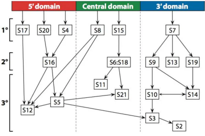
The assembly of the 30S subunit is a multistep process (Figure 1.3) that starts with transcription of the 16S ribosomal RNA (rRNA) and the synthesis of the ribosomal proteins (r-proteins). Folding of the 16S rRNA starts before transcription is completed. This process is coupled with modifications to the RNA and processing of the precursor sequences (Shajani et al., 2011; Connolly et al., 2009). The binding of the 21 r-proteins to the 16 rRNA stabilizes rRNA and suppresses its misfolding (Hosokawa et al., 1966; Traub and Nomura, 1968, 1969; Woodson et al., 2008, 2011).
Vintage experiments by Nomura (Hosokawa et al., 1966; Traub and Nomura, 1968, 1969) and more recent experiments from the Williamson and Woodson laboratories (Talkington et al. 2005; Adilakshmi et al. 2008) have defined the hierarchy and kinetic pathway of the 21 r-proteins binding to mature 16S rRNA in vitro. They have shown that six primary r-proteins bind the naked 16S rRNA, while secondary r-proteins require one or more primary r-proteins binding. Finally, the tertiary r-proteins bind after a temperature-dependent conformational step that is dependent on the binding of secondary r-proteins (Traub and Nomura, 1968; Shajani et al., 2011) (Figure 1.4).
Figure 1.3 Biogenesis pathways of the bacterial ribosomal subunits 30S (left; pdb code 2AVY; RNA shown in gray and r-proteins in red) and 50S (right; PDB code 2AW4; RNA colored gray and r-proteins purple). The biogenesis of these subunits start with the transcription of primary rRNA transcripts, which contain 16S, 23S and 5S rRNA sequences and proceeds through a series of ill-defined steps. Several ribosomal biogenesis factors, shown in the central box, facilitate the ribosomal assembly through a coordinated series of maturation events.
This hierarchy in protein binding leads to a cooperative assembly, ensuring each complex forms completely. Cooperativity mostly arises from structural changes in the 16S rRNA induced by the progressive addition of proteins (Culver et al., 2003). The 30S proteins make few base-specific contacts with the rRNA, but recognize the shape of the folded RNA (Brodersen et al., 2002). Co-folding of the RNA and the r-proteins increases the specificity of the assembly (Williamson et al., 2005). Williamson and colleagues, monitoring the binding rates and activation energies of all the 30S ribosomal proteins simultaneously, revealed that the assembly can proceed along multiple alternative pathways, due to the existence of an ensemble of multiple intermediate states from distinct assembly pathways, with no evidence of a single rate-limiting step (Williamson et al., 2005; Talkington et al., 2005; Mulder et al., 2010). They also showed that protein binding to the rRNA drives conformational rearrangements that stabilize the native fold of the 30S subunit (Talkington et al., 2005). This paradigm has been strengthened by Woodson and coworkers who showed multiple early folding nucleation events and induced fit of protein–rRNA complexes (Adilakshmi et al., 2008).
In line with the co-transcriptional nature of ribosome assembly, r-protein binding rates follow the 5’ to 3’ directionality of rRNA transcription, such that r-proteins bind rapidly to the 5’ 16S rRNA domain that forms the 30S body, and more slowly to the 3’ domain that forms the 30S head (Talkington
Figure 1.4 The Nomura assembly map. A few proteins, referred to as primary binding proteins (1°), bind directly to the nascent 16S rRNA. The binding of secondary binding proteins (2°) depend on primary binders wheras the tertiary binding proteins (3°) are sequential to secondary binders. The map is divided into 5′ (red), central (green), and 3′ (blue) domains on the basis of binding position relative to the 16S rRNA.
et al., 2005). Overall the folding of the 5’ and central domains is more robust than that of the 3’ domain due to redundant and alternative assembly pathways, while assembly of the 3’ domain follows a more restrictive pathway that is susceptible to interference and kinetic traps (Xu et al., 2010). Multiple assembly pathways increase the flexibility of the assembly process, while accessory factors and modification enzymes chaperone the late stages of assembly and control the quality of the mature subunits.
During 30S ribosomal subunit biogenesis, assembly factors prevent accumulation of misfolded intermediate states of low free energy that slowly convert into mature 30S subunits. Four protein factors, RsgA (also known as YjeQ), Era, RbfA and RimM, are involved in the late stages of the 30S subunit maturation (Jomaa et al., 2011; Leong et al., 2013; Guo et al., 2013; Jeganathan et al., 2015; Thurolow et al., 2016). Recent work indicates that these factors bind the 30S subunit at or near the decoding center and aid its folding (Lopez-Alonso et al., 2017; Razi et al., 2017; Sharma et al., 2015; Datta et al., 2007); however, the precise mechanisms and the functional interplay among them are very complex and are not yet well understood.
1.2 GTPases and ribosome biogenesis
Guanine nucleotide-binding proteins or G proteins are well-known molecular switches that control several key cellular events (Bourne et al., 2009). Extensive structural and biochemical studies have contributed to our current understanding of their roles in protein synthesis, signaling events leading to cell proliferation and differentiation, endocytosis, protein trafficking, cytoskeletal rearrangement and cell motility (Bourne et al., 2009).
During the genome sequencing revolution in late 1990s it became clear that bacteria harbored several GTPases that had no known function but were
homologous to proteins in eukaryotic organisms. Prior to discover that many of these proteins were implicated in ribosome biogenesis, several groups had shown that many of these uncharacterized GTPases were essential for bacterial growth and had potential links to cell cycle and metabolic pathways (Britton et al., 1998; Morimoto et al., 2002). Although these proteins were initially classified as part of the Ras superfamily of GTPases, nevertheless they are distinct as they have additional domains that are lacking in many of the small monomeric GTPases. Most of these extra domains mediate the interaction of the GTPases with the ribosome through direct binding to rRNA and/or to ribosomal proteins. Mutations affecting the ribosome-associated GTPases, as well as in many Ras superfamily GTPases in eukaryotes, have pleiotropic phenotypes indicating potential connections between the ribosome and the cell cycle, stress, cell growth and nutrient availability.
The GTPases involved in ribosome assembly (referred to as RA-GTPases hereafter) have been studied in multiple bacterial species and share some general features: 1) most of these proteins are essential for growth and, in cases in which null mutations have been made, the mutants show an impaired phenotype including reduced levels of 70S ribosomes in the cell, due to improper assembly of the individual subunits; 2) Many, but not all, RA-GTPases are conserved throughout evolution and homologs can be found in most eukaryotic genomes, including human; 3) The bacterial RA-GTPases interact with ribosome subunits, usually in a GTP-dependent manner. Among the RA-GTPases, RsgA, Era and YqeH are involved in the maturation of the 30S subunit maturation while YihA, RbgA, Der and Obg in the maturation of the 50S subunit (Goto et al., 2013).
1.2.1 General features of GTPases
Monomeric GTPases function as molecular switches, with the GTP-bound form corresponding to the “ON” state and the GDP-bound form to the “OFF” state. Although the bacterial RA-GTPases share many conserved features with traditional GTPases, some unique aspects are highlighted below.
GTPases have conserved motifs recognizable at the sequence and structural levels (Vetter and Wittinghofer, 2001). These include the G1-G2-G3-G4-G5 motifs with G2 and G3 contained in the so-called switch I and switch II regions. These motifs coordinate the binding of guanine nucleotides (G motifs) and the positioning of an Mg++ ion and a water molecule for efficient hydrolysis. The highly mobile switch regions, which show different conformations based on the GTP/GDP bound, are often the sites at which effector proteins bind to propagate the downstream GTP mediated signal events (Wittinghofer, Vetter, 2011). The intrinsic rate of GTP hydrolysis is usually low and additional factors are required to speed it up. These include factors such as GTPase activating proteins (GAPs) and guanine nucleotide exchange factors (GEFs). For several RA-GTPases, the ribosome may act as a GAP factor and, in few cases, also a role as a GEF has been proposed, hovewer, the molecular details of these functions have not been fully characterized to date.
The conserved functional 18–20 kDa G domain has a common structure and switch mechanism and consists of a six-stranded β-sheet and five helices on both sides (Vetter and Wittinghofer, 2001). The conserved sequence elements surround the nucleotide-binding site and are designed as G1 (GxxxxGK[S/T]), G2 (T), G3 (DxxG), G4 (N/TKxD) and a weakly conserved G5 sequence motif (often SAK). The loop containing G1, named the P-loop (for phosphate binding) and originally termed the Walker A motif,
interacts with the β- and γ-phosphate groups of GTP. The loops containing G2 and G3, termed switch I and switch II, respectively, make contact with the γ-phosphate and undergo a structured/unstructured shift upon GTP hydrolysis. The aspartate of DxxG in G3 (also called the Walker B motif) makes a water-mediated contact to the Mg++ ion, which is required for GTP hydrolysis in most Ras-like and other G proteins. G4 and G5 are instead the major determinants for guanine-base specificity (Vetter and Wittinghofer, 2001). Comparison of available structures of GTPases, in both the di- and triphosphate bound state, confirmed a canonical interaction with nucleotides and led to the conclusion that the switch mechanism is also conserved (Tu et al., 2009, 2011; Foucher et al., 2012). The conformational changes during GTP/GDP transition are confined to switch I and switch II (Milburn et al., 1990; Wittingover and Vetter, 2011). However, in multidomain proteins, these regions are often located in the interdomain interface such that the GTP/GTP transition can induce changes in the relative orientation of G flanking domains (Wittingover and Vetter, 2011).
1.2.2 Circulary permutated GTPases
A subset of RA-GTPases contains a unique circular permutation of the GTP binding domain. These atypical circularly permuted GTPases (cpGTPases) are grouped into distinct subfamilies, represented by the proteins YlqF, YqeH, YjeQ and YawG (Leipe et al., 2002). In the aminoacid sequence of these proteins the occurrence of sequence motifs follows the order G4-G5-G1-G2-G3, instead of the usual order G1-G2-G3-G4-G5 observed in canonical GTPases (Anand et al., 2006). Despite such a variation at the primary sequence level, which should lead to different topological connections between secondary structure elements, the three dimensional fold is well preserved (Shin et al., 2004). Although there are some structural
differences between cpGTPases and traditional GTPases in terms of the GTP binding pocket, the circular permutation does not dramatically alter the way these proteins interact with guanine nucleotides. In these proteins the circular permutation of G region relocates Switch-II to the C-terminus and leaves it unfastened. Since nucleotide-binding and hydrolytic activity require Switch-II to be held rigidly, an additional domain or a super secondary structure able to the proper positioning of Switch-II is required (Anand et al., 2006). As a consequence of the only permutation observed in nature is the creation of a new C-terminus following the DxxG motif in Switch-II. Such feature of cpGTPases confers two advantages: first, the Switch-II is properly positioned and oriented to favour guanine nucleotide binding and hydrolysis. Second, this coupling allows the propagation of conformational changes, mediated by GTP hydrolysis and associated with Switch-II, to the C-terminal domain thus regulating its biochemical functions (Anand et al., 2006).
1.2.3 HAS family of GTPases and mechanism of GTP hydrolysis
All the GTPases involved in ribosome biogenesis are members of the hydrophobic aminoacid substituted (HAS) family of GTPases (Mishra et al., 2005). In classical GTPases, such as Ras, a conserved glutamine residue (Gln61 designated as the Glncat) is located in the switch II. Hydrolysis of GTP is due to a nucleophilic attack by a water molecule and the role of Glncat is to stabilize the transition state by orienting the relative positions of the nucleophilic water and the γ-phosphate (Mishra et al., 2005). The importance of the catalytic glutamine in GTP hydrolysis is well documented (Vetter and Wittinghofer, 2001) and its mutation to hydrophobic aminoacids is reported to be oncogenic in Ras. Because of this substitution, the HAS-GTPases are believed to use an alternative mechanism for GTP hydrolysis (Mishra et al., 2005).
Another feature that remains unclear for the RA-GTPases is how the subsequent stabilization of the transition state (TS) is achieved. In the Ras system, RasGAPs stimulate GTP hydrolysis by supplying an additional residue, the “arginine-finger” (ArgGAP), into the active site, which is responsible for ~2,000-fold acceleration of the GTPase reaction by direct electrostatic stabilization of developing negative charges in the transition state (Scheffzek et al., 1997). The ArgGAP is provided either in cis by the same molecule or in trans by a different molecule and the scenarios are well known (Mishra et al., 2005). Up to now, RA-GTPases seem to be an exception among Ras-related G proteins, as the search for an arginine-finger or an analogous catalytic element has been unsuccessful (Rodnina et al., 2009). It has been proposed that some RA-GTPases use a monovalent cation (M+ ion) as a structural and catalytic cofactor (Kuhle et al., 2014). These RA-GTPases coordinate a M+ ion next to the GTP-γ-phosphate in a conserved coordination shell, where it forms a structurally relevant component of the catalytic center. The M+ ion adds another positive charge to the preorganized active site of GTPases that together with the invariant lysine of the P-loop and the Mg++ ion forms a triangle of positively charged moieties around the GTP molecule. Therefore, the M+ ion would be in a suitable position to neutralize negative charges of the TS in the γ-phosphate as well as the designated leaving group (GDP). This suggests that the M+ ion might function as the catalytic element that contributes to rapid GTP hydrolysis by providing electrostatic stabilization for the TS, in analogy to the arginine-finger in the Ras-RasGAP system or the M+ ion in MnmE (Kuhle et al., 2014).
The cation-dependent GTPases can be placed into two distinct groups, according to their behaviour in vitro: those that are stimulated by potassium
ions but not by sodium ions (potassium-selective cation-dependent GTPases) and those that are stimulated by both potassium and sodium ions (sodium-accomodating cation-dependent GTPases) (Ash et al., 2012). Moreover, it has been shown that the intrinsic activities of many bacterial RA-GTPases, YqeH (Anand et al., 2010), Era (Rafay et al., 2012), RbgA (Achila et al., 2012) and Der (Foucher et al., 2012) are enhanced by potassium ions, wheras ObgE activity is stimulated by sodium ions (Gkekas et al., 2017).
1.2.4 Role of GTPases in ribosomal assembly
The next major advancement in the field of RA-GTPases will come from determining how these proteins participate in ribosome biogenesis at molecular level. Few possible mechanisms by which RA-GTPases could function in ribosome assembly have been proposed: i) RA-GTPases could serve to recruit other assembly factors during key points of the assembly process; ii) RA-GTPases may control rRNA structure by regulating the activity of RNA helicases; iii) RA-GTPases could serve as RNA helicases itself and directly unwind or refold rRNA during the assembly process; iv) RA-GTPases could serve to determine whether a particular step in assembly has been properly achieved prior to the occurrency of the following phase (Britton et al., 2009). Under this light, energy from GTP hydrolysis can be used to regulate delivery or removal of proteins to nascent ribosomes as well as to promote a conformational rearrangement within nascent ribosomes. Moreover, GTPases could act as reversible “placeholders”, regulating r-proteins binding to nascent ribosome, as nutrient and enviromental sensors, by perception the cell metabolic state reflected in the GTP/GDP ratio, or as “checkpointers” by preventinting the entry of the 70S into the translational processes if not properly mature (Karbstein et al., 2007). Therefore, RA-GTPases perform essential functions in the assembly of ribosome and a clear
picture of how they work at the molecular level is critical to understand the general process of ribosome biogenesis.
1.3 Ribosome small subunit-dependent GTPase A (RsgA)
Ribosome small subunit-dependent GTPase A (RsgA), also named YjeQ /YloQ/CpgA, is a ribosome assembly factor that intervenes during the late stages of 30S maturation (Daigle et al., 2002; Himeno et al., 2004; Jomaa et al., 2011). RsgA is broadly conserved among bacteria but absent in eukaryotes (Daigle et al., 2002; Leipe et al., 2002). RsgA has low intrinsic GTPase activity that is stimulated by the 30S subunit and the 70S ribosome, but not by the 50S subunit (Himeno et al., 2004). RgsA is found associated with ribosome at very low stoichiometry (1:200) in vivo (Daigle and Brown 2004) and in vitro binds stably to the 30S subunit in the presence of GDPNP, a nonhydrolyzable GTP analogue, but not GTP or GDP (Daigle and Brown 2004; Himeno et al., 2004). In the presence of GDPNP-RsgA, 70S ribosomes dissociated into their subunits, suggesting an intersubunit localization of the factor (Himeno et al., 2004).
1.3.1 Structural features
RsgA belongs to the TRAFAC (translation factors) class of GTPases and, within this class, to the YlqF/YawG sub-family. YlqF/YawG members share the common characteristic of a circularly permutated GTP binding site in which the canonical G motifs (G1-G2-G3-G4-G5), mediating the nucleotide binding and hydrolysis (Bourne et al., 1991), are circularly permutated and adopt a G4–G5–G1–G2–G3 pattern (Shin et al., 2004; Levdikov et al., 2004;
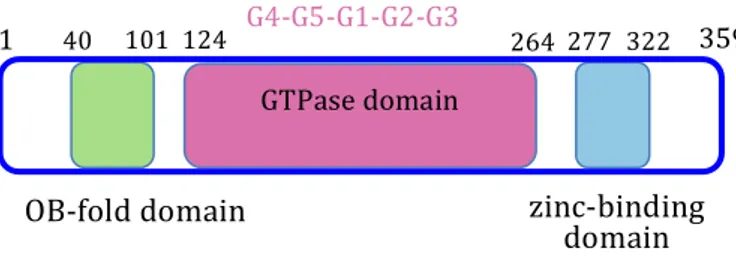
Nichols et al., 2007). G1 is characterized by the consensus sequence GxxxxGKS/T in which the lysine side chain is responsible for phosphate binding; G2 has only a threonine residue highly conserved and is located in the so called switch I loop; G3, located in the switch II loop, contains the DxxG motif in which the aspartic acid residue is involved in Mg++ coordination; G4, characterized by the N/TKxD motif, is responsible for guanine specificity together with the G5 region that is only weakly conserved. In RsgA, the G domain is located within two additional regions: an oligonucleotide/oligosaccharide binding-fold domain (OB-domain) and a zinc-binding domain (Daigle et al., 2002; Nichols et al., 2007; Shin et al., 2004). The OB-fold consists of five antiparallel β-strands defining a β-barrel. The zinc-binding domain is composed of four helices in which the central pair, together with the intervening loop, defines a zinc-binding site. In RsgA structure the switches I and II and are ideally positioned to propagate conformational changes between the GTPase domain and the other two domains (Razi et al., 2017).
1.3.2 RsgA and the 30S subunit
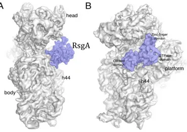
High resolution structural information on the E. coli RsgA-30S complex, achived very recently by cryo-EM studies (Razi et al., 2017; Lopez-Alonzo et al., 2017), allowed the unequivocal positioning of RsgA with respect to the 30S subunit (Figure 1.5).
In the GTP-bound form, RsgA binds all three domains of the subunit. The OB-fold contacts the body of the 30S, whereas the zinc-finger domain anchors the protein to both the head and platform domains. Finally, the GTPase domain covers the decoding center almost completely and contacts the platform through a long loop (Figure 1.6). In particular, the OB-fold interacts with the 30S mainly through helix 18 and helix 44, the GTPase domain contacts helix 44, mainly through switch I and switch II, and helix 24 in the platform, and the C-terminal zinc binding domain anchors the protein to the head through helices 29 and 30 and to the platform by contacting helix 45 (Razi et al., 2017). In the GDP form of RsgA the OB-domain is the dominant interface for 30S contact. Accordingly, it has been proposed that the OB-domain acts as an anchoring point to tether RsgA to the 30S subunit (Lopez-Alonso et al., 2017). The interaction of RsgA with the 30S subunit places the GTPase domain in direct contact with the upper part of helix 44
Figure 1.5 Cryo-EM structure of the 30S-RsgA complex. The side view (A) and the front view (B) of the 30S-RsgA complex are shown. Important landmarks of the 30S subunit, in gray, as well as the three domains of RsgA, in purple, are indicated.
[Figure adapted from Razi et al., 2017] RsgA
that represents the ribosomal motif undergoing the largest conformational change upon RsgA binding.
RsgA RsgA RsgA
As previously suggested, RsgA binds the 30S subunit close to the decoding center, in a position that is incompatible with that of all the three translation initiation factors (IF1, IF2, and IF3) (Carter et al., 2001; Simonetti et al., 2008), as well as A- and P-site tRNAs (Selmen et al., 2006) and the 50S subunit. Recent studies suggest functional interplay between RsgA and other factors involved in the 30S maturation process, like Era, RbfA, and RimM (Inoue et al., 2006; Campbell and Brown, 2008; Goto et al., 2011). Era is of particular interest because of the genetic interaction of this gene with RsgA: overexpression of Era suppresses defects in growth and ribosome maturation of a rsgA-null mutant (Campbell and Brown, 2008). Era performs its function in conjunction with RsgA; in particular they both assist the processing of the 3’ end of the precursor 17S rRNA. The binding sites of Era and RsgA to the 30S subunit do not overlap, thus simultaneous binding of the two proteins is possible. Moreover, RsgA promotes the release of RbfA from the mature 30S subunit (Goto et al., 2011). RbfA is a small protein that binds the 30S subunit at the junction of the head and body, and its binding alter the position of helices 44 and 45 (Datta et al., 2007). The binding of RsgA to the 30S subunit has a stabilizing effect in the upper region of helix 44. This is the same rRNA motif that appears severely disrupted on RbfA binding (Razi et al., 2017). Therefore, it is likely that binding of RsgA to the 30S subunit
Figure 1.6. The cryo-EM structure and atomic model of RsgA bound to the 30S subunit. The zoom-in view of the density representing RsgA in the cryo-EM map of the 30S-RsgA complex with the atomic model of RsgA superimposed in the cryo-EM map is shown on the left. The equivalent close-up view of the atomic model of the complex with the assembling factor binding to the decoding center of the 30S subunit is shown on the right. The rRNA and r-proteins interacting with RsgA are labeled. The panels shown different views of the 30S-RsgA complex, the side view (A), the front view (B) and the platform view (C). [Figure adapted from Razi et al., 2017]
forces helix 44 back into the normal decoding position and triggers the release of RbfA. RsgA also distorts the binding site for IF1, which binds the 30S subunit in the cleft between helix 44 and protein S12 (Carter et al., 2001). In addition, the RsgA binding site partially overlaps with the interaction site of IF2 (Simonetti et al., 2008) and the C-terminal domain of IF3 (Dallas and Noller 2001). Therefore, RsgA might assists ribosome maturation by preventing premature formation of the translation initiation complex (Joomaa et al., 2011). These observations suggest that RsgA might be a general checkpoint protein in the late stage of the 30S subunit biogenesis, not only to release RbfA and other biogenesis factors from the nascent 30S subunit, but also to block the binding of players in translation initiation to the premature 30S subunit. Moreover, RsgA has a role in destabilizing kinetically trapped assembly intermediate as it induces local conformational changes in the 30S structure and disrupts binding of several tertiary r-protein binding, like S2, S3, S12 and S21 (Lopez-Alonso et al., 2017). However, to ultimately describe the existing functional relationships between these factors, additional structural, genetic and biochemical studies of RsgA and other assembly factors are needed.
1.3.3 GTPase activity
Although the GTPase activity of RsgA was characterized early on (Daigle et al., 2002, 2004), the role of this activity in the overall function of RsgA and its regulation remains unclear. The GTPase activity of RsgA is stimulated by over 100-fold in the presence of mature 30S subunits (Daigle et al., 2002, 2004; Himeno et al., 2004). Although the structure does not reveal what triggers this stimulation, it is possible that the specific conformation of helix 44 may stimulate the GTPase activity in RsgA. Recent structural information provides also important clues on the catalytic mechanism of RsgA and on the
30S mediated GTPase activity stimulation (Daigle et al., 2002, 2004; Himeno et al., 2004). Lopez-Alonso and coworkers showed that the catalytic residue in RsgA (performing function that parallel Gln61 in Ras) is an histidine (His248 in E. coli sequence) located in the switch 1 loop. The 16S rRNA, in particular the upper region of h44, plays a key role in mantaining His248 in position suitable for the catalitic events; the same role has been proposed for the 23S rRNA in activating EF-Tu (Lopez-Alonzo et al., 2017). Conformational changes in the GTPase domain caused by GTP hydrolysis, ready propagated to neighboring domains, may lead to reorganization of the interface of the complex, causing an overall decrease in the binding affinity of RsgA (Razi et al., 2017) and its release from the 30S subunit. Therefore, the GTPase activity of RsgA may functions as a sensor to facilitate the release of the protein from the 30S subunit once RsgA has performed its functions (Daigle and Brown 2004).
1.3.4 Role of RsgA in 30S subunit maturation
Biochemical and genetic studies over last decade have provided a growing body of evidence implicating a role for RsgA in the late stages of 30S biogenesis (Jomaa et al., 2011; Daigle et al., 2004; Himeno et al., 2004). Involvement of RsgA in ribosome maturation has been firsly suggested by the phenotype arising from rsgA deletion. Disruption of the gene for RsgA in Escherichia coli affects cell growth, subunit association and processing of the 16S rRNA (Himeno et al., 2004), whereas in Bacillus subtilis it has been shown to affects the growth and morphology of the bacterial cells (Campbell et al., 2005; Cladiere et al., 2006). RsgA deletion or inactivation of its ribosome small subunit-dependent GTPase activity provides Escherichia coli cells with resistance to high salt stress, suggesting a functional connection of the ribosome with the cellular mechanisms of salt tolerance. In addition to an
altered growth rate, rsgA deletion in S. aureus results in reduced virulence in mouse models (Cambell, 2006), implicating that RsgA is as a valid antibacterial target. Despite the acquired knowledge, the specific roles of RsgA in ribosomal assembly remain elusive. It is known that RsgA promotes the subunits assembly in vivo by changing the kinetics of the assembly process (Shanjani et al., 2011) and facilitating the incorporation of ribosomal proteins, especially those tertiary proteins with very slow binding rates (Talkington et al., 2005), during the late-stage 30S subunit maturation. RsgA plays a role as a checkpoint protein in the mature 30S subunit by testing the ability of the 30S subunit to perform proofreading before the subunit is released to the pool of active ribosomes. Another possible checkpoint role of RsgA is to block the binding of initiation factors to premature 30S subunits and ensuring quality control of the 30S subunit production (Guo et al., 2011). RsgA promotes dissociation of RbfA from the 30S subunit and as such facilitates the docking of the penultimate 16S rRNA helix, h44, on to the body of the 30S subunit (Guo et al., 2011; Jeganathan et al., 2015). Indeed, ribosomes purified from rsgA depleted strains are characterized by a distorted decoding center where h44/h45/h24 are not juxtaposed, preventing these particles from associating with the 50S subunit and engaging in translation (Jomaa et al., 2011). Moreover, aminoglycoside antibiotics, such as neomycin, which bind in the decoding center on the interface side of the 30S subunit, inhibit the ribosome dependent GTPase activity of RsgA (Campbell et al., 2005). In contrast chloramphenicol, which binds to the 50S subunit had no effect on its activity (Himeno et al., 2004). However, the role of the GTPase activity in these functions of RsgA remains unclear.
Interestingly, RsgA has been identified as a target for the stringent response nucleotides (p)ppGpp in S. aureus (Corrigan et al., 2016) and in E.coli
(Zhang et al., 2018). These studies revealed that RsgA, as well as other GTPases involved in ribogenesis, is inhibited by the stringent response nucleotides suggesting a possible mechanism by which the stringent response alarmones can control cell proliferation by interfering with ribosome assembly to inhibit cell growth and promote antimicrobial tolerance.
With the increased importance of ribosome biogenesis as a potential anti-microbial target, the chemical basis of RsgA activity becomes more important (Comartin et al., 2006; Nikolay et al., 2016; Stokes et al., 2014). Additional work and future structures of RsgA alone or in complex with GDP, GTP or transition state analogues as well as in complex with preribosomal particles or 30S-RAs particle, not only would clarify the molecular mechanisms of how this protein assists the maturation of the functional core of the 30S subunit (Razi et al., 2017) but also will be essential to explore the possibility to target RsgA for bacterial infection treatment.
1.4 Targeting ribosomal assembly as novel antibacterial strategy
The widespread and wasteful use of antibiotics in agricolture and clinical applications has strengthened the spread of resistant of and often multi-resistant bacteria via horizontal gene transfer (Davies et al., 2010). In addition to acquired resistance, some bacterial species have an intrinsic or innate resistance to different classes of antibiotics essentially carried out by three mechanisms: restricted uptake and efflux; drug inactivation and changes in targets (Lambert et al., 2002).
Existing antibiotics have limited chemical diversity and few mechanisms of action, making research on novel antibacterial targets a critical factor in
fighting multidrug resistance in bacteria (Poehlsgaard and Douthwaite, 2005). The ribosome and proteins involved in the translational process are among the main antibiotic targets. Crystal structures of naturally produced antibiotics and their semi-synthetic derivatives bound to ribosome have provided unparalleled insight into their mechanisms of action, and have also facilitated the design of more effective compounds for targeting multidrug-resistant bacteria. Many chemically diverse antibiotic compounds target the ribosome at surprisingly few locations, which results in overlap between many of their binding sites. Given the fundamental importance of the rRNA in the translation mechanism, it is not surprising that most ribosome inhibitors target the rRNA-rich surfaces on the 30S and 50S subunits (Figure 1.7). The 30S subunit is targeted by drugs that include tetracycline and aminoglycosides, which hinder the subunit in carrying out its function of deciphering the genetic information encoded in the mRNA (Poehlsgaard and Douthwaite, 2005). On the 50S subunit, most of the antibiotics binding sites cluster at or near the peptidyl-transferase centre (PTC), where peptide-bond formation occurs. The binding sites of macrolides are located near the PTC within the ribosomal exit tunnel, preventing elongation of most nascent chains (Wilson et al., 2014).
The plethora of recent structures of antibiotics in complex with the ribosome has highlighted how resistance emerges through mutation or modification of the drug binding sites (Wilson, 2014; Poehlsgaard and Douthwaite, 2005). Indeed, structural studies provided insight about how existing drugs might be improved or novel drugs created. Derivatizing existing drugs to improve interaction at their binding site is an approach that has received considerable attention in the pharmaceutical industry (Wilson, 2014). Moreover, rational approaches based on crystallographic data have been applied to the design of new aminoglycosides and to the development of hybrid drugs (Poehlsgaard and Douthwaite, 2005). These novel drugs target the same sites as the parent compounds but with improved properties (Wilson, 2014).
A considerable challenge still remains to identify and target unexploited sites with novel drugs (Poehlsgaard and Douthwaite, 2005). The prospect of blocking ribosome function by preventing the assembly of subunits has come to light, supported by recent studies of non-ribosomal proteins involved in this process (Comartin and Borwn, 2006). Accumulating evidence indicates that the proteins like Era, Obg, RsgA, YlqF and RimM may be crucial to bacterial ribosome assembly and therefore they may represent novel targets for modernantibacterial drug discovery (Comartin and Borwn, 2006). These assembly factors are small, soluble and amenable to X-ray crystallography, to determine their structure, and cryo-electron microscopy to analyze their interaction with the ribosome. Furthermore, the pleiotropic effects of their inhibition may offer multiple ways to inhibit cell growth through the
Figure 1.7. Overview of antibiotic binding sites on the 30S and 50S subunits. Major antibiotic binding sites on the 30S (A) and 50S subunits (B) are indicated and shown as red spheres; the names of antibiotics classes bound to each site are listed.
impairment of a single target, an attractive feature that might limit the emergence of resistance (Maguire, 2009). Moreover, structures of additional antibiotic-ribosome complexes from diverse species will shed further light on the factors that govern species specificity, which should lead to the development of more selective or broader spectrum antimicrobials.
In conclusion, we find ourselves in a new era of ribosome and antibiotic research. With multi-drug resistance in bacteria being continuous threat to public health, there is enormous interest in rational approaches for the discovery of molecules belonging to new chemical classes and/or displaying novel mechanisms of action that could block protein translation (Comartin and Borwn, 2006).
1.5. Pseudomonas aeruginosa
Among the many bacteria that cause thread to human health Pseudomonas aeruginosa (P. aeruginosa) is of particular interest. P. aeruginosa is a Gram-negative bacterium that causes infections in individuals suffering from immune deficiency, severe burns and cystic fibrosis (Sousa and Pereira, 2014). Moreover, P. aeruginosa is responsible for 10-15 % of the nosocomial infections worldwide (Blanc et al., 1998). These infections are hard to treat due to the natural resistance of the P. aeruginosa strains, as well as to the remarkable ability of acquiring further mechanisms of resistance to multiple groups of antimicrobial agents (Stateva and Yordanov, 2009). P. aeruginosa is an highly adaptable organism; it can grow on a wide variety of substrates and alter its lifestyle in response to changes in the surrounding environment. During infection, P. aeruginosa generate a series of adaptive responses to facilitate its survival and colonization in the hostile host environment,
including the alteration of surface antigens, an increase in antibiotic resistances and the regulation of metabolic pathways (Hogardt and Heesemann, 2010). In addition, P. aeruginosa is particularly prone to maintain its survival in the host by promoting biofilm formation (Rybtke et al., 2015).
P. aeruginosa is intrinsically resistant to many structurally unrelated antimicrobial agents (Mesaros et al., 2007) and represents a phenomenon of bacterial resistance, since all known mechanisms of antimicrobial resistance can be seen in it: derepression of chromosomal AmpC b-lactamase (also known as cephalosporinase); production of plasmid or integron-mediated b-lactamases of different molecular classes; diminished outer membrane permeability; overexpression of various efflux pumps with wide substrate specificity; synthesis of aminoglycoside-modifying enzymes; and structural alterations of topoisomerases II and IV determining quinolone resistance (Stateva and Yordanov, 2009). Worryingly, often these mechanisms exist simultaneously conferring combined resistance to many strains (McGowan, 2006). The extensive use of antibiotics to treat P. aeruginosa, as well as the emergence of mutator variants, has generated the selective pressure to resistance development (Lambert et al., 2002), leading to serious therapeutic problems as well as the urgency for identification of new potential targets for the development of innovative antibacterial startegies (Stateva and Yordanov, 2009).
2. Aim of the thesis
The increase in antibiotic resistance among pathogenic bacterial strains presents a significant health threat. So far, the main efforts to combat antibiotic resistance are focused on the development of new antibiotics targeting protein biosynthesis. Ribosome, the large molecular machine responsible for this process, and proteins involved in the translational process represent ideal targets of molecules with antibacterial activity.
As discussed in the Introduction, it is now clear that ribosome assembly in vivo is an intricate and finely tuned process promoted by the action of several proteins acting as assembly factors, whose precise role is still largely unknown. Small GTPases represent the largest class of ribosome assembly factors in bacteria and are emerging as possible targets to be explored for the development of novel antibacterial strategies. Among them, of particular interest is the Ribosome small subunit-dependent GTPase A (RsgA). The characterization of RsgA from the human pathogenic bacterium Pseudomonas aeruginosa (PaRsgA) represents the main focus of this PhD thesis.
RsgA is a late-stage ribosome biogenesis factor involved in the 30S subunit maturation, broadly conserved among bacteria but absent in eukaryotes. Despite the large amount of biochemical, structural and genetic data on RsgA achieved in the last decade, its mechanism of action is still not completely understood.
The main goal of this work is the determination of the PaRsgA structure by X-ray crystallography. To date, no structure is available for RsgA from this opportunistic pathogen. This knowledge will allow investigate the molecular
features for the recognition of GDP and GTP as well as the key determinants for the mechanism of GTP hydrolysis.
Moreover, an accurate kinetic analysis of PaRsgA interaction with GDP and GTP, together with a detailed functional characterization of PaRsgA, provided the determination of substrates affinity and biochemical parameters of GTP hydrolysis.
The results obtained will pave the way for future experiments aimed at the characterization of the binding mechanism underlying ribosome recognition and to get key insight the GTPase activity of PaRsgA in the presence of other assembly factors and/or the ribosomal particle.
3. Materials and methods
3.1 Protein Expression and Purification
PaRsgA synthetic gene (Eurofins Genomics) was cloned into expression vector pET28a(+) between restriction sites NdeI and BamHI. Escherichia coli cells, strain BL21(DE3) (Biolabs, Ipswich, MA), transformed with the expression vectors, were grown to A600 ~ 0.6 in Luria-Bertani (LB) medium supplemented with kanamycin at 37°C. Expression was induced by addition of 1mM isopropyl-1-thio-ß-D-galactopyranoside (IPTG) and cells were further grown at 20°C for 16 hours. Harvested cells were lysed in a buffer containing 20mM Tris-HCl pH 8.0, 20mM MgCl2, Benzonase nuclease (500U) (Sigma Aldrich), Protease Inhibitor Cocktail Tablet (Roche, Basel, CH) and sonicated. After centrifugation, supernatant was supplemented with 20mM Imidazole and 0.5M NaCl, and loaded on a HisTrap FF (GE Healthcare) equilibrated with the same buffer. Proteins were eluted by a linear gradient of 20mM Tris-HCl pH 8.0, 0.5M NaCl plus imidazole (from 20mM to 1M). Fractions containing PaRsgA, were collected, diluted 10-fold and loaded on an anion exchange column (Q-Sepharose Fast Flow, GE Healthcare) equilibrated with 20mM Tris-HCl pH 8.0. After elution with NaCl, fractions containing the protein were monitored by UV absorption at 280 nm and SDS-PAGE, pooled and concentrated. Sample was buffer exchanged to 20mM Tris-HCl pH 8.0, 200mM NaCl (Storage Buffer) using a PD10 column (GE-Healthcare) and stored at -20 °C. For crystallization experiments protein was further purified by gel filtration on a fast-performance liquid chromatography (FPLC) column (Superdex 75 10/300; GE Healthcare).
3.2 Protein crystallization and structure solution
Purified PaRsgA (concentrated up to 20 mg/ml) was subjected to crystallization procedure using vapour-diffusion technique. Crystallization trials have been performed using the automatic crystallization platform (Phenix-Art Robbins) and different commercial sparse-matrix screens. Promising hits were obtained by using the poly(acrylic acid sodium salt) 5100 as precipitating agent. To improve crystals shape, size and quality various parameters have been screened. High quality crystals were obtained by mixing 2 µl of the protein solution (20 mg/ml) with 3 µl of the reservoir solutions containing 28-30% poly(acrylic acid sodium salt) 5100, 0.1M Hepes pH 7.5, 0.02M MgCl2 and 5% PEG 200. Crystals were cryoprotected increasing the poly(acrylic acid sodium salt) 5100 up to 34 % and flash-frozen. Diffraction data were collected at cryogenic temperature (100 K) at the XRD1 beamline of ELETTRA Synchrotron (Trieste, Italy). Data were indexed and integrated with XDS (Kabsch et al., 2010). The structure was solved by molecular replacement using the structure of RsgA from Salmonella typhimurium (pdb code: 2RCN; Nichols et al., 2007) as search model. Molecular replacement as well as refinement procedure was carried out using Phenix (Adams et al., 2010). For the refinement the initial model was subjected to rigid body refinement and, subsequently, to several cycle of refinement and manual visual inspecting and rebuilding using COOT (Emsley et al., 2010). Final model, containing a GDP molecule in the active site and a Zn atom, was refined to an Rwork = 0.248 and an Rfree= 0.286. Data collection and refinement parameters are reported in Table 1. The coordinates and structure factors have been deposited in the Protein Data Bank with accession number 6H4D. Structural superpositions were performed with COOT (Emsley et al., 2010). Figures were prepared with Chimera (Pettersen et al., 2004).
3.3 Preparation of nucleotide-free PaRsgA
PaRsgA protein was purified with the GDP bound to the active site (GDP-PaRsgA). Preparation of nucleotide-free protein was carried out in two-step as reported in Ford et al., 2009. Briefly, GDP was firstly degraded by 1U/mg of alkaline phosphatase (Roche) and replaced with a non hydrolysable GTP-analogue, Gpp(CH2)p (Sigma-Aldrich), by incubating the protein at 25 °C with 1,5 fold excess of Gpp(CH2)p for 16 hours. After, 0.002U of snake venum phosphodiesterase (Sigma-Aldrich) per mg of Gpp(CH2)p-PaRsgA was then added to the solution and incubated one hour at 25°C. Free nucleotide was removed by PD10 column (GE-Healthcare) equilibrated with the Storage Buffer. Enzymes used to obtain the nucleotide-free RsgA were inactivated by 4 steps of rapid freezing-defreezing in liquid nitrogen. The complete removal of GDP was verified by ion-pair reversed-phase HPLC (RP-HPLC) analysis, using a PrevailTM C18 column (GRACE), equilibrated with 100 mM KH2PO4/K2HPO4 pH 5.8, 10 mM tetrabutylammonium bromide, 8.5% acetonitrile buffer (HPLC buffer) and monitoring the nucleotides absorbance at 254 nm.
3.4 CD and fluorescence spectroscopy
All circular dichroism (CD) experiments were performed in sodium phosphate buffer 20mM pH 7.2, NaCl 150mM using a Jasco J710 instrument (Jasco Inc., Easton, MD, USA) equipped with a Peltier apparatus for temperature control. Static spectra were collected at 20 °C. Proteins concentration was 10 µM. Spectra were collected using a quartz cell with 1-mm optical path length (Hellman, Plainview, NY, USA) and a scanning speed of 100 nm/min. The reported spectra are the average of three scans.
The spectral contribution of buffers was subtracted as appropriate. Thermal denaturation experiments were performed using a quartz cell with 1-mm optical path length and monitoring the variation of CD signal at 210 nm. Temperature was progressively increased, in 1°C/min steps, from 20 °C to 90 °C.
Thermodynamic stabilities of bound PaRsgA and the nucleotide-free protein (6 µM) were determined at 25 °C by equilibrium urea-induced denaturation experiments, monitoring the change of intrinsic fluorescence emission. Equilibrium denaturations were carried on a Fluoromax-4 spectrofluorometer (Jobin-Yvon) monitoring the Trp fluorescence emission upon addition of urea between 300 and 400 nm at 25 °C in sodium phosphate buffer 20mM pH7.2, NaCl 150mM. Assuming a standard two-state model, the urea-induced denaturation transitions were fitted to the Eq. (1)
(1) ΔGd = mD-N (D–D50 )
where ΔGd is the free energy of folding at a concentration D of denaturant, mD-N is the slope of the transition (proportional to the increase in solvent-accessible surface area from the native to the denatured state) and D50 is the midpoint of the denaturation transition. An equation which takes into account the pre- and post-transition baselines was used to fit the observed unfolding transition. The Kaleidagraph software was used for graphical representation.
3.5 Binding kinetic measurements
Binding kinetic experiments were performed on an SX-18 stopped-flow instruments (Applied Photophysics) using the nucleotide-free PaRsgA and fluorescent GDP and GTP analogues (2′ or 3′ mant-GDP and mant-GTP (Life
Technologies)). The excitation wavelength was 355 nm (2.3 nm slit) and the fluorescence emission was measured using a 420 nm cut off filter at 25 °C. Fluorescence intensity of the mant moiety depends on the hydrophobicity of its environment, thus binding of protein to nucleotides leads to an increase of mGDP/mGTP intrinsic fluorescence. Kinetic parameters of PaRsgA-nucleotides association were measured using 1 µM of PaRsgA-nucleotides (concentration after mixing) and different concentration of protein (from 0.2 to 5 µM /after mixing) in 50mM Tris-HCl pH 7.5, 50 mM NaCl. The increase in fluorescence intensity of mGDP or mGTP upon PaRsgA binding was monitored over time. Three to five separate curves were averaged for each concentration of protein, and the averaged curves were fitted to a single exponential function, yielding the observed rate constant kobs. The association rate constants (kon), for both GDP and GTP binding, were obtained from the slope of the plot of kobs versus the protein concentration. Dissociation rate constants (koff) were determined by displacement experiments in which nucleotide-free PaRsgA (2 µM), pre-incubated with 5 µM of mGDP/GTP, was rapidly mixed with an excess of non-fluorescent GDP or GTP. Rates of m-nucleotides release were monitored as a decrease in fluorescence intensities. The resulting time traces were fitted on a single exponential, yielding koff. The equilibrium dissociation constant KD was calculated from the ratio of koff and kon. The GraphPad Prism software was used for graphical representation.
3.6 GTPase activity
The intrinsic GTPase activity of PaRsgA was assessed measuring the GTP-GDP conversion over the time by ion-pair reversed-phase HPLC (RP-HPLC)
analysis.
To define the experimental condition in which perform the quantitative analysis, PaRsgA (5 µM) was incubated with 400 µM GTP (steady state conditions) at 25°C in Tris 20 mM pH 8.0 containing different concentrations (up to 300 mM) of NaCl or KCl, as well as in absence of salt, in presence of 10 mM MgCl2. The GDP production was measured after 120 minutes
incubation. For the quantitative determination of catalytic parameters, 5µM of PaRsga was incubated with different GTP concentrations (50-1200 µM) at 25°C in Tris 20 mM pH 8.0, NaCl 200 mM, in presence of 10 mM MgCl2. At different time intervals the reactions were stopped and the GDP production was monitored. Finally, to investigate the role of the cation over PaRsga GTPase activity, PaRsgA (5 µM) was incubated with 400 µM GTP (steady state conditions) at 25°C in Tris 20 mM pH 8.0 containing 200 mM of different monovalent cations (NaCl, KCl, NH4Cl, LiCl and CsCl), in presence of 10 mM MgCl2. The GDP production was measured after 120
minutes incubation.
The reactions were stopped by adding the proteinase K (Sigma) and by heating 10 minutes at 95 °C. The denatured protein was removed by centrifugation and the supernatant applied to the PrevailTM C18 column. The GDP production was monitored by separation of the nucleotides (GTP and GDP) using a PrevailTM C18 column (GRACE, 150 x 4.6 mm) attached to an HLPC AZURA® system (KNAUER). The nucleotides were eluted using a buffer solution containing 100 mM KH2PO4/K2HPO4 pH 5.8, 10 mM tetrabutylammonium bromide, 8.5% acetonitrile buffer, as mobile phase. Nucleotides were detected by their absorbance at 254 nm and peak areas were converted to concentration using a standard curve derived from known
nucleotides concentrations. For the catalytic parameters estimation, the initial reaction rates (vo) were obtained as the slope of the linear fit of the initial
linear period of the reaction in plot showing the GDP concentration versus time. V0s were plotted as a function of GTP concentration and fitted with the Michaelis-Menten equation. The GraphPad Prism and the Kaleidagraph software were used for graphical representation.
4. Results
4.1 PaRsgA expression and purification
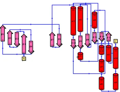
Rsga from Pseudomonas aeruginosa (PaRsgA; Uniprot: Q9HUL3) is a soluble protein of 339 aminoacids (MW = 39241 Da), composed of three domains: a N-terminal Oligonucleotide/Oligosaccharide Binding (OB) domain, a central circularly permutated GTPase (cpGTPase) domain and a C-terminal Zinc-binding domain (Figure 1).
PaRsgA was expressed in E. coli BL21 (DE3) and purified to homogeneity (Figure 4.2A) by using various chromatographic approaches (affinity, ion-exchange and size-exclusion chromatography) (see Material and Methods). The UV spectrum of the purified protein shows a blunt peak around 280 nm (Figure 4.2B), suggesting the presence of a nucleotide bound to the purified protein.
Figure 4.1 Domains organization of PaRsgA. The N-terminal OB-fold domain is shown in green, the central cpGTPase domain in pink and the C-terminal zinc-finger domain in cyan.
GTPase domain
1 359
OB-folddomain zinc-binding
domain
G4-G5-G1-G2-G3
In order to confirm the presence of a co-purified nucleotide and to unveil its nature, we developed a protocol based on an ion-pair reversed-phase HPLC (RP-HPLC) analysis, using a PrevailTM C18 column and monitoring the absorbance of the sample at 254 nm. Since the polarity of nucleotides increases with the number of phosphate groups, nucleoside triphosphates, unlike nucleoside mono- and diphosphates are weakly retained on reversed-phase chromatography with a conventional mobile reversed-phase. Conversely, by using an ion pairing reagent, such as tertrabutylammonium bromide (TBAB), nucleoside diphosphates were retained better than mono-phosphates although more weakly than nucleoside triphosphates. In this case, the separation is based on the formation of ion pair(s) between the positively charged ion-pair Figure 4.2 SDS PAGE of PaRsgA purification (A). The diffrerent lines are representative of the purification steps, carried out with various cromatography approaches: 1) marker, 2) supernatant, 3) affinity 4) ionic exchange and 5) size exclusion cromatography. (B) UV-spectrum of PaRsgA co-purified with a nucleotide bound.
1 2 3 4 5 A B 62 49 38 28 18 14
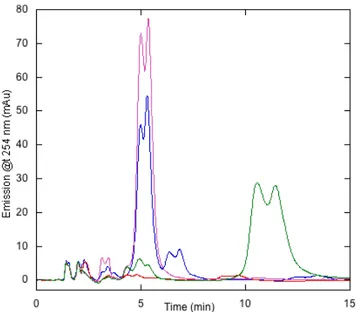
reagent and the negatively charged nucleotide. Since this ion-pair RP-HPLC method enables the efficient separation of guanine nucleotides, it was possible to identify them by referring to the retention times established by the corresponding standards. Representative RP-HPLC chromatogram of purified PaRsgA (Figure 4.3) shows a prominent peak eluting at the retention time corresponding to GDP. The calibration curve of the GDP standard (Figure 4.14) obtained by plotting the peak areas against the concentration of the analyte allows to estimate that PaRsgA is purified with approximately 80% of GDP nucleotide bound.
Figure 4.3 Identification of the nucleotide bound to PaRsgA and validation of the nucleotide-free form production through reverse phase cromatograpy. Nucleotide-bound
PaRsgA is shown in blue, nucleotide-free PaRsgA in red, GDP standard (50uM) in
4.2 PaRsgA structural characterization
4.2.1 PaRsgA crystallization, data collection and structure determination
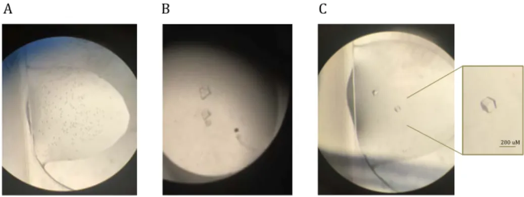
Purified PaRsga was concentrated up to 20 mg/ml and subjected to crystallization procedure using the vapour-diffusion technique. Crystallization trials have been performed using commercial sparse-matrix screens and an automatic crystallization platform (Phenix-Art Robbins) at 21°C. Initial promising hits (multiple small crystals or microcrystalline precipitates) were obtained with the INDEX screen (Hampton Research) where the poly(acrylic acid sodium salt) 5100 is present as precipitating agent (Figure 4.4A-B).
To improve crystal quality, we performed different experiments in which various parameters have been screened, such as protein concentration, the crystallization buffer and pH, the presence of additional salts or “additive agents” to slow down the crystallization speed as well as the protein/reservoir solution ratio in the drop. Moreover, different crystallization techniques, such as seeding performed with microcrystals and crystallization under oil (also at 4°C), were attempted in order to improve the diffraction quality. A wide array of PaRsgA crystals were analysed through X-ray diffraction with different synchrotron light source (Elettra, Trieste; Bessy II Berlin; ESRF, Grenoble). The best diffracting crystal (2.9Å resolution) was obtained by mixing 2 µl of the protein solution (20 mg/ml) with 3 µl of the reservoir solutions containing 30% poly(acrylic acid sodium salt) 5100, 0.1 M Hepes pH 7.5, 0.02 M MgCl2 and 5% PEG 200 (Figure 4.4C).
Diffraction data were collected at 100K (after cryoprotecting the crystal by increasing the poly(acrylic acid sodium salt) 5100 concentration up to 34 %) at the XRD1 beamline of the ELETTRA synchrotron (Trieste, Italy). PaRsgA crystal belongs to the space group P4132, with unit cell parameters of a=b=c= 146.4 Å. One monomer of PaRsgA is present per asymmetric unit. PaRsgA structure (pdb code: 6H4D) was solved by molecular replacement using the structure of RsgA from Salmonella typhimurium (pdb code: 2RCN; Nichols et al., 2007) as a search model and refined as reported in the experimental section. The final model, containing a GDP molecule and a Zn atom, was refined to an Rwork = 0.248 and an Rfree= 0.286. Data collection and refinement parameters are reported in Table 1.
A B C
200 uM
Figure 4.4. GDP-PaRsgA microcrystals (A) and multiple crystals (B). GDP-PaRsgA crystals (C) obtained by the optimized crystallization condition: 28-30% poly(acrilyc acid sodium salt) 5100, 0.1M Hepes pH 7.5, 0.02M MgCl
4.2.2 Overall structure
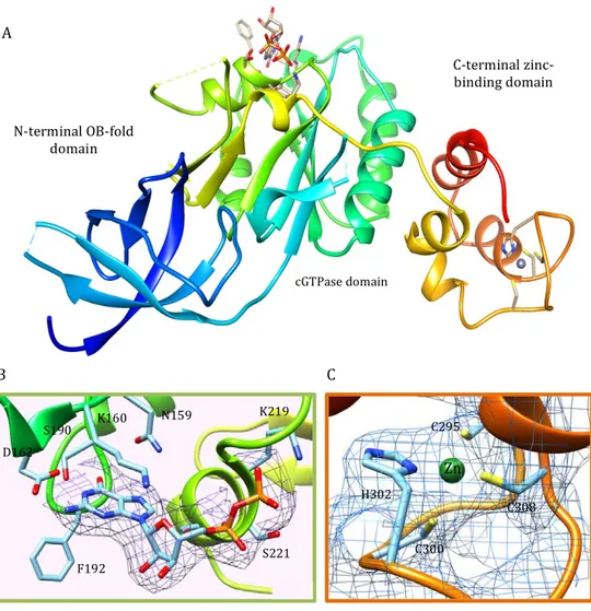
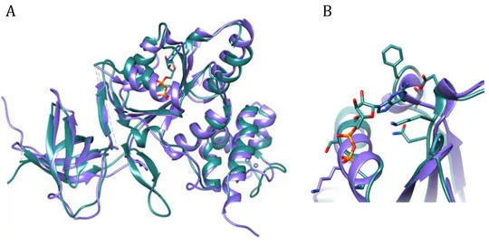
Crystal structure of PaRsgA is depicted in Figure 4.5A and, as reported in the previous section, consists of three domains: a N-terminal Oligonucleotide/ Oligosaccharide Binding (OB) domain, a central circularly permutated domain (cpGTPase), and a C-terminal Zn-binding domain.
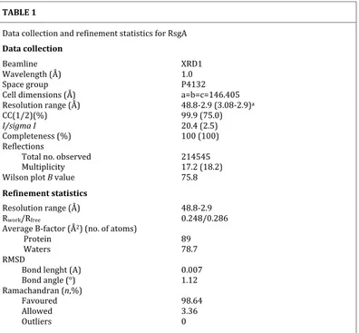
Table 1. Data collection and refinement statistics for PaRsgA. TABLE 1 Data collection and refinement statistics for RsgA Data collection Beamline XRD1 Wavelength (Å) 1.0 Space group P4132 Cell dimensions (Å) a=b=c=146.405 Resolution range (Å) 48.8-2.9 (3.08-2.9)a CC(1/2)(%) 99.9 (75.0) I/sigma I 20.4 (2.5) Completeness (%) 100 (100) Reflections Total no. observed 214545 Multiplicity 17.2 (18.2) Wilson plot B value 75.8 Refinement statistics Resolution range (Å) 48.8-2.9 Rwork/Rfree 0.248/0.286 Average B-factor (Å2) (no. of atoms) Protein 89 Waters 78.7 RMSD Bond lenght (A) 0.007 Bond angle (°) 1.12 Ramachandran (n,%) Favoured 98.64 Allowed 3.36 Outliers 0 aValues in parentheses refer to the highest resolution shell
The quality of the diffraction data is good and electron density is clearly visible for the most of residues with the exception of residues belonging to the N-terminal region (NTE, res: 1-39), residues 90-91, the switch I region
Figure 4.5 Cartoon representation of PaRsgA structure (A) colored in rainbow. Close view of the GDP binding region (B). 2Fobs-Fcalc density map is contoured at 1σ and the GDP and GDP contacting residues are shown in stick representation. Close view of the zinc-binding region (C). 2Fobs-Fcalc density map is contoured at 1σ and the Zn is represented as a green sphere and Zn contacting residues are shown in stick representation. C300 C308 C295 Zn H302 F192 D162 S190 K160 N159 K219 S221 N-terminal OB-fold domain C-terminal zinc-binding domain cGTPase domain A B C