Abstract. –OBJECTIVE: Increase in ACL (an-terior cruciate ligament) reconstructions has led to a higher prevalence of patients with postoper-ative symptoms which require investigation. We aimed to investigate the utility of magnetic reso-nance imaging (MRI) and computer tomography (CT) in determining tunnel size and graft obliqui-ty after single bundle ACL reconstruction.
PATIENTS AND METHODS: A retrospective comparison was made on 29 symptomatic knees after anatomic single bundle (trans AM) and transtibial ACL reconstructions which had both MRI and CT scans at an average of 1.3 years postoperatively (2 months-5.7 years). We com-pared CT and MRI (T2 sequence) tunnel size and graft obliquity estimates using Pearson correla-tion and t-test. We also compared MRI’s of ACL reconstructed knees with hamstrings or patellar autografts, which were confirmed by operative protocol as either antero-medial (AM) technique (n=21) or trans-tibial (TT) technique (n=19). The surgeries were performed for an average of 6.29 (4-10) years for the TT group and 1.3 (0-3) years for the AM group, respectively. The graft inclina-tion was measured relative to the tibial plateau using DICOM software. Statistical analysis used the mean value for each case and the data were processed using the non-parametric Kruskal-Wallis test to determine the difference in graft obliquity and tunnel placement.
RESULTS: Tunnel size estimates correlate well between CT and MRI on axial scans: R2=0.795 and 0.630 for femur and tibia respectively. The po-sition of the tunnels and graft obliquity were found to differ on MRI images in both coronal and sagittal planes. Coronal graft obliquity averaged 72.38° (ranging from 69° to 76°) using the AM technique and 75.47° (ranging from 72° to 78°) with TT technique. Sagittal graft inclination angle was 54.5 (51-58.5) and 63.68 (59-69.5) respectively. MRI proves to be the most useful imaging method in determining outcome after ACL reconstruction.
Anterior cruciate ligament reconstruction and
determination of tunnel size and graft obliquity
D. VERMESAN, F. INCHINGOLO
4, J.M. PATRASCU, I. TROCAN,
R. PREJBEANU, S. FLORESCU, G. DAMIAN
3, V. BENAGIANO
2,
A. ABBINANTE
1, M. CAPRIO
1, R. CAGIANO
1, H. HARAGUS
Department of Orthopedics and Trauma, University of Medicine and Pharmacy “Victor Babes”, Timisoara, Romania
1Department of Biomedical Sciences and Human Oncology, Medical School, University of Bari, Italy 2Department of Basic Medical Sciences, Neurosciences and Sensory Organs, Medical School,
University of Bari, Italy
3Faculty of Medicine, Pharmacy and Dental Medicine, West University “Vasile Goldis”, Arad, Romania 4Department of Interdisciplinary Medicine, Medical School, University of Bari, Italy
However, for a better revision of the ACL recon-structions, CT can offer a clearer image of tunnels and bone stock. A more anatomical graft position-ing increases obliquity in coronal and sagittal planes and, thus, becomes difficult to assess both tunnels in a single slice.
CONCLUSIONS: The anatomic single bundle reconstruction technique has been found to more accurately reproduce the femoral footprint and the orientation of the graft compared to the TT technique where the appropriate tibial tunnel placement resulted in a more vertical graft. Key Words:
ACL, MRI, CT tunnel size, Graft obliquity.
Introduction
The introduction of the anatomic ACL (anteri-or cruciate ligament of the knee) reconstruction technique changed the way surgeons drill the femoral tunnel, by using an antero-medial (AM) portal drilling technique instead of the traditional trans-tibial (TT) technique. Subsequently it was changed the positioning of the tunnels and the re-sulting obliquity of the graft, positioning it in a more anatomical fashion. One of the first materi-als evaluating the neoligament obliquity, in ACL reconstructed patients, found a continuous and homogeneous graft similar to the native ACL, but with a more vertical position that does not recre-ate the normal sagittal obliquity. Nevertheless, these more vertical grafts were found to still con-trol anterior posterior knee displacement1. Ahn et
al2 showed a lot of interest towards MRI
(mag-netic resonance imaging) evaluation after ACL reconstructions. When AM and TT techniques
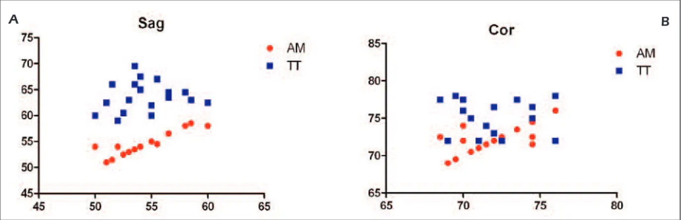
Figure 1. Tunnels position and Graft obliquity data on both sagittal(A) and coronal (B) planes.
A B
were compared, they also found a significant ver-tical angle in the coronal and sagittal plane for the TT reconstructions compared with the native ACL. In addition, the more horizontally can be the angle of the tibial tunnel, the closer can be the result, compared to the native ACL. When the tibial remnant stump was preserved, magnetic resonance imaging showed significantly larger grafts with progressive remodeling and no in-crease in the incidence of cyclops lesions3.
Fur-ther researches could provided additional data to support the femoral tunnel drilling through the AM portal. When compared to the TT technique, it could offer better function and closer obliquity with respect to the native contralateral ACL4-6.
With regard to the tibial tunnel aperture, the re-sults are somewhat inconclusive between the two techniques. It has been shown that trans-tibial technique places the tibial tunnel in the same po-sition as the AM procedure or more posterior in order to achieve a better placement of the femoral tunnel5-8. The overall increase in ACL
re-constructions has led to an its higher prevalence in the population and, thus, in an increased oc-currence of patients with postoperative symp-toms requiring investigation. The majority of cas-es lead for revisions rcas-esulted caused by bad tun-nel positioning with secondary impingement and/or instability. Most of the times this is caused by surgical and technical mistakes9. One study6,
reviewing patients for ACL revisions, found that 88% of knees resulted with graft outside the na-tive tibial and femoral insertions. Many of them were entirely on the intercondylar femoral roof and one third extended posterior to the anterior cruciate ligament tibial attachment with TT tech-nique6. In addition, almost half of the patients
un-derwent one or more times in revisions without
correction of the misplaced tunnels and, there-fore, still with subsequent failures6. In addition to
MRI, 3D volume could make the computer to-mography (CT) an useful tool for planning accu-rate femoral tunnel positioning when aiming for anatomic ACL reconstruction. The direct inser-tion of the ACL is located in the depression be-tween the resident’s ridge and the articular carti-lage margin on the lateral femoral condyle10. The
same images are useful to evaluate the current tunnel positions and determine the revision oper-ative strategy11. Given these premises, we aimed
to investigate the utility of different imaging techniques (MRI, CT) in determining tunnel size, graft obliquity and complications after ACL re-constructions in the setting of preoperative plan-ning of a single bundle anatomic revision.
Patients and Methods
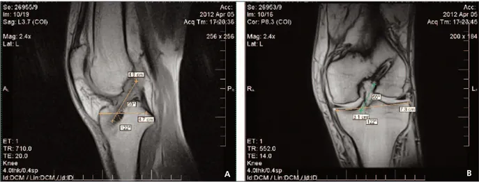
From our database, selected MRI scannings of 40 ACL reconstructed knees with hamstrings or patellar autogenous graft which were confirmed by operative protocol as either AM technique (n=21) or TT technique (n=19). All indexed proce-dures were single bundle reconstructions, which have been performed by different surgeons using various fixation devices with an average of 6.29 (4-10) years ago for the TT group and 1.3 (0-3) years ago for the AM group. The MRI scans were given blinded, regarding the surgical technique, to two experienced examiners: a knee surgeon with over 150 ACL reconstructions per year and a radi-ologist from a knee and sports clinic; they were asked to determine the graft inclination relative to the tibia using Efilm DICOM viewer software (Figure 1A and B). The null hypothesis was that
Figure 2. MRI/CT correlation of femoral tunnel size on axial views at the gape level.
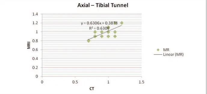
Figure 3. MRI/CT correlation of tibial tunnel size on axial views at the gape level.
CT tunnel size and the graft obliquity values using the Pearson correlation and t-test compared to MRI (T2) (Figures 2 and 3).
Statistical Analysis
Statistical comparison between MRI sagittal and coronal plane obliquity for the AM portal and the TT portal using the one-tailed t test (data processed with SPSS, IBM) gave the following results, summarized in the following sagittal and coronal (Table I).
there were no differences between the two groups. For statistical analysis we used the mean value for each case and processed the data using the non-parametric Kruskal-Wallis test to determine the difference in graft obliquity and tunnel placement in use. In addition, we retrospectively compared 29 symptomatic knees after anatomic single bun-dle (trans AM) and trans-tibial ACL reconstruc-tions that had both MRI and CT scans at an aver-age of 1.3 years postoperatively (2 months-5.7 years). We analyzed the correlation between the
Results
Tunnels position and graft obliquity were found to differ on images in both sagittal and coronal planes (Figure 1A and B). Coronal graft obliquity averaged 72.38° (ranging from 69° to 76°) using the AM technique Figure 4) and 75.47° (ranging from 72° to 78°) with TT tech-nique. Sagittal graft inclination angle was 54.5 (51-58.5) and 63.68 (59-69.5) respectively (Fig-ure 5). We determined a statistically significant difference in graft obliquity and tunnel angles with a more anatomical favorable position in AM technique and in vertical results for TT tech-nique. Radiological measurements showed less variance between the two groups. Axial measure-ments of the tunnel size at the level of the aper-ture were compared between MRI and CT. Re-sults showed no correlation (R2 = 0.795 and 0.630 for the femur and tibia, respectively) (Fig-ures 2 and 3). By comparison with the classical TT technique, the tibial tunnel placement result-ed in a more vertical graft than native ACL
(Fig-ures 6). The 3D VRT CT was found to be a reli-able way to determine tunnel placement as far as 5.7 years after the index surgery (Figure 7). On the other hand, MRI was a consistent and reliable method to determine the status of the neoliga-ment (re-rupture or integration) as well as poten-tial meniscal and cartilage associated lesions (Figure 8). Comparative MRI’s of the above postoperative cases depicting a ruptured and a healed graft respectively after 2 and 5 years (Fig-ure 9).
Discussion
MRI proves to be the most useful imaging method in determining the outcome after ACL reconstruction. It gives reliable information on graft healing, integrity, length, position, inclina-tion angle, obliquity, impingement syndromes and potential associated lesions. It may even identify ACL impingement against PCL (poste-rior cruciate ligament) with the knee extended, Sagittal (degrees) Coronal (degrees)
AM (n=21) 54.50 (SEM = 0.48) 72.38 (SEM = 0.41)
TT (n=19) 63.68 (SEM = 0.64) 75.47 (SEM = 0.52)
p(AM vs TT) < 0.001 < 0.001
95% CI -10.78 to -7.59 -4.42 to -1.76
Table I. MRI sagittal and coronal plane obliquity for the AM portal and the TT portals.
AM: femoral tunnel drilled through the anteromedial portal; TT: femoral tunnel drilled through the tibial tunnel, n: number of subjects; SEM: standard error of mean; CI: confidence interval.
Figure 4.A, B, Sagittal and coronal T2 MRI exemplifying the trans AM graft obliquity measurements related to the tibial plateau.
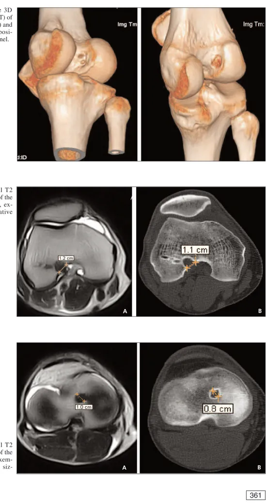
Figure 5. Comparative 3D VRT (volume rendered CT) of the over the top (trans TT) and anatomical (trans AM) posi-tioning of the femoral tunnel.
Figure 6. A, B, Axial T2 MRI and CT at the level of the femoral tunnel aperture, ex-emplifying the comparative sizing.
A B
A B
Figure 7. A, B, Axial T2 MRI and CT at the level of the tibial tunnel aperture, exem-plifying the comparative siz-ing.
which cannot be detected by conventional arthroscopy12. A more anatomical graft
position-ing increases obliquity in coronal and sagittal planes and thus becomes difficult to assess both tunnels in a single slice. However, for a revision of ACL reconstructions, CT scans can offer a clearer imaging of tunnels and bone. A more anatomical graft positioning (trans AM) increas-es obliquity in both coronal and sagittal planincreas-es and it becomes difficult to assess both tunnels in a single slice (Figure 5). 3D CT reconstructed volumes (VRT) are reliable and can be used to assess the tunnel position regarding stability and outcome13. Although not as widely used, 3D
MR imaging were proved accurate and could al-so be used for pre-operative templating in anatomic ACL reconstruction14. The anatomic
single bundle reconstruction technique has been found to more accurately reproduce the femoral footprint and the orientation of the graft. By comparison with the classical TT technique, the tibial tunnel placement resulted in a more verti-cal graft than native ACL (Figure 4). Nonethe-less, in many cases normal graft obliquity is not restored with either technique. This later finding is similar to that of Hantes et al5and it may be
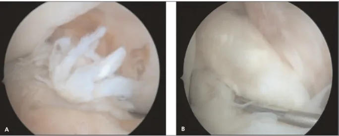
caused by the single bundle technique itself. The current standard requires large footprints to Figure 8.A, B, Arthroscopic views of the below cases through the lateral portal, depicting a ruptured and a healed graft, re-spectively (at 2 and 5 years postoperatively).
A B
Figure 9.A, B, Compara-tive MRI’s of the above cases depicting a ruptured and a healed graft respectively (at 2 and 5 years postoperatively).
be restored by double bundle procedure in order to provide a closer resemblance to the native ar-chitecture. Nevertheless, for smaller knees and footprints the double-bundle is no longer con-sidered superior15. It is universally accepted that
ACL reconstruction via trans-tibial technique fails to accurately position femoral and tibial tunnels within the natural insertion site16. In
ad-dition, freedom of femoral drilling through the AM portal will also allow for a more anterior placement of the tibial tunnel8. This, in turn,
will lead to a more oblique and natural graft placement with improved restoration of anato-my and stability with ACL reconstruction com-pared with conventional trans-tibial drilling techniques17.
Conclusions
MRI proves to be the most useful imaging method in determining an outcome after ACL reconstruction. However, for a revision of ACL reconstructions, CT could offer a clearer image of tunnels and bone stock. The anatomic single bundle reconstruction technique has been found to more accurately reproduce the femoral foot-print and the orientation of the graft compared to the TT technique, where the appropriate tib-ial tunnel placement resulted in a more vertical graft.
––––––––––––––––––––
Acknowledgements
Thanks to Cristina Nuta, MD, for contributing to the postop-erative physical therapy.
–––––––––––––––––-––––
Conflict of Interest
The Authors declare that there are no conflicts of interest.
References
1) AYERZAMA, MÚSCOLODL, COSTA-PAZM, MAKINOA,
RONDÓN L. Comparison of sagittal obliquity of the reconstructed anterior cruciate ligament with native anterior cruciate ligament using magnetic resonance imaging. Ar throscopy 2003; 19: 257-261.
2) AHNJH, LEE SH, YOOJC, HAHC. Measurement of the graft angles for the anterior cruciate ligament reconstruction with transtibial technique using
postoperative magnetic resonance imaging in comparative study. Knee Surg Sports Traumatol Arthrosc 2007; 15: 1293-1300.
3) AHN JH, LEE SH, CHOISH, LIMTK. Magnetic reso-nance imaging evaluation of anterior cruciate liga-ment reconstruction using quadrupled hamstring tendon autografts: comparison of remnant bundle preser vation and standard technique, Am J Sports Med 2010; 38: 1768-1777.
4) ABEBE ES, MOORMAN CT 3RD, DZIEDZIC TS, SPRITZER
CE, COTHRAN RL, TAYLOR DC, GARRETT WE JR, DE -FRATELE. Femoral tunnel placement during anteri-or cruciate ligament reconstruction: an in vivo imaging analysis comparing transtibial and 2-inci-sion tibial tunnel-independent techniques, Am J Sports Med 2009; 37: 1904-1911.
5) HANTES ME, ZACHOS VC, LIANTSIS A, VENOUZIOU A, KARANTANAS AH, MALIZOS KN. Differences in graft orientation using the transtibial and anteromedial portal technique in anterior cruciate ligament re-construction: a magnetic resonance imaging study. Knee Surg Spor ts Traumatol Ar throsc 2009; 17: 880-886.
6) MARCHANT BG, NOYES FR, BARBER-WESTINSD, FLECK
-ENSTEIN C. Prevalence of nonanatomical graft placement in a series of failed anterior cruciate ligament reconstructions, Am J Sports Med 2010; 38: 1987-1996.
7) FRANKRM, SEROYERST, LEWISPB, BACHBR JR, VERMA
NN. MRI analysis of tibial position of the anterior cruciate ligament. Knee Surg Sports Traumatol Arthrosc 2010; 18: 1607-1611.
8) YEJX, SHENGS, ZHOUHB, XUW, XIEZG, DONGQR,
XUYJ. Arthroscopic reconstruction of the anterior cruciate ligament with the LARS artificial liga-ment: thirty-six to fifty-two months follow-up study. Eur Rev Med Pharmacol Sci 2013; 17: 1438-1446.
9) CARSON EW, ANISKO EM, RESTREPO C, PANARIELLO
RA, O'BRIEN SJ, WARREN RF. Revision anterior cruciate ligament reconstruction: etiology of fail-ures and clinical results, J Knee Surg 2004; 17: 127-132.
10) IWAHASHIT, SHINOK, NAKATAK, OTSUBOH, SUZUKI T, AMANOH, NAKAMURAN. Direct anterior cruciate lig-ament insertion to the femur assessed by histol-ogy and 3-dimensional volume-rendered comput-ed tomography, Arthroscopy 2010; 26(Suppl): S13-20.
11) VAN ECK CF, SCHREIBER VM, LIU TT, FU FH. The anatomic approach to primary, revision and aug-mentation anterior cruciate ligament reconstruc-tion. Knee Surg Sports Traumatol Arthrosc 2010; 18: 1154-1163.
12) FU J I M O T O E, SU M E N Y, DE I E M, YA S U M O T O M,
KOBAYASHI K, OCHI M. Anterior cruciate ligament graft impingement against the posterior cruciate ligament: diagnosis using MRI plus three-dimen-sional reconstruction software. Magn Reson Imaging 2004; 22: 1125-1129.
13) HARAGUSH, PREJBEANUR, RAMADANIF. Revision ACL Surger y in Prejbeanu R (Ed) Atlas of knee arthroscopy, Springer-Verlag London 2015, pg 103-129.
14) HANY, KURZENCWYGD, HARTA, POWELLT, MARTINEAU
PA. Measuring the anterior cruciate ligament’s footprints by three-dimensional magnetic reso-nance imaging. Knee Surg Sports Traumatol Arthrosc 2012; 20: 986-995.
15) SIEBOLD R. The concept of complete footprint restoration with guidelines for single- and
dou-ble-bundle ACL reconstr uction. Knee Surg Sports Traumatol Arthrosc 2011; 19: 699-706. 16) PREJBEANU R, HARAGUSH, RAMADANI F. The Anterior
Cruciate Ligament in Prejbeanu R (Ed) Atlas of knee arthroscopy, Springer-Verlag London 2015, pg 47-101.
17) VERMESAN D, PREJBEANU R, LAITIN S, GEORGIANU V,
HARAGUSH, NITESCUS, TATULLOM, TATTOLIM, CAPRIO
M, CAGIANO R. Meniscal tears left in situ during anatomic single bundle anterior cruciate ligament reconstruction. Eur Rev Med Pharmacol Sci 2014; 18: 252-256