Pediatr Radiol (1990) 20:594-597
Pediatric
Radiology
9 Springer-Verlag 1990Radiographic findings in hereditary multiple exostoses
and a new theory of the pathogenesis of exostoses
U. E. Pazzaglia 1 , L. P e d r o t t i 1, G. Beluffi 2 , V. Monaf63 and S. Savasta 3 1 Clinica Ortopedica dell'Universitfi di Pavia, I.R.C.C.S. Policlinico San Matteo, 2 Servizio di Radiodiagnostica Policlinico San Matteo, and
3 Clinica Pediatrica dell'Universit5 di Pavia, Pavia, Italy Received: 20 February 1990; accepted: 29 March 1990
A b s t r a c t . Analysis of 330 exostoses in 18 patients affected by hereditary multiple exostoses disease suggested a new classification of exostoses as eccentric or full-thickness. Radiographically arrest of metaphyseal remodeling with failure of coning and persistence of the primary meta- physeal trabeculae was evident in full-thickness exos- toses. Similar bone lesions can be obtained experimen- tally with inhibitors of bone turn-over. A localized, peripheral defect in remodeling over a limited time can give a satisfactory explanation also for the origin of eccen- tric exostoses. T h e thesis that this is the basic mechanism of exostosis formation is presented.
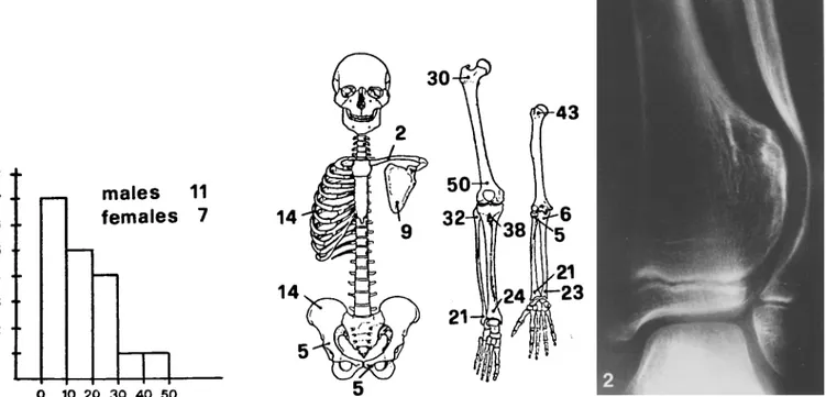
Fig.1. Distribution of age and localization of exostoses in the stu- died patients
Fig. 2. R.L., 10-years old. Eccentric exostosis of the distal tibia with remodeling of the fibula around exostosis
Multiple exostoses ( H M E ) is a well known hereditary dis- ease characterized by great variability in the n u m b e r of exostoses, which is often very large, as well as in their shape, size and effects on bone growth. Exostoses also occur as solitary lesions which are not familial; they are usually classified as sessile or peduncolated according to the extension of their implant (type of attachment). The presence in a single patient with H M E of pedunculated and sessile eccentric exostoses and the club-shaped thickening of the metaphyses suggest a c o m m o n under- lying defect, the nature of which cannot be explained by previous pathogenetic theories.
John H u n t e r first described multiple exostoses in his lectures on the Principles of Surgery delivered in 1786/87 and noted that as in rickets bone ends were enlarged; this led him to suppose that the two conditions were related [4].
A century later Virchow [15] suggested separation and rotation of a portion of the epiphyseal cartilage as the
8 7 6 5 4 3 2 1 1 o
males
11
females 7
N 10 2 0 3 0 4 0 5 03O
43
U. E. Pazzaglia et al: Hereditary multiple exostoses 595
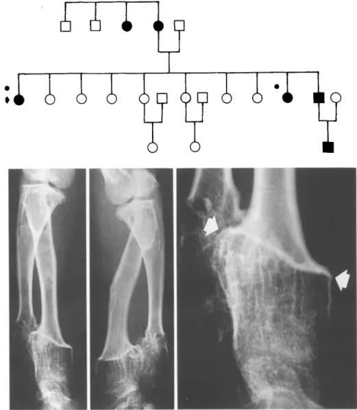
Fig. 3. E R A., 12-years-old, 6 family members with HME. Antero-posterior and lateral views of the forearm showing a fail-thickness exostosis of the distal, radial metaphysis. The arrows indicate the line of arrest of metaphyseal remodeling. The cylindrical metaphysis is composed of thin trabeculae with a prevailing longitudinal pattern. A full-thickness exostoses of the ulna is also evident. Shortening of both the ulna and radius is present p a t h o g e n e t i c m e c h a n i s m o f exostosis. I n 1914 M i i l l e r p o s - t u l a t e d a t h e o r y b a s e d o n t h e o b s e r v a t i o n o f c h o n d r o g e n i c cells in t h e p e r i o s t e a l l a y e r [8] a n d s h o r t l y a f t e r w a r d s , in 1920, K e i t h d r e w a t t e n t i o n t o t h e m e c h a n i s m o f m e t a - p h y s e a l r e m o d e l i n g a n d t h e r e l a t e d a n a t o m i c a l s t r u c t u r e s a n d p o s t u l a t e d a d e f e c t o f t h e p e r i c h o n d r a l ring, a s t r u c - t u r e w h i c h is n e c e s s a r y to limit t h e g r o w t h o f c a r t i l a g e a n d p r e v e n t u n d u e t r a n s v e r s e g r o w t h [5]. I n t h e p r e s e n t s t u d y i n t e r p r e t a t i o n o f t h e r a d i o g r a p h i c f e a t u r e s o f 330 e x o s t o s e s in H M E l e a d s us to p r o p o s e a n e w c l a s s i f i c a t i o n a n d n e w p a t h o g e n e t i c t h e o r y o f e x o s - toses. A s s o l i t a r y a n d m u l t i p l e l e s i o n s a r e h i s t o l o g i c a l l y similar, it s e e m s r e a s o n a b l e to a s s u m e t h a t this t h e o r y is a p p l i c a b l e t o all e x o s t o s e s . Materials and m e t h o d s
A skeletal survey was obtained in 18 patients from 13 families with HME seen in the Orthopedic Department of the University of Pavia. Eleven patients were seen for the first time in childhood before achieving skeletal maturity and seven were adult relatives. The age distribution of the subjects and localization of the exostoses are shown in Fig. 1.
Radiographic findings
A total of 330 exostoses was found in these patients and the number of exostoses per patient ranged from 4 to 28 with a mean of 18 per pa- tient. The shape and volume of the exostoses was very variable and their appearance was often quite bizarre. Analysis of their radio- graphic appearance suggested a new classification as eccentric or full-thickness.
Eccentric exostoses have lost their connection with the adjacent growth cartilage; they probably migrate from the metaphysis as bone grows and in older patients can reach the diaphysis. Similar features are seen in solitary exostoses. Eccentric exostoses do not interfere with growth in length of the bone from which they arise. In the forearm or leg remodeling may occur in the bone adjacent to the growing mass (Fig. 2), or compression of the adjacent cartilage by the exostosis may cause a growth defect with tilting of the epi- physeaI plate,
Full-thickness exostoses involve the entire circumference of the metaphysis. They are frequent in HME but have never been re- ported in solitary exostoses. The metaphysis is cylindrical with a line of arrest of metaphyseal remodeling clearly evident on the X-rays (Fig. 3). The trabecular pattern of the metaphysis is composed of thin longitudinal trabeculae which represent the calcified intercolumnar septa of the growth plate cartilage and primary metaphyseal trabe- culae which have not undergone remodeling. Cartilage growth potential is severely compromised and results in shortening of the bone. As usually occurs with eccentric exostoses, growth recovery
596
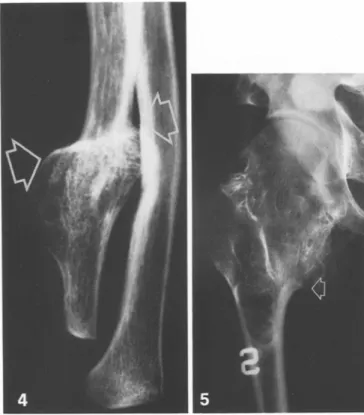
Fig.4. S.A., 6-years old. Recovery of growth and remodeling of a full-thickness exostosis of the ulna. The arrows indicate the line of arrest of metaphyseal remodeling. There has been some reorganiza- tion of the enlarged area and the longitudinal trabecular pattern is no longer evident
Fig.5. O.G., 18-yearsold. Coxavalgain a full-thickness exostosisin- volving the upper femur. The arrow indicates the line of arrest of metaphyseal remodeling. The arched trabecular systems of the neck are replaced by longitudinal trabeculae
and remodeling was observed in one case of HME in this series (Fig.4).
Others deformities characteristic of HME such as coxa valga (Fig. 5), bending of the radius, displacement of the radial head, epi- physeal tilting in varus or valgus can be explained by different growth rates of the bones in biosseous limb segments (forearm and leg) [1, 12].
Discussion
Of fundamental importance to admission in the present study is the prerequisite of a family history of H M E , a criterion which should assure exclusion of all cases of endochondromatosis, a disease which is not transmitted genetically. Indeed, most older case reports of H M E in- clude examples of multiple endochondromatosis or O1- lier's disease [13]; although a relationship between the two conditions was postulated in 1925 by Stocks and Bar- rington [14] it has since been established that they are separate entities. The criteria currently used for clas- sifying exostoses are difficult to apply to the bizarre and complex features of full-thickness exostoses probably due to the fact that such criteria are based mainly on de- scription of solitary exostoses or eccentric exostoses in HME. It is in fact difficult to postulate a theory of the
U. E. Pazzaglia et al: Hereditary multiple exostoses origin of exostoses which can explain the relationship be- tween sessile and pedunculated exostoses showing eccen- tric growth and full-thickness exostoses not showing ec- centric growth but maintaining the same width as the epiphysis. Hunter's suggestion that rickets and exostoses are related is in part true. Indeed the two conditions have in c o m m o n an arrest of remodeling but differ in as much as calcification is inhibited in rickets but not in HME. Virchow's theory is supported by the recent experimental production of exostoses after transplantation of epi- physeal line cells under the periosteum [2] but does not give a satisfactory explanation of the arrest of meta- physeal remodeling in full-thickness exostoses, a defect also shared by Mueller's theory [8]. The theory of a de- fective perichondral ring as suggested by Keith supposes a lack of coordination between endochondral ossifica- tion, longitudinal growth and perichondral ring forma- tion [5]. According to this theory it is held that growth plate cartilage increases its transverse diameter by appo- sition from the overlying perichondrium [3, 9].
Langeskjold on the contrary produced evidence that the epiphyseal cartilage expands peripherally by intersti- tial growth and that the outer layers of cartilage cells are transformed into the proliferative layer of the periosteum [6]. The origin of exostoses is consequently referred to the persistent chondrogenic properties of these cells in the periosteum.
Eccentric exostoses have a histologic appearance simi- lar to that of the bone from which they arise with an outer layer of cortical bone and an inner marrow cavity; the cartilage cap repeats the columnar arrangement of the epiphyseal plate with all the phases of endochondral ossi- fication. Unfortunately, the histology of full-thickness exostoses is unknown; however, radiographically arrest of metaphyseal remodeling with failure of coning and per- sistence of the primary metaphyseal trabeculae is evident even in the absence of histologic documentation. These
A
B
C
Fig. 6 A-C. Diagrams illustrating the effects of an inhibitor of osteo- clastic resorption (EHDP) on the shape of tibial metaphysis in the rat (modified from Schenk et al. 1973): A the conical shape of the metaphysis results from resorption of the external metaphyseal trabeculae; the outline of the proximal tibia is superimposed at three subsequent times (black line, dark grey line, gray line); B complete inhibition of metaphyseal remodeling produces a cylindrically shaped metaphysis; C effects of a transitory, localized arrest of metaphyseal remodeling; growth potential of the exostosis results from the detachment and subsequent rotation of the fragment of growth plate cartilage
U. E. Pazzaglia et al: Hereditary multiple exostoses
features can be observed in all such exostoses and in our opinion represent the basic mechanism of exostoses for- mation. A similar appearance can be obtained experimen- tally with inhibitors of bone turn-over such as diphospho- nates, the main difference being that the latter also inhibit the calcification process [11]. In the same experimental model reduction in longitudinal growth of bones has been observed [10]; this finding fits with the shortening of bones observed with full-thickness exostoses and suggests the presence of amechanism regulating resorption at the bot- tom of the growth plate and rate of cell proliferation at the top. A n arrest of remodeling satisfactorily explains all the features observed in exostoses involving the entire meta- physeal area.
With regard to eccentric exostoses, according to this hypothesis, a localized peripheral defect in remodeling over a limited time gives rise to an eccentric piece of growth cartilage which with growth loses its connection with the original epiphyseal plate. This situation is repre- sented in the figure illustrating the effect of inhibition of osteoclastic resorption in Shenk's experimental model [11] (Fig. 6). T h e most difficult p r o b l e m with this model is satisfactory explanation of the rotation and eccentric growth of eccentric exostoses, since the linear arrange- ment of cartilage cells is no longer parallel to that of the original growth plate. However, if arrest of remodeling is transitory and perich0ndral resorption recommences, as demonstrated by the conical shape of the metaphysis in eccentric exostoses, the detached portion of cartilage will no longer be restrained by Ranvier's perichondral ring and in this situation tilting is easily foreseeable due to pro- gression of the resorption/apposition process at the base [13] and to the growth potential of the unrestrained carti- lage cap at the d o m e of the exostosis. An interaction be- tween exostosis and periosteum can also explain the slower migration rate as compared to that of the original growth plate [7] as well as the rotation of the cartilage cap. Most bone pathology textbooks include exostoses and H M E in the sections on tumors but our theory of their pa- thogenesis suggests t h a t t h e y should be better considered among the genetic chondrodysplasias.
References
597
1. Bethge JFJ (1963) Hereditare, multiple Exostosen und ihre pathogenetische Deutung. Arch Orthop Unfall Chir 54:667 2. D'Ambrosia R, Ferguson AB (1968) The formation of osteo-
chondroma by epiphyseal cartilage transplantation. Clin Orthop 61:103
3. Ham AW (1957) Histology. J. B. Lippincott Co, Philadelphia 4. Hunter J (1786-i787) Lectures on the Principles of Surgery.
Quoted by Solomon L, 1961
5. Keith A (191%1920) Studies on the anatomical changes which accompany certain growth disorders of the human body. J Anat 54:101
6. Langenskjold A (1947) Normal and pathological bone growth in the light of the development of cartilaginous loci in chondrodys- plasia. Acta Chir Scand 95:367
7. Maroteaux R Lamy M (1960) La maladie exostosante. Sem Hop Paris 3:172
8. Mueller E (1914) Uber heredit~ire multiple cartilaginare Exosto- sen und Ecchondrosen. Beitr Path Anat 57:232
9. Lacroix P (1951) The organization of bones. Churchill, London 10. Russell RGG, Kisling AM, Casey PA, Fleisch H, Thornton J,
Schenk R, Williams DA (1973) Effect of Diphosphonate and Calcitonin on the chemistry and quantitative histology of rat bone. Calc Tissue Res 11:179
11. Schenk R, Merz WA, Muhlbauer R, Russell RGG, Fleisch H (1973) Effect of Ethane-l-hydroxy-l.l-diphosphonate (EHDP) and Dichloromethylen diphosphonate (C12MDP) on the calcifi- cation and resorption of cartilage and bone in the tibial epiphysis and metaphysis of rats. Calc Tissue Res 11:196
12. Solomon L (1961) Bone growth in diaphyseal aclasis. J Bone Joint Surg 43 B: 700
13. Solomon L (1963) Hereditary multiple exostosis. J Bone Joint Surg 45 B: 292
14. Stocks R Barrington A (1925) Hereditary disorders of bone de- velopment. The treasury of human inheritance, vol. 3. University Press, Cambridge
15. Virchow R (1875) Uber die Entstehung des Enchondroma und seine Beziehung zur Ecchondrosis und Exostosis cartilaginea. Mon Ber Kgl Preuss Akad Wiss, p 760, qoted by Bethge, 1963 Prof. U.E. Pazzaglia
Clinica Ortopedica dell'Universit~ di Pavia I.R.C.C.S. Policlinico San Matteo
Via Taramelli, 3 1-27100 Pavia Italy