290 THE JOURNAL OF BONE AND JOINT SURGERY
PERIOSTEAL
CHONDROMA
OF
THE
HUMERUS
LEADING
TO
SHORTENING
A CASE REPORT
UGO E. PAZZAGLIA, L. CECILIANI
From the Clinica Ortopedica deli’ Universita di Pavia
We report a case of periosteal chondroma of the proximal humerus with multiple cartilaginous masses extending around two-thirds of the metaphysial circumference. The humerus was short, presumably because the tumour interfered with growth at the epiphysial plate.
Periosteal chondroma is a benign tumour, many cases of which have been reported since Lichtenstein and Hall (I 952) described it as arising between periosteum and cortex. It is widely recognised that the radiographs are easily misinterpreted and thought to be those of a chondrosarcoma (Lichtenstein and Hall 1952; Spjut el al.
1971). The tumour is often situated in the proximal humerus as were four of the eight cases reported by Rockwell, Saiter and Enneking (1972).
The following case is unusual because of the short humerus, and the multiple cartilaginous masses affecting more than two-thirds of the shaft circumference.
CASE REPORT
A I 3-year-old boy presented with a painless hard mass on the proximal shaft of his right humerus; the right humerus was 6 cm shorter than the left (Figs I and 2). The parents had noticed the shortening six months previously.
Radiographs on admission showed an irregular erosion of the cortex on the medial, the anterior and the lateral aspects of the shaft, with some sclerotic reaction (Figs 3 and 4). Metaphysial remodelling proximal to the tumour was absent, but distally the bone was normal in shape. A radiographic survey showed the rest of the skeleton to be normal.
Scintigraphy showed diffuse, weak uptake in the right humerus at the level of the lesion (Fig. 7). Arteriography did not demonstrate the nutrient artery and showed the mass to be poorly vascularised (Fig. 5).
The provisional diagnosis was low-grade chondro-sarcoma.
U. E. Pazzaglia, MD, Assistant in Orthopaedics L. Ceciliani, MD, Professor of Orthopaedics
Clinica Ortopedica dell’Universita di Pavia, Via Taramelli, 1-27100
Pavia, Italy.
Requests for reprints should be sent to Dr U. E. Pazzaglia. f) 1985 British Editorial Society of Bone and Joint Surgery
030l-620X/85/2057 $2.00
______
At operation three distinct masses were found beneath the periosteum: the largest on the medial aspect of the shaft, and the other two on the lateral and anterior aspects (Fig. 6). The affected portion of bone was removed en bloc and replaced by a free fibular autograft.
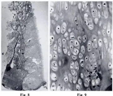
Gross examination showed three masses of cartilage between the bone and the periosteum. Histologically these were composed of benign cartilage (Figs 8 and 9).
The fIbular graft resorbed completely in four months; consequently a vascularised fibular graft was inserted and this consolidated in two months.
DISCUSSION
The difficulty in distinguishing between periosteal chon-droma and chondrosarcoma is well recognised. Radio-graphs can be misleading: in this case the three separate cartilaginous masses, involving the circumference of the humerus extensively, gave the appearance of a lesion involving both cortex and medulla, leading to the
.,.
-.
Fig. 7
Figure 8-Cartilage mass lying between the cortical bone and the
periosteum (haematoxylin and eosin, x I .2). Figure 9-Benign aspect of the cartilage (haematoxylin and eosin, x 200).
PERIOSTEAL CHONDROMA OF THE HUMERUS LEADING TO SHORTENING 291
VOL. 67-B, No. 2, MARCH 1985
Scintigram showing slightly increased uptake in the right
humerus.
Fig. 6
Figures 3 and 4-Anteroposterior and lateral
views of the right proximal humerus. Figure
5-The arteriogram does not demonstrate the
nutrient artery of the humerus. Figure 6-The
diagram illustrates the position of the three car-tilaginous masses.
The authors are grateful to Dr P. D. Byers for his advice and assistance with this paper.
REFERENCES
Dahlin DC. Bone tumours: general aspects and data on 6221 cases. 3rd
ed. Springfield, Illinois: CC Thomas, 1978.
Langenski#{246}ld A, Edgren W. The growth mechanism of the epiphysial cartilage in the light of experimental observations. Acta Orthop
Scand 1949:19: 19-24.
Lichtenstein L, Hall JE. Periosteal chondroma: a distinctive benign cartilage tumor. J Bone Joint Surg [Am] 1952;34-A:691-7.
Moed BR, Lamont RL. Unicameral bone cyst complicated by growth retardation: report of three cases. J Bone Joint Surg [Am] 1982: 64-A: 1379-8 1.
Nelson JP, Foster RJ. Solitary bone cyst with epiphyseal involvement: a case report. Cliii Orthop 1976;1l8:147-50.
Pratt CWM. Observations on osteogenesis in the femur of the foetal
rat. JAnat 1957:91:533-44.
Rockwell MA, Saiter ET, Enneking WF. Periosteal chondroma. J Bone
Joint Surg [Am] 1972:54-A: 102-8.
Shapiro F. Ollier’s disease: an assessment of angular deformity,
shortening and pathological fracture in twenty-one patients. J
Bone Joint Surg [Am] 1982;64-A:95-I03.
Spjut HJ, Dorfman HD, Fechner RE, Ackerman LV. Tumors of bone
and cartilage. (Atlas of tumor pathology, second series, fasc. 5)
Washington, DC: Armed Forces Institute ofPathology, 1971.
292 U. E. PAZZAGLIA, L. CECILIANI
THE JOURNAL OF BONE AND JOINT SURGERY
presumptive diagnosis of chondrosarcoma. It was only by gross and microscopic examination that the diagnosis of periosteal chondroma was established.
Periosteal chondroma is said to arise from the periosteum (Spjut et a!. 1971; Dahlin 1978). The four humeral lesions reported by Rockwell et a!. (1972) were all located proximally; they were said not to be anatomi-cally related to epiphysial cartilage. This was true also in our case where the tumour mass was about 2.5 cm from the growth plate. Nevertheless, the shortening of the humerus focuses attention on the proximal growth plate.
Humeral shortening in our patient was first ob-served six months before admission. Bone growth must have been slowed before this, but not much before, otherwise there would have been greater discrepancy in length. It seems reasonable to assume that the delay in growth began two or three years before presentation.
Distal to the tumour there was a normally re-modelled metaphysis; but proximally remodelling was lacking. Presumably, therefore, the bone developed normally until the tumour interfered with metaphysial remodelling. Possibly the inhibition of metaphysial re-modelling coincided with the onset of reduced growth of the epiphysial plate. Retardation of growth has been reported as a rare complication of unicameral bone cysts (Nelson and Foster 1976; Moed and Lamont 1982); remarkably, in all three of Moed and Lamont’s cases the humeral proximal metaphysis was affected. A slowing of growth of the epiphysial plate and consequent shorten-ing can occur in Ollier’s disease when the full width of the metaphysis is involved; angular deformity occurs when only part of the metaphysis is affected (Shapiro 1982). These growth defects have been ascribed either to an abnormality of the epiphysial cartilage, or to restriction of growth of the epiphysial plate by an abnormally thick periosteal sleeve, arising as a reaction to the lesion (Shapiro 1982).
A third hypothesis is possible: bone growth depends on the proliferative cell layer of the epiphysial plate (Langenski#{246}ld and Edgren 1949; Pratt 1957), and any interference with this, such as the diminution of blood supply, will reduce growth. In the present case the nutrient artery of the humerus was not seen on arterio-graphy; however, disruption of the blood supply was not confirmed at operation so we cannot say if this was the cause of the growth disturbance.
Study of this case has led us to think that, in retrospect, less radical treatment would have been suffi-cient and that it might have been better to retain the strip of normal cortex.