Abstract - Hypocellular or hypoplastic myelodysplastic syndromes (HMDS) are a distinct subgroup accounting for 10–15% of all MDS patients, that are characterized by the presence of bone marrow (BM) hypocellularity, various degree of dysmyelopoiesis and sometimes abnormal karyotype.
Laboratory and clinical evidence suggest that HMDS share several immune-mediated pathogenic mechanisms with acquired idiopathic aplastic anemia (AA).
Different immune-mediated mechanisms have been documented in the damage of marrow hematopoietic progenitors occurring in HMDS; they include oligoclonal expansion of cytotoxic T lymphocytes (CTLs), polyclonal expansion of various subtypes of T helper lymphocytes, overexpression of FAS-L and of the TNF–related apoptosis-inducing ligand (TRAIL), underexpression of Flice-like inhibitory protein long isoform (FLIPL) in marrow cells as well as higher release of Th1 cytokines, such as interferon-gamma (IFN-γ) and tumor necrosis factor-alpha (TNF-α). It has also been documented that some HMDS patients have higher frequency of polymorphisms linked both to high production of proinflammatory cytokines such as TNF-α and transforming growth factor-β and to the inhibition of T-cell mediated immune responses such as interleukin-10, further suggesting that immune-mediated mechanisms similar to those seen in AA patients may also operate in HMDS.
Clinically, the strongest evidence for immune–mediated hematopoietic suppression in some HMDS is the response to immunosuppression including mainly cyclosporine, anti-thymocyte globulin and/or cyclosporine, or alemtuzumab.
Here we review all these immune mechanisms as well as the influence of this deranged cellular and humoral immunologic mileau on the initiation and possible progression of MDS. All these observations are pivotal not only for a better understanding of MDS pathophysiology, but also for their immediate clinical implications, eventually leading to the identification of MDS patients who may benefit from immunosuppression.
Keywords: hypoplastic myelodysplastic syndrome, immune system, bone marrow microenvironment
I. OVERLAPPING AND DIFFERENCIAL FEATURES BETWEEN APLASTIC ANEMIA, HYPOCELLULAR
AND NORMO/HYPERCELLULAR MDS Myelodysplastic syndromes (MDS) are a heterogeneous group of diseases characterized by impairment of cellular differentiation (also defined as ineffective hematopoiesis) progressive peripheral cytopenias and increased risk of developing acute myeloid leukemia (AML) [1]. Although the
French-American-British (FAB) and World Health Organization (WHO) classification systems do not take into account hypoplastic or hypocellular myelodysplastic syndromes (HMDS) as a defined category of MDS, being HMDS probably considered the expression of a transitional status of other MDS categories, they appear to be a distinct clinicopathologic entity [2].
HMDS accounts for 10–15% of all MDS [3-5] and are characterized by the following features: age-corrected bone marrow hypoplasia (ie cellularity less than 30% under age of 60 years or cellularity less than 20% for older than 60 years) [4], marked dyserythropoiesis, both dysgranulopoiesis and dysmegakariopoiesis [4,5], macro-cytosis, severe neutropenia and thrombocytopenia [3-6], frequent abnormal karyotype [6-8], low rate of progression to acute leukemia, and poor response to conventional therapeutic approach for MDS [6].
Recently, Bennett and Orazi described several others marrow morphological criteria, detectable by bone biopsy histological analysis, useful to help HMDS diagnosis, such as the presence of dysplastic megakaryocytes [4-5] within the disorganized microarchitecture of MDS marrow [4], the detection of fibrosis [4,8], and the immunohistochemical identification of aggregates or clusters of blasts in the central intertrabecular region of marrow, also defined as abnormally localized immature myeloid precursor cells (ALIP) [4,5,9-10].
According to FAB and WHO classification systems, the majority of HMDS cases fall into refractory anemia (RA) and refractory cytopenias with multilineage dysplasia (RCMD) categories [3-8]. In comparison to RA and RCMD, HMDS patients typically are younger, more frequently display a severe neutropenia and thrombocytopenia and a lower percentage of blasts, as well as even less frequently show karyotypically abnormal dysplastic marrow cells. Although Tuzuner et al. documented no difference in prognosis between HMDS and normo-/hypercellular MDS [11], several other studies have reported a more favorable overall survival in the subgroup of HMDS patients [6,8,12].
The distinction between HMDS and AA is even more problematic than that with RA and RCMD when marrow is sparcely cellular with an overall cellularity less than 20% and when these findings are associated with the presence of mast cells and reactive lymphocytes, sometimes organized in small lymphoid clusters, similar to those observed in AA bone marrow biopsies [4,5].
The presence of a clear dysmegakaryopoiesis and dysgranulopoiesis, but not of a mild isolated dyserythropoiesis, usually also found in AA, the detection of karyotypic and fluorescent in situ hybridization (FISH)
Immunological derangement in Hypocellular Myelodysplastic Syndromes
B Serio
1, AM Risitano
2, V Giudice
1, N Montuori
3, C Selleri
11Hematology and Hematopoietic Stem Cell Transplant Center, Department of Medicine and Surgery, University of Salerno, Italy; 2 Hematology, Department of Clinical Medicine and Surgery, and 3Department of Translational
Medical Sciences, Federico II University of Naples, Italy ([email protected])
abnormalities, as well as the identification of any sideroblast, of clusters of blasts, and of an increased number of marrow fetal hemoglobin (HbF)-positive erythroblasts, distinctly address toward a diagnosis of HMDS [13].
However, recognized HMDS karyotypic and FISH abnormalities, such as trisomy 8, trisomy 1q, 20q deletion and monosomy 7, can be also be found, although less frequently, in AA patients in particular throughout their clinical course [6-8]. Additional clonal molecular defects such as mutations in the RNA component of telomerase (TERC) or in the telomerase reverse transcriptase enzyme (TERT) genes and several other microdeletions, assessed by single nucleotide polymorphism (SNP) array–based karyotyping, have been documented in both HMDS and AA patients [14].
The separation of AA and HMDS is even more difficult when clonal cytogenetic markers are absent. Quantification of marrow CD34+ cells by immunohistochemistry and flow-cytometry has been reported to help in distinguishing between AA and HMDS [15-20]. Matsui et al. have demonstrated that the mean percentage of CD34+ cells in AA patients is significantly lower than those of HMDS patients [15]. Noteworthy, we previously documented that, in addition to the defect in more mature committed progenitor cells, also most immature hematopoieitic stem cells, measured as secondary colony-forming cells (CFC) after 5 weeks of long-term bone marrow culture (LTBMC), were affected by disease process in HMDS [18,19]. However, although marrow and circulating CD34+ cells and secondary CFC numbers were significantly higher in HMDS than in AA, we found that there was a high degree of overlap between these two diseases, clearly demonstrating that secondary CFC numbers in either marrow and peripheral blood
could not help to distinguish AA from HMDS in an individual patient [17,18].
Recently, Tripathi et al. also reported that circulating blood lymphocytes of AA patients had significantly lower S-phase fraction (SPF) and aneuploidy in comparison to HMDS patients, suggesting that SPF and aneuploidy could be a further parameter to differentiate AA from HMDS patients [21].
In addition, we documented that HMDS and RA patients show a severe deficit in marrow and circulating committed (CD34+ cells and primary CFC), and immature progenitor cells (such as secondary CFC), compared to normal donors, implying that immature hematopoietic stem cell compartment is affected by disease process in HMDS. However, despite the dramatic difference in marrow cellularity, both more committed and immature progenitor cells showed no significant differences between HMDS and RA patients (Figure 1) [17].
II. IMMUNOLOGIC DERANGEMENT IN MDS AND HMDS
It is widely recognized that genetic, epigenetic, apoptotic and differentiation abnormalities characterizing MDS hematopoietic progenitors may be initiated and/or supported by a significant derangement of immunological microenvironment in some MDS patients. The immune effector mechanisms involved in the complex physiopathology of MDS include not only marrow and circulating immune cell changes, but also dysregulation in their cytokine expression and release. The role of the
Figure 1. Severe deficit of marrow and circulating CD34+ cells and secondary CFC in HMDS and RA patients. Each bar represents mean
progenitors ± SD of progenitor cells in normal controls (n=20), in tot HMDS (n=11) and in RA (n=20) patients. Figure 1A. Mean circulating CD34+
cells in normal controls, in HMDS, and RA patients: 113±12, 54±11 and 64±8, respectively; p < 0.05 between normal control vs HMDS and RA patients, p > 0.05 between HMDS and RA patients; mean bone marrow (BM) CD34+ cells in normal controls, in HMDS, and RA patients: 135.6±21,
15.4±4.2 and 23.2±4.7, respectively; p < 0.05 between normal control vs HMDS and RA patients, p > 0.05 between HMDS and RA patients. Figure
1B. Mean circulating secondary CFC in normal controls (n=12), HMDS (n=18) and RA (n=30) patients: 16.6±2.5, 5.9±3.4 and 12.0±3.6, respectively;
p < 0.05 between normal control vs HMDS and RA patients, p > 0.05 between HMDS and RA patients; and mean BM secondary CFC in normal controls, HMDS and RA patients: 146.6±38.0, 17.5±6.4 and 21.2±6.6, respectively; p < 0.05 between normal control vs HMDS and RA patients, p >
0.05 between HMDS and RA patients.
Abbreviations. CFC = colony-forming cells; HMDS = hypoplastic myelodysplastic syndrome; MNC = mononuclear cells; TNC = total nucleated cells;
main changes in the immune cell compartment and in the cytokine profile in MDS, including HMDS, is discussed below.
Effector immune cell changes
Similar to AA, it has been recently documented by several authors, using different T-cell-receptor (TCR) repertoire molecular analysis techniques, that oligoclonal expansion of cytotoxic T cells (CTL) expressing specific TCR variable beta (Vβ) chain, with unique hypervariable complementarity determining region 3 (CDR3), is detectable in blood and marrow of some MDS patients, regardless of the presence of a hypocellular or normo/hypercellular MDS marrow [23,24].
It has been reported that some MDS patients may show a decrease of such immunodominant CTL clonotypes after immune-suppressive treatment concurrently with blood count improvement, suggesting that these CTL clonotypic-specific assays may be used for monitoring disease activity and response to immunosuppressive therapy [25,26].
The presence of these dominant T-cell clonotypes in blood and marrow of about 90% of MDS patients, as well as the documentation that the number of TCR-Vβ families with skewed CDR3 constitutes about 10% of the total T-cell population in MDS patients, suggest that these dominant clonal CTL in MDS are likely the result of an antigen-driven dominant immune response, and hypothesize their pivotal pathophysiologic role in T-cell mediated inhibition of hematopoietic progenitors in MDS patients [24-26]. Formal proof of this hypothesis has been achieved in AA patients, showing that these dominant T-cell clonotypes exert potent cytotoxicity against AA autologous marrow progenitor cells, but have not been demonstrate in MDS so far due to the difficulty of obtaining sufficient numbers of autologous target MDS cells [22,23].
The antigens driving the immune attack on hematopoietic progenitors of AA and possibly of some MDS patients could be similar, but unfortunately they are remain unknown, as well as the possible primary abnormalities of hematopoietic progenitors leading to the breaking of immune self-tolerance [24,27].
Recently, Sloand et al. have documented increased numbers of Vβ CTL subfamilies in marrow and blood of MDS with trisomy 8, suggesting that these immunodominant CTL, able to selectively kill trisomy 8 MDS cells in vitro, may account for the higher responsiveness of MDS patients with trisomy 8 to immunosuppressive therapy, compared with other MDS subtypes [6,28,29].
Furthermore, Sloand et al. have demonstrated, by microarray analysis [30], that CD34+ MDS cells with trisomy 8 overexpress the Wilms tumor protein 1 (WT1) and that circulating CD8+ CTL cells, recognizing WT1126– 134 peptides, may be detected, by tetramer analysis, in this MDS population, suggesting that WT1 may operate as a neoantigen on trisomy 8 MDS cells triggering the expansion of WT1-specific CD8+ T cells and leading to
autoimmune suppression of MDS clone and likely of residual normal marrow cells [27,30,31]. However, it is not possible to exclude that other neoantigens or the overexpression of other self-antigens presented by trisomy 8 MDS cells, such as neutrophil elastase (NE), proteinase 3 (P3) and the human leukocyte antigen (HLA)-A2 restricted nonameric peptide (PR1), detectable also in other myeloid malignant cells, might elicit the expansion of such antigens-specific CD8+ CTL [29,31].
Two main pathways in CTL-mediated cytotoxicity have been described: Ca2+-dependent perforin/granzyme-mediated apoptosis, and Ca2+-independent Fas ligand (FAS-L)/Fas-receptor (Fas-R) mediated apoptosis [32-37]. Previously, we have reported that Fas-L is slightly increased on CD3+ cells of HMDS and RA patients, compared with normal controls [17]. Moreover, we and others have documented that CD34+ cells of HMDS, as well as of low/intermediate-1 risk and trisomy 8 MDS patients, overexpress Fas-R further suggesting a central role of Fas-L/Fas-R-mediated apoptosis in ineffective hematopoiesis of some MDS [17,28,32]. However, we did not found any differential sensitivity of circulating and marrow hematopoietic progenitors patients to Fas-L between RA and HMDS in vitro, supporting the hypothesis that similar mechanisms for CD34+ cell impairment may be involved in these two diseases [17]. Recently, it has also been reported that decreased expression of Fas-R, or Fas-associated via death domain (FADD), detected when low-risk MDS progress to AML, may be related to FAS gene silencing mediated by DNA methylation at nuclear factor kB (NF-kB) binding sites [38].
Several abnormalities have been also reported within various CD4+ subsets in MDS patients, including HMDS [3,6,39]. Similar to CTL, increased number of individual Vβ subfamilies have been detected in CD4+ T cells of MDS patients, independent of their International Prognostic Scoring System (IPSS) category; conversely to CTL, CD4+ T cells do not show increased frequencies of CDR3 immunodominant clones, indicating that expanded Vβ subfamilies are polyclonal in MDS patients [40].
We and others previously found that CD4+ T helper (Th) cells producing interferon-gamma (IFN-γ), playing a central role in promoting and maintaining CTL responses in several immune-mediated diseases, are increased in HMDS and low-risk MDS patients, compared to healthy controls; however, a high degree of overlap in IFN-γ producing CD4+ Th1 cells was present between these two diseases [17,41].
Other CD4+ T helper cell subsets, such as regulatory T-cells (Treg), Th17 and Th22, regulating CTL activity, have been described contributing to derangement of immunological microenvironment in MDS [42-46].
Impaired proliferative capacity and bone marrow trafficking of Treg, implying an enhancement of autoimmune processes, have been documented in low-risk MDS [47-51]. By contrast, high-risk MDS show increased Treg numbers, resulting in deficient anti-tumor immunity,
Figure 2. Main pathways involved in immune-mediated apoptosis of hematopoietic progenitor cell compartment in lower risk MDS and in immune-escape of MDS clone in higher risk MDS. See paragraph II, immunologic derangement in HMDS and MDS for details. Abbreviations. APAF-1 = apoptotic protease activating factor-1; CM Treg = central memory T regulatory cell; CTL = cytotoxic T lymphocyte; DC =
dendritic cell; EM Treg = effector memory T regulatory cell; FADD = Fas-Associated protein with Death Domain; FLICE = FADD-like interleukin-1 beta-converting enzyme; FLIP = Flice-like inhibitory protein; HSC = hematopoietic stem cell; FAS-L = FAS ligand ; INF-γ = interferon gamma; INFsR
= interferon receptor; iNOS = inducible nitric oxide synthase; IRF-1 = interferon regulatory factor-1; KIR = Killer Ig-like receptors; MDSC = myeloid derived suppressor cell; MHC = major histocompatibility complex; NF-kB = nuclear factor kB; NK = natural killer cell; NO = nitric oxide; TCR = T cell receptor; TGFβ = transforming growth factor b; TGBR = TGFβ receptor; TH1 = lymphocyte T helper 1; TH17 = lymphocyte T helper 17; TNF-α =
tumor necrosis factor alpha; TRAIL = TNF–related apoptosis-inducing ligand; TRAIL-R = TNF–related apoptosis-inducing ligand receptor; TRAF2 = TNF receptor associated factor 2.
favoring MDS progression into AML [43,45]. Concordantly, total marrow Treg levels have been reported to be inversely related with CD8+ cytotoxic
T-cell recruitment, degree of dyserythropoiesis, and the need for erythropoietin treatment [51]. In addition, it has been recently described that also high-risk MDS patients with normal number of total Treg may often show expansion of effector memory Tregs (TregEM: CD3+, CD4+, FOXP3+, CD25+, CD127dim, CD27−, CD45RA−), a highly suppressive Treg subset arising from central memory Treg (TregCM: CD3+, CD4+, FOXP3+, CD25+, CD127dim, CD27+, CD45RA−), which may promote MDS clone escape from immunosurveillance and finally AML progression [50]. Indeed, increased Treg EM frequency in MDS patients was reported independently associated with a higher number of marrow blast cells and decreased overall survival [48].
The role of Th17 cells in MDS is still controversial. Kordasti et al. reported higher levels of Th17 cells in low-risk MDS patients and their correlation with increased apoptosis of marrow hematopoietic cells [41]. Conversely, Bouchliou documented decreased numbers and functional impairment of Th17 cells in patients with low/intermediate-1 risk MDS, and increased numbers in patients with intermediate-2/high risk MDS [47]. By contrast, concomitant expansion of peripheral Th22 and Th17 population have been more recently documented in high-risk MDS, as compared to low-risk MDS patients. This finding suggests that these two CD4+ Th subsets, which mediate the suppression of immune anti-tumor responses, may favor MDS clone immune escape during MDS progression (Figure 2) [42] .
In addition to the above mentioned effector cells, that are all involved in adaptive immune responses, also cells of innate immune responses seem equally contribute to the pathogenesis of MDS; they include natural killer (NK) cells and myeloid-derived suppressor cells (MDSCs).
NK cells may mediate inhibition of marrow hematopoietic progenitors in low-risk MDS through direct killing and production of cytokines, such as IFN-γ and TNF-α [52-55]. Higher frequencies of NK cells have been documented in low-risk MDS. Moreover, in HMDS patients, we found a decreased frequency of long cytoplasmic tail of killer cell immunoglobulin-like receptor KIR2DL3, mediating inhibitory signals [56,57].
Only recently, it has also been documented that Lin– HLA-DR–CD33+ MDSCs, which play an important role in suppressing T-cell responses during inflammation and in cancer, were markedly expanded in blood and marrow of MDS patients, contributing both to ineffective granulopoiesis of MDS patients, through the production of inflammatory molecules, such as TGF-β, NO, IL-10 and arginase, and to promoting T cell tolerance favoring MDS progression [58-61].
Cytokine dysregulation
Expression and secretion profile of multiple cytokines, such as tumor necrosis factor alpha (TNF-α), TNF–related apoptosis-inducing ligand (TRAIL), IFN-γ, Flice-like inhibitory protein (FLIP), transforming growth
factor beta (TGF-β), IL4, IL6, IL10 and IL17, produced by the above described immune effector cells and other components of microenvironment, have been found dysregulated in some MDS, further suggesting their contribution to the pathophysiology of this disease [3,6,17,62].
CD4+ and CD8+ T lymphocytes, mainly from low-risk MDS patients, consistently show higher expression of TNF-α, as compared to healthy controls; in addition, increased serum levels of this cytokine has been documented in lower risk MDS [17,63,64]. Furthermore, TRAIL, usually not expressed by healthy marrows cells, is overexpressed in MDS cells inducing preferentially apoptosis of cytogenetically dysplastic cells by upregulating their agonistic receptors 1/2 and likely downregulating expression and/or function of long isoform of the cytoplasmic inhibitor of apoptosis FLIP [65,66]. Interestingly, when we analyzed polymorphisms at positions -308 of the promoter region of TNF-α, we found in MDS patients a high frequency of G/A genotype, which has been associated with increased expression and production of TNF-α [56].
IFN-γ, has been frequently found overexpressed in marrow and peripheral blood cells from low-risk MDS patients, as above described; in addition, IFN-γ has been even more frequently found increased in the serum of these patients. On the other hand, we have previously documented that in vitro blockade of IFN-γ improves autologous marrow colony formation in HMDS and RA patients [17,56]. Furthermore, elevated IL-12 and IL-17 levels, enhancing IFN-γ production by CD4+ and CD8+ T lymphocytes, were recently described in lower-risk MDS patients [67]. Noteworthy, IFN-γ, in case of inhibition of interferon regulatory factor-1 (IRF-1), a tumor suppressor gene frequently inactivated in high risk MDS, may induce aberrant stimulatory signals in MDS clone [18,53,54]. Conversely, high levels of IRF-1 have been found associated with a favorable prognosis and an increase of autoimmune phenomena in MDS [68]. However, in contrast to AA patients, we did not find, using single nucleotide polymorphisms (SNPs) analysis, increased frequencies of the hypersecretory genotype T/T of the IFN-γ both in HMDS and normo/hypercellular MDS [56].
In addition to IFN-γ and TNF-α, TGF-β is reported to be another potent inhibitor of hematopoiesis triggering apoptosis of both early and late hematopoietic progenitors. Both TNF-α and IFN-γ, as well as, TGF-β may stimulate the expression of inducible nitric oxide synthase (iNOS) by normal MDS cells and marrow microenvironment, contributing to ineffective hematopoiesis in MDS [17,52,53,69]. IFN-γ, TNF-α, and TGF-β can all induce apoptosis of primary human hematopoietic progenitors mediated by p38 mitogen-activated protein kinase (MAPK), which has been found overexpressed in low-risk MDS [69-71].
Moreover, direct evidence of hyperactivation of the TGF-β pathway in MDS has been recently proven by
showing that smad2, a downstream mediator of TGF-β receptor I kinase (TBRI) activation, is overexpressed in gene expression profiles of MDS CD34+ cells and is constitutively activated in marrow progenitors of low-risk MDS patients [69]. Noteworthy, as previously documented for AA patients, when we examined the frequency of TGF-β polymorphisms, MDS population showed a higher rate of GG codon 25 variant genotype and of TT codon 10 variant, consistent with a high secretory phenotype [56]. When MDS patients were subdivided according to marrow cellularity, we found that hypocellular MDS were characterized by a higher prevalence for G/G genotype, further suggesting that immune-mediated mechanisms similar to those seen in AA patients may also operate in hypoplastic MDS patients, leading to depletion of early and late hematopoietic progenitor cells [56].
In HMDS patients, but not in AA patients, analysis of promoter polymorphisms of IL-10 showed increased prevalence of G/A genotype variant at position –1082, functionally associated with decreased production of IL-10 levels [56]. Defective IL-IL-10 levels frequently described in low-risk MDS may allow enhanced production of Th1 cytokines responsible for increased apoptosis and decreased progenitor cell differentiation and proliferation in MDS patients. Conversely, high serum levels of IL-10, inhibiting T-cell mediated immune responses, have been reported in high-risk patients (Figure 2) [72].
III. CURRENT IMMUNOSUPPRESSIVE STRATEGIES IN MDS
Immunosuppression with horse antithymocyte globulin (ATG) and cyclosporine A (CyA) is the standard of care for AA patients lacking a low-risk transplant procedure, resulting in a durable overall response rate (ORR) in about 60-70% of patients [73].
Immunosuppression was first used with some success in HMDS patients and then also applied to normo- or hyper-cellular MDS [74,75]. Since about half of all deaths in low-risk MDS are related to complications of cytopenia rather than leukemic evolution, immunosuppression in such studies has been widely used in these subtypes of MDS patients [3,6].
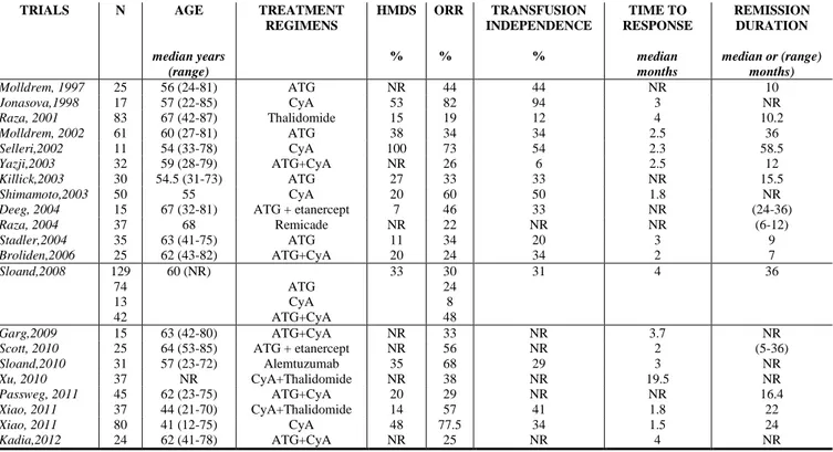
Main studies with current immunosuppressive treatments in MDS patients are summarized in Table 1. However, as a complete discussion on the results of the immunosuppresssion in MDS patients is beyond the scope of this article, the reader is referred to others extensive review on this topic [3,6,76-78].
CyA-based treatment studies have documented that CyA induces, both in hypocellular and normo-/ hyper-cellular MDS patients, fast (cumulative time to response 2 months) and sustained (about 2 years) hematological improvement in about 60% of MDS (cumulative ORR: 62%, range 8-82%) with an estimated overall survival (OS) of 28 months. In addition, CyA allows to achieve
TABLE 1. MAIN CLINICAL TRIALS WITH IMMUNOSUPPRESSIVE THERAPY IN MDS
TRIALS N AGE median years (range) TREATMENT REGIMENS HMDS % ORR % TRANSFUSION INDEPENDENCE % TIME TO RESPONSE median months REMISSION DURATION median or (range) months) Molldrem, 1997 25 56 (24-81) ATG NR 44 44 NR 10 Jonasova,1998 17 57 (22-85) CyA 53 82 94 3 NR Raza, 2001 83 67 (42-87) Thalidomide 15 19 12 4 10.2 Molldrem, 2002 61 60 (27-81) ATG 38 34 34 2.5 36 Selleri,2002 11 54 (33-78) CyA 100 73 54 2.3 58.5 Yazji,2003 32 59 (28-79) ATG+CyA NR 26 6 2.5 12 Killick,2003 30 54.5 (31-73) ATG 27 33 33 NR 15.5 Shimamoto,2003 50 55 CyA 20 60 50 1.8 NR
Deeg, 2004 15 67 (32-81) ATG + etanercept 7 46 33 NR (24-36)
Raza, 2004 37 68 Remicade NR 22 NR NR (6-12) Stadler,2004 35 63 (41-75) ATG 11 34 20 3 9 Broliden,2006 25 62 (43-82) ATG+CyA 20 24 34 2 7 Sloand,2008 129 74 13 42 60 (NR) ATG CyA ATG+CyA 33 30 24 8 48 31 4 36 Garg,2009 15 63 (42-80) ATG+CyA NR 33 NR 3.7 NR
Scott, 2010 25 64 (53-85) ATG + etanercept NR 56 NR 2 (5-36)
Sloand,2010 31 57 (23-72) Alemtuzumab 35 68 29 3 NR Xu, 2010 37 NR CyA+Thalidomide NR 38 NR 19.5 NR Passweg, 2011 45 62 (23-75) ATG+CyA 20 29 NR NR 16.4 Xiao, 2011 37 44 (21-70) CyA+Thalidomide 14 57 41 1.8 22 Xiao, 2011 80 41 (12-75) CyA 48 77.5 34 1.5 24 Kadia,2012 24 62 (41-78) ATG+CyA NR 25 NR 4 NR
Abbreviations. ATG = antithymocyte globulin; CyA = cyclosporine A; HMDS = hypoplastic myelodysplastic syndrome; NR = not reported; ORR =
transfusion independence in 58% of MDS patients. CyA was generally well tolerated, requiring drug withdrawal in a minority of patients, most of whom due to renal toxicity. However, we and others documented even higher hematological responses in hypocellular MDS [17,75,79-81].
Various clinical trials have investigated the role of horse (h) or rabbit (r) ATG alone [74,80,82-84] and in combination with CyA in MDS [80,85-89]. Approximately 30% of younger patients with lower risk MDS, expressing the HLA-DR15 allele and showing hypocellular bone marrow, achieved a rapid (time to response about 2.8 months) and prolonged hematological response with an estimated OS of 41 months [74,80,82-89]. As reported by the largest ATG-based treatment study of National Institutes of Health (NIH) on MDS, combination therapy of ATG with CyA, in comparison to ATG alone, is able both to further improve hematological responses allowing to achieve approximately 44% of ORR and to decrease the relapse rate [80]. In this and other ATG-based treatment study it has been reported that MDS patients relapsing post-ATG may be again responsive to CyA treatment [3,6].
In contrast to AA, although the comparative efficacy of hATG versus rATG has not been formally studied in MDS, it seems that there is no significant difference between the two sources of ATG in terms of hematological responses or adverse effects [84,90]. In addition, both CSA- and ATG-based treatments appeared do not increase progression to acute leukemia.
Alemtuzumab is an anti-CD52 monoclonal antibody, which has been successfully used in the treatment of AA patients failing initial immunosuppression with ATG and not eligible for transplant [91]. Recently, alemtuzumab monotherapy was used in a pilot study of 32 MDS patients, selected on the basis of likely responsiveness to immunosuppression due to younger age, low IPSS score, and the presence of HLA-DR15. A sustained ORR was documented among 77% and 57% of patients with intermediate-1 and intermediate-2 MDS, respectively, with a median time to response of about 3 months. Noteworthy, four of seven patients with karyotypic abnormalities at diagnosis had complete cytogenetic remission, including one patient with monosomy 7 [92].
In addition, we should mention that the therapeutic TNF-α blockade with anti-TNF-α monoclonal antibodies, soluble TNF-α receptors, and chemical inhibitors of TNF, such as thalidomide, have been disappointing when used as monotherapy [93-97]. More recently, combination of thalidomide with CyA [98,99], as well as of soluble TNF-α receptor etanercept with ATG [100,101] in MDS patients seems to increase hematological responses in MDS patients.
IV. CONCLUSION
Distinguishing HMDS from AA is clinically relevant since the incidence of progression to acute leukemia is
higher in HMDS. Differential diagnosis between these two diseases is still challenging, especially when dysplastic cells are difficult to detect due to marked hypocellularity of specimens and karyotipic abnormalities are not found. Also the more advanced SNP technology do not help to differentiate between these two diseases given that similar molecular abnormalities may be detected both in AA and MDS, including HMDS.
The autoimmune pathogenesis of MDS, including HMDS, is multifactorial and still unraveled. Several laboratory and clinical studies have provided evidence that some MDS seem likely to be related to derangement in the complex cross-talk between immunological microenvironment and marrow hematopoietic stem cells, resulting on one side in ineffective hematopoiesis related to immune-mediated apoptosis of normal hematopoietic progenitors, and on the other side in triggering clonal expansion of dysplastic progenitor cells leading to acute leukemia development.
Mechanisms of immune damage of hematopoietic progenitors, mediated by increased numbers of CTL, IFN-γ producing CD4+ cells and Th17 cells, increased levels of pro-apoptotic cytokines, as well as by decreased numbers of Treg, are predominantly involved in HMDS and low-risk MDS. By contrast, mechanisms developing an immunosuppressive marrow microenvironment favoring MDS clone escape, mainly associated with increased numbers of TregEM, MDSCs and anti-apoptotic cytokines, as well as with NK cell dysfunction, may contribute to high-risk MDS and acute leukemia progression.
Clinically, the best evidence for immune-mediated impairment of hematopoietic progenitor cell compartment in MDS patients is the improvement of peripheral cytopenia and survival after CyA- and ATG- based regimens, or alemtuzumab. Although the response to immunosuppression has been documented more often in HMDS patients, it may occurs also in lower-risk MDS patients. Other predictive factors recognized for response to immunosuppression in MDS include mainly younger age, HLA-DR15 expression, shorter duration of red cell transfusion dependence [102] and trisomy 8.
Based on the mechanisms described above, such better understanding of innate and adaptive immune responses involved in MDS pathophysiology, may pave the way for the development of novel immunotherapeutic approaches. Indeed, therapeutic strategies interfering with immune-mediated apoptosis of hematopoietic progenitor cell compartment in lower risk MDS and with immune-evasion of MDS clone in higher risk MDS, are already under evaluation in phase I/II clinical trials and are available for investigation in MDS patients [72,103-106].
ACKNOWLEDGMENT
The work was supported in part by Associazione Italiana contro le Leucemie, Linfomi e Mieloma (AIL), Sezione Marco Tulimieri di Salerno.
Conflict-of-interest disclosure: The authors declare no competing financial interests.
REFERENCES
[1] Fenaux P, Adès L. How we treat lower-risk myelodysplastic syndromes. Blood 2013;121(21):4280-4286.
[2] Vardiman J. The classification of MDS: from FAB to WHO and beyond. Leuk Res 2012;36(12):1453-1458. [3] Calado RT. Immunologic aspects of hypoplastic myelodysplastic syndrome. Semin Oncol 2011;38(5):667-672.
[4] Bennett JM, Orazi A. Diagnostic criteria to distinguish hypocellular acute myeloid leukemia from hypocellular myelodysplastic syndromes and aplastic anemia: recommendations for a standardized approach. Haematologica 2009;94(2):264-268.
[5] Marisavljevic D, Cemerikic V, Rolovic Z, Boskovic D, Colovic M. Hypocellular myelodysplastic syndromes: clinical and biological significance. Med Oncol 2005;22(2):169-175.
[6] Sloand EM. Hypocellular myelodysplasia. Hematol Oncol Clin North Am 2009;23(2):347-360.
[7] Koh Y, Lee HR, Song EY, Kim HK, Kim I, Park S, Park MH, Kim BK, Yoon SS, Lee DS. Hypoplastic myelodysplastic syndrome (h-MDS) is a distinctive clinical entity with poorer prognosis and frequent karyotypic and FISH abnormalities compared to aplastic anemia (AA). Leuk Res 2010;34(10):1344-1350.
[8] Huang TC, Ko BS, Tang JL, Hsu C, Chen CY, Tsay W, Huang SY, Yao M, Chen YC, Shen MC, Wang CH, Tien HF. Comparison of hypoplastic myelodysplastic syndrome (MDS) with normo-/hypercellular MDS by International Prognostic Scoring System, cytogenetic and genetic studies. Leukemia 2008;22(3):544-550.
[9] Fohlmeister I, Fischer R, Mödder B, Rister M, Schaefer HE. Aplastic anemia and the hypocellular myelodysplastic syndrome: histomorphological, diagnostic and prognostic features. J Clin Pathol 1985;38:1218-1224.
[10] De Wolf-Peeters C, Stessens R, Desmet V, Tricot G, Verwilghen RL. The histological characterization of ALIP in the myelodysplastic syndromes. Pathol Res Pract 1986;181:402-407.
[11] Tuzuner N, Cox C, Rowe JM, Watrous D, Bennett JM. Hypocellular myelodysplastic syndromes (MDS): new proposals. Br J Haematol 1995;91:612– 617. [12] Greenberg P, Cox C, LeBeau MM, Fenaux P, Morel P, Sanz G, Sanz M, Vallespi T, Hamblin T, Oscier D, Ohyashiki K, Toyama K, Aul C, Mufti G, Bennett J. International scoring system for evaluating prognosis in myelodysplastic syndromes. Blood 1997;89(6):2079-2088.
[13] Choi JW, Fujino M, Ito M. F-blast is a useful marker for differentiating hypocellular refractory anemia from aplastic anemia. Int J Hematol 2002;75:257–260.
[14] Kirwan M, Vulliamy T, Marrone A, Walne AJ, Beswick R, Hillmen P, Kelly R, Stewart A, Bowen D, Schonland SO, Whittle AM, McVerry A, Gilleece M, Dokal I. Defining the pathogenic role of telomerase mutations in myelodysplastic syndrome and acute myeloid leukemia. Hum Mutat 2009;30(11):1567-1573. [15] Matsui WH, Brodsky RA, Smith BD, Borowitz MJ, Jones RJ. Quantitative analysis of bone marrow CD34 cells in aplastic anemia and hypoplastic myelodysplastic syndromes. Leukemia 2006;20(3):458-462.
[16] Orazi A, Albitar M, Heerema NA, Haskins S, Neiman RS. Hypoplastic myelodysplastic syndromes can be distinguished from acquired aplastic anemia by CD34 and PCNA immunostaining of bone marrow biopsy specimens. Am J Clin Pathol 1997;107:268–274.
[17] Selleri C, Maciejewski JP, Catalano L, Ricci P, Andretta C, Luciano L, Rotoli B. Effects of cyclosporine on hematopoietic and immune functions in patients with hypoplastic myelodysplasia: in vitro and in vivo studies. Cancer 2002;95(9):1911-1922.
[18] Sato T, Kim S, Selleri C, Young NS, Maciejewski JP. Measurement of secondary colony formation after 5 weeks in long-term cultures in patients with myelodysplastic syndrome. Leukemia 1998;12(8):1187-1194.
[19] Maciejewski JP, Selleri C, Sato T, Anderson S, Young NS. A severe and consistent deficit in marrow and circulating hematopoietic cells (long-term culture-initiating cells) in acquired aplastic anemia. Blood 1996;88:1983–1991.
[20] Maciejewski JP, Kim S, Sloand E, Selleri C, Young NS. Sustained long-term hematologic recovery despite a marked quantitative defect in the stem cell compartment of patients with aplastic anemia after immunosuppressive therapy. Am J Hematol 2000 Oct;65(2):123-131.
[21]Tripathi P, Tripathi AK, Kumar A, Ahmad R, Balapure AK, Vishwakerma AL. Diagnostic and prognostic values of S-phase fraction and aneuploidy in patients with bone marrow aplasia. Indian J Hematol Blood Transfus 2009;25(1):10-16.
[22] Risitano AM, Maciejewski JP, Selleri C, Rotoli B. Function and malfunction of hematopoietic stem cells in primary bone marrow failure syndromes. Curr Stem Cell Res Ther 2007;2(1):39-52.
[23] Risitano AM, Maciejewski JP, Green S, Plasilova M, Zeng W, Young NS. In-vivo dominant immune responses in aplastic anaemia: molecular tracking of putatively pathogenetic T-cell clones by TCR beta-CDR3 sequencing. Lancet 2004;364(9431):355-364.
[24] Maciejewski JP, O'Keefe C, Gondek L, Tiu R. Immune-mediated bone marrow failure syndromes of progenitor and stem cells: molecular analysis of cytotoxic T cell clones. Folia Histochem Cytobiol 2007;45(1):5-14. [25] Wlodarski MW, Gondek LP, Nearman ZP, Plasilova M, Kalaycio M, Hsi ED, Maciejewski JP. Molecular strategies for detection and quantitation of clonal cytotoxic T-cell responses in aplastic anemia and myelodysplastic syndrome. Blood 2006;108(8):2632-2641.
[26] Kochenderfer JN, Kobayashi S, Wieder ED, Su C, Molldrem JJ. Loss of T-lymphocyte clonal dominance in patients with myelodysplastic syndrome responsive to immunosuppression. Blood 2002;100(10):3639-3645. [27] Barrett AJ, Sloand E. Autoimmune mechanisms in the pathophysiology of myelodysplastic syndromes and their clinical relevance. Haematologica 2009;94(4):449-451.
[28] Sloand EM, Pfannes L, Chen G, Shah S, Solomou EE, Barrett J, Young NS. CD34 cells from patients with trisomy 8 myelodysplastic syndrome (MDS) express early apoptotic markers but avoid programmed cell death by up-regulation of antiapoptotic proteins. Blood 2007;109(6):2399-2405.
[29] Sloand EM, Mainwaring L, Fuhrer M, Ramkissoon S, Risitano AM, Keyvanafar K, Lu J, Basu A, Barrett AJ, Young NS. Preferential suppression of trisomy 8 compared with normal hematopoietic cell growth by autologous lymphocytes in patients with trisomy 8 myelodysplastic syndrome. Blood 2005;106(3):841-851. [30] Chen G, Zeng W, Miyazato A, Billings E, Maciejewski JP, Kajigaya S, Sloand EM, Young NS. Distinctive gene expression profiles of CD34 cells from patients with myelodysplastic syndrome characterized by specific chromosomal abnormalities. Blood 2004;104(13):4210-4218.
[31] Sloand EM, Melenhorst JJ, Tucker ZC, Pfannes L, Brenchley JM, Yong A, Visconte V, Wu C, Gostick E, Scheinberg P, Olnes MJ, Douek DC, Price DA, Barrett AJ, Young NS. T-cell immune responses to Wilms tumor 1 protein in myelodysplasia responsive to immunosuppressive therapy. Blood 2011;117(9):2691-2699.
[32] Sloand EM, Kim S, Fuhrer M, Risitano AM, Nakamura R, Maciejewski JP, Barrett AJ, Young NS. Fas-mediated apoptosis is important in regulating cell replication and death in trisomy 8 hematopoietic cells but not in cells with other cytogenetic abnormalities. Blood 2002;100(13):4427-4432.
[33] Gyan E, Frisan E, Beyne-Rauzy O, Deschemin JC, Pierre-Eugene C, Randriamampita C, Dubart-Kupperschmitt A, Garrido C, Dreyfus F, Mayeux P, Lacombe C, Solary E, Fontenay M. Spontaneous and Fas-induced apoptosis of low-grade MDS erythroid precursors involves the endoplasmic reticulum. Leukemia 2008;22(10):1864-1873.
[34] Deeg HJ, Beckham C, Loken MR, Bryant E, Lesnikova M, Shulman HM, Gooley T. Negative regulators of hemopoiesis and stroma function in patients with myelodysplastic syndrome. Leuk Lymphoma 2000;37(3-4):405-414.
[35] Selleri C, Sato T, Raiola AM, Rotoli B, Young NS, Maciejewski JP. Induction of nitric oxide synthase is involved in the mechanism of fas-mediated apoptosis in haemopoietic cells. Br J Haematol 1997;99:481–489. [36] Claessens YE, Bouscary D, Dupont JM, Picard F, Melle J, Gisselbrecht S, Lacombe C, Dreyfus F, Mayeux P, Fontenay-Roupie M. In vitro proliferation and differentiation of erythroid progenitors from patients with
myelodysplastic syndromes: evidence for Fas-dependent apoptosis. Blood 2002;99(5):1594-1601.
[37] Ettou S, Audureau E, Humbrecht C, Benet B, Jammes H, Clozel T, Bardet V, Lacombe C, Dreyfus F, Mayeux P, Solary E, Fontenay M. Fas expression at diagnosis as a biomarker of azacitidine activity in high-risk MDS and secondary AML. Leukemia 2012;26(10):2297-2299.
[38] Ettou S, Humbrecht C, Benet B, Billot K, d'Allard D, Mariot V, Goodhardt M, Kosmider O, Mayeux P, Solary E, Fontenay M. Epigenetic control of NF-κB-dependent FAS gene transcription during progression of myelodysplastic syndromes. Mol Cancer Res 2013;11(7):724-735.
[39] Li X, Xu F, He Q, Wu L, Zhang Z, Chang C. Comparison of immunological abnormalities of lymphocytes in bone marrow in myelodysplastic syndrome (MDS) and aplastic anemia (AA). Intern Med 2010;49(14):1349-1355.
[40] Fozza C, Longinotti M. Are T-cell dysfunctions the other side of the moon in the pathogenesis of myelodysplastic syndromes? Eur J Haematol 2012;88(5):380-387.
[41] Kordasti SY, Afzali B, Lim Z, Ingram W, Hayden J, Barber L, Matthews K, Chelliah R, Guinn B, Lombardi G, Farzaneh F, Mufti GJ. IL-17-producing CD4(+) T cells, pro-inflammatory cytokines and apoptosis are increased in low risk myelodysplastic syndrome. Br J Haematol 2009;145(1):64-72.
[42] Shao LL, Zhang L, Hou Y, Yu S, Liu XG, Huang XY, Sun YX, Tian T, He N, Ma DX, Peng J, Hou M. Th22 cells as well as Th17 cells expand differentially in patients with early-stage and late-stage myelodysplastic syndrome. PLoS One 2012;7(12):e51339.
[43] Fozza C, Longinotti M. The role of T-cells in the pathogenesis of myelodysplastic syndromes: passengers and drivers. Leuk Res 2013;37(2):201-203.
[44] Ghiringhelli F, Ménard C, Terme M, Flament C, Taieb J, Chaput N, Puig PE, Novault S, Escudier B, Vivier E, Lecesne A, Robert C, Blay JY, Bernard J, Caillat-Zucman S, Freitas A, Tursz T, Wagner-Ballon O, Capron C, Vainchencker W, Martin F, Zitvogel L. CD4+CD25+ regulatory T cells inhibit natural killer cell functions in a transforming growth factor-beta-dependent manner. J Exp Med 2005;202(8):1075-1085.
[45] Ralainirina N, Poli A, Michel T, Poos L, Andrès E, Hentges F, Zimmer J. Control of NK cell functions by CD4+CD25+ regulatory T cells. J Leukoc Biol 2007;81(1):144-153.
[46] Bontkes HJ, Ruben JM, Alhan C, Westers TM, Ossenkoppele GJ, van de Loosdrecht AA. Azacitidine differentially affects CD4(pos) T-cell polarization in vitro and in vivo in high risk myelodysplastic syndromes. Leuk Res 2012;36(7):921-930.
[47] Bouchliou I, Miltiades P, Nakou E, Spanoudakis E, Goutzouvelidis A, Vakalopoulou S, Garypidou V, Kotoula V, Bourikas G, Tsatalas C, Kotsianidis I. Th17 and Foxp3(+) T regulatory cell dynamics and distribution
in myelodysplastic syndromes. Clin Immunol 2011;139(3):350-359.
[48] Mailloux AW, Sugimori C, Komrokji RS, Yang L, Maciejewski JP, Sekeres MA, Paquette R, Loughran TP Jr, List AF, Epling-Burnette PK. Expansion of effector memory regulatory T cells represents a novel prognostic factor in lower risk myelodysplastic syndrome. J Immunol 2012;189(6):3198-3208.
[49] Fozza C, Longu F, Contini S, Galleu A, Virdis P, Bonfigli S, Murineddu M, Gabbas A, Longinotti M. Patients with early-stage myelodysplastic syndromes show increased frequency of CD4+CD25+CD127(low) regulatory T cells. Acta Haematol 2012;128(3):178-182. [50] Mailloux AW, Epling-Burnette PK. Effector memory regulatory T-cell expansion marks a pivotal point of immune escape in myelodysplastic syndromes. Oncoimmunology 2013;2(2):e22654.
[51] Alfinito F, Sica M, Luciano L, Pepa RD, Palladino C, Ferrara I, Giani U, Ruggiero G, Terrazzano G. Immune dysregulation and dyserythropoiesis in the myelodysplastic syndromes. Br J Haematol 2010;148(1):90-98.
[52] Maciejewski J, Selleri C, Anderson S, Young NS. Fas antigen expression on CD34+ human marrow cells is induced by interferon gamma and tumor necrosis factor alpha and potentiates cytokine-mediated hematopoietic suppression in vitro. Blood 1995;85(11):3183-3190. [53] Sato T, Selleri C, Young NS, Maciejewski JP. Hematopoietic inhibition by interferon-gamma is partially mediated through interferon regulatory factor-1. Blood 1995;86(9):3373-3380.
[54] Sato T, Selleri C, Young NS, Maciejewski JP. Inhibition of interferon regulatory factor-1 expression results in predominance of cell growth stimulatory effects of interferon-gamma due to phosphorylation of Stat1 and Stat3. Blood 1997;90(12):4749-4758.
[55] Maciejewski JP, Selleri C, Sato T, Cho HJ, Keefer LK, Nathan CF, Young NS. Nitric oxide suppression of human hematopoiesis in vitro. Contribution to inhibitory action of interferon-gamma and tumor necrosis factor-alpha. J Clin Invest 1995;96(2):1085-1092.
[56] Serio B, Selleri C, Maciejewski JP. Impact of immunogenetic polymorphisms in bone marrow failure syndromes. Mini Rev Med Chem 2011;11(6):544-552. [57] Howe EC, Wlodarski M, Ball EJ, Rybicki L, Maciejewski JP. Killer immunoglobulin-like receptor genotype in immune-mediated bone marrow failure syndromes. Exp. Hematol 2005;33:1357-1362.
[58] Chen X, Eksioglu EA, Zhou J, Zhang L, Djeu J, Fortenbery N, Epling-Burnette P, Van Bijnen S, Dolstra H, Cannon J, Youn J, Donatelli SS, Qin D, De Witte T, Tao J, Wang H, Cheng P, Gabrilovich DI, List A, Wei S. Induction of myelodysplasia by myeloid-derived suppressor cells. J Clin Invest 2013;123(11):4595–4611. [59] Jiang HJ, Fu R, Wang HQ, Li LJ, Qu W, Liang Y, Wang GJ, Wang XM, Wu YH, Liu H, Song J, Guan J, Xing LM, Ruan EB, Shao ZH. Increased circulating of myeloid-derived suppressor cells in myelodysplastic syndrome. Chin Med J (Engl) 2013;126(13):2582-2584.
[60] Gabrilovich DI, Nagaraj S. Myeloid-derived suppressor cells as regulators of the immune system. Nat Rev Immunol 2009;9(3):162-174.
[61] Zhou L, Nguyen AN, Sohal D, Ying Ma J, Pahanish P, Gundabolu K, Hayman J, Chubak A, Mo Y, Bhagat TD, Das B, Kapoun AM, Navas TA, Parmar S, Kambhampati S, Pellagatti A, Braunchweig I, Zhang Y, Wickrema A, Medicherla S, Boultwood J, Platanias LC, Higgins LS, List AF, Bitzer M, Verma A. Inhibition of the TGF-beta receptor I kinase promotes hematopoiesis in MDS. Blood 2008;112(8):3434-3443.
[62] Allampallam K, Shetty VT, Raza A. Cytokines and MDS. Cancer Treat Res 2001;108:93–100.
[63] Kitagawa M, Saito I, Kuwata T, Yoshida S, Yamaguchi S, Takahashi M, Tanizawa T, Kamiyama R, Hirokawa K. Overexpression of tumor necrosis factor (TNF)-alpha and interferon (IFN)-gamma by bone marrow cells from patients with myelodysplastic syndromes. Leukemia 1997;11(12):2049-2054.
[64] Stifter G, Heiss S, Gastl G, Tzankov A, Stauder R. Over-expression of tumor necrosis factor-alpha in bone marrow biopsies from patients with myelodysplastic syndromes: relationship to anemia and prognosis. Eur J Haematol 2005;75(6):485-491.
[65] Benesch M, Platzbecker U, Ward J, Deeg HJ, Leisenring W. Expression of FLIP(Long) and FLIP(Short) in bone marrow mononuclear and CD34+ cells in patients with myelodysplastic syndrome: correlation with apoptosis. Leukemia 2003;17(12):2460-2466.
[66] Secchiero P, Zauli G. Tumor-necrosis-factor-related apoptosis-inducing ligand and the regulation of hematopoiesis. Curr Opin Hematol 2008;15(1):42-48. [67] Zhang Z, Li X, Guo J, Xu F, He Q, Zhao Y, Yang Y, Gu S, Zhang Y, Wu L, Chang C. Interleukin-17 enhances the production of interferon-γ and tumour necrosis factor-α by bone marrow T lymphocytes from patients with lower risk myelodysplastic syndromes. Eur J Haematol 2013;90(5):375-384.
[68] Pinheiro RF, Metze K, Silva MR, Chauffaille Mde L.The ambiguous role of interferon regulatory factor-1 (IRF-1) immunoexpression in myelodysplastic syndrome. Leuk Res 2009;33(10):1308-1312.
[69] Navas TA, Mohindru M, Estes M, Ma JY, Sokol L, Pahanish P, Parmar S, Haghnazari E, Zhou L, Collins R, Kerr I, Nguyen AN, Xu Y, Platanias LC, List AA, Higgins LS, Verma A. Inhibition of overactivated p38 MAPK can restore hematopoiesis in myelodysplastic syndrome progenitors. Blood 2006;108(13):4170-4177. [70] Navas TA, Mohindru M, Estes M, Ma JY, Sokol L, Pahanish P, Parmar S, Haghnazari E, Zhou L, Collins R, Kerr I, Nguyen AN, Xu Y, Platanias LC, List AA, Higgins LS, Verma A. Inhibition of overactivated p38 MAPK can restore hematopoiesis in myelodysplastic syndrome progenitors. Blood 2006;108(13):4170-4177. [71] Sokol L, Cripe L, Kantarjian H, Sekeres MA, Parmar S, Greenberg P, Goldberg SL, Bhushan V, Shammo J, Hohl R, Verma A, Garcia-Manero G, Li YP, Lowe A, Zhu J, List AF. Randomized, dose-escalation study of the p38α MAPK inhibitor SCIO-469 in patients with
myelodysplastic syndrome. Leukemia 2013;27(4):977-980.
[72] Aggarwal S, van de Loosdrecht AA, Alhan C, Ossenkoppele GJ, Westers TM, Bontkes HJ. Role of immune responses in the pathogenesis of low-risk MDS and high-risk MDS: implications for immunotherapy. Br J Haematol 2011;153(5):568-581.
[73] Risitano AM. Immunosuppressive therapies in the management of acquired immune-mediated marrow failures. Curr Opin Hematol 2012;19(1):3-13.
[74] Molldrem JJ, Caples M, Mavroudis D, Plante M, Young NS, Barrett AJ. Antithymocyte globulin for patients with myelodysplastic syndrome. Br J Haematol 1997;99(3):699-705.
[75] Jonásova A, Neuwirtová R, Cermák J, Vozobulová V, Mociková K, Sisková M, Hochová I. Cyclosporin A therapy in hypoplastic MDS patients and certain refractory anaemias without hypoplastic bone marrow. Br J Haematol 1998;100(2):304-309.
[76] Sloand EM, Barrett AJ. Immunosuppression for myelodysplastic syndrome: how bench to bedside to bench research led to success. Hematol Oncol Clin North Am 2010;24(2):331-341.
[77] Barrett AJ, Sloand EM. Immunosuppressive therapy for myelodysplastic syndromes: refining the indications. Curr Hematol Malig Rep 2008;3(1):23-28.
[78] Parikh AR, Olnes MJ, Barrett AJ. Immunomodulatory treatment of myelodysplastic syndromes: antithymocyte globulin, cyclosporine, and alemtuzumab. Semin Hematol 2012 Oct;49(4):304-11. [79] Shimamoto T, Tohyama K, Okamoto T, Uchiyama T, Mori H, Tomonaga M, Asano Y, Niho Y, Teramura M, Mizoguchi H, Omine M, Ohyashiki K. Cyclosporin A therapy for patients with myelodysplastic syndrome: multicenter pilot studies in Japan. Leuk Res 2003;27(9):783-788.
[80] Sloand EM, Wu CO, Greenberg P, Young N, Barrett J. Factors affecting response and survival in patients with myelodysplasia treated with immunosuppressive therapy. J Clin Oncol 2008;26(15):2505-2511.
[81] Xiao L, Qi Z, Qiusheng C, Li X, Luxi S, Lingyun W. The use of selective immunosuppressive therapy on myelodysplastic syndromes in targeted populations results in good response rates and avoids treatment-related disease progression. Am J Hematol 2012;87(1):26-31. [82] Molldrem JJ, Leifer E, Bahceci E, Saunthararajah Y, Rivera M, Dunbar C, Liu J, Nakamura R, Young NS, Barrett AJ. Antithymocyte globulin for treatment of the bone marrow failure associated with myelodysplastic syndromes. Ann Intern Med 2002;137(3):156-163. [83] Killick SB, Mufti G, Cavenagh JD, Mijovic A, Peacock JL, Gordon-Smith EC, Bowen DT, Marsh JC. A pilot study of antithymocyte globulin (ATG) in the treatment of patients with 'low-risk' myelodysplasia. Br J Haematol 2003;120(4):679-684.
[84] Stadler M, Germing U, Kliche KO, Josten KM, Kuse R, Hofmann WK, Schrezenmeier H, Novotny J, Anders O, Eimermacher H, Verbeek W, Kreipe HH, Heimpel H, Aul C, Ganser A. A prospective, randomised, phase II
study of horse antithymocyte globulin vs rabbit antithymocyte globulin as immune-modulating therapy in patients with low-risk myelodysplastic syndromes. Leukemia 2004;18(3):460-465.
[85] Yazji S, Giles FJ, Tsimberidou AM, Estey EH, Kantarjian HM, O'Brien SA, Kurzrock R. Antithymocyte globulin (ATG)-based therapy in patients with myelodysplastic syndromes. Leukemia 2003;17(11):2101-2106.
[86] Broliden PA, Dahl IM, Hast R, Johansson B, Juvonen E, Kjeldsen L, Porwit-MacDonald A, Sjoo M, Tangen JM, Uggla B, Oberg G, Hellstrom-Lindberg E. Antithymocyte globulin and cyclosporine A as combination therapy for low-risk non-sideroblastic myelodysplastic syndromes. Haematologica 2006;91(5):667-670.
[87] Garg R, Faderl S, Garcia-Manero G, Cortes J, Koller C, Huang X, York S, Pierce S, Brandt M, Beran M, Borthakur G, Kantarjian H, Ravandi F. Phase II study of rabbit anti-thymocyte globulin, cyclosporine and granulocyte colony-stimulating factor in patients with aplastic anemia and myelodysplastic syndrome. Leukemia 2009;23(7):1297-1302.
[88] Passweg JR, Giagounidis AA, Simcock M, Aul C, Dobbelstein C, Stadler M, Ossenkoppele G, Hofmann WK, Schilling K, Tichelli A, Ganser A. Immunosuppressive therapy for patients with myelodysplastic syndrome: a prospective randomized multicenter phase III trial comparing antithymocyte globulin plus cyclosporine with best supportive care--SAKK 33/99. J Clin Oncol 2011;29(3):303-309.
[89] Kadia TM, Borthakur G, Garcia-Manero G, Faderl S, Jabbour E, Estrov Z, York S, Huang X, Pierce S, Brandt M, Koller C, Kantarjian HM, Ravandi F. Final results of the phase II study of rabbit anti-thymocyte globulin, ciclosporin, methylprednisone, and granulocyte colony-stimulating factor in patients with aplastic anaemia and myelodysplastic syndrome. Br J Haematol 2012;157(3):312-320.
[90] Scheinberg P, Nunez O, Weinstein B, Scheinberg P, Biancotto A, Wu CO, Young NS. Horse versus rabbit antithymocyte globulin in acquired aplastic anemia. N Engl J Med 2011;365(5):430-438.
[91] Risitano AM, Schrezenmeier H. Alternative immunosuppression in patients failing immunosuppression with ATG who are not transplant candidates: Campath (Alemtuzumab). Bone Marrow Transplant 2013;48(2):186-190.
[92] Sloand EM, Olnes MJ, Shenoy A, Weinstein B, Boss C, Loeliger K, Wu CO, More K, Barrett AJ, Scheinberg P, Young NS. Alemtuzumab treatment of intermediate-1 myelodysplasia patients is associated with sustained improvement in blood counts and cytogenetic remissions. J Clin Oncol 2010;28(35):5166-5173.
[93] Deeg HJ, Gotlib J, Beckham C, Dugan K, Holmberg L, Schubert M, Appelbaum F, Greenberg P. Soluble TNF receptor fusion protein (etanercept) for the treatment of myelodysplastic syndrome: a pilot study. Leukemia 2002;16(2):162-164.
[94] Raza A. Anti-TNF therapies in rheumatoid arthritis, Crohn's disease, sepsis, and myelodysplastic syndromes. Microsc Res Tech 2000;50(3):229-235.
[95] Raza A, Candoni A, Khan U, Lisak L, Tahir S, Silvestri F, Billmeier J, Alvi MI, Mumtaz M, Gezer S, et al. Remicade as TNF suppressor in patients with myelodysplastic syndromes. Leuk Lymphoma 2004;45:2099-2104.
[96] Maciejewski JP, Risitano AM, Sloand EM, Wisch L, Geller N, Barrett JA, Young NS. A pilot study of the recombinant soluble human tumour necrosis factor receptor (p75)-Fc fusion protein in patients with myelodysplastic syndrome. Br J Haematol 2002;117(1):119-126.
[97] Raza A, Meyer P, Dutt D, Zorat F, Lisak L, Nascimben F, du Randt M, Kaspar C, Goldberg C, Loew J, Dar S, Gezer S, Venugopal P, Zeldis J. Thalidomide produces transfusion independence in long-standing refractory anemias of patients with myelodysplastic syndromes. Blood 2001;98:958 – 965.
[98] Xu ZF, Qin TJ, Zhang Y, Liu KQ, Hao YS, Xiao ZJ. [Cyclosporine A in combination with thalidomide for the treatment of patients with myelodysplastic syndromes.] Zhonghua Xue Ye Xue Za Zhi 2010;31(7):451-455. [99] Xiao Z, Xu Z, Zhang Y, Qin T, Zhang H, Fang L. Cyclosporin A and thalidomide in patients with myelodysplastic syndromes: Results of a pilot study. Leuk Res 2011;35(1):61-65.
[100] Deeg HJ, Jiang PY, Holmberg LA, Scott B, Petersdorf EW, Appelbaum FR. Hematologic responses of patients with MDS to antithymocyte globulin plus etanercept correlate with improved flow scores of marrow cells. Leuk Res 2004;28(11):1177-1180.
[101] Scott BL, Ramakrishnan A, Fosdal M, Storer B, Becker P, Petersdorf S, Deeg HJ. Anti-thymocyte globulin plus etanercept as therapy for myelodysplastic syndromes (MDS): a phase II study. Br J Haematol 2010;149(5):706-710.
[102] Saunthararajah Y, Nakamura R, Wesley R, Wang QJ, Barrett AJ. A simple method to predict response to immunosuppressive therapy in patients with myelodysplastic syndrome. Blood 2003;102(8):3025-3027.
[103] Bachegowda L, Gligich O, Mantzaris I, Schinke C, Wyville D, Carrillo T, Braunschweig I, Steidl U, Verma A. Signal transduction inhibitors in treatment of myelodysplastic syndromes. J Hematol Oncol 2013;6:50. [104] Epling-Burnette PK, McDaniel J, Wei S, List AF. Emerging immunosuppressive drugs in myelodysplastic syndromes. Expert Opin Emerg Drugs 2012;17(4):519-541.
[105] List AF. New therapeutics for myelodysplastic syndromes. Leuk Res 2012;36(12):1470-1474.
[106] Duong VH, Padron E, List AF, Komrokji RS. Multikinase inhibitors for treating high-risk myelodysplastic syndromes: can this be brought into clinical practice? Expert Rev Hematol 2013;6(5):485-487.