R
ecent advances in cancer biology have opened a new era of molecular targeting therapy, including tyrosine kinase inhibitors (TKIs) targeting epidermal growth factor receptor (EGFR) that have a significant treatment advan-tage over cytotoxic agents for the treatment of advanced non–small cell lung cancer (NSCLC) (1,2). More recently, an entirely new group of drugs has been shown to be ben-eficial to patients with advanced-stage cancer (3–6). This includes immune checkpoint inhibitors (ICIs) that pro-long survival and improve quality of life in patients with advanced-stage cancers including NSCLCs (7). In a U.S.ICIs increased from 1.54% in 2011 to 43.63% in 2018; patients’ response to ICIs was 0.14% in 2011 and in-creased to 12.46% in 2018.
Since the first report from Japan of severe acute lung injury in patients with NSCLC treated with gefitinib (9), pneumonitis associated with molecular targeting agents has attracted considerable attention. More recently, ICIs such as nivolumab and pembrolizumab have demon-strated to be associated with toxicities often termed im-mune-related adverse effects, including pneumonitis as one of the clinically significant and potentially
life-threaten-Chest CT Diagnosis and Clinical Management of
Drug-related Pneumonitis in Patients Receiving
Molecular Targeting Agents and Immune Checkpoint
Inhibitors: A Position Paper from the Fleischner Society
Takeshi Johkoh, MD, PhD* • Kyung Soo Lee, MD, PhD* • Mizuki Nishino, MD, MPH •William D. Travis, MD • Jay H. Ryu, MD • Ho Yun Lee, MD, PhD • Christopher J. Ryerson, MD, MAS • Tomás Franquet, MD, PhD • Alexander A. Bankier, MD, PhD • Kevin K. Brown, MD • Jin Mo Goo, MD, PhD • Hans-Ulrich Kauczor, MD • David A. Lynch, MB • Andrew G. Nicholson, MD • Luca Richeldi, MD, PhD • Cornelia M. Schaefer-Prokop, MD, PhD • Johny Verschakelen, MD, PhD • Suhail Raoof, MD •
Geoffrey D. Rubin, MD, MBA • Charles Powell, MD • Yoshikazu Inoue, MD, PhD • Hiroto Hatabu, MD, PhD
From the Department of Radiology, Kansai Rosai Hospital, Amagasaki, Japan (T.J.); Department of Radiology, Samsung Medical Center (K.S.L., H.Y.L.) and Department of Health Sciences and Technology, SAIHST (H.Y.L.), Sungkyunkwan University School of Medicine, 81 Irwon-ro, Gangnam-gu, Seoul 06351, South Korea; Department of Imaging, Dana-Farber Cancer Institute, Boston, Mass (M.N.); Department of Radiology (M.N.) and Center for Pulmonary Functional Imaging (H.H.), Brigham and Women’s Hospital, Harvard Medical School, Boston, Mass; Department of Pathology, Memorial Sloan-Kettering Cancer Center, New York, NY (W.D.T.); Division of Pulmonary and Critical Care Medicine, Mayo Clinic, Rochester, Minn (J.H.R.); Department of Medicine, University of British Columbia and Centre for Heart Lung Innovation, St. Paul’s Hospital, Vancouver, Canada (C.J.R.); Department of Radiology, Hospital de Sant Pau, Universitat Autònoma de Barcelona, Barcelona, Spain (T.F.); Department of Radiology, University of Massachusetts Medical Center, Worcester, Mass (A.A.B.); Departments of Medicine (K.K.B.) and Radiology (D.A.L.), National Jewish Health, Denver, Colo; Department of Radiology, Seoul National University College of Medicine, Seoul, Korea (J.M.G.); Diagnostic and Interventional Radiol-ogy, University Hospital Heidelberg, Translational Lung Research Center Heidelberg, member of the German Center of Lung Research, Heidelberg, Germany (H.U.K.); Department of Histopathology, Royal Brompton and Harefield NHS Foundation Trust and National Heart and Lung Institute, Imperial College, London, England (A.G.N.); Complex Operative Unit of Pneumology, Fondazione Policlinico Universitario A. Gemelli IRCCS, Catholic University of the Sacred Heart, Rome, Italy (L.R.); Department of Radiology, Meander Medical Center, Amersfoort, the Netherlands (C.M.S.P.); Department of Radiology, University Hospitals Leuven, Leuven, Belgium (J.V.); Division of Pulmonary and Critical Care Medicine, Lenox Hill Hospital, Northwell Health System, New York, NY (S.R.); Department of Radiology, Duke Univer-sity School of Medicine, Durham, NC (G.D.R.); Department of Pulmonary, Critical Care and Sleep Medicine, Icahn School of Medicine at Mount Sinai, New York, NY (C.P.); and Clinical Research Center, National Hospital Organization Kinki-Chuo Chest Medical Center, Sakai, Osaka, Japan (Y.I.). Received August 14, 2020; revision requested September 22; final revision received October 6; accepted November 5. Address correspondence to K.S.L. (e-mail: [email protected]).
*T.J. and K.S.L. contributed equally to this work. Conflicts of interest are listed at the end of this article.
Radiology 2021; 00:1–17 • https://doi.org/10.1148/radiol.2021203427 • Content codes:
Use of molecular targeting agents and immune checkpoint inhibitors (ICIs) has increased the frequency and broadened the spec-trum of lung toxicity, particularly in patients with cancer. The diagnosis of drug-related pneumonitis (DRP) is usually achieved by excluding other potential known causes. Awareness of the incidence and risk factors for DRP is becoming increasingly important. The severity of symptoms associated with DRP may range from mild or none to life-threatening with rapid progression to death. Imaging features of DRP should be assessed in consideration of the distribution of lung parenchymal abnormalities (radiologic pattern approach). The CT patterns reflect acute (diffuse alveolar damage) interstitial pneumonia and transient (simple pulmonary eosinophilia) lung abnormality, subacute interstitial disease (organizing pneumonia and hypersensitivity pneumonitis), and chronic interstitial disease (nonspecific interstitial pneumonia). A single drug can be associated with multiple radiologic patterns. Treatment of a patient suspected of having DRP generally consists of drug discontinuation, immunosuppressive therapy, or both, along with supportive measures eventually including supplemental oxygen and intensive care. In this position paper, the authors provide diag-nostic criteria and management recommendations for DRP that should be of interest to radiologists, clinicians, clinical trialists, and trial sponsors, among others.
This article is a simultaneous joint publication in Radiology and CHEST. The articles are identical except for stylistic changes in keeping with each journal’s style. Either version may be used in citing this article.
Published under a CC BY 4.0 license.
Methodology and Literature Search
The international multidisciplinary panel included 19 experts in interstitial lung diseases (ILDs) and lung cancer (10 radiolo-gists, seven pulmonoloradiolo-gists, and two pathologists). All were Fleischner Society members, working with two expert medical librarians who were nonmembers.
The panel developed key questions believed to be important for the diagnosis and management of DRP based on their clini-cal experience in the diagnosis and management of pneumonitis. In brief, coleaders drafted provisional questions. After discussion by all members of the writing committee, the questions were fi-nalized. Likewise, answer paragraphs for each key question were drafted by key writing members, which were circulated, revised, and approved by all committee members.
The PubMed literature search strategy is shown in Appendix E1 (online) (search date: June 1, 2020). Combined free-text key-words and controlled vocabulary terms were used. The filters for the search were applied, and the filters included Humans [spe-cies], English [language], and journal articles [article types]. The number of search results totaled 926 items not including abstract only or editorial only. With the 926 items at hand, the key writ-ing members wrote sections in their charges in consideration of the given questions. The cited items included 106 original arti-cles and seven reviews. During circulation, review, proofreading, and revision processes among the writing committee members, we added eight case report references. We included 121 refer-ences consisting mainly of original articles (n = 106), relevant reviews (n = 7), and case reports (n = 8), with relevant citations provided to the writing committee for incorporation into the evidence summary where appropriate.
Clinical Features of DRP
The incidence of DRP varies widely among published studies. A recent population-based study from France (25) estimated an incidence of 1.2 per 100 000 per year and a prevalence of 2.6 per 100 000. DRP accounts for 2.5%–5% of prevalent cases of ILD (25–27). Cancer drugs (eg, bleomycin) are the most common cause of DRP, followed by drugs for autoim-mune diseases (eg, methotrexate), amiodarone, and antibiot-ics (eg, nitrofurantoin), based on a recent systematic review (27). Additionally, awareness of the incidence and risk factors of pneumonitis related to specific anticancer agents is increas-ing in importance.
DRP can manifest as a variety of clinical syndromes of lung injury (27–31). The onset of illness may be acute, insidious, and sometimes delayed with a long latent period (eg, beyond 10 years in some cases of carmustine-induced pulmonary fibrosis) after completion of drug treatment (27,28). The clinical symp-toms are generally nonspecific, including dyspnea, cough, mal-aise, and low-grade fever. Some patients may be asymptomatic even in the presence of diffuse pulmonary opacities. Lung aus-cultation often reveals crackles but may be normal. It is difficult to clinically distinguish DRP from lung disease of other causes such as infections, pulmonary hemorrhage, pulmonary edema, radiation-induced pneumonitis, and metastases. In addition, evaluation for cardiovascular etiologies including heart failure,
Abbreviations
DAD = diffuse alveolar damage, DRP = drug-related pneumonitis, EGFR = epidermal growth factor receptor, HP = hypersensitivity pneu-monitis, ICI = immune checkpoint inhibitor, ILD = interstitial lung disease, NSCLC = non–small cell lung cancer, NSIP = nonspecific inter-stitial pneumonia, OP = organizing pneumonia, PD-1 = programmed cell death protein 1, PD-L1 = programmed death ligand 1,TKI = ty-rosine kinase inhibitor
Summary
Increasing use of molecular targeting agents and immune checkpoint inhibitors has increased the frequency and broadened the spectrum of lung toxicity, particularly in patients with cancer. In this position pa-per from the Fleischner Society, the authors provide diagnostic crite-ria and management recommendations of drug-related pneumonitis for radiologists, clinicians, clinical trialists, and trial sponsors.
Essentials
n The CT patterns in drug-related pneumonitis (DRP) reflect acute
(diffuse alveolar damage) interstitial pneumonia and transient (simple pulmonary eosinophilia) lung abnormality, subacute inter-stitial disease (organizing pneumonia and hypersensitivity pneu-monitis), and chronic interstitial disease (nonspecific interstitial pneumonia).
n The diagnostic criteria include newly identified pulmonary
paren-chymal opacities at imaging, temporal association of presentation with the initiation of a systemic therapeutic agent, and the exclu-sion of other likely causes.
n Management of DRP consists of drug discontinuation,
immu-nosuppressive therapy, or both, along with supportive measures including supplemental oxygen and intensive care.
scans during cancer treatment allows detailed evaluations of both presymptomatic and symptomatic drug-related pneumo-nitis (DRP) and characterization of its radiologic manifesta-tions in patients with different cancer types treated with dif-ferent agents.
The reports on pneumonitis among patients with NSCLC receiving EGFR-targeted TKIs, or EGFR-TKIs, included acute fatal events (13–23). Moreover, the recent introduction of novel molecular targeting agents such as third-generation EGFR-TKIs, newer anaplastic lymphoma kinase inhibitors, BRAF inhibitors, antibody-drug conjugates, and use of ICI combination regimens has brought new challenges to DRP (24). Despite many reports on this topic, there is limited published guidance on the evalua-tion and treatment of patients with DRP secondary to treatment with ICIs.
In this position paper from the Fleischner Society, we identi-fied specific questions for diagnosis and management of DRP in patients receiving molecular targeting agents and ICIs. We conducted a systematic search to identify supporting evidence on these topics that had been published since the introduction of EGFR-TKIs in 2003. Based on the review of the literature combined with expert opinions, this position paper provides a description of radiologic patterns; a set of proposed diagnostic criteria; and management recommendations for DRP, which will be of value in improving the understanding of the pathophysiol-ogy, prognosis, and management of this diverse group of condi-tions. We believe that this position paper will be of interest to a diverse group of health care providers and clinical researchers.
pected outcomes for the individual patient. These issues need to be discussed with the patient in a shared decision-making process that incorporates the individual patient’s values and preferences.
An adequate lung biopsy performed with bronchoscopic (forceps or cryobiopsy) or surgical approach (preferably, video-assisted thoracoscopic biopsy) can demonstrate the histopatho-logic pattern of lung injury (eg, nonspecific interstitial pneu-monia [NSIP], organizing pneupneu-monia [OP], or diffuse alveolar damage [DAD]) in patients suspected of having DRP. However, the features seen on lung biopsy are unlikely to confirm the di-agnosis of DRP because these histopathologic patterns are non-specific and can be seen with other causes, including infections. A biopsy may sometimes be useful to exclude recurrent malig-nancy, given that tumors can manifest as diffuse lung infiltration and mimic ILD.
Bronchoscopic cryobiopsy yields larger samples of lung tissue but is associated with a higher rate of bleeding and pneumotho-rax compared with forceps biopsy (39). Overall in-hospital mor-tality after surgical lung biopsy for ILD in the United States was found to be 6.4% in a recent analysis of a national data set (40). The in-hospital mortality rate was 1.7% for elective operations compared with 16.0% for nonelective operations. Possible need for surgical lung biopsy should be entertained early rather than late in the clinical course because severe respiratory dysfunction and dependence on mechanical ventilation increase the mortal-ity rate associated with surgical lung biopsy (41).
More often, bronchoscopy with bronchoalveolar lavage is performed to exclude infections (including opportunistic, my-cobacterial, and viral), alveolar hemorrhage, or metastatic and/ or lymphangitic spread (cancer cells). However, bronchoalveolar lavage fluid–derived differential cell count is often overlapping and nonspecific (27,28,42,43). Nonetheless, bronchoalveolar lavage may yield specimens diagnostic of infection (eg, Pneumo-cystis) or malignancy and bronchoalveolar lavage fluid–derived differential cell counts may provide diagnostic clues (eg, eosino-philia) as to the underlying pathologic process.
When Should Suspected Offending Drugs Be Stopped in Patients Suspected of Having DRP?
Discontinuation of the suspected drug is advisable for patients with severe or progressive lung disease (eg, with worsening to grade 2 or 3 [Common Terminology Criteria for Adverse Events]) for which DRP is deemed a possible or likely cause of the clinical presentation, while additional studies are being performed for diagnostic clarification. Improvement follow-ing cessation of drug administration without glucocorticoid therapy would strongly support the diagnosis of DRP in the absence of other more likely explanations emerging from the diagnostic work-up (27,43).
It may be appropriate to closely monitor patients while continuing therapy when the lung injury is not severe or pro-gressive, particularly asymptomatic patients with isolated ra-diologic changes (grade 1 pneumonitis), as discussed in the following sections regarding specific agents including mecha-nistic target of rapamycin, or mTOR, inhibitor and third-generation EGFR-TKIs (32). It is important to acknowledge pulmonary embolism, pulmonary veno-occlusive disease, and
other forms of pulmonary hypertension needs to be considered in this setting. The clinical suspicion for DRP arises from the temporal relationship between drug exposure and the onset of clinical presentation (27,28). In most patients, the diagnosis of DRP is unlikely to be made with certainty even after extensive clinical evaluation, including lung biopsy.
The increasing use of molecular targeting agents and ICIs has broadened the spectrum of lung toxicity encountered clini-cally, especially in patients with cancer. This is exemplified by immune-related adverse effects in patients treated with ICIs, which manifest as a wide array of organ toxicities, including pneumonitis (11,32,33). These toxicities are thought to be the result of general immunologic activation, including autoim-mune response.
The severity of symptoms associated with DRP may range from mild to life-threatening with rapid progression to death. The Common Terminology Criteria for Adverse Events pub-lished by the National Cancer Institute of the National Institutes of Health provides standardized definitions for grading the se-verity of organ toxicity (34). The grades for pneumonitis include grade 1 (asymptomatic), grade 2 (symptomatic), grade 3 (severe symptoms), grade 4 (life-threatening respiratory compromise), and grade 5 (death related to adverse event).
Laboratory tests such as serologic testing and microbial cul-tures may help to establish infectious or other etiologies for pul-monary infiltrates but are not useful in specifically diagnosing DRP (12,27–29,35,36). Similarly, pulmonary function testing commonly demonstrates a restrictive pattern (reduced forced vi-tal capacity and/or tovi-tal lung capacity) along with a reduced dif-fusion capacity, which is the typical pattern seen in ILD. When present, it is helpful in assessing the degree of pulmonary impair-ment, but it does not contribute to confirming the diagnosis of DRP (27–29).
When Should Chest CT Be Performed to Confirm the Diagnosis in Patients Suspected of Having DRP?
In patients receiving drugs potentially causing pulmonary toxicity, chest CT (and particularly thin-section CT; section thickness of 2.0–2.5 mm or less) plays an important role in evaluating the appearance, the progression, and the resolution of pulmonary abnormalities (37). CT should be performed as early as possible when DRP is suspected and in the presence of a positive temporal relationship between drug exposure and symptom onset. CT may allow early detection of the DRP while it is still at a reversible stage, or it may help to identify findings indicating other etiologies that can explain the symp-toms of the patients (38). CT is also essential to evaluate the presence of other common causes (eg, community-acquired or health care–associated pneumonia) for the nonspecific clinical manifestations of DRP.
Should Lung Biopsy Be Performed to Confirm the Diagnosis in Patients Suspected of Having DRP?
Whether lung biopsy should be performed in patients sus-pected of having DRP depends on the clinical context, alterna-tive diagnoses being considered, benefit-risk analysis, and
ex-Imaging Features of DRP
Most DRP is diagnosed on routine follow-up CT scans for monitoring in patients with cancer; specific protocol-based scans targeted to depict DRP are not obtained. In addition, given the unique nature (eg, nature of diagnostic exclusion, symptomless cases, and wide differential diagnoses, etc) of the entity, defining the specific protocol for CT study is difficult and impractical. Thinner-section (2.0–2.5 mm or less in sec-tion thickness) and contiguous CT scans are recommended usually with intravenous contrast agent injection. For thorough image analysis, not only transverse but also coronal reformat-ted images are needed. Follow-up chest CT is useful to assess the changes of DRP findings and response to DRP treatment (36,54). However, the details of follow-up scans including time intervals depend on the clinical context (severity of symptoms and clinical follow-up course, etc).
The CT features of DRP associated with systemic therapeutic agents should be systematically described, including distribution and patterns of parenchymal abnormalities and presence of in-dividual features including ground-glass opacities, airspace con-solidation, reticular opacities, centrilobular nodules, interlobular septal thickening, honeycombing, and traction bronchiectasis.
Each drug can be associated with multiple injury patterns at CT (Fig 1) (55), which are typically not specific. In most situa-tions, clinicians rely on the temporal relationships between the administration of drugs and the onset of symptoms, along with the exclusion of other potential causes of lung injury, particularly infections and metastatic diseases (28).
What Are the CT Patterns of DRP?
DRPs have various histologic patterns and diverse CT findings (31,56,57). Although CT and histologic patterns coincide in only half of patients with DRP (57), the CT pattern reflects the extent and distribution of lung abnormalities and helps to predict changes in terms of prognostication. Some of the com-monly described patterns include interstitial pneumonia either as NSIP, OP, DAD, hypersensitivity pneumonitis (HP), and simple pulmonary eosinophilia (Table 1) (58–61). Often, im-aging demonstrates more than one pattern (Fig 1); in this case the dominant pattern is typically reported. Other drug-related lung diseases such as granulomatous pneumonitis, vasculitis, alveolar proteinosis, constrictive (obliterative) bronchiolitis, and veno-occlusive disease are uncommon (32,59) and dem-onstrate diverse CT features; thus, they are difficult to clas-sify into one of the aforementioned common CT patterns. The DRP CT patterns are nonspecific for either drug reaction in general or the reaction to a particular drug. Consequently, the diagnosis of DRP is based on a combination of clinical, radiologic, and histologic (when necessary) findings in a pa-tient who has received a drug known or suspected to cause the abnormalities.
Radiologic NSIP pattern.—NSIP pattern consists of patchy or diffuse areas of ground-glass opacity (58,59), typically with peripheral and lower lung zone predominance. With pro-gression, evidence of fibrosis including reticulation, traction the life-threatening nature of the malignancies treated with the
implicated drugs, the benefits of the therapy, and the uncer-tainties regarding the impact of medication discontinuation in this setting. Thus, it is appropriate for clinicians to discuss these issues at a multidisciplinary conference (see later section on Multidisciplinary Diagnosis of DRP) and with patients to reach a shared decision regarding preferred course of action. Additionally, major clinical guidelines based on the consensus of multidisciplinary and multiorganizational panels are avail-able for specific entities such as ICI-related pneumonitis, and should be considered for patient treatment in specific clinical settings when relevant (44–47).
Does Improvement with Glucocorticoid Therapy Confirm the Diagnosis of DRP?
Clinical improvement subsequent to glucocorticoid therapy does not definitively confirm the diagnosis of DRP because other inflammatory processes that are not drug related (eg, radiation pneumonitis) may also respond to glucocorticoid therapy. In addition, improvement may merely be coincidental and due to a self-limited event (eg, aspiration pneumonia) with spontaneous recovery. Nonetheless, glucocorticoid therapy is commonly used in the management of patients suspected of having DRP, especially if the lung injury is severe or progres-sive, and response to glucocorticoid therapy would support a diagnosis of DRP (vs progression of underlying cancer, for example) in the absence of a better alternative explanation (27,45,48,49). Improvement with glucocorticoid therapy may also obviate invasive diagnostic maneuvers such as broncho-scopic or surgical lung biopsy.
Should Rechallenge with a Suspected Drug Be Performed to Confirm the Diagnosis of DRP?
It is rarely appropriate to rechallenge with the suspected drug to confirm the diagnosis, especially when the lung toxicity has been severe or if there were substantial residual abnormalities at chest imaging (45,50). An exception may be considered when lung toxicity has been mild and transient, particularly if alternative cancer therapies are unlikely to be effective.
What Prognostic Factors Should Be Considered in Patients with DRP?
Prognostic factors in patients with DRP include acute onset, severity of lung toxicity (eg, hypoxemia), response to treatment (eg, drug withdrawal), older age, current or prior smoking his-tory, preexisting lung disease, other comorbidities, and the status of the underlying cancer (27,35). There are no scoring schemes currently available that integrate these factors into a predictive model. The prognosis associated with DRP also var-ies depending on the specific drug and the type of underlying cancer. There are emerging reports of beneficial effects from drug-related toxicities on tumor response to therapy, especially in those who are treated with newer molecular targeting cancer therapy or ICIs (51,52). This interesting possibility should be further investigated in a disease-specific and therapy-specific manner (53).
chovascular in distribution (Fig 2) (62). The abnormalities are usually bilateral and symmetric, with predominant lower-lung bronchiectasis, and occasionally honeycombing are identified
(Table 1). In some patients, fibrosis is predominantly
peribron-Figure 1: Images show gefitinib-related pneumonitis with different CT patterns in two different patients with lung cancer. (a, b) Lung window
images of CT scans obtained at levels of right inferior pulmonary vein (a) and liver dome (b), respectively, show several areas of parenchymal
consolidation or poorly defined nodules (arrows) in right lung. Lesions showed migration and waxing and waning in extent for 3 months and then disappeared (not shown here), features compatible with simple pulmonary eosinophilia pattern. Patient was a 53-year-old woman with lung adeno-carcinoma, positive for missense mutation in exon 21. (c, d) CT scans obtained at levels of right middle lobar bronchus (c) and basal trunk (d),
re-spectively, demonstrate parenchymal opacity in both lungs along bronchovascular bundles (thick arrows) or subpleural lungs (arrowheads), features compatible with organizing pneumonia pattern. Also note primary lung cancer (open arrows) in right middle lobe and hilar and subcarinal lymph node enlargement (thin arrows). Patient was a 46-year-old man and had non-small cell lung cancer on biopsy, positive for none of the epidermal growth factor receptor mutations.
Table 1: Radiologic Patterns of Drug-related Pneumonitis at Chest CT
Pattern Imaging Manifestations at Chest CT
NSIP (see Fig 2) Starting from patchy areas of GGO, progression to irregular reticular opacities, architectural distortion, and traction bronchiectasis, with or without associated areas of consolidation; bilateral and symmetric, with predominant lower-lung involvement OP (see Figs 1c, 1d, 3, and 4) Multifocal patchy alveolar opacities typically with peribronchovascular and/or peripheral
distribution; may demonstrate reversed halo sign
HP (see Fig 5) Poorly defined small centrilobular nodules, bilateral GGO, large or lobular areas of decreased attenuation and vascularity (mosaic attenuation)
DAD (AIP/ARDS pattern) (see Fig 6) Extensive bilateral areas of GGO and dependent airspace consolidation in exudative phase; traction bronchiectasis and decreased lung volumes in organizing and fibrotic phases Simple PEo (see Figs 1a, 1b, and 7) Nonsegmental consolidation or GGO, unilateral or bilateral; transient and migratory;
spontaneous resolution within 4 weeks is common
Note.—AIP = acute interstitial pneumonia, ARDS = acute respiratory distress syndrome, DAD = diffuse alveolar damage, GGO = ground-glass opacity, HP = hypersensitivity pneumonitis, NSIP = nonspecific interstitial pneumonia, OP = organizing pneumonia, PEo = pulmo-nary eosinophilia.
and peripheral involvement (58,63). A NSIP pattern has been reported in patients undergoing treatment with gefitinib or er-lotinib (Table 2) (16).
Radiologic OP pattern.—Radiologic OP pattern is character-ized by areas of consolidation often in a predominantly periph-eral or peribronchovascular distribution (Table 1) (56,59,64). Radiologic OP pattern may occur in patients treated with ICIs (Fig 3), EGFR-TKIs (Fig 1), mTOR inhibitors (Fig 4), and anaplastic lymphoma kinase inhibitors (Table 2) (56,58,65). Radiologic HP pattern.—HP pattern shows small, poorly defined centrilobular nodules with or without widespread areas of ground-glass opacity or lobular areas of decreased attenuation and vascularity (Fig 5). Radiologic HP pattern may occur after treatment with gefitinib or erlotinib (66), mTOR inhibitors, and ICIs (Table 2) (54,65).
Radiologic DAD pattern.—DAD pattern demonstrates ex-tensive bilateral areas of ground-glass opacity and depen-dent airspace consolidation with traction bronchiectasis at chest CT with their proportion depending on disease phases (exudative, organizing, and fibrotic) (67). The extent of ground-glass opacity and traction bronchiectasis increases as the disease evolves (68). This pattern has been reported in patients treated with EGFR-TKIs, anaplastic lymphoma ki-nase inhibitors, and ICIs (Fig 6) (12,58,69). This radiologic
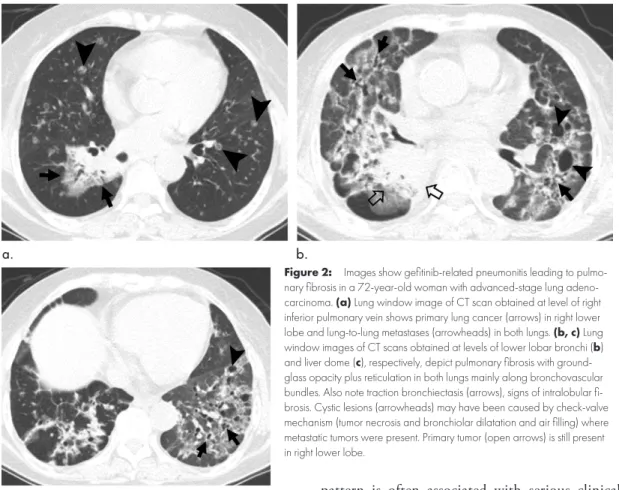
Figure 2: Images show gefitinib-related pneumonitis leading to
pulmo-nary fibrosis in a 72-year-old woman with advanced-stage lung adeno-carcinoma. (a) Lung window image of CT scan obtained at level of right
inferior pulmonary vein shows primary lung cancer (arrows) in right lower lobe and lung-to-lung metastases (arrowheads) in both lungs. (b, c) Lung
window images of CT scans obtained at levels of lower lobar bronchi (b)
and liver dome (c), respectively, depict pulmonary fibrosis with
ground-glass opacity plus reticulation in both lungs mainly along bronchovascular bundles. Also note traction bronchiectasis (arrows), signs of intralobular fi-brosis. Cystic lesions (arrowheads) may have been caused by check-valve mechanism (tumor necrosis and bronchiolar dilatation and air filling) where metastatic tumors were present. Primary tumor (open arrows) is still present in right lower lobe.
pattern is often associated with serious clinical outcome from pneumonitis, thus requiring awareness of this pattern among radiologists (Table 2) (32).
Radiologic simple pulmonary eosinophilia pattern.—Sim-ple pulmonary eosinophilia pattern demonstrates nonseg-mental consolidation or ground-glass opacity that can be unilateral or bilateral. The lung abnormalities are usually transient and migratory, and the prognosis is excellent; spontaneous resolution within 4 weeks is common (Figs 1, 7) (70,71). The pulmonary eosinophilia pattern is seen in osimertinib therapy (72).
CT Characteristics Associated with Specific Classes of Cancer Therapy
With the recent rapid advances of cancer therapy, pneumo-nitis related to novel agents have been increasingly described, along with their CT patterns. Many studies, as described in the following sections, applied the concept of CT pattern–based approach similar to the one described above, indicating a wide-spread use and applicability of this approach. It should also be noted that the concept and approach to DRP continue to evolve, as more novel agents are translated into the clinical set-tings and provide newer sets of challenges for the diagnosis, monitoring, and treatment.
Molecular Target Agents
EGFR-TKI Therapy.—In 2003, four cases of severe DAD pat-tern in patients treated with gefitinib were reported; among
(74) reporting clinically significant effects of pneumonitis re-lated to EGFR-TKIs. Serum proteomic markers and genetic polymorphisms have been studied as candidates to explain the higher incidence of pneumonitis in Japanese patients com-pared with others; however, no conclusive results have been obtained (73,75–77). Genetic and environmental factors that contribute to the development of EGFR-TKI pneumonitis re-main to be understood.
NSIP (Fig 2), OP (Fig 1), DAD (Fig 6), and HP (Fig 5) patterns have been reported with EGRF-TKIs. Poor progno-sis is expected when there is a short interval between the ini-tiation of the targeting therapy and the onset of pneumonitis, when the CT findings are represented by a DAD pattern, and when there is preexisting ILD (58). Severe and potentially fatal four patients, two recovered with steroids and two died due to
the DRP (9). In a recent report of a meta-analysis (73) of 153 trials worldwide including 15 713 patients with NSCLC and EGFR-TKI therapy, the overall incidence of DRP was 1.12% for all grades, 0.61% for high-grade pneumonitis, and 0.20% for grade 5 pneumonitis. When the incidence of pneumonitis was compared among the multiple factors including EGFR-TKI agents, treatment lines, EGFR mutation status, trial phases, and countries in the meta-analyses, significantly higher incidence rates were noted among Japanese studies compared with non-Japanese studies for all grades (4.77% vs 0.55%; P , .001), high grade (2.49% vs 0.37%; P , .001), and grade 5 pneumonitis (1.00% vs 0.18%; P , .001) (Table 2). These findings provide further support to a previous Japanese study
Table 2: Incidence and Patterns of Drug-related Pneumonitis Caused by Molecular Targeting Agents and Immune Checkpoint Inhibitors
Drug Incidence: All-Grade Pneumonitis (%) Incidence: High-Grade (Grade 3–4) Pneumonitis (%) Radiologic Patterns
EGFR inhibitors*† OP, DAD (AIP/ARDS),
HP, NSIP, PEo
Erlotinib Overall: 1.12 (0.79, 1.58) Overall: 0.61 (0.40, 0.93)
Gefitinib Japan: 4.77 (3.84, 5.91) Japan: 2.49 (1.77, 3.50)
Afatinib† Non-Japan: 0.55 (0.32, 0.92) Non-Japan: 0.37 (0.21, 0.64)
Osimertinib‡ 3.01 (1.85, 4.85) 0.56 (0.18, 1.73)
ALK inhibitors* OP, DAD (AIP/ARDS)§
Alectinib Overall: 2.14 (1.37, 3.34) Overall: 1.33 (0.80, 2.21)
Brigatinib Japan: 6.25 (3.97, 9.70) Japan: 3.31 (1.66, 6.47)
Ceritinib Non-Japan: 1.14 (0.33, 3.92) Non-Japan: 0.39 (0.03, 5.19)
Crizotinib … …
PD-1 inhibitors║
Nivolumab Monotherapy: 2.7 (1.9, 3.6) Monotherapy: 0.8 (0.4, 1.2)
Pembrrolizumab Combination therapy: 6.6 (4.7, 8.7)# Combination therapy: 1.7 (0.8, 2.9)#
PD-L1 inhibitors** OP, DAD (AIP/ARDS),
HP, NSIP
Atezolizumab 1.3 (0.8, 1.9) 0.4 (0, 0.8)††
Durvalumab … …
Avelumab … …
Note.—Modified from reference 24. Data in parentheses are 95% CIs. Drug-related pneumonitis from mechanistic target of rapamy-cin inhibitors, CD20 antibodies, and ipilimumab were not tabulated owing to lack of robust meta-analysis data. AIP = acute interstitial pneumonia, ALK = anaplastic lymphoma kinase, ARDS = acute respiratory distress syndrome, DAD = diffuse alveolar damage, EGFR = epidermal growth factor receptor mutation, HP = hypersensitivity pneumonitis, NSCLC = non–small cell lung cancer, NSIP = nonspecific interstitial pneumonia, OP = organizing pneumonia, PD-1 = programmed cell protein death 1, PD-L1 = programmed death ligand 1, PeO = pulmonary eosinophilia. Source.—References 54, 58, 73, 84, 85, 89, 90, 120.
* Incidence rates are meta-analyses of trials of NSCLC treated with single-agent therapy.
† Incidence is among patients treated with EGFR inhibitors without prior exposure to EGFR-directed therapy.
‡ Data include patients who received osimertinib after previous treatment with conventional EGFR inhibitors. Overall incidence of
pneu-monitis was 4% in a recent phase 3 first-line treatment of osimertinib for EGFR-mutant NSCLC.
§ In addition to these common patterns, “pulmonary edema–like shadows” characterized by bilateral ground-glass appearance, thickening
of the interlobular septa and the bronchovascular bundles distributed predominantly in the side of the pulmonary hilum, and occasional bilateral pleural effusion have been described in ALK-related pneumonitis.
║ Incidence rates are based on the meta-analyses of PD-1 inhibitor trials for melanoma, NSCLC, and renal cell carcinomas.
# Incidence rates are based on the meta-analyses of combination therapy regimens of PD-1 inhibitor, combined with ipilimumab or peptide
vaccines, for patients with melanoma.
** Incidence rates are based on the meta-analyses of single-agent PD-L1 inhibitor trials for NSCLC.
carcinoma (56). In a retrospective study of 22 patients, eight (36%) developed DRP with areas of ground-glass opacity and consolidation (80). In 178 patients with advanced renal cell car-cinoma, 52 patients (29%) developed DRP (81). In 46 patients with metastatic renal cell carcinoma (21 with temsirolimus and 25 with everolimus), CT evidence of pneumonitis was seen in 14 patients (30%). Stable disease by using Response Evaluation Criteria in Solid Tumors criteria was achieved in 12 (86%) of 14 patients who developed radiologic pneumonitis compared with 14 (44%) of 32 without pneumonitis (P , .01) (53).
In 66 patients with advanced neuroendocrine tumors who were treated with everolimus, DRP was reported in 14 (21%) patients (OP pattern in eight, NSIP pattern in five, and HP pattern in one) (Fig 4) (65). In 40 patients with Waldenstrom macroglobulinemia being treated with everolimus, 23 (58%) pa-tients developed DRP, with a radiologic OP pattern in 16 and NSIP pattern in seven (82).
According to the management guideline by Albiges et al (83), asymptomatic patients with mTOR pneumonitis and radiologic changes only (grade 1) may continue mTOR inhibitor therapy without dose adjustment at the treating physician’s discretion. However, patients should be informed of any signs of worsening to look out for, which would require contacting their physician. pneumonitis in patients treated with ICIs plus EGFR-TKIs has
been recently reported (56,78).
Emerging observations indicate that a milder form of lung reaction to EGFR-TKIs may manifest at imaging only without clinical symptoms, especially in the setting of the newer EGFR-TKI treatments. A novel type of drug-related pulmonary phe-nomenon called transient asymptomatic pulmonary opacities has been described in up to 20% of patients with NSCLC treated with the third-generation EGFR-TKI osimertinib (Fig 8) (72,79). Transient asymptomatic pulmonary opacities are de-scribed as localized pulmonary opacities mostly with radiologic simple pulmonary eosinophilia pattern, which resolves with-out any treatment during continued osimertinib therapy with a median 6-week duration (Figs 1, 8). Interestingly, patients who developed this apparent grade 1 pneumonitis had longer progression-free survival and overall survival compared with patients without transient asymptomatic pulmonary opacities, indicating the potential association between drug-related phe-nomena and treatment benefits.
mTOR inhibitors.—DRP is frequently seen in patients with mTOR inhibitors, including temsirolimus and everolimus. Temsirolimus has been approved for treatment of renal cell
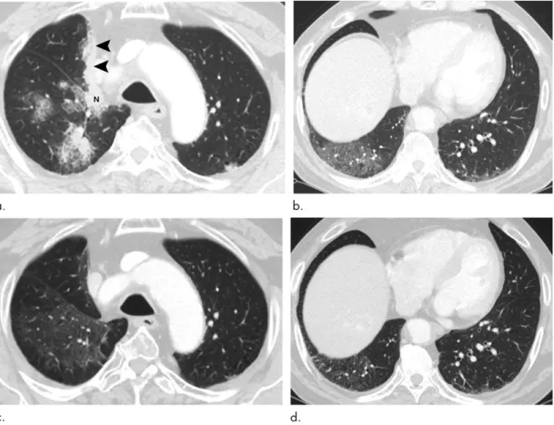
Figure 3: Images show pembrolizumab-related pneumonitis with organizing pneumonia pattern in a 68-year-old man with lung
ad-enocarcinoma. (a, b) Lung window images of CT scans obtained at levels of aortic arch (a) and liver dome (b), respectively, show patchy
parenchymal consolidation, ground-glass opacity, and nodules in both lungs. Also note right hilar nodal enlargement (N) and tumor pleural seeding (arrowheads). Patient received right upper lobectomy and following adjuvant concurrent chemotherapy-radiation therapy 3 years before current CT examination. Pembrolizumab was given 14 days prior to this CT examination. (c, d) Follow-up CT scans obtained 1.5
months after and at similar levels to a and b, respectively, demonstrate much decreased extent and attenuation of lung lesions. Patient
CD20 antibody.—Rituximab, a B-cell–depleting monoclonal antibody, has been reported to cause pulmonary toxicity. In a systematic review of 21 clinical trials and 40 case reports and/ or series, 121 patients were reported to have DRP. The most common indication for the drug therapy was diffuse large B-cell lymphoma. The DRP occurred more frequently in male patients and most commonly in the 5th and 6th decades of life. Rituximab-related pneumonitis was fatal in 18 (15%) of 121 cases and showed DAD pattern at CT (86).
ICI Therapy
The U.S. Food and Drug Administration has approved agents including ipilimumab (cytotoxic T-lymphocyte–associated protein 4 inhibitor), nivolumab, and pembrolizumab (pro-grammed cell death protein 1 [PD-1] inhibitors), as well as atezolizumab and durvalumab (programmed death ligand 1 [PD-L1] inhibitors) to treat different types of advanced cancer (87). In this setting, ICI therapy is associated with a variety of immune-related adverse effects that can affect any organ (6,56). The initial reports have described a spectrum of radio-logic patterns of interstitial pneumonias and clinical courses (Table 2) (12,35,36,88).
Anaplastic lymphoma kinase inhibitors.—Severe acute pneu-monitis in patients receiving crizotinib therapy for advanced NSCLC has been reported (69). In the recent meta-analysis (84) of 18 trials with 2261 patients with anaplastic lymphoma kinase inhibitor monotherapy and advanced NSCLC, the over-all incidence of pneumonitis was 2.14% for over-all grades, 1.33% for high-grade pneumonitis (grade 3 or above), and 0.22% for grade 5 pneumonitis. Similar to the EGFR-TKI study, Japanese cohorts showed a higher incidence of anaplastic lymphoma ki-nase–inhibitor pneumonitis for all grades (6.25% vs 1.14%; P , .001) and grade 3 and above pneumonitis (3.31% vs 0.39%; P , .001), compared with non-Japanese cohorts from multi-ple countries other than Japan (Table 2) (84). In postmarket-ing surveillance of crizotinib therapy in Japan, the incidence of pneumonitis associated with crizotinib therapy was 5.8% for all grades, and 3.5% for grade 3 or greater pneumonitis. In 27% of patients with pneumonitis, CT findings were suggestive of the presence of DAD. Age 55 years or older, Eastern Cooperative Oncology Group performance status between 2 and 4, smoking history, previous or concomitant ILD, and comorbid pleural ef-fusion were noted as significant risk factors for crizotinib-related pneumonitis (85).
Figure 4: Images show everolimus (mechanistic target of
ra-pamycin)–related pneumonitis with organizing pneumonia pattern in a 68-year-old woman with breast cancer lung metastasis. (a) Lung
window of CT scan obtained at level of liver dome shows metastatic lung nodule from breast cancer. (b) Lung window image obtained
at level of 15 mm inferior to a and 2.5 months after everolimus use
demonstrates parenchymal opacity composed of ground-glass opacity and consolidation (arrows) in bilateral lower lung zones. (c) Coronal
monotherapy. In another meta-analysis (90) of 19 clinical trials of PD-1 inhibitors and PD-L1 inhibitors as single-agent therapy in NSCLC, the incidence was higher in patients treated with PD-1 inhibitors compared with those treated with PD-L1 inhibitors (3.6% vs 1.3%, respectively; P = .001), providing valuable insight for optimal clinical selection of these agents given the overlapping approved indications of PD-1 and PD-L1 inhibitors. A subanalysis of patients with NSCLC treated with pembrolizumab in the phase I KEYNOTE-001 trial demonstrated that the overall incidence of In a meta-analysis (89) including 4496 patients from 20
sin-gle-tumor-type trials of PD-1 inhibitor including 12 melanoma studies, five NSCLC studies, and three renal cell carcinoma stud-ies, the overall incidence of pneumonitis during PD-1 inhibitor monotherapy was 2.7% (95% CI: 1.9, 3.6) for all grades and 0.8% (95% CI: 0.4, 1.2) for grade 3 or higher pneumonitis. The inci-dence of PD-1–related pneumonitis was higher in patients with NSCLC or renal cell carcinoma compared with that in patients with melanoma, and during combination therapy compared with
Figure 5: Images show docetaxel-related pneumonitis with hypersensitivity pneumonitis pattern in a 62-year-old woman with breast cancer. (a, b) Lung window of CT
scans obtained at levels of great vessels (a) and cardiac ventricle (b), respectively, show patchy and wide areas of ground-glass opacity and some small nodular lesions
(arrowheads in b) in both lungs. Also note area of lobular hypoattenuation (open arrow in b) in left lower lobe. Patient had been undergoing docetaxel chemotherapy after
right mastectomy and sentinel lymph node dissection. (c, d) Coronal images also demonstrate areas of ground-glass opacity, small nodules (arrowheads), and lobular
areas (open arrow) of mosaic perfusion in both lungs. (e) Transverse and (f) coronal-reformatted CT images obtained at similar levels to and 6 months after a and c,
re-spectively, and with discontinuation of docetaxel therapy, show disappeared lung lesions.
Figure 6: Images show erlotinib-related pneumonitis with diffuse alveolar damage pattern in a 40-year-old man with an advanced-stage lung
adenocarcinoma. (a, b) Lung window images of CT scans obtained at levels of aortic arch (a) and cardiac ventricles (b), respectively, and after
erlotinib therapy, depict diffuse ground-glass opacity in entire right lung, features compatible with diffuse alveolar damage. Also note masses (arrows) in left lung, lung-to-lung metastatic nodules (arrowheads) in both lungs, and a large amount of pericardial effusion (open arrows).
of lung injury or in whom the differential diagnosis raises the consideration of markedly different therapeutic strategies (eg, drug toxicity vs infection or malignancy).
What Histologic Characteristics Should Be Documented in Lung Biopsies Performed for DRP?
Lung biopsies should be evaluated for patterns of interstitial pneu-monia by using criteria within the American Thoracic Society/ European Respiratory Society International Multidisciplinary Classification of the Idiopathic Interstitial Pneumonias (94,95) including cellular and fibrotic NSIP, usual interstitial pneumo-nia, OP (including acute fibrinous subtype), lymphoid interstitial pneumonia and DAD, as well as bronchocentric inflammatory changes (including hypersensitivity pneumonia) and noncaseat-ing granulomas (96). Diffuse malignant infiltration, mimicknoncaseat-ing or coexisting with ILD, should be ruled out. In addition, depend-ing on the morphologic features and the clinical settdepend-ing, infectious agents such as bacteria, fungi, mycobacteria, or viral agents should be searched for by using special stains where indicated.
What Histologic Features Are Most Suggestive of DRP?
Although any of the above histologic patterns can be seen in DRP, there is a more frequent overlap of patterns, coexistent tissue eosinophilia, chronic interstitial inflammation, lym-phoid aggregates, and pleuritis compared with idiopathic cases showing similar histologic patterns. However, these same fea-tures are not specific because they are also seen in connective tissue disease–related lung disease.
There are limited published data on the pathologic features of pulmonary toxicity in ICIs and targeting molecular therapies; hence, most of the cases are diagnosed based on clinical and CT features only. Nonetheless, the main pathologic features de-scribed include cellular and/or fibrosing interstitial pneumonia, OP, HP, DAD, and pulmonary eosinophilia (35,54).
What Information Is Available from Bronchoalveolar Lavage Fluid Analysis?
Infectious organisms can be identified with bronchoalveolar lavage fluid cultures (27). In 12 (46%) of 26 patients with pneumonitis was 3.8%. A higher incidence was noted in patients
with a history of asthma or chronic obstructive pulmonary disease (5.3%) and in those with a history of thoracic radiation (6.0%) (6,91). A retrospective study enrolling 1826 patients with can-cer reported 64 (3.5%) cases of ICI-related pneumonitis, which more commonly occurred in men and former or current smok-ers, with a median age of 59 years. In this series, 66% of patients with pneumonitis had grade 2 or 3, 9% had grade 4, and 9% had grade 5 (fatal) pneumonitis. An earlier onset was noted in lung cancer versus melanoma (median of 2.1 months vs 5.2 months; P = .02). OP (23%) was the most common pattern followed by HP pattern (16%) (87,92). Moreover, in a recent meta-analysis of fatal toxicities related to ICIs, DRP was identified as the most common toxicity leading to PD-1/PD-L1–related mortality, ac-counting for 35% of all deaths (88,93). In a study of 20 patients with DRP among 170 patients with melanoma, lung cancer, and lymphoma treated with PD-1 inhibitors, OP pattern (Fig 3) was found in 13 patients and was the most common pattern, followed by NSIP pattern in three, HP pattern in two, and DAD pattern (acute respiratory distress syndrome/acute respiratory distress syn-drome pattern in the study referring to the American Thoracic Society/European Respiratory Society classification of idiopathic interstitial pneumonias) in two patients (54). The CT patterns were associated with the toxicity grades of pneumonitis as defined by Common Terminology Criteria for Adverse Events; DAD pattern had the highest grades, followed by OP pattern, whereas NSIP and HP patterns had lower grades, indicating the utility of CT pattern–based approach in assessing the severity of ICI-related pneumonitis (54). Seventeen patients received corticosteroid ther-apy, and three also received infliximab treatment. Seven patients were retreated, of whom two developed DRPs again. One patient demonstrated pneumonitis flare-up on tapering of corticosteroid intake without retreatment with ICI or any other agents, further indicating the complex nature of the entity and importance of im-aging follow-up of these patients (54).
Pathologic Analysis
Lung biopsy may be indicated in patients in whom the clinical and radiologic picture do not clearly point to a specific pattern
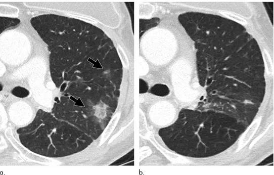
Figure 7: Images show osimertinib-related pneumonitis with simple pulmonary eosinophilia pattern in a 60-year-old man with lung
adenocarci-noma. (a) Lung window image obtained at level of distal main bronchi shows multifocal opacity (arrows) in right upper lobe and superior segment
of left lower lobe during osimertinib therapy. (b) CT scan obtained at similar level to and 2 months after a demonstrates that opacity lesions in both
shown to be effective in other disciplines (99). Multidisci-plinary diagnosis is particularly important in patients sus-pected of having DRP because there is no individual feature that is required or sufficient for the diagnosis of DRP. The multidisciplinary diagnosis ap-proach typically involves clini-cians, radiologists, and pathol-ogists (if biopsy is performed) and can be used in clinical set-tings and trials with centralized review of adverse events. Be-cause DRP is often observed in more acute or subacute clinical settings, the actual consulta-tions among subspecialties may happen as informal and formal communication by using tele-phone or virtual conference. In more chronic or difficult cases, it may be discussed formally at a multidisciplinary diagnosis conference. It is important that multidisciplinary discussion shall happen in the clinical context of the need for clinical management of the patients suspected of having DRP.
Description of Central Review for the Multidisciplinary Diagnosis of DRP
To determine the accurate incidence of DRP in clinical trials and postmarketing surveillance, the cases diagnosed by each physician in primary investigation site should be evaluated by using a process of central review (100,101) to achieve uniform criteria through accurate and consistent data. The strategy at the time of the review should be based on a multidisciplinary diagnosis approach, involving a multidisciplinary team consist-ing of at least one chest physician, one oncologist, one chest radiologist, and (if a biopsy is available) one pathologist. More-over, it is essential to use a mutually agreed diagnostic checklist (refer to Appendix E2 [online] for record of multidisciplinary discussion and Appendix E3 [online] for objective evaluation of chest CT and ILD) consistently throughout an individual study or cohort. At first, each radiologist and chest physician should independently evaluate the case, followed by the subse-quent multidisciplinary discussion among the experts to reach a consensus.
Management
Pharmacovigilance, or drug safety monitoring, plays an important role in identifying, understanding, and prevent-ing adverse drug reactions. The World Health Organization Program for International Drug Monitoring (VigiAccess) provides an international forum for collaboration in phar-macovigilance, collecting data from real-world settings. All drug-related adverse effects should be declared to the phar-everolimus treatment and initial diagnosis of DRP,
bronchoal-veolar lavage fluid cultures enabled a diagnosis of Pneumocystis jirovecii pneumonia (97). Cell count of bronchoalveolar lavage fluid in DRP may disclose raised lymphocyte, neutrophil, and eosinophil numbers. Particularly in pulmonary eosinophilia pattern, cell count helps to make a diagnosis of the disease by documenting eosinophilia. However, the cell count is not spe-cific, because similar results could be seen in other inflamma-tory and infectious conditions (27,28,42,43).
Proposal for Diagnostic Criteria
Camus et al (98) proposed the following diagnostic criteria for DRP: (a) exposure to the causative drug, (b) development of pulmonary infiltrates, (c) meticulous exclusion of all other pos-sible causes, (d) dechallenge producing measurable improve-ment in symptoms and imaging, and (e) rechallenge causing worsening. However, some patients do not have improvement with dechallenge, and rechallenge is often impossible in many clinical settings, thus making these criteria impractical in many patients.
Therefore, we propose the following criteria: (a) newly iden-tified pulmonary parenchymal opacities at CT or chest radiog-raphy, commonly in a bilateral nonsegmental distribution; (b) temporal association of presentation with the initiation of a sys-temic therapeutic agent; and (c) exclusion of other likely causes (Table 3) (see also Figs E1–E5 [online]).
Proposed Method for Central Review
Introduction of Multidisciplinary Diagnosis
The process of multidisciplinary diagnosis is by means of inter-active multidisciplinary discussion, an approach that has been
Figure 8: Images show osimertinib-related transient asymptomatic pulmonary opacities in a 65-year-old woman with
lung adenocarcinoma. (a) Lung window image of CT scan obtained at level of left main bronchus shows focal opacity
areas (arrows) in left upper lobe during osimertinib therapy. (b) CT scan obtained at similar level to and 2 months after a demonstrates that opacities seen at CT scan (a) have disappeared nearly completely without any therapy for opacity
Table 3: Clinical, Pathologic, and Radiologic Features of DRP Compared with Pneumonia, Diffuse Alveolar Hemorrhage, Pulmo-nary Edema, Radiation Pneumonitis, and PulmoPulmo-nary Metastases
Variable Clinical Features Relevant Factors Pathologic Features Radiologic Features
DRP Asymptomatic to
acutely progressive dyspnea, and cough with or with-out fever
Temporal relationship between drug exposure and onset of disease; improvement with drug cessation
OP, DAD, cellular and fibrotic NSIP, granulomatous interstitial pneumonia, PEo, and lymphoid interstitial pneumonia
Various interstitial pneumonia patterns including OP, DAD, NSIP, HP, and PEo Pneumonia (see Fig E1 [online]) Fever, chill, productive cough, myalgia, headache
Varying disease patterns depending on patients’ immune status;
immunocompetent versus immunocompromised status; positive microbiology culture or polymerase chain reaction test; improvement with antibiotic treatment
Filling of alveolar spaces by exudate of edema fluid and neutrophil (lobar); patchy peribronchiolar inflammation with less abundant edema formation (bronchopneumonia); and mononuclear inflammatory cell infiltrate in alveolar septa and interstitial tissue surrounding small parenchymal vessels (interstitial pneumonia) Lobar pneumonia, bronchopneumonia, and interstitial pneumonia patterns; atypical
pneumonia (septic emboli, abscess, and chronic pneumonia such as actinomycosis or chronic necrotizing pulmonary aspergillosis) Diffuse alveolar hemorrhage (see Fig E2 [online]) Hemoptysis (two-thirds of patients), anemia and diffuse opacity at imaging
Injury to alveolar-capillary microcirculation (eg, microscopic polyangiitis), circulating autoantibody (eg, ANCA), coagulation disorders
Intraalveolar hemorrhage, hemosiderin-laden macrophages in alveolar spaces and interstitium, and occasional focal or diffuse areas of capillaritis
Bilateral patchy opacities in middle and lower lung zones on chest radiographs; diffuse or geographic ground-glass opacities/ consolidation at CT Pulmonary edema (see Fig E3 [online]) Dyspnea, cough, frothy sputum (sometimes) Hydrostatic (cardiac or renal failure) and permeability edema (DAD)
Expansion of connective tissue space around conducting airways, accompanying vessels, and interlobular septa (hydrostatic edema); alveolar space and interstitial edema; hyaline membrane formation and proliferation of type II cells
Hazy opacities, Kerley lines, batwing appearance in hydrostatic edema; patchy and widespread areas of parenchymal opacities in permeability edema and their evolutional change; pleural effusion (more frequently in hydrostatic edema) Radiation pneumonitis (see Fig E4 [online])
Dyspnea, dry cough, chest pain with or without fever (low grade) Temporal relationship to radiation exposure (3–12 weeks after irradiation)
Airspace and interstitial edema, proceeding to poorly defined consolidation, DAD and type II cell hyperplasia; evolutional changes to radiation fibrosis; HP or OP pattern away from radiation portal can occur
Opacities within radiation portal or roughly within area of high-dose radiation; ground-glass opacity and OP pattern away from radiation portal Pulmonary lymphangitic carcinomatosis (see Fig E5 [online]) Progressively worsening dyspnea, cough
Most commonly with gastric, breast, lung, and pancreas cancers
Thickening of bronchovascular bundles and septae, related to proliferation of neoplastic cells, interstitial inflammation and fibrosis (desmoplastic reaction) and lymphatic dilatation by edema or tumor section (mucin)
Linear or reticulonodular lesions on chest radiographs; ground-glass opacities; septal thickening (smooth or nodular), bilateral asymmetric or unilateral; pleural effusion at CT Note.—ANCA = antinuclear cytoplasmic antibody, DAD = diffuse alveolar damage, DRP = drug-related pneumonitis, HP = hypersensitiv-ity pneumonitis, NSIP = nonspecific interstitial pneumonia, OP = organizing pneumonia, PEo = pulmonary eosinophilia. Source.—Refer-ence 121.
without ILD. Furthermore, greater CT extent of preexisting ILD portended higher risk of fatal outcome.
Does Glucocorticoid Therapy Improve Clinical Outcome in Patients with DRP?
Glucocorticoid therapy is often used in patients with DRP to ameliorate and to expedite the recovery of lung injury (27,45,48). This strategy is commonly used when DRP is moderate to severe and of acute or fulminant onset. However, this practice is based on retrospective studies and expert opinion because no clinical trial has been performed to prove the efficacy of glucocorticoid therapy in the treatment of patients with DRP (113).
Conclusion
This position paper of the Fleischner Society summarizes simpli-fied diagnostic criteria, CT pattern approach, and management recommendation of drug-related pneumonitis (DRP) in the emerging era of molecular targeting agents and cancer immu-notherapy, by using a multidisciplinary approach. The diagnosis and management of DRP will continue to evolve with the ad-vancement of treatments, and a radiologic pattern approach with multidisciplinary diagnosis will remain crucially important for the optimal treatment of the patients.
Acknowledgments: We are grateful for the librarians Myung-Ah Shim and Jaero Park for their dedicated support of manuscript formatting. Both librarians are work-ing at the Samsung Medical Information & Media Services of Samsung Medical Center located in Seoul, South Korea.
Author contributions: Guarantors of integrity of entire study, T.J., K.S.L., T.F., Y.I., H.H.; study concepts/study design or data acquisition or data analysis/inter-pretation, all authors; manuscript drafting or manuscript revision for important intellectual content, all authors; approval of final version of submitted manuscript, all authors; agrees to ensure any questions related to the work are appropriately resolved, all authors; literature research, T.J., K.S.L., M.N., J.H.R., H.Y.L., T.F., K.K.B., J.M.G., H.U.K., L.R., C.M.S.P., C.P., H.H.; clinical studies, T.J., K.S.L., M.N., J.H.R., H.Y.L., J.V., Y.I., H.H.; and manuscript editing, T.J., K.S.L., M.N., W.D.T., J.H.R., H.Y.L., C.J.R., A.A.B., K.K.B., J.M.G., H.U.K., D.A.L., A.G.N., L.R., C.M.S.P., J.V., S.R., G.D.R., C.P., Y.I., H.H.
Disclosures of Conflicts of Interest: T.J. disclosed no relevant relationships.
K.S.L. Activities related to the present article: disclosed no relevant relationships. Activities not related to the present article: receives royalties from Elsevier, Lippin-cott, and Springer. Other relationships: disclosed no relevant relationships. M.N. Activities related to the present article: disclosed no relevant relationships. Activities not related to the present article: is a consultant for Daiichi Sankyo and Astra-Zeneca; has grants/grants pending with Merck, Canon Medical Systems, Daiichi Sankyo, and AstraZeneca; received payment for lectures including service on speak-ers bureaus from Roche. Other relationships: disclosed no relevant relationships.
W.D.T. disclosed no relevant relationships. J.H.R. disclosed no relevant relation-ships. H.Y.L. disclosed no relevant relationrelation-ships. C.J.R. disclosed no relevant re-lationships. T.F. disclosed no relevant rere-lationships. A.A.B. Activities related to the present article: disclosed no relevant relationships. Activities not related to the present article: is a consultant for Daiichi Pharmaceutical and Olympus Medical; received payment for lectures including service on speakers bureaus from Olympus Medical; receives royalties from Elsevier. Other relationships: disclosed no relevant relationships. K.K.B. Activities related to the present article: disclosed no relevant relationships. Activities not related to the present article: is DMC chair of Bioge and Humanetics; is member of scientific advisory board for Galecto, Third Pole, Galapa-gos, Boehringer Ingelheim, Theravance, Lifemax, Pliant, Blade Therapeutics, Open Source Imaging Consortium, Huitai Biomedicine, Lilly, Dispersol, and DevPro Bi-opharma; has grants/grants pending with NHLBI. Other relationships: disclosed no relevant relationships. J.M.G. Activities related to the present article: disclosed no relevant relationships. Activities not related to the present article: has grants/grants pending with Infinitt Healthcare and Dongkook Lifescience. Other relationships: disclosed no relevant relationships. H.U.K. Activities related to the present article: disclosed no relevant relationships. Activities not related to the present article: has
macovigilance program, which is the cornerstone of the alert system. With clinicians’ reports on adverse drug reactions, the incorporation of clinical and pharmacologic information could help to avoid the unnecessary exposure to adverse drug reactions (102,103).
In general, delayed diagnosis of DRP is associated with higher severity of lung injury and less reversibility, resulting in residual lung damage (ie, fibrosis) (27,48). Thus, early diagno-sis and cessation (except some drugs; refer to previous sections regarding specific agents) of the offending drug intake promote optimal outcomes in patients with DRP. In addition, glucocor-ticoids are commonly administered to facilitate the resolution of lung injury, particularly for those severely affected (National Cancer Institute grade 3–4 pneumonitis) as assessed by symp-toms, gas exchange derangements, and radiologic abnormalities (27,35,45,48). These patients usually require hospitalization for their initial treatment and monitoring (35,45). Supportive mea-sures, including supplemental oxygen and noninvasive or inva-sive mechanical ventilator support, may be needed.
For the management of ICI-related pneumonitis, the Na-tional Comprehensive Cancer Network, American Society of Clinical Oncology, Society for Immunotherapy of Cancer, and European Society for Medical Oncology guidelines (45– 47,104,105) recommend discontinuing ICI therapy for any grade of pneumonitis and recommend treating grade 2 pneumo-nitis with corticosteroids. Permanent discontinuation of ICIs is suggested for grade 3–4 pneumonitis by all guidelines. For those without improvement on corticosteroids after 48 hours, inflix-imab, mycophenolate mofetil, or intravenous immunoglobulin may be used (45–47,104,105).
Is Preexisting ILD a Risk Factor for DRP and Does Preexisting ILD Lead to Worse Outcomes in Patients with DRP?
Preexisting ILD is a risk factor for the development of DRP (27,28,48). Multiple studies on the incidence of lung toxicity associated with antineoplastic agents have demonstrated that preexisting ILD is associated with a higher likelihood of DRP, sometimes described as an acute exacerbation of preexisting ILD (28,74,80,106–112). For example, the odds ratio for developing DRP in patients with lung cancer treated with chemotherapy or gefitinib ranges from 4.8 to 25.3 (depending on the severity of ILD) in patients with preexisting ILD compared with those without preexisting ILD (74). A recent systematic review (113) on DRP found that preexisting ILD is an independent risk factor for DRP with a wide spectrum of therapeutic agents. However, some uncertainty remains on whether preexisting ILD is a risk factor for the development of ICI-associated pneumonitis in particular, largely due to the exclusion of patients with preexisting ILDs from clinical trials of ICIs. Recent studies have suggested preexisting fi-brotic changes at CT are associated with an increased risk of anti-PD-1–related pneumonitis in patients with NSCLC (114,115).
Several studies demonstrated worse outcome related to DRP among patients with preexisting ILD compared with those without (74,109,110,112,116–119). For example, Ku-doh et al (110) reported the odds ratio for fatal outcomes from DRP related to chemotherapy or gefitinib therapy to be 2.27 for patients with preexisting ILD compared with those