International Expert Consensus Document on
Takotsubo Syndrome (Part I): Clinical
Characteristics, Diagnostic Criteria, and
Pathophysiology
Jelena-Rima Ghadri
1
, Ilan Shor Wittstein
2
, Abhiram Prasad
3
, Scott Sharkey
4
,
Keigo Dote
5
, Yoshihiro John Akashi
6
, Victoria Lucia Cammann
1
, Filippo Crea
7
,
Leonarda Galiuto
7
, Walter Desmet
8,9
, Tetsuro Yoshida
10
, Roberto Manfredini
11
,
Ingo Eitel
12
, Masami Kosuge
13
, Holger M. Nef
14
, Abhishek Deshmukh
3
,
Amir Lerman
3
, Eduardo Bossone
15
, Rodolfo Citro
15
, Takashi Ueyama
16†
Domenico Corrado
17
, Satoshi Kurisu
18
, Frank Ruschitzka
1
, David Winchester
19
,
Alexander R. Lyon
20,21
, Elmir Omerovic
22,23
, Jeroen J. Bax
24
, Patrick Meimoun
25
,
Guiseppe Tarantini
17
, Charanjit Rihal
3
, Shams Y.-Hassan
26
, Federico Migliore
17
,
John D. Horowitz
27
, Hiroaki Shimokawa
28
, Thomas Felix Lu
¨ scher
29,30
, and
Christian Templin
1
*
International Experts: Jeroen J. Bax, Eduardo Bossone, Victoria Lucia Cammann,
Rodolfo Citro, Domenico Corrado, Filippo Crea, Walter Desmet, Ingo Eitel,
Leonarda Galiuto, Jelena-Rima Ghadri, Thomas Felix Lu
¨ scher, Alexander R. Lyon,
Roberto Manfredini, Patrick Meimoun, Federico Migliore, Holger M. Nef,
Elmir Omerovic, Frank Ruschitzka, Guiseppe Tarantini, Christian Templin,
Shams Y-Hassan (European sites); Abhishek Deshmukh, Amir Lerman,
Abhiram Prasad, Charanjit Rihal, Scott Sharkey, David Winchester,
Ilan Shor Wittstein (USA sites); Yoshihiro John Akashi, Keigo Dote,
Masami Kosuge, Satoshi Kurisu, Hiroaki Shimokawa, Takashi Ueyama,
Tetsuro Yoshida (Asian sites); John D. Horowitz (Australian site)
1
University Heart Center, Department of Cardiology, University Hospital Zurich, Zurich, Switzerland;2
Department of Medicine, Johns Hopkins University School of Medicine,
Baltimore, MD, USA;3Division of Cardiovascular Diseases Mayo Clinic, Rochester, MN, USA;4Cardiovascular Research Division, Minneapolis Heart Institute Foundation,
Minneapolis, MN, USA;5
Department of Cardiology, Hiroshima City Asa Hospital, Hiroshima, Japan;6
Division of Cardiology, Department of Internal Medicine, St. Marianna
University School of Medicine, Kawasaki, Japan;7Department of Cardiovascular Sciences, Catholic University of the Sacred Heart, Rome, Italy;8Department of Cardiovascular
Medicine, University Hospitals Leuven, Leuven, Belgium;9
Department of Cardiovascular Sciences, University of Leuven, Leuven, Belgium;10
Department of Cardiovascular
The opinions expressed in this article are not necessarily those of the Editors of the European Heart Journal or of the European Society of Cardiology.
This paper was guest edited by Bernard J. Gersh (Mayo Clinic,[email protected]).
* Corresponding author. Tel:þ41 44 255 9585, Fax: þ41 44 255 4401, Email:[email protected]
†
Deceased.
VCThe Author(s) 2018. Published by Oxford University Press on behalf of the European Society of Cardiology.
This is an Open Access article distributed under the terms of the Creative Commons Attribution Non-Commercial License (http://creativecommons.org/licenses/by-nc/4.0/), which permits non-commercial re-use, distribution, and reproduction in any medium, provided the original work is properly cited. For commercial re-use, please contact [email protected]
doi:10.1093/eurheartj/ehy076
..
..
..
..
..
..
..
..
..
..
..
..
..
..
..
..
..
..
..
..
..
..
..
..
..
..
..
..
..
..
..
..
..
..
..
..
..
..
..
..
..
..
..
..
..
..
.
Medicine, Onga Nakama Medical Association Onga Hospital, Fukuoka, Japan;11Clinica Medica, Department of Medical Sciences, University of Ferrara, Ferrara, Italy;12University
Heart Center Luebeck, Medical Clinic II, Department of Cardiology, Angiology and Intensive Care Medicine, University of Luebeck, Luebeck, Germany;13
Division of Cardiology,
Yokohama City University Medical Center, Yokohama, Japan;14Department of Cardiology, University Hospital Giessen, Giessen, Germany;15Heart Department, University
Hospital “San Giovanni di Dio e Ruggi d’Aragona”, Salerno, Italy;16
Department of Anatomy and Cell Biology, Wakayama Medical University School of Medicine, Wakayama,
Japan;17Department of Cardiac, Thoracic, and Vascular Sciences, University of Padua Medical School, Padova, Italy;18Department of Cardiovascular Medicine, Hiroshima
University Graduate School of Biomedical and Health Sciences, Hiroshima, Japan;19
Division of Cardiovascular Disease, Department of Medicine, University of Florida, Gainesville,
FL, USA;20NIHR Cardiovascular Biomedical Research Unit, Royal Brompton Hospital, London, UK;21National Heart and Lung Institute, Imperial College, London, UK;
22
Department of Cardiology, Sahlgrenska University Hospital, Gothenburg, Sweden;23
Department of Molecular and Clinical Medicine, Institute of Medicine, Sahlgrenska
Academy, Gothenburg University, Gothenburg, Sweden;24Department of Cardiology, Leiden University Medical Center, Leiden, The Netherlands;25Department of Cardiology
and Intensive Care, Centre Hospitalier de Compiegne, Compiegne, France;26
Department of Cardiology, Karolinska University Hospital, Huddinge, Stockholm, Sweden;
27
Department of Cardiology, Basil Hetzel Institute, Queen Elizabeth Hospital, University of Adelaide, Adelaide, Australia;28Department of Cardiovascular Medicine, Tohoku
University Graduate School of Medicine, Sendai, Japan;29
Center for Molecular Cardiology, Schlieren Campus, University of Zurich, Zurich, Switzerland; and30
Department of Cardiology, Royal Brompton & Harefield Hospital and Imperial College, London, UK
Received 2 June 2017; revised 23 November 2017; editorial decision 30 January 2018; accepted 17 April 2018; online publish-ahead-of-print 29 May 2018
Takotsubo syndrome (TTS) is a poorly recognized heart disease that was initially regarded as a benign condition. Recently, it has been
shown that TTS may be associated with severe clinical complications including death and that its prevalence is probably underestimated.
Since current guidelines on TTS are lacking, it appears timely and important to provide an expert consensus statement on TTS. The
clini-cal expert consensus document part I summarizes the current state of knowledge on cliniclini-cal presentation and characteristics of TTS and
agrees on controversies surrounding TTS such as nomenclature, different TTS types, role of coronary artery disease, and etiology. This
consensus also proposes new diagnostic criteria based on current knowledge to improve diagnostic accuracy.
...
Keywords
Takotsubo syndrome
•
Broken heart syndrome
•
Takotsubo definition
•
Acute heart failure
•
Consensus
statement
•
InterTAK Diagnostic Criteria
Outline
History . . . 2033
Nomenclature . . . 2033
Epidemiology . . . 2034
Symptoms and signs . . . 2035
Diagnostic criteria. . . 2035
Pathophysiology . . . 2036
Sympathetic stimulation. . . 2036
Potential pathophysiological effects of enhanced
sympathetic stimulation . . . 2036
Plaque rupture . . . 2036
Multi-vessel epicardial spasm. . . 2036
Microcirculatory dysfunction . . . 2036
Catecholamine toxicity on cardiomyocytes. . . 2037
Activation of myocardial survival pathways . . . 2037
Predisposition and risk factors . . . 2038
Hormonal factors. . . 2038
Genetic factors . . . 2038
Psychiatric and neurologic disorders . . . 2038
Triggers. . . 2039
Emotional stressors. . . 2039
Physical stressors . . . 2039
Absence of identifiable causes . . . 2039
Types of takotsubo syndrome . . . 2039
Chronobiology. . . 2042
References. . . 2042
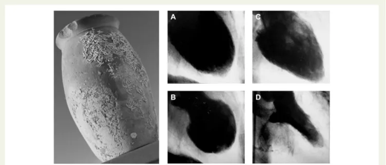
History
The term takotsubo syndrome (TTS) was first introduced when Sato
et al.
1published their report of five cases in a Japanese medical
textbook in 1990. The first TTS case of this series was managed in
1983 in the Hiroshima City Hospital (Figure
1
). A 64-year-old female
presented with acute chest pain consistent with acute myocardial
in-farction (AMI), typical electrocardiographic (ECG) changes, but normal
coronary arteries and an unusual appearance of the left ventricle (LV)
with a narrow neck and apical ballooning during systole. Interestingly,
the marked wall motion abnormalities on left ventriculography
dis-appeared after 2 weeks. Over time TTS was more frequently
diag-nosed in Japan. Therefore, it was first assumed that this disorder only
affected people of Asian descent, as TTS was completely unknown to
the Western world until the first cases were published from French
and American research groups in the late 1990s.
2,3Desmet et al.
4introduced the first patient case series in Caucasians using the term
‘takotsubo’.
Takotsubo syndrome gained international awareness among
re-searcher and physicians when Wittstein et al.
5reported their findings
in the New England Journal of Medicine in 2005. Since then TTS has
been more frequently recognized worldwide but still remains an
underappreciated and often misdiagnosed disorder.
6,7Nomenclature
Takotsubo syndrome derived its name from the Japanese word for
octopus trap, due to the shape of the LV at the end of systole and has
been described under a remarkable number of different names in the
literature including ‘broken heart syndrome’, ‘stress cardiomyopathy’,
and ‘apical ballooning syndrome’.
8No single term precisely describes
the heterogeneous ventricular appearance with which this syndrome
can occur. To date, consensus has not been reached on the
nomencla-ture. The term ‘takotsubo’ is widely used in acknowledgement of the
..
..
..
..
..
..
..
..
..
..
..
..
..
..
..
..
..
..
..
..
..
..
..
..
..
..
..
..
..
..
..
..
..
..
..
..
..
..
..
..
..
..
..
..
..
..
..
..
..
..
Japanese physicians who initially described this disorder.
1However, in
contrast to other cardiomyopathies that are usually not transient in
na-ture, TTS is characterized by a temporary wall motion abnormality of
the LV and shares common features with acute coronary syndrome
(ACS) [similar symptoms at presentation, ECG abnormalities, elevated
cardiac biomarkers as well as a comparable in-hospital mortality with
ST-segment elevation myocardial infarction (STEMI) and non-STEMI]
specifically in terms of a microvascular ACS form.
9Among different
eti-ologies of heart failure such as coronary artery disease (CAD),
tachyarrhtyhmias etc. TTS includes a wide spectrum of emotional or
physical triggers resulting also in left ventricular dysfunction. Therefore,
it is best described as a ‘syndrome’ and the term ‘takotsubo syndrome’
seems most appropriate.
9,10,11Epidemiology
Since the initial report by Japanese cardiologists 25 years ago, TTS has
been increasingly recognized in diverse countries across six continents.
Takotsubo syndrome is estimated to represent approximately 1–
3%
12,13of all and 5–6%
14of female patients presenting with suspected
STEMI. The Nationwide Inpatient Sample discharge records from
2008 using the International Classification of Diseases revealed that
TTS accounts for 0.02% of hospitalizations in the United States.
15Recurrence rate of TTS is estimated to be 1.8% per-patient year.
16Based on the published literature about 90%
16,17of TTS patients are
women with a mean age of 67–70 years,
16,18and around 80% are older
than 50 years (Figure
2
).
16Women older than 55 years have a five-fold
greater risk of developing TTS than women younger than 55 years and
a 10-fold greater risk than men.
15With growing awareness of TTS,
male patients are diagnosed more often, especially after a physical
trig-gering event.
19TTS has also been described in children
20,21with the
youngest reported TTS patient being a premature neonate born in the
28th gestational week.
22Current data on racial differences are
incon-sistent and large-scale studies are lacking. However, it has been
re-ported that TTS seems to be uncommon in African–Americans and
Hispanics,
23while most of the cases reported in the United States
have been Caucasians.
15,24Furthermore, it has been reported that
patients of African-American descent have more in-hospital
complica-tions such as respiratory failure, stroke and require more frequently
mechanical ventilation compared to Caucasians and Hispanics.
25With
regard to ECG differences, it has been shown that QT prolongation as
well as T-wave inversion are more often reported in
African-American women with TTS.
26Of note, regarding gender differences
the TTS prevalence in men appears to be higher in Japan.
19The
preva-lence of TTS appears to be higher in patients with non-emotional
trig-gers admitted to intensive care units.
27Moreover, it is likely that
Figure 1
Historical Japanese octopus trap (left). Courtesy of Dr Templin, University Hospital Zurich, Zurich, Switzerland. Left ventriculogram of
the first reported case of takotsubo syndrome. Diastole (A) and systole (B) during the acute phase of takotsubo syndrome. Recovery of left
ventricu-lar wall motion abnormality two weeks after the event (C and D). Courtesy of Dr Dote, Hiroshima City Asa Hospital, Hiroshima, Japan.
Figure 2
Age and sex distribution of patients with takotsubo
syn-drome. Reprinted with permission from Templin et al.
16..
..
..
..
..
..
..
..
..
..
..
..
..
..
..
..
..
..
..
..
..
..
..
..
..
..
..
..
..
..
..
..
..
..
..
..
..
..
..
..
..
..
..
..
..
..
..
..
..
..
..
..
subclinical TTS cases remain undetected, especially in
non-percutaneous coronary intervention centres.
28Symptoms and signs
The most common symptoms of TTS are acute chest pain, dyspnoea,
or syncope and thus indistinguishable from AMI at the first glance.
16However, in some patients, TTS may be diagnosed incidentally by
new ECG changes or a sudden elevation of cardiac biomarkers.
Clinical manifestation of TTS induced by severe physical stress may
be dominated by the manifestation of the underlying acute illness. In
this regard, patients with ischaemic stroke or seizure-triggered, TTS
had less frequent chest pain,
29,30which could be explained by
im-paired consciousness, neurologic complications, or a sudden
haemo-dynamic deterioration. In contrast, patients with emotional stress
factors had a higher prevalence of chest pain and palpitations.
31Importantly, a subset of TTS patients may present with symptoms
arising from its complications, e.g. heart failure, pulmonary oedema,
stroke, cardiogenic shock, or cardiac arrest.
Diagnostic criteria
The diagnosis of TTS is often challenging because its clinical
pheno-type may closely resemble AMI regarding ECG abnormalities and
bio-markers.
32While a widely established non-invasive tool allowing a
rapid and reliable diagnosis of TTS is currently lacking, coronary
angi-ography with left ventriculangi-ography is considered the gold standard
diagnostic tool to exclude or confirm TTS.
Abe et al.
33introduced the first diagnostic criteria for TTS in 2003.
One year later, a dedicated group of cardiologists from the Mayo
Clinic proposed their diagnostic criteria.
34In 2006, the American
College of Cardiology and American Heart Association classified
TTS as a primary acquired cardiomyopathy.
35In 2008, the revised
version of the Mayo Clinic Diagnostic Criteria was published
incor-porating neurogenic stunned myocardium.
32Furthermore, the
au-thors defined different TTS sub-types and highlighted that
obstructive coronary lesions may occasionally be present
concomi-tantly.
32The Mayo Clinic Diagnostic Criteria are the most widely
known, but exceptions to the rule [e.g. the presence of CAD] are
poorly appreciated among physicians and cardiologists. More
re-cently, other research groups have proposed slightly different criteria
for TTS, i.e. the Japanese Guidelines,
36the Gothenburg criteria,
37the
Johns Hopkins criteria,
38the Tako-tsubo Italian Network proposal,
39the criteria of the Heart Failure Association (HFA) TTS Taskforce of
the European Society of Cardiology (ESC),
10as well as the criteria
recommended by Madias.
40Thus, there is a lack of a worldwide
consensus.
41Based on current knowledge, we have developed
new international diagnostic criteria (InterTAK Diagnostic Criteria,
Table
1
) for the diagnosis of TTS that may help to improve
identifica-tion and stratificaidentifica-tion of TTS. The most important changes with
accompanying rationale include:
(i) Pheochromocytoma is a neuroendocrine tumour derived from
enterochromaffin cells of the adrenal gland that may lead to a
‘cat-echolamine storm’ with LV dysfunction, ECG abnormalities, and
increased biomarkers as well as hypercontraction of sarcomeres and
contraction band necrosis indistinguishable from TTS.
42Notwithstanding, most of the diagnostic criteria have excluded
pheochromocytoma as a specific cause of TTS.
32–34,36,37,40The
Japanese criteria emphasize that pheochromocytoma is a TTS-like
myocardial dysfunction.
36Pheochromocytoma is also included as a
secondary cause of TTS in the diagnostic criteria of the HFA of the
ESC.
10(ii) Concomitant CAD is reported with a prevalence ranging from
10–29%.
16,43,44In this regard, patients with TTS and obstructive
CAD are often misdiagnosed as classical ACS and differentiation can
be challenging.
45Therefore, the presence of CAD should not be
con-sidered as an exclusion criterion as acknowledged by the modified
Table 1
International Takotsubo Diagnostic Criteria (InterTAK Diagnostic Criteria)
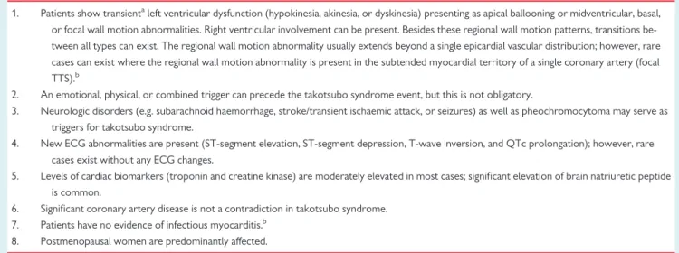
1. Patients show transientaleft ventricular dysfunction (hypokinesia, akinesia, or dyskinesia) presenting as apical ballooning or midventricular, basal, or focal wall motion abnormalities. Right ventricular involvement can be present. Besides these regional wall motion patterns, transitions be-tween all types can exist. The regional wall motion abnormality usually extends beyond a single epicardial vascular distribution; however, rare cases can exist where the regional wall motion abnormality is present in the subtended myocardial territory of a single coronary artery (focal TTS).b
2. An emotional, physical, or combined trigger can precede the takotsubo syndrome event, but this is not obligatory.
3. Neurologic disorders (e.g. subarachnoid haemorrhage, stroke/transient ischaemic attack, or seizures) as well as pheochromocytoma may serve as triggers for takotsubo syndrome.
4. New ECG abnormalities are present (ST-segment elevation, ST-segment depression, T-wave inversion, and QTc prolongation); however, rare cases exist without any ECG changes.
5. Levels of cardiac biomarkers (troponin and creatine kinase) are moderately elevated in most cases; significant elevation of brain natriuretic peptide is common.
6. Significant coronary artery disease is not a contradiction in takotsubo syndrome. 7. Patients have no evidence of infectious myocarditis.b
8. Postmenopausal women are predominantly affected.
a
Wall motion abnormalities may remain for a prolonged period of time or documentation of recovery may not be possible. For example, death before evidence of recovery is captured.
b
Cardiac magnetic resonance imaging is recommended to exclude infectious myocarditis and diagnosis confirmation of takotsubo syndrome.
..
..
..
..
..
..
..
..
..
..
..
..
..
..
..
..
..
..
..
..
..
..
..
..
..
..
..
..
..
..
..
..
..
..
..
..
..
..
..
..
..
..
..
..
..
..
..
..
..
..
..
..
..
..
..
..
..
..
..
..
..
..
..
..
..
..
..
..
..
..
..
..
..
..
..
..
..
..
..
..
..
..
..
..
..
..
.
Mayo Clinic Diagnostic Criteria.
32In such patients, the wall motion
abnormalities usually extend beyond the territory of the involved
coronary artery. Furthermore, TTS may co-exist with ACS
46and it
has been reported that ACS itself may trigger TTS.
47–50(iii) There are rare cases in which the regional wall motion
abnor-mality corresponds to the distribution of a single coronary
ar-tery.
16,32,51This holds true for the focal TTS type mostly involving an
anterolateral segment.
16,51Therefore, the criteria should not exclude
cases in which the wall motion abnormalities are restricted to the
distribution of a single coronary artery. In this situation, a clear
differentiation of TTS, ACS, or myocarditis requires cardiac magnetic
resonance imaging demonstrating myocardial oedema rather than
late gadolinium enhancement in case of TTS.
52Pathophysiology
Sympathetic stimulation
The precise pathophysiological mechanisms of TTS are incompletely
understood, but there is considerable evidence that sympathetic
stimulation is central to its pathogenesis. An identifiable emotionally
or physically triggering event precipitates the syndrome in most
cases,
16and TTS has been associated with conditions of
catechol-amine excess (e.g. pheochromocytoma,
53central nervous system
dis-orders
54) and activated specific cerebral regions.
55Clinical features
of TTS and the various ballooning patterns can be caused by
intraven-ous administration of catecholamines and beta-agonists.
56Although
it has been shown that patients with TTS triggered by emotional
stress have markedly elevated levels of catecholamines compared to
patients with Killip Class III myocardial infarction,
5others
57could not
replicate this finding most likely due to methodological issues. In line
with a sympathetic stimulation, elevated norepinephrine levels in the
coronary sinus have been found in TTS patients, suggesting an
in-crease in the local release of myocardial catecholamines.
58Accordingly, analyses of heart rate variability have also demonstrated
a sympathetic predominance and marked depression of
parasympa-thetic activity during the acute phase.
59Microneurographic studies
confirmed increased muscle sympathetic nerve activity and
decreased spontaneous baroreflex control of sympathetic tone in
some TTS patients,
60as did myocardial scintigraphy using
123I-meta-iodobenzylguanidine.
61Furthermore, abnormalities in myocardial
sympathetic function can persist for months after recovery of LV
sys-tolic function.
62These abnormalities appear to induce an interstitial
mononuclear inflammatory response and occasionally contraction
band necrosis.
5Several animal models have also supported the central role of
ad-renergic stimulation in TTS.
63–65In rats, LV apical ballooning can be
provoked by immobilization stress and attenuated by alpha- and
beta-receptor blockade.
66Furthermore, in a more recent and novel
rat model, it was possible to demonstrate that the administration of
different catecholamines instigates the various ventricular ballooning
patterns by an afterload-dependent mechanism.
67Potential pathophysiological effects of
enhanced sympathetic stimulation
Although enhanced sympathetic stimulation is central to TTS, the
mechanism by which catecholamine excess precipitates myocardial
stunning in the variety of regional ballooning patterns that
character-ize this syndrome is unknown. Several hypotheses have been
pro-posed as follows:
Plaque rupture
It has been suggested that transient ischaemia induced by plaque
rup-ture followed by rapid lysis may cause myocardial stunning in patients
with apparent non-obstructed CAD at angiography. Indeed,
eccen-tric atherosclerotic plaques in the mid-portion of the left anterior
descending (LAD) coronary artery have been reported, but
intravas-cular ultrasound and optical coherence tomography have failed to
identify ruptured plaques in the vast majority of TTS patients.
68–70Furthermore, this explanation is very unlikely as patients with TTS
exhibit wall motion abnormalities extending beyond single coronary
vascular territories and also sometimes include the right ventricle. In
addition, the apical ballooning phenotype is known to occur in the
ab-sence of a wraparound LAD and this coronary anatomical variant is
not more prevalent in TTS than in the control group.
71Multi-vessel epicardial spasm
Sympathetically mediated epicardial spasm has been proposed as a
potential cause in TTS. Takotsubo syndrome may be associated with
endothelial dysfunction and other conditions of abnormal vasomotor
function such as migraine or Raynaud’s phenomenon.
72Similarly,
endothelium-dependent dilation is reduced after emotional stress
and prevented by endothelin antagonists.
73At presentation, patients
with TTS have marked impairment in brachial artery flow-mediated
dilation compared to those with infarction or healthy controls, which
gradually improves over several weeks.
74In the early recovery
period, predisposition to coronary vasospasm using intracoronary
acetylcholine was demonstrated in some, but not all TTS patients.
75Furthermore, it has been suggested that the pattern of LV
dysfunc-tion in patients with TTS may require involvement of specific
coron-ary side branches.
76Similarly, myocardial bridging in the LAD has
been considered.
77Although epicardial coronary vasoconstriction
may contribute to TTS in a subset of patients,
1,78the vast majority of
patients do not show any evidence of epicardial spasm even with use
of provocative agents.
Furthermore, endothelial dysfunction is often associated with
oxi-dative stress, and studies suggest that this may play a role in
myocar-dial dysfunction in TTS. A recent study by Zhang et al.
79found that
hydrogen sulfide relieved cardiac dysfunction in animal models by
decreasing oxidative stress. It has been reported that the level of
oxi-dative stress correlates to the extent of myocardial dysfunction in
TTS patients in the acute recovery phase. Nanno et al.
80measured
8-hydroxy-2’-deoxyguanosine (8-OHdG) and norepinephrine levels in
TTS patients compared with AMI patients. They found that 8-OHdG
levels changed proportionately with wall motion score and plasma
levels of norepinephrine were twice as high in TTS patients as in AMI
patients.
Microcirculatory dysfunction
Catecholamines and endothelin exert their vasoconstrictor effects
primarily in the coronary microvasculature where a
1-receptors
81
and endothelin receptor type A predominate suggesting that acute
microcirculatory dysfunction may have a central role in TTS.
..
..
..
..
..
..
..
..
..
..
..
..
..
..
..
..
..
..
..
..
..
..
..
..
..
..
..
..
..
..
..
..
..
..
..
..
..
..
..
..
..
..
..
..
..
..
..
..
..
..
..
..
..
..
..
..
..
..
..
..
..
..
..
..
..
..
..
..
..
..
..
..
..
..
..
..
..
..
..
..
..
..
..
..
..
..
.
Furthermore, acutely TTS exhibits decreased microRNA (miRNA)
125a-5p as well as increased plasma levels of its target endothelin-1
in line with the microvascular spasms hypothesis.
82Microvascular
blood flow may be reduced in the acute phase of TTS as is coronary
flow reserve.
83–87Similarly, increased thrombolysis in myocardial
in-farction (TIMI) frame counts and abnormal grades of TIMI myocardial
perfusion have been noted.
11,88In the acute phase, intravenous administration of adenosine has
been shown to transiently improve myocardial perfusion, wall
mo-tion score index, and left ventricular ejecmo-tion fracmo-tion (LVEF) in TTS,
suggesting that intense microvascular constriction plays a major role
in the pathophysiology.
89In addition, the notion of acute
microcircu-latory dysfunction in TTS as a contributing pathophysiological factor
secondary to enhanced sympathetic stimulation is supported by
endomyocardial biopsies revealing apoptosis of microvascular
endo-thelial cells.
90Microcirculatory dysfunction in the acute phase of TTS
is transient and its recovery appears to correlate with improved
myo-cardial function.
Cold pressor testing 1–3 years after the acute episode results in an
elevation of catecholamines and transient apical and mid-LV wall
motion abnormalities.
91Mental stress or reactive hyperaemia result
in lower vasomotor responses, but higher catecholamine levels in
women with TTS compatible with impaired vascular reactivity and
endothelial function.
92Similarly, in women with a history of TTS
coronary vasomotion to acetylcholine is impaired.
93Impaired
micro-vascular endothelial function was observed in virtually all patients
with TTS.
Catecholamine toxicity on cardiomyocytes
Transient LV dysfunction in TTS could also result from direct effects
of catecholamines on cardiomyocytes. Endomyocardial biopsies
re-vealed occasional contraction band necrosis, which is generally
observed in clinical settings of extreme catecholamine production
such as pheochromocytoma or subarachnoid haemorrhage,
associ-ated with hypercontracted sarcomeres, dense eosinophilic
trans-verse bands, and interstitial mononuclear inflammation as a reflection
of myocyte injury.
38Catecholamines can decrease myocyte viability
through cyclic adenosine monophosphate (cAMP) mediated Ca
2þoverload as it may occur in TTS. Sarcoplasmic-Ca
2þ-adenosine-tri-phosphatase (SERCA2a) gene expression is downregulated and that
of sarcolipin upregulated, while phospholamban is dephosphorylated
in TTS.
94Thus, an increased phospholamban/SERCA2a ratio could
re-sult in contractile dysfunction due to decreased Ca
2þ-affinity.
95Indeed, intense G-protein stimulated b
1-adrenergic receptor signalling
modulates gene expression via the cAMP responsive element binding
protein-1 and nuclear factor of activated T-cells signalling pathways.
95In rodent heart failure models, administration of isoproterenol
yields apical fibrosis,
96and abnormalities of apical contraction and
metabolism,
97features known to occur in dysfunctional apical
seg-ments during the acute phase in TTS using fludeoxyglucose-positron
emission tomographic studies.
98,99In animal models, intracellular lipid
droplets accumulate in cardiomyocytes in response to high doses of
catecholamines
100as in endomyocardial biopsies of TTS patients
dur-ing the acute phase, but not after recovery.
101In a rat model of TTS
myocardial perfusion in dysfunctional segments appears preserved,
challenging microvascular spasm as a primary mediator.
102In the mammalian LV b-adrenergic receptor density is highest in
the apex, while sympathetic innervation is the lowest
63,103–105sug-gesting that it may be more sensitive to high levels of catecholamines
which may reduce not only coronary blood flow, but at high levels
paradoxically also exert negative inotropic effects
63,104due to a
‘molecular switch’ of the b
2-adrenergic receptor from the positive
inotropic G
sto the negatively inotropic G
ipathway.
106–108
Since
the b
2-adrenoceptor is linked via G
iactivation to stimulation of
endothelial nitric oxide (NO) synthase, it seems possible, that
peroxynitrate mediated nitrosative stress could lead to negative
inotropy and inflammation in TTS. Indeed, TTS patients have
markers of increased NO signalling
109and post-mortem hearts of
TTS patients also demonstrate markers of increased nitrosative
stress.
110Peroxynitrite release would also result in activation of
poly(ADP-ribose)-transferase-1, which might contribute to the
myocardial energetic impairment, which has recently been reported
in patients with TTS.
111Endomyocardial biopsies in patients with TTS
further suggest that these anti-apoptotic pathways are activated
acutely.
112A polymorphism of the G-protein receptor kinase 5
(GRK5) gene L41Q that blunts b
2-G
itrafficking appears common
in TTS.
113On the other hand, a larger study failed to support the
conclusions of this study.
114In summary, current evidence suggests that TTS is caused by an
acute release of catecholamines from either sympathetic nerves,
the adrenal medulla, or as drug therapy and occurs primarily in
subjects with increased susceptibility of the coronary
microcircu-lation and of cardiac myocytes to the stress hormones leading
to prolonged but transient LV dysfunction with secondary
myocardial inflammation.
Activation of myocardial survival pathways
The severe wall motion abnormalities seen in TTS are transient
sug-gesting that protective mechanisms are likely to operate to preserve
myocardial integrity. Two different mechanisms might elicit
myocar-dial protection. The first one is represented by adrenoceptor-related
protective mechanisms. Indeed, supra-physiological levels of
epineph-rine trigger b
2-adrenoceptor to switch from G
sto G
icoupling, thus
causing a negative inotropic response, which limits the degree of
acute myocardial injury in response to the catecholamine surge.
107The second mechanism is represented by the phosphoinositide
3-kinase/protein kinase B (AKT) survival pathway, which has been
found to be transiently activated during the acute phase of TTS.
112AKT is critical for postnatal cardiac growth and coronary
angiogen-esis. Also, its downstream targets, especially the mechanistic target
of rapamycin and glycogen synthase kinase 3 (GSK3), are
well-established regulators of metabolism, proliferation, and cell survival.
Cell survival is achieved through various mechanisms: (i) direct
inhibition of apoptosis, (ii) inhibition of proapoptotic transcriptional
factors, (iii) enhancement of anti-apoptotic transcriptional factors,
and (iv) enhancement of cell metabolism by inhibition of the GSK3.
The demonstration that down-regulation of myocardial function is
a protective mechanism caused by a severe reduction of perfusion is
confirmed by several clinical studies showing ‘inverse
perfusion-metabolism mismatch,’ which is typically observed during myocardial
stunning.
115..
..
..
..
..
..
..
..
..
..
..
..
..
..
..
..
..
..
..
..
..
..
..
..
..
..
..
..
..
..
..
..
..
..
..
..
..
..
..
..
..
..
..
..
..
..
..
..
..
..
..
..
..
..
..
..
..
..
..
..
..
..
..
..
..
..
..
..
..
..
..
..
..
..
..
..
..
..
..
..
..
..
..
..
..
..
.
Predisposition and risk factors
Psychological and physical stressors are universal and affect virtually
all individuals throughout their life. However, very few people
de-velop TTS and even fewer experience recurrent episodes. These
ob-servations support the existence of risk factors that may make
certain individuals more susceptible to TTS. Predisposition and risk
factors for TTS are reviewed below:
Hormonal factors
The striking preponderance of postmenopausal females suggests a
hormonal influence. Potentially, declining oestrogen levels after
menopause increase the susceptibility to TTS in women.
116Indeed,
women older than 55 years have an almost five-fold risk of
develop-ing TTS compared to those younger than 55 years.
15Oestrogens can
influence vasomotor tone via up-regulation of endothelial NO
syn-thase.
117Also, there is evidence that oestrogens can attenuate
catecholamine-mediated vasoconstriction and decrease the
sympa-thetic response to mental stress in perimenopausal women.
118,119In
women with subarachnoid haemorrhage, low levels of oestradiol
have been associated with an increased risk of LV wall motion
abnormalities.
120In ovariectomized rats subjected to
immobiliza-tion stress, ECG and contractile abnormalities can be induced
and attenuated with oestrogen supplementation.
121However,
systematic data demonstrating a clear link between oestrogen
levels and the development of TTS are lacking so far.
Genetic factors
A genetic predisposition to TTS has been suggested by a report of five
cases of familial TTS, two in mother-daughter pairs
122,123and three in
pairs of sisters.
124–126Takotsubo syndrome does not appear to have a
multigenerational Mendelian inheritance pattern. Hence, it is likely that
a genetic predisposition (if present) may interact with environmental
factors, polygenic aetiology and/or recessive susceptibility alleles.
Polymorphisms in adrenergic genes indeed affect receptor function
and downstream signalling,
127and this raises the possibility that their
distribution may differ in TTS patients. Indeed, functional variants of
ad-renergic receptor genes have been associated with the magnitude of
cardiac dysfunction in patients with subarachnoid haemorrhage
128and
pheochromocytoma,
129conditions which can trigger TTS.
b
1-adrenergic receptor (amino acid position 389) and b
2-adrener-gic receptor (amino acid position 27) variants were associated with a
greater release of troponin I and a
2-adrenergic receptor deletion
(del322–325) with reduced LVEF.
128However, a
2c
-adrenergic
re-ceptor and b
1-adrenergic receptor polymorphisms do not seem to
differ between TTS and controls.
130In contrast, a different
distribu-tion of b
1-receptor polymorphisms Arg389Gly [homozygous
argin-ine (Arg)/Arg] is more frequently found in TTS, while b
2-receptor
polymorphisms Gln27Glu [homozygous glutamine (Gln)/Gln] were
found more frequently in healthy controls, and no difference was
observed in the b
2-receptor Arg16Gly variant between groups.
131Furthermore, similar genetic polymorphisms in the b
1-adrenergic
re-ceptor and the b
2-adrenergic receptor were noted in TTS and
con-trols, while a higher frequency of rs17098707 polymorphism in the
GRK5 gene was found in TTS patients.
113Unfortunately, these
stud-ies provide conflicting results and are limited in their gene-targeted
approach and incomplete in genetic characterization of the complex
adrenergic signalling network. Whole-exome sequencing in 28 TTS
subjects revealed no difference in allele frequency or burden
be-tween TTS subjects and population controls.
132As such, these data
do not provide strong evidence for a genetic predisposition in TTS,
but lend support to genetic heterogeneity and a potential polygenic
susceptibility conferring a cumulative effect on dysregulation of
ad-renergic pathways. Most of the published studies were conducted in
small cohorts and much larger cohorts are required to evaluate the
genetics of TTS comprehensively.
Borchert et al.
133have investigated a genetic predisposition for
TTS by creating the first ‘takotsubo in a dish’ model by using
TTS-specific induced pluripotent stem cell-derived cardiomyocytes
(iPSC-CMs). This model recapitulates some of the pathophysiology
observed in patients during the acute phase of TTS allowing further
exploration of underlying mechanisms.
134They found an overactive
b-adrenergic pathway and higher sensitivity of catecholamines in TTS
iPSC-CMs and TTS engineered heart muscle.
133Interestingly,
recep-tor desensitization and different b
1/b
2-adrenoreceptor responses
shed further light on the mechanisms of TTS. Based on this
TTS-model future treatment targets should be identified to rescue
pa-tients with TTS.
133,134Psychiatric and neurologic disorders
A high prevalence of psychiatric and neurologic disorders has been
re-ported in patients with TTS. In an age- and sex-matched comparison
between patients with TTS and ACS, rates of psychiatric or
neuro-logic disorders were substantially higher in TTS.
16In this regard, 27%
had an acute, former, or chronic history of neurologic disorders and
42% had a psychiatric diagnosis with half of them suffering from
de-pression.
16Indeed, anxiety and depression appear more common in
TTS than in patients with STEMI or healthy controls
135and in a
pro-spective study, the prevalence of depression and anxiety was 78%,
much higher than in patients with ACS.
136Patients with TTS also
ap-pear to have a high prevalence of type-D-personality, which is
charac-terized by negative emotions and social inhibition, and which has been
associated with an increased cardiovascular risk.
137However, another
study found no difference in the personality profile and stress coping
skills
between
TTS
patients
and
population
controls.
138,139Interestingly, in a recent study comparing the signature of circulating
miRNAs in TTS and STEMI, miR-16 and miR-26a, known to be
associ-ated with neuropsychiatric conditions, were significantly upregulassoci-ated
in TTS.
82Psychological disorders may thus have a pathogenic role. Of
note, depressed patients have an exaggerated norepinephrine
re-sponse to emotional stress,
140and a subset of patients has an
increased spillover and decreased reuptake of norepinephrine.
Similarly, patients with panic disorder and anxiety have a decreased
catecholamine reuptake due to impairment of norepinephrine
reup-take transporters.
141On the other hand, antidepressants, e.g. selective
norepinephrine reuptake inhibitors, may facilitate myocardial stunning
by increasing local levels of catecholamines.
142This increased
thetic response to acute stress combined with greater cardiac
sympa-thetic sensitivity may make patients with mood disorders and anxiety
susceptible to stress-related cardiac dysfunction.
Takotsubo syndrome has been reported to occur after neurologic
disorders especially stroke,
143subarachnoid haemorrhage,
144and
seizures.
29Histopathological findings of autopsied patients with
sud-den unexpected death in epilepsy revealed contraction band
..
..
..
..
..
..
..
..
..
..
..
..
..
..
..
..
..
..
..
..
..
..
..
..
..
..
..
..
..
..
..
..
..
..
..
..
..
..
..
..
..
..
..
..
..
..
..
..
..
..
..
..
..
..
..
..
..
..
..
..
..
..
..
..
..
..
..
..
..
..
..
..
..
..
..
..
..
..
..
..
..
..
..
..
..
..
.
necrosis,
145abnormalities also found in autopsied patients with
TTS.
146It has been demonstrated that regions of the insular or
pos-terior fossa are mainly affected in patients with ischaemic stroke and
epileptic events.
147This suggests that neurologic or psychiatric
condi-tions may serve as predisposing factors for the development of TTS.
Furthermore, a heart-brain interaction has been proposed in TTS. In
this regard, substantial structural differences between TTS and
healthy controls have been shown including the limbic network
comprising the insula, amygdala, cingulate cortex, and hippocampus,
all of which are strongly involved in the control of emotional
process-ing, cognition, and the autonomic nervous system.
148Triggers
A hallmark of TTS is its association with a preceding stressful event.
Initially, most reported triggers involved an emotional trauma.
1As
TTS became more known, an association with physical stressors was
also noted as well as TTS cases that occur in the absence of an
evi-dent stressor.
16,149A systematic illustration of preceding emotional
and physical stressors is shown in Figure
3
.
Physical triggers are more common than emotional stress
fac-tors.
16Interestingly, male patients are more often affected from a
physical stressful event, while in women an emotional trigger can be
more frequently observed.
16Of note, precipitating triggers may
rep-resent a combination of emotional and physical issues
16(e.g. panic
at-tack during a medical procedure), as well as environmental triggers
such as long-term exposure to aircraft noise
150. On the other hand,
about one-third of patients presents without evidence of an
identifi-able preceding stressful event.
151In hospitalized patients, TTS may have an atypical presentation and
manifest itself by tachycardia, hypotension, heart failure, elevation of
cardiac biomarkers, or ECG abnormalities. It has been reported that
patients with in-hospital TTS are more frequently males and have a
higher prevalence of in-hospital death compared to patients with
out-of-hospital TTS.
152This suggests that out-of-hospital TTS often occurs
in the absence of a critical medical problem, while in-hospital TTS is
preceded mainly by chronic comorbidities or acute medical illnesses.
Emotional stressors
Psychological triggers represent a range of traumatic emotions
including grief (e.g. death of a family member, friend, or pet),
interper-sonal conflicts (e.g. divorce or family estrangement), fear and panic
(e.g. robbery, assault, or public speaking), anger (e.g. argument with a
family member or landlord), anxiety (e.g. personal illness, childcare,
or homelessness), financial or employment problems (e.g. gambling
loss, business failure, or job loss), or embarrassment (e.g. legal
pro-ceedings, infidelity, incarceration of family member, defeat in a
com-petitive event).
149Natural disasters such as earthquakes
153,154and
floods
155are also associated with an increase in TTS events.
However, emotional triggers are not always negative as positive
emotional events can also provoke TTS (e.g. surprise birthday party,
winning a jackpot, and positive job interview)
156as shown in Figure
3
.
This entity has been described as the ’happy heart syndrome.
156Physical stressors
Physical stressors may be related to physical activities (for instance
heavy gardening
157or sports
158), medical conditions, or
proce-dures such as acute respiratory failure (e.g. asthma,
159end-stage
chronic obstructive lung disease
160), pancreatitis,
161cholecyst-itis,
162pneumothorax,
163traumatic injury,
164sepsis,
165thyrotoxi-cosis,
166malignancy
also
including
chemotherapy
167and
radiotherapy,
168pregnancy,
169Caesarean section,
170lightning
strike,
171near drowning,
172hypothermia,
173cocaine,
174alcohol
175or opiate withdrawal,
176and carbon-monoxide poisoning.
177Exogenous drugs in terms of catecholamines
56,178and
sympatho-mimetic drugs
56,179may also act as triggers for TTS including
dobutamine stress testing,
180electrophysiological testing
181(with
isoproterenol or epinephrine) and beta-agonists for asthma or
chronic obstructive lung disease.
179,182Also, acute coronary artery
obstruction might act also as a trigger for TTS.
47Nervous system conditions (e.g. stroke,
143head trauma,
183mi-graine
72, intracerebral haemorrhage,
184or seizures
29) also represent
an important trigger in the acute onset of TTS.
Endogenous catecholamine spillover related to
pheochromocy-toma serves as a distinct physical trigger.
Absence of identifiable causes
Recognition that TTS may occur spontaneously has demonstrated
the inappropriateness of the term ‘stress cardiomyopathy’ to
de-scribe the entire spectrum of TTS. Whether the clinical course differs
for this subset is unknown, and levels of catecholamines and related
hormones have not been reported.
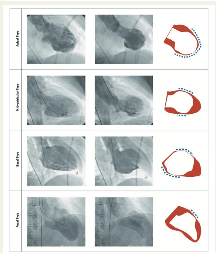
Types of takotsubo syndrome
Although several anatomical TTS variants have been described four
major types can be differentiated based on the distribution of regional
wall motion abnormalities as shown in
Figure 4
.
16,51The most
com-mon TTS type and widely recognized form is the (i) apical ballooning
type also known as the typical TTS form, which occurs in the majority
of cases.
16,51Over the past years, atypical TTS types have been
increasingly recognized.
51These include the (ii) midventricular, (iii)
basal, and (iv) focal wall motion patterns.
51Recently, it has been
dem-onstrated that patients suffering from atypical TTS have a different
clinical phenotype.
51These patients are younger, suffer more often
from neurologic comorbidities, have lower brain natriuretic peptide
values, a less impaired LVEF, and more frequent ST-segment
depres-sion compared to typical TTS.
51,185In-hospital complication rate is
similar between typical and atypical types, while 1-year mortality is
higher in typical TTS.
51After adjustment for confounders, LVEF
<45%, atrial fibrillation, neurologic disorders but not TTS phenotype
were independent predictors of death.
51Beyond 1-year, long-term
mortality is similar in typical and atypical TTS phenotypes, therefore,
patients should be equally monitored and treated.
51The basal
phenotype has been reported to be associated with the presence of
pheochromocytoma,
186epinephrine-induced TTS,
178or
subarach-noid haemorrhage
187consequently, these conditions should be
considered in this particular setting.
Besides the four major TTS types, other morphological variants
have been described including the biventricular (apical type and right
..
..
..
..
..
..
.
ventricular involvement),
19isolated right ventricular,
188,189and global
form.
190Global hypokinesia as a manifestation of TTS is difficult to
prove given the very broad differential diagnoses including conditions
such as tachycardia-induced cardiomyopathy. Right ventricular
involvement is present in about one-third of TTS patients and may be
a predictor for worse outcome.
191The true prevalence of the
iso-lated right ventricular form is unknown since little attention is paid to
the right ventricle in daily clinical echo routine.
Figure 3
Emotional and physical stress factors precipitating takotsubo syndrome. Reprinted, modified, and translated with permission from
Schlossbauer et al.
7COPD, chronic obstructive pulmonary disease; PRES, posterior reversible encephalopathy syndrome; TIA, transient ischaemic
attack.
Figure 4
The four different types of takotsubo syndrome during diastole (left column) and systole (middle column). The right column depicts
diastole in red and systole in white. The blue dashed lines demonstrate the region of the wall motion abnormality. Reprinted and modified with
per-mission from Templin et al.
16..
..
..
..
..
..
..
..
..
..
..
..
..
..
..
..
..
..
..
..
..
..
..
..
..
..
..
..
..
..
..
..
..
..
..
..
..
..
..
..
..
..
..
..
..
..
..
..
..
..
..
..
..
..
..
..
..
..
..
..
..
..
..
..
..
..
..
..
..
..
..
..
..
..
..
..
..
..
..
..
..
..
..
..
..
..
.
Patients with recurrent TTS can demonstrate different wall
motion patterns at each event,
192,193suggesting that left
ventricu-lar adrenergic receptor distribution does not explain different
TTS types.
Chronobiology
A growing body of evidence reveals that acute cardiovascular events
are not distributed randomly over time, but instead depend on the
time of day, day of the week, and months/season of the year.
194–197Several studies have investigated chronobiological features of TTS.
Two studies reported a peak in the morning
198,199and afternoon
hours,
200while others failed to show a statistically significant
tem-poral pattern.
201Two studies observed the highest frequency on
Monday
196,202and a third investigation has not found a weekly
variation.
198Most conducted studies reported a summer
prefer-ence for TTS,
24,203while one study reported a winter peak.
204Hence, conflicting results about the chronobiological pattern of
TTS exist.
Funding
J.R.G. has received a research grant “Filling the gap” from the University
of Zurich.
Conflict of interest: none declared.
References
1. Sato H. Tako-tsubo-like left ventricular dysfunction due to multivessel coronary spasm. In: K Kodama, K, Haze M Hori, eds. Clinical Aspect of Myocardial Injury: From Ischemia to Heart Failure. Tokyo: Kagakuhyoronsha Publishing Co; 1990. p56–64; (Article in Japanese).
2. Pavin D, Le Breton H, Daubert C. Human stress cardiomyopathy mimicking acute myocardial syndrome. Heart 1997;78:509–511.
3. Sharkey SW, Shear W, Hodges M, Herzog CA. Reversible myocardial contrac-tion abnormalities in patients with an acute noncardiac illness. Chest 1998;114: 98–105.
4. Desmet WJ, Adriaenssens BF, Dens JA. Apical ballooning of the left ventricle: first series in white patients. Heart 2003;89:1027–1031.
5. Wittstein IS, Thiemann DR, Lima JA, Baughman KL, Schulman SP, Gerstenblith G, Wu KC, Rade JJ, Bivalacqua TJ, Champion HC. Neurohumoral features of myocardial stunning due to sudden emotional stress. N Engl J Med 2005;352: 539–548.
6. Templin C, Napp LC, Ghadri JR. Takotsubo syndrome: underdiagnosed, under-estimated, but understood? J Am Coll Cardiol 2016;67:1937–1940.
7. Schlossbauer SA, Ghadri JR, Templin C. Takotsubo-Syndrom—ein ha¨ufig ver-kanntes Krankheitsbild. Praxis (Bern 1994) 2016;105:1185–1192.
8. Sharkey SW, Lesser JR, Maron MS, Maron BJ. Why not just call it tako-tsubo cardiomyopathy: a discussion of nomenclature. J Am Coll Cardiol 2011;57: 1496–1497.
9. Luscher TF, Templin C. Is takotsubo syndrome a microvascular acute coronary syndrome? Towards of a new definition. Eur Heart J 2016;37:2816–2820. 10. Lyon AR, Bossone E, Schneider B, Sechtem U, Citro R, Underwood SR,
Sheppard MN, Figtree GA, Parodi G, Akashi YJ, Ruschitzka F, Filippatos G, Mebazaa A, Omerovic E. Current state of knowledge on takotsubo syndrome: a position statement from the taskforce on takotsubo syndrome of the heart failure Association of the European Society of Cardiology. Eur J Heart Fail 2016; 18:8–27.
11. Pelliccia F, Sinagra G, Elliott P, Parodi G, Basso C, Camici PG. Takotsubo is not a cardiomyopathy. Int J Cardiol 2018;254:250–253.
12. Prasad A, Dangas G, Srinivasan M, Yu J, Gersh BJ, Mehran R, Stone GW. Incidence and angiographic characteristics of patients with apical ballooning syn-drome (takotsubo/stress cardiomyopathy) in the HORIZONS-AMI trial: an ana-lysis from a multicenter, international study of ST-elevation myocardial infarction. Catheter Cardiovasc Interv 2014;83:343–348.
13. Bybee KA, Prasad A, Barsness GW, Lerman A, Jaffe AS, Murphy JG, Wright RS, Rihal CS. Clinical characteristics and thrombolysis in myocardial infarction
frame counts in women with transient left ventricular apical ballooning syn-drome. Am J Cardiol 2004;94:343–346.
14. Redfors B, Vedad R, Angeras O, Ramunddal T, Petursson P, Haraldsson I, Ali A, Dworeck C, Odenstedt J, Ioaness D, Libungan B, Shao Y, Albertsson P, Stone GW, Omerovic E. Mortality in takotsubo syndrome is similar to mortality in myocardial infarction—a report from the SWEDEHEART registry. Int J Cardiol 2015;185:282–289.
15. Deshmukh A, Kumar G, Pant S, Rihal C, Murugiah K, Mehta JL. Prevalence of Takotsubo cardiomyopathy in the United States. Am Heart J 2012;164: 66–71 e1.
16. Templin C, Ghadri JR, Diekmann J, Napp LC, Bataiosu DR, Jaguszewski M, Cammann VL, Sarcon A, Geyer V, Neumann CA, Seifert B, Hellermann J, Schwyzer M, Eisenhardt K, Jenewein J, Franke J, Katus HA, Burgdorf C, Schunkert H, Moeller C, Thiele H, Bauersachs J, Tscho¨pe C, Schultheiss H-P, Laney CA, Rajan L, Michels G, Pfister R, Ukena C, Bo¨hm M, Erbel R, Cuneo A, Kuck K-H, Jacobshagen C, Hasenfuss G, Karakas M, Koenig W, Rottbauer W, Said SM, Braun-Dullaeus RC, Cuculi F, Banning A, Fischer TA, Vasankari T, Airaksinen KEJ, Fijalkowski M, Rynkiewicz A, Pawlak M, Opolski G, Dworakowski R, MacCarthy P, Kaiser C, Osswald S, Galiuto L, Crea F, Dichtl W, Franz WM, Empen K, Felix SB, Delmas C, Lairez O, Erne P, Bax JJ, Ford I, Ruschitzka F, Prasad A, Lu¨scher TF. Clinical features and outcomes of takotsubo (stress) cardiomyopathy. N Engl J Med 2015;373:929–938.
17. Schneider B, Athanasiadis A, Stollberger C, Pistner W, Schwab J, Gottwald U, Schoeller R, Gerecke B, Hoffmann E, Wegner C, Sechtem U. Gender differ-ences in the manifestation of tako-tsubo cardiomyopathy. Int J Cardiol 2013;166: 584–588.
18. Roshanzamir S, Showkathali R. Takotsubo cardiomyopathy a short review. Curr Cardiol Rev 2013;9:191–196.
19. Aizawa K, Suzuki T. Takotsubo cardiomyopathy: Japanese perspective. Heart Fail Clin 2013;9:243–247.
20. Berton E, Vitali-Serdoz L, Vallon P, Maschio M, Gortani G, Benettoni A. Young girl with apical ballooning heart syndrome. Int J Cardiol 2012;161:e4–e6. 21. Otillio JK, Harris JK, Tuuri R. A 6-year-old girl with undiagnosed
hemophago-cytic lymphohistiocytosis and takotsubo cardiomyopathy: a case report and re-view of the literature. Pediatr Emerg Care 2014;30:561–565.
22. Rozema T, Klein LR. Takotsubo cardiomyopathy: a case report and literature review. Cardiol Young 2016;26:406–409.
23. Nascimento FO, Larrauri-Reyes MC, Santana O, Pe´rez-Caminero M, Lamas GA. Comparison of stress cardiomyopathy in hispanic and non-hispanic pa-tients. Rev Esp Cardiol (Engl Ed) 2013;66:67–68.
24. Regnante RA, Zuzek RW, Weinsier SB, Latif SR, Linsky RA, Ahmed HN, Sadiq I. Clinical characteristics and four-year outcomes of patients in the Rhode Island Takotsubo Cardiomyopathy Registry. Am J Cardiol 2009;103:1015–1019. 25. Franco E, Dias A, Koshkelashvili N, Pressman GS, Hebert K, Figueredo VM.
Distinctive electrocardiographic features in African Americans diagnosed with takotsubo cardiomyopathy. Ann Noninvasive Electrocardiol 2016;21:486–492. 26. Qaqa A, Daoko J, Jallad N, Aburomeh O, Goldfarb I, Shamoon F. Takotsubo
syndrome in African American vs. non-African American women. West J Emerg Med 2011;12:218–223.
27. Park JH, Kang SJ, Song JK, Kim HK, Lim CM, Kang DH, Koh Y. Left ventricular apical ballooning due to severe physical stress in patients admitted to the med-ical ICU. Chest 2005;128:296–302.
28. Ghadri JR, Ruschitzka F, Luscher TF, Templin C. Takotsubo cardiomyopathy: still much more to learn. Heart 2014;100:1804–1812.
29. Stollberger C, Wegner C, Finsterer J. Seizure-associated Takotsubo cardiomy-opathy. Epilepsia 2011;52:e160–e167.
30. Jung JM, Kim JG, Kim JB, Cho KH, Yu S, Oh K, Kim YH, Choi JY, Seo WK. Takotsubo-like myocardial dysfunction in ischemic stroke: a hospital-based registry and systematic literature review. Stroke 2016;47:2729–2736. 31. Song BG, Yang HS, Hwang HK, Kang GH, Park YH, Chun WJ, Oh JH. The
im-pact of stressor patterns on clinical features in patients with tako-tsubo cardio-myopathy: experiences of two tertiary cardiovascular centers. Clin Cardiol 2012; 35:E6–E13.
32. Prasad A, Lerman A, Rihal CS. Apical ballooning syndrome (tako-tsubo or stress cardiomyopathy): a mimic of acute myocardial infarction. Am Heart J 2008;155:408–417.
33. Abe Y, Kondo M, Matsuoka R, Araki M, Dohyama K, Tanio H. Assessment of clinical features in transient left ventricular apical ballooning. J Am Coll Cardiol 2003;41:737–742.
34. Bybee KA, Kara T, Prasad A, Lerman A, Barsness GW, Wright RS, Rihal CS. Systematic review: transient left ventricular apical ballooning: a syndrome that mimics ST-segment elevation myocardial infarction. Ann Intern Med 2004;141: 858–865.
35. Maron BJ, Towbin JA, Thiene G, Antzelevitch C, Corrado D, Arnett D, Moss AJ, Seidman CE, Young JB; American Heart Association; Council on Clinical Cardiology, Heart Failure and Transplantation Committee; Quality of Care and