Role of Integrase Acetylation in HIV-1 Replication
Cycle and Search for Acetylation Inhibitors
Paola Valentini
PhD Thesis in Molecular Biology
Supervisor:
Prof. Anna Cereseto
Scuola Normale Superiore, Pisa
HIV-1 integrase catalyzes the integration of the viral DNA into the genome of the host cells. This irreversible event is crucial to the pathogenesis of the infection and complicates its eradication both by the immune systems and by pharmacological treatments.
The mode of action of this viral enzyme is still not completely characterized, although full understanding of some key aspects, as the mechanism of integration site selection, are relevant both for the development of new anti-integrase drugs and for potential application of HIV-derived vectors for gene therapy.
Our group has demonstrated that integrase is post-translationally acetylated by two cellular histone-acetyl transferases (HATs), chromatin-modifying enzymes whose major role is that of transcriptional co-activators. Integrase acetylation is important for the viral infectivity and interaction with HATs might be one of the determinants of HIV-1 preferential integration in actively transcribed genomic regions.
Integrase is a poorly exploited target of anti-HIV drugs, while traditional therapies based on combinations of reverse transcriptase inhibitors and protease inhibitors are facing the rapid diffusion of multi-drugs resistant viral variants. This pushes research towards new drugs and new targets, including integrase and, even better, its interactions with cellular cofactors like, for instance, HATs.
This thesis deals with the selection of novel inhibitors of integrase acetylation, to be used as lead compound for the development of new generation anti-integrase drugs.
A selective inhibitor of integrase acetylation was identifyied through in
studies led to the rational design of a smaller set of compounds, whose activity was tested with in vitro and in vivo assays. Finally, one molecule was chosen for further studies with HIV-1 derived lentiviral vectors. This cinnamoil compound was able to inhibit integrase acetylation in the virus and reduced viral integration in infected cells. In a reciprocal experiment, viral vectors containing hyper-acetylated integrase were generated by trans-incorporation of fusion integrase-HAT proteins, or of isolated HAT domains. The enhanced infectivity of these virions confirmed the role of acetylation for integrase function.
Table of contents Page
1 – Introduction 1
1.1 HIV-1 and AIDS: epidemiology and disease 2
1.2 HIV-1 virion structure 4
1.2.1 Gag poliprotein 6
1.2.2 Env gene 8
1.2.3 Pol poliprotein 9
1.2.3.1 Integrase 10
Integrase structure 10
Integrase enzymatic activity 13
Integrase multimerization 14
Cellular proteins interacting with integrase 14
1.2.4 Regulatory and accessory proteins 22
1.3 HIV-1 replication cycle 26
1.3.1 Integration 31
1.3.2 Integration site selection 34
1.4 Current antiretroviral therapies 39
1.4.1 Integrase inhibitors 42
1.4.2 Integrase inter-face inhibitors 43
1.5 Integrase post-translational modifications 45
1.6 Histone post-translational modifications: acetylation 46
1.7 Histone Acetyl-Transferases: p300 and GCN5 49
1.8 Acetylation of non-histone proteins 55
1.8.1 Acetylation and protein function 56
1.8.2 Acetylation of viral proteins 57
derivatives 61
1.9.2 Synthetic HAT inhibitors 65
2 – Aims of the thesis and experimental strategy 69
3 – Materials and methods 73
3.1 Plasmids 74
3.2 Antibodies 74
3.3 Recombinant proteins production and purification 75
3.4 In vitro acetylation assay to test the efficacy of curcumin and
its derivatives. 76
3.5 In vivo acetylation assays to test the efficacy of curcumin
derivatives in mammalian cells 78
3.6 Strand Transfer assay 78
3.7 Cell culture and transfection 79
3.8 Lentiviral vectors production 79
3.9 Stable and transient knockdown of GCN5 expression 80
3.10 HIV-1 infectivity assays 80
3.11 RT-Q-PCR analysis 81
3.12 Western blotting 83
4 – Results 85
4.1 A new class of small molecules is able to inhibit p300 Hystone
Acetyl-Transferase 87
4.1.1 Screening for new HAT inhibitors 87
4.1.2 Efficacy of inhibitors 1b and 2c on different HATs and
in cell culture conditions. 94
4.2 In vivo inhibition of integrase acetylation 98
HAT inhibitor 2c
4.3.2 Infectivity in cells treated with the HAT inhibitor 2c 106
4.4 Transient and stable knockdown of p300 and GCN5 108
4.5 Infectivity of hyperacetylated virions 111
4.5.1 Generation of virions containing hyper-acetylated integrase through IN-HAT chimeras
trans-incorporation
111
4.5.2 Generation of virions containing hyper-acetylated
integrase through HAT domains trans-incorporation 113
5 – Discussion 119
5.1 A new class of small molecules is able to inhibit p300 Hystone
Acetyl-Transferase 120
5.2 Integrase acetylation inside the viral particles 122
5.3 Molecular engineering of viral particles containing
hyper-acetylated integrase 123
5.4 Importance of integrase acetylation during the replication cycle
of HIV-1 125
5.5 Inhibitors of integrase acetylation as potential lead compounds
for the design of second generation integrase inhibitors 126
6 – Conclusions and future directions 129
1.1 HIV-1 and AIDS: epidemiology and disease
Human immunodeficiency virus type 1 (HIV-1) is the main cause of HIV disease, which can progress with variable dynamics to its end stage, the Acquired Immunodeficiency Sindrome (AIDS). AIDS and HIV-1 infection represent global health problems and complex scientific dilemmas, which raise enormous social, ethical and economical issues, thus they are obvious targets for drug discovery.
First reported in 1981 in a small number of patients, after three decades AIDS has become a major epidemic, which account for about 33 million people infected worldwide, according to the 2010 UNAIDS Report on the global AIDS Epidemic (UNAIDS, 2010).
The clinical profile of the infection caused by HIV is specific. Upon an initial HIV-1 infection, there is a period of strong viral replication and immune activation, which results in a relatively low steady state of viraemia. Afterwards, the infection enters a chronic stage, characterized by a limited virus replication and absence of evident symptoms of disease. This phase can persist for many years, ultimately leading to an irreversible damage of the immune system characterized by a total loss of CD4+ T cells. This results in the onset of the AIDS stage, wherein repeated opportunistic infections can become lethal for the vast majority of untreated patients. In a very small proportion of infected patients, the so-called ‘long-term non-progressors’, the CD4 T cells count remains stable and normal, and no signs of disease occur. These individuals are able to control viral replication to low levels without undergoing antiretroviral treatments and represent one of the models of immune control of HIV-1 (Pantaleo, 1995). Elite controllers or suppressors (ES) represent a distinct subset of untreated patients, who appear to be able to control viral replication at undetectable levels (Thiébaut, 2011; Blankson, 2010; Hatano, 2009).
HIV-1 seems to be highly adapted for life in the host, taking advantage of cellular machinery to promote replication and transmission while possessing adequate equipment for immune evasion strategies (Douek, 2002; Kwong, 2002; Yue, 2005). In most individuals HIV-1 induces a generalised immune activation that involves not only the main target of infection (i.e. CD4+ T lymphocytes and monocyte/macrophages) but also B lymphocytes, natural killer cells, and antigen-presenting cells (Lawn, 2001).
Human Immunodeficiency Virus–1 is a member of the lentivirus genus of the Retroviridae family, a large group of single stranded RNA viruses endowed with the unique property of retro-transcribing their RNA into complementary cDNA, a process that is carried out by a virus-encoded enzyme called Reverse Transcriptase (RT).
According to The Universal Virus Database of the International Committee on Taxonomy of Viruses (ICTVdB) the Retroviridae family is currently classified into 7 genera.
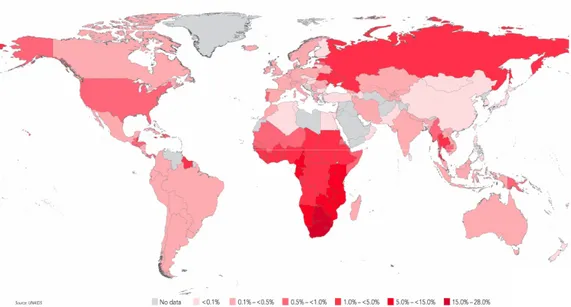
Figure 1-1. A global view of HIV-1 infection. According to the World Health Organization, 34 million people were living with HIV-1 at the end of 2010 (WHO – UNAIDS).
One of the features that distinguish HIV-1, as well as the other lentiviruses, from other members of the retroviridae family, is their ability to productively infect non-dividing, terminally differentiated cells, without a requirement for cell passage through mitosis to establish productive infections (Lewis, 1994).
1.2 HIV-1 virion structure
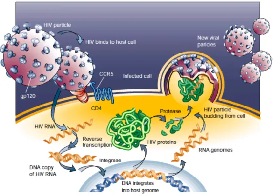
Like other retroviruses, HIV is an enveloped virus with a central, cone-shaped core surrounded by a lipid bilayer enriched in cholesterol and sphingomyelin, derived from the membrane of the host cell (Chan, 1998; Liao, 2001; Pierson, 2003; Krogstad, 2003). Embedded in this viral envelope are the 2 envelope glycoproteins, gp120 Surface (SU), exposed to the extra-cellular environment and the gp41 Trans-Membrane (TM) anchoring protein, as well as numerous cellular membrane proteins derived from the infected cells. A protein shell composed of many copies of the matrix (MA) protein separates the viral lipid envelope by the capsidic core. The core is composed of approximately 2,000 molecules of the 24 kD capsid (CA) protein (Gelderblom and Gottlinger, http://www.hiv.lanl.gov; Krogstad, 2003). The viral genome is composed of 2 copies of the positive-sense ribonucleic acid (RNA) packaged within this core. The RNA molecules are held together as a dimer, coated and protected by multiple copies of the nucleocapsid (NC) protein. The viral core also contains the viral integrase (IN) and reverse transcriptase (RT) proteins, which play essential roles in early steps of virus replication (Kaplan, 2002).
The retroviral genome is about 9-kb of RNA, and encodes nine open reading frames. Three of these encode the Gag, Pol, and Env polyproteins, which are subsequently proteolyzed into individual proteins common to all retroviruses by the viral protease (Coffin, 1997; Zuckerman, 2004).
The four Gag proteins, matrix (MA or p17), capsid (CA or p24), nucleocapsid (NC or p7), and p6, and the two Env proteins, gp120 and gp41, are structural components that make up the core of the virion and outer membrane envelope, respectively.
The Pol gene encodes three enzymes that define the replicative strategy of the retrovirus: reverse transcriptase (RT) copies the viral RNA genome into DNA, and integrase (IN) mediates the insertion of that DNA into the genomic DNA of an infected cell to establish the provirus (and persistent infection). The Figure 1-2. Genetic organization of HIV-1 (from the ICTV database)
The ~9.7 kb provirus comprises two identical LTRs (long terminal repeats) flanking the internal unique sequence. The 5′ LTR is a promoter for transcription; the 3′ LTR ensures polyadenylation. Genome regions encoding Gag, Pol and Env and the accessory proteins are shown.
third enzyme, protease (PR), is necessary for maturation of virions into an infectious form.
Of the remaining six regulatory/accessory genes of HIV-1, tat and rev are crucial for virus replication, whereas vif, vpr, vpu, and nef are thought to have modulatory functions on the immune system in vivo (often in a species-specific manner).
1.2.1 Gag poliprotein
Gag is a multidomain polypeptide that constitutes the major structural constituent of all retroviruses. Indeed, Gag is capable of assembling into virus-like particles when expressed in various cell types in the absence of other viral constituents (Gheysen, 1989). HIV-1 Gag is synthesized as a precursor polyprotein, Pr55Gag, which consists of four major domains. Concomitant with or soon after virion budding, Pr55Gag is cleaved by the virally-encoded protease (Gelderblom, 1991) into its mature products p17 matrix, p24 capsid, p7 nucleocapsid, the carbossi (C)-terminal p6, and several small polypeptides including p1 and p2.
Matrix (p17, MA), situated at the amino (N)-terminal domain of the gag polyprotein (Freed, 1998), is, in mature virions, a 132-aa polypeptide (Göttlinger, 1989; Bryant, 1990), which forms a protective shell associated directly with the inner layer of the viral membrane (Gelderblom, 1991).
The matrix protein serves several functions in the viral replication cycle. MA is important for targeting Gag and Gag-Pol precursor polyproteins to the plasma membrane prior to viral assembly (Flint, 2004). In addition, this protein appears to help incorporate Env glycoproteins into viral particles (Mammano, 1995). Furthermore, MA is part of the pre-integration complexes (PICs) (Bukrinsky,
1993) and contains two nuclear localization signals (NLS) (Haffar, 2000) that may facilitate the nuclear import (Gallay, 1995). It has been recently shown that HIV-1 MA displays biological activities also outside infected cells, in particular it is able to activate the transcription factors c-Myc and CREB in human B cells, suggesting a potential mechanism of B cell lymphomagenesis during HIV-1 infection (Li, 2010).
Capsid (p24, CA) is the second component of the Gag polyprotein and forms the core shell of the HIV-1 viral particle with about 2000 molecules per virion (Scarlata, 2003). This protein is responsible for the morphogenesis of the mature, cone-shaped core and for assembly and particle production (Dorfman, 1994). Capsid is also important for infectivity, by participating in viral uncoating, and it has been reported to be the major determinant for the unique ability of HIV-1 to access the nucleus independent of the cell cycle (Yamashita, 2007). Two cellular proteins, cyclophilin A (CypA) and TRIM5α, regulate infection at the uncoating step. CypA binds capsid acting as a viral cofactor, increasing the viral infectivity (Kootstra, 2003; Saphire, 2002; Towers, 2007). Indeed CypA may participate as an uncoating factor and modulate CA disassembly (Li, 2009) or protect the viral core by binding of cellular restriction factors (Sokolskaja, 2006), leading to an increased infectivity. Interestingly, in African Green Monkey the interaction between CypA and HIV-1 CA decreases infectivity, as it facilitates restriction mediated by TRIM5α. This is due to the existence, in old world primates, but not in humans, of a TRIM5α-CypA fusion protein, which is responsible for the post-entry restriction (Sayah, 2004; Sokolskaja, 2004). In human cells, instead, TRIM5 and CypA seem to act independently one from the other (Sokolskaja, 2006; Hatziioannou, 2005).
Nucleocapsid (NC) protein is the third component of the Gag polyprotein and it is complexed to the genomic RNA inside the viral core. The NC domain
is required for genomic RNA packaging and primer placement and it has a role in viral RNA dimerization (Frankel, 1998; Adamson, 2007; Bampi, 2004). The mature NC protein, which is released in a late cleavage reaction, plays a major role in assuring the specificity and efficiency of reverse transcription and is also important for other events in the virus life-cycle including maturation of the genomic RNA dimer, integration of proviral DNA into the host genome and budding (Popova, 2010). NC’s function in virus replication is correlated with its ability to act as a nucleic acid chaperone (Williams, 2001).
P6 protein comprises the C-terminal 51 amino acids of Gag and is important for incorporation of Vpr during viral assembly (Cohen, 1990). In addition, p6 is required for efficient viral particle release (Demirov, 2002; Huang, 1995; Stuchell, 2004).
1.2.2 Env gene
The Env gene encodes the mature TransMembrane gp41 (TM) and the Surface gp120 (SU) envelope glycoproteins, cleaved by cellular enzymes from the gp160 precursor (Zuckerman, 2004). The cellular enzyme responsible for the processing of the gp160 precursor is furin or a furin-like protease (Hallenberger, 1992). Cleavage of gp160 is required for Env-induced fusion activity and virus infectivity (Freed, 1989; McCune, 1988). The proteins gp120 and gp41 are located on the viral membrane surface and their function is to bind the CD4 receptor of the target cells and mediate fusion between viral and cellular membranes, respectively (Frankel, 1998).
1.2.3 Pol poliprotein
The Pol poliprotein harbors the viral enzymes protease, reverse transcriptase and integrase, which are processed by cleavage by the viral protease. These three enzymes are not active in their monomeric forms, but need to oligomerize as dimers or tetramers to be catalitically active.
Reverse Transcriptase (RT) protein catalyzes both RNA-dependent and DNA-dependent DNA polymerization reactions and contains an RNase H domain that cleaves the RNA portion of RNA-DNA hybrids generated during the reaction (Coffin, 1997). RT is characterized by a high error rate when transcribing RNA into DNA, since it lacks a proofreading function (Coffin, 1997).
Protease (PR) is activated during or shortly after budding of virions from the cell, and cleaves Gag into the virus structural proteins. Cleavage occurs sequentially and in a highly ordered manner.
The first cleavage event catalyzed by PR during or immediately after virion release from the cell serves to release PR itself from the Gag-Pol polyprotein. Following its own cleavage from the precursor, the dimeric enzyme cleaves a number of sites in both Gag and Gag-Pol. PR activity does not seem to target a consensus sequence, but it appears to cleave different targets with varying efficiencies, so that Gag cleavage takes place as an ordered, step-wise cascade. Mutations in Gag that disrupt the ordered nature of PR-mediated processing severely disrupt virus assembly or subsequent maturation (Krausslich, 1991).
1.2.3.1 Integrase
HIV-1 integrase (IN) is an essential viral enzyme that is required to catalyze the specific and efficient insertion of the viral DNA product of reverse
transcription into the host cell genomic DNA (Bushman, 1990; Goff, 1992; Vink, 1993).
Integrase participates also in other steps of the viral replication cycle, playing a role in the uncoating of the viral core (Leavitt, 1996; Nakamura, 1997; Li, 2009; Briones 2010), in nuclear import of the viral DNA (Gallay, 1997; Tsurutani, 2000; Ikeda, 2004) and in viral DNA synthesis (Masuda, 1995; Engelman, 1995; Wu, 1999).
Integrase structure
The integrase enzyme is a 288 amino acids, 32 KDa protein encoded by the 3′-end of the pol gene and approximately 50-100 copies of the integrase enzyme are packaged per virion particle (Flint, 2003).
Integrase is comprised of three structural and functional domains: an N-terminal domain (NTD), a catalytic core domain (CCD), and a C-N-terminal domain (CTD). The functional integrase enzyme is composed of integrase homodimers that are proposed to further associate with each other to form a multimer complex in solution (Ellison, 1995; Engelman, 1993).
The N-terminal domain (NTD) of integrase includes amino acids 1 to 50 and it contains two pairs of highly conserved hystidines (residues 12 and 16) and cysteines (residues 40 and 43) that form a zinc finger motif that has been demonstrated in vitro to involve the chelation of zinc ions (Burke, 1992; Zheng, 1996). The N-terminal domain is required for high-order multimerization that is stimulated by zinc (Zheng, 1996; Cai, 1997). A zinc atom is required for proper NTD folding and it is necessary for optimal enzymatic activity (Burke, 1992; Zheng, 1996).
The core domain (CCD) contains the catalytic site and consists of aminoacids 50 to 212. It is characterized by three invariant and essential acidic
residues (D64, D116 and E152), which forms the catalytic triad indispensable for the enzymatic activity. Crystal structures of several integrase catalytic core domains, obtained as dimers or trimers (Goldgur, 1999; Lubkowski, 1998; Chen, 2000; Molteni, 2001), show that it consists of five β-sheets flanked by six α-helices that are connected by flexible loops.
The less conserved carboxy-terminal domain (CTD) consists of 76 aminoacids (aminoacids 212 to 288) and adopts an SH3-like fold (aminoacids 220 to 270) (Cai, 1997; Eijkelenboom, 1995). The C terminus of IN is the less conserved region of the protein and it is required both for 3’end processing and Figure 1-3. Structure of HIV-1 Integrase. (A) Green and cyan: inner residues of the IN tetramer, engaged with viral DNA; blue and yellow: outer IN CCDs domain; Magenta: cellular DNA; Orange: viral DNA. (B) Resection of the upper IN dimer from A, highlighting the position of canonical IN domains: The three domains appear to be stably folded when prepared separately. The amino-terminal domain is characterized by pairs of histidine and cysteine residues (HHCC) that are universally conserved among retroviral integrases. The core domain contains the catalytic site, which includes the so-called catalytic triad, formed by universally conserved and essential residues: an aspartate, and at some distance another aspartate and a glutamic acid, separated by 35 amino acids (DD35E) (Krishnan, 2010; Coffin, 1997).
integration activity (Coffin, 1997). Hindmarsh and colleagues showed that an HIV-1 IN fragment representing residues 235 to 288 binds nonspecifically to DNA (Hindmarsh, 1999). Interpreting the DNA binding activity of integrase CTD is not obvious, since integration involves two different DNA substrates, which have different structural requirements: the viral cDNA and the host genomic DNA (Coffin, 1997). The isolated CTD binds well to simple linear double-stranded DNA oligonucleotides (Engelman, 1994; Lutzke, 1994; Vink, 1993), suggesting that it may contribute to binding the viral cDNA ends (att sites) (Coffin, 1997). In addition, the C-terminus seems to enhance the multimerization of IN (Hindmarsh, 1999) (Asante-Appiah, 1999; Engelman, 1999). The CTD domain contains 3 lysines residues which are acetylated by both p300 and GCN5 histone acetyl-transferases (K264, K266 and K273) (Cereseto, 2005) and a fourth lysine acetylated exclusively by GCN5 (Terreni, 2010).
The crystal structure of a full-lenght retroviral integrase (from the Prototype Foamy Virus) has been recently characterized. Hare and colleagues showed that the retroviral intasome (the nucleoprotein complex needed for integration of the viral DNA into the host genome) is comprised of an integrase tetramer that tightly binds the two viral DNA extremities (Hare, 2010).
The N- and C-terminal domains of IN are essential for proper interactions with substrates and they are needed for 3’ processing and strand transfer, presumably because without them the CCD cannot correctly position the viral cDNA termini at the active site (Chiu, 2004).
Integrase enzymatic activity
Following reverse transcription, IN catalyzes a series of reactions to integrate the viral genome into a host chromosome. Initially, in a reaction termed 3’-processing, IN removes two or three nucleotides from one or both viral DNA ends to expose the 3'hydroxyl groups of the invariant CA dinucleotides. Next, after import of the viral DNA into the nucleus, IN inserts both 3'ends of the viral DNA into opposing strands of cellular genomic DNA (Coffin, 1997).
Mechanistically and structurally, IN belongs to a diverse family of polynucleotidyl transferases (Dyda, 1994), which notably includes RNaseH (Nowotny, 2005), the transposases from Tn5 (Davies, 2000) and eukaryotic mobile element Mos1 (Richardson, 2009; Jaskolski, 2009; Nowotny, 2009; Engelman, 1991). The reactions catalysed by these enzymes proceed by SN2-type nucleophilic substitution, assisted by divalent metal cofactors (Nowotny, 2005; Engelman, 1991). In retroviral integrase, a pair of divalent metal cations (Mg or Mn) is thought to be coordinated by three carboxylates of the invariant DD35E motif within the catalytic core domain (CCD). In vivo, integrase acts within a large nucleoprotein complex that contains viral DNA and several virus- and host cell-derived components called the Pre-Integration Complex (PIC). PICs include viral proteins, such as the viral matrix, vpr and nucleocapsid (Miller, 1997), and several host proteins, such as barrier to autointegration (BAF [Lin, 2003]), high mobility group proteins (HMGs [Farnet, 1997]), and LEDGF/p75 (Llano, 2004) (a detailed list of integrase cofactors is the subject of a dedicated paragraph ahead).
Integrase multimerization
Studies with purified recombinant protein and model DNA substrates indicated that integrase does not function in its monomeric form, but individual protein monomers establish complementary contacts both with DNA substrates and with the other integrase subunits, to form the functional nucleoprotein complexes (Engelman, 1993; van Gent, 1993; van den Ent, 1999; Zhao, 2008; Kessl, 2009; Hare, 2010). Although a dimeric protein is sufficient to process each 3’-end, a tetramer is needed to carry out the concerted integration of both viral ends (Faure, 2005; Guiot, 2006; Li, 2006; Hare, 2010). A dynamic interaction between integrase subunits is essential for the assembly of the fully functional nucleoprotein complex and restricting the molecular movement of individual subunits within a multimer could compromise catalytic processes. Cellular proteins interacting with integrase
Because of retroviruses’ limited genome size and content, each step in the elaborate replication cycle of HIV-1 requires the assistance of multiple host proteins. In particular the factors described hereafter have been shown, at different extents, to have a role at the integration step.
Ini1 (Integrase Interactor 1), also known as hSNF5, was first identified as a cellular cofactor of IN by two-hybrids screening (Kalpana et al., 1994). Ini-1 is the human homolog of yeast SNF5, a transcriptional activator and component of the chromatin remodeling SWI/SNF complex (Carlson, 1994) and it was similarly shown to be part of the mammalian SWI/SNF complex (Wang, 1996). The exact role of Ini1 in HIV-1 replication is presently unclear. The first reports about it shown that recombinant Ini1 directly binds Integrase and stimulates IN catalytic activity in vitro (Kalpana, 1994).
Interestingly, evidence for a possible role for Ini1 in the post-integration steps of HIV-1 replication is stronger. Indeed, Ini1 is incorporated into the virions and it is necessary for efficient viral particle production (Yung, 2004), while a cytoplasmic fragment of Ini1 (S6), when over-expressed, was able to interact with IN in the context of the Gag-pol precursor and it was reported to inhibit viral particle production, thus suggesting a role for Ini-1 during the late stage of HIV-1 replication (Yung, 2001).
On the other hand, another report suggested a role for Ini-1 as an inhibitor of the early steps of HIV-1 replication. Maroun and co-workers, showed that siRNA mediated silencing of SWI/SNF complex expression increased the formation of 2-LTR circles and integrated forms of viral DNA, (Maroun, 2006). In fact a single amino acid change, K71R, in integrase reduced its ability to interact with Ini1, leading to an increased viral infectivity (Maroun, 2006).
Ku, a chromatin-associated protein which is part of the double-stranded DNA break recognition and repair system known as non-homologous end-joining (NHEJ), has also been identified in PICs (Li, 2001; Lin, 2003). This protein seems to enhance viral DNA circularization in infected cells after reverse transcription. In this way Ku might protect cells from apoptosis induced by linear unintegrated viral cDNA forms, allowing the remaining integrated viral DNA copies to efficiently complete the viral replication cycle (Li, 2001).
BAF-1 (Barrier to autointegration 1), is a small DNA-binding protein identified as a component of the MLV and HIV-1 PIC (Chen, 1998; Suzuki, 2002; Lin, 2003; Mansharamani, 2003). The association of BAF-1 with the PIC might be mediated by interactions with DNA, Gag or IN. BAF-1 seems to function by bridging and condensing DNA helices (Zheng, 2000; Umland, 2000) and by doing so on viral DNA it would render it inaccessible to autointegration reactions. Indeed, removing BAF-1 from the PIC by using a
high-salt wash activates the suicidal autointegration of the viral termini into internal sites on the viral DNA in cis. With the same bridging mechanism, BAF could promote anchoring of the PIC to the target DNA, as demonstrated by the fact that its presence promotes the integration to target DNA in trans (Suzuki, 2002; Lee, 1998).
LAP2α (lamina-associated polypeptide 2α) is a laminin-associated component of the nuclear envelope and it is another host component of the PICs. LAP2α binds to BAF-1 (Shumaker, 2001) and promotes productive PIC integration (Suzuki, 2004). LAP2α is required for infection by MLV, and by HIV-1 entering the cell using its own envelope protein but not by HIV-1 pseudotyped with VSV G protein (Suzuki, 2004).
Emerin, a component of the inner nuclear membrane, has been reported to be necessary for HIV-1 infection of nondividing macrophages and dividing HeLa cells (Jacque, 2006). However, Emerin role in HIV-1 infection is controversial (Shun, 2007; Mulky, 2008). The localization of emerin in the nuclear membrane is mediated by BAF-1 (Haraguchi, 2001; Lee, 2001), and is regulated by BAF-1 phosphorylation (Bengtsson, 2006; Hirano, 2005). Emerin itself is phosporilated by the ERK2/MAP Kinase (Bukong, 2010). According to Jacque and coworkers, Emerin seems to function to mediate the association of the PIC with chromatin after nuclear entry of the PIC, thereby enhancing viral DNA integration. The association of emerin with viral DNA is mediated by BAF, which binds to the PIC in the cytoplasm, and the two proteins seem to work together to promote HIV-1 integration into chromatin (Jacque, 2006).
HMGA1 and HMGA2 (high mobility group chromosomal protein A1 and A2), are non-histone DNA-binding proteins that can modulate transcriptional regulation and chromatin structure (Farnet, 1997). They have sequence-specific binding sites on chromatin and seem to function by facilitating the binding of
transcription factors to the cellular genome (Thomas, 2001). HMGA1 and HMGA2 have been identified within the PICs of MLV and HIV-1, and have the capability to favor retroviral integration (Farnet, 1997; Li, 2000). However, a recent report investigating the role of these cellular proteins during the viral replication cycle has indicated that they actually are dispensable for retroviral integration, probably due to redundancy with other factors (Beitzel, 2003).
EED (Embryonic Ectoderm Development protein) is a chromatin-remodeling protein. It belongs to the broadly conserved Polycomb family of proteins, and has recently been found to interact with integrase (Violot, 2003). Like BAF and HMGs, EED is associated to condensed chromatin. According to data acquired so far, these proteins’ primary effect is on donor viral DNA, and not on the acceptor cellular genome; however, this coincidence raises the hypothesis of a major involvement of these factors at the level of the integration site in vivo. This involvement could also be indirect, with these proteins acting as bridges for the interaction with other factors (e.g. transcriptional factors) that, in turn, could favor integration, as well as transcription.
Components of the DNA damage response system, including DNA-PK, ATM, ATR, Ku80 and XRCC4/ligase IV, have all been suggested to be important for HIV-1 DNA integration (Daniel, 1999; Smith, 2006). Recent work indicates that these proteins are not directly involved in the integration reaction (Ariumi, 2005; Dehart, 2005), though they are probably required to induce the post-integration DNA repair systems that are responsible for filling in the single-stranded gaps and sealing the nicks that are left at the sites of viral DNA insertion by IN.
LEDGF/p75 (lens epithelium-derived growth factor), assists the integration process by tethering integrase to the host chromosomal DNA (Maertens, 2003; Cherepanov, 2003). LEDGF/p75 is a ubiquitously and
constitutively expressed nuclear transcriptional co-activator and it is the 530 amino acid product of the gene PSIP1 (Ge, 1998). A 333-aminoacids splice variant, LEDGF/p52 (p52), shares p75’s N-terminal 325 residues. The two proteins differ in their C-terminal portion, derived by alternative splicing, which consists of 8 amino acids in the case of p52 and 205 amino acids for p75 (Ge, 1998). The C-terminal portion of LEDGF/p75 contains the integrase binding domain (IBD) (Cherepanov, 2004). Both LEDGF/p52 and LEDGF/p75 are chromatin-associated proteins, which have been implicated in transcriptional regulation, cell survival and autoimmunity. P52 seems to be the more active one and also has a more restricted intranuclear distribution during the different phases of the cell cycle (Nishizawa, 2001).
LEDGF/p75 is necessary for the nuclear and chromatin localization of PICs for HIV-1 and other lentiviruses. To exert this function, LEDGF/p75 acts as a receptor that tethers HIV integrase to chromatin and stabilize it by protecting it from degradation, while strongly influencing the genome-wide distribution of HIV integration. In the absence of LEDGF/p75, lentiviral IN proteins are cytoplasmic (Maertens, 2003). LEDGF/p75 enhances in vitro strand transfer activity of integrase from HIV and from other lentiviruses, but have no effect on integrase from other types of retroviruses (Cherepanov, 2007). LEDGF’s feature common to its viral and cellular roles is its ability to act as a molecular adaptor and tether proteins to the chromatin fiber.
Transportin-SR2 (TRN-SR2), one of the alternate splicing product of the gene TNPO3, is a protein belonging to the importin-beta family of proteins (Lai, 2000; Kataoka, 1999) and it has been recently identified as the nuclear import factor of HIV in cycling cell lines as well as in macrophages (Christ, Thys, 2008). TRN-SR2 was first identified as the shuttle transporter for the splicing factors SR (Serine/Arginine) proteins (Lai, 2000; Lai, 2001), but we
now know that it can shuttle other proteins as well, as it does for instance with the RNA-binding motif protein 4 (RBM4) (Lai, 2003). In the study of Christ et al. it was shown that TRN-SR2 interacts with the viral integrase (Christ, 2008; Thys, 2011; Luban, 2008) and that silencing of TRN-SR2 interferes with HIV replication by inhibiting the nuclear import of viral particles (Christ, 2008; Thys, 2011). These findings are confirmed by two independent genome-wide RNAi screenings (Brass, 2008; Konig, 2008). Notably, the impact of TRN-SR2 on nuclear import is specific for lentiviruses, as it does not seem to influence MoMLV, which is known to be dependent on nuclear envelope breakdown during mitosis (Christ, 2008; Levin, 2010). Recently Ocwieja et al. suggested that the steps of HIV import through the nuclear pore may influence subsequent integration site preference both in non-cycling and in dividing cells (Ocwieja, 2011), showing that in transportin knockdown cells the distribution patterns of integration site was altered for HIV but not for MLV infections, in line with first results by Christ et al.. In particular they showed that, in the absence of TRN-SR2, HIV redirects its integration preference towards chromosomal regions with low gene density, as opposite to its usual behaviour, thus originating less productive infection events.
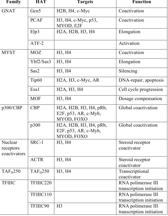
P300 histone acetyl transferase, a well-known transcriptional co-activator, has been discovered by our group as an integrase cofactor (Cereseto, 2005), which binds and acetylates HIV integrase, positively influencing viral DNA integration and infectivity. These findings were later confirmed by two other groups (Topper, 2007; Apolonia, 2007) and by subsequent studies by our group (Allouch, 2009; Terreni, 2010) and are discussed ahead throghout this thesis.
KAP1, a protein involved in transcriptional silencing thanks to its interaction with other chromatin modifying proteins (among which the histone deacetylase complex HDAC1), has been recently identified as a novel HIV-1 restriction factor, targeting specifically acetylated integrase (Allouch, 2011). It has been demonstrated that KAP1 binds to acetylated integrase and recruits HDAC1, which in turns deacetylates integrase and reduces integration. The authors propose a model for the virus to escape KAP1 restriction, in which KAP1 is inactivated by phosphorilation operated by ATM, a protein involved in the DNA double-strand break repair system, which is activated upon HIV-1 infection.
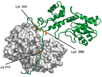
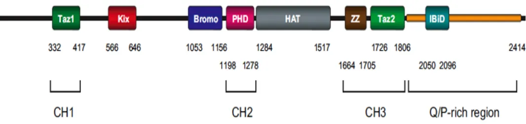
Figure 1-4. Three-dimensional models of IN complexed with p300. IN is represented in green and p300 in light grey. The three lysine residues in the C-terminal domain of IN that are acetylated by both GCN5 and p300 (Lys 264, Lys 266, and Lys 273) are shown in yellow. P300 is rendered as surface, while IN as a cartoon to highlight the C-terminal unfolded portion which inserts in the binding pockets of the HAT (Terreni, 2010).
1 – Int roduc ti on C of ac to r O th er n am es Ph ys io lo gi ca l r ol e R ol e in H IV -1 in fe ct io n R ef er en ce s st imu la te s IN c at al yt ic a ct iv ity in v itr o Ka lp an a, 1 99 4 as e In te ra ct or 1 (I ni 1) hS N F5 C hr oma tin re mo de llin g, tra ns cr ip tio na l s ile nc in g vi rio n as se mb ly Yu ng , 2 00 4 XR C C do ub le -s tra nd ed D N A br ea k re co gn iti on a nd re pa ir (n on -ho mo lo go us D N A en d jo in in g) en ha nc e th e ci rc ul ar iz at io n of v ira l D N A Li , 2 00 1 r t o au to in te gr at io n (B A F1 ) BA N F1 D N A-br id gi ng p ro te in . At ta ch in g ch ro ma tin to th e in ne r nu cl ea r me mb ra ne ? pr omo te s th e in te gr at io n to ta rg et D N A in tr an s Su zu ki , 2 00 2 Le e, 1 99 8 W ils on , 2 00 2 a-as so ci at ed p ol yp ep tid e 2 α ) TMP O ; LE M D 4; la m in in -a ss oc ia te d co mp on en t o f th e nu cl ea r e nv el op e pr omo te s pr od uc tiv e in te gr at io n by b in di ng to BA F1 Sh uma ke r, 20 01 Su zu ki , 2 00 4 in EM D ; LE M D 5; co mp on en t o f t he in ne r n uc le ar me mb ra ne me di at es a ss oc ia tio n of P IC s w ith c hr oma tin Ja cq ue , 2 00 6 m ob ili ty g ro up c hr om os om al n A 1 (H MG A 1) H M G I(Y ) mo du la tio n of tr an sc rip tio na l re gu la tio n an d of c hr oma tin st ru ct ur e st imu la te s in te gr at io n; re du nd an t? Fa rn et , 1 99 7 Li , 2 00 0 Be itz el , 2 00 3 yo ni c Ec to de rm D ev el op m en t n (E ED ) H EE D ; W AI T-1 ch ro ma tin -re mo de lin g pr ot ei n in te ra ct s w ith In te gr as e an d w ith v ira l D N A; in te gr at io n si te s el ec tio ns ?; in te ra ct io n w ith o th er c of ac to rs ? Vi ol ot , 2 00 3 K , A TM, A TR , K u8 0 an d 4/ lig as eI V co mp on en ts o f t he D N A da ma ge re sp on se s ys te m po st -in te gr at io n D N A re pa ir Sm ith , 2 00 6 Ar iu m i, 20 05 D eh ar t, 20 05 e pi th el iu m -d er iv ed g ro w th (L ED G F/ p7 5) PS IP 1 tra ns cr ip tio na l r eg ul at io n, c el l su rv iv al a nd a ut oi m mu ni ty te th er s th e IN to th e ho st c hr omo so ma l D N A Ma er te ns , 2 00 3 C he re pa no v, 2 00 3 or tin -S R 2 (T R N -S R 2) TN PO 3; IP O 12 ; MT R 10 A sh ut tli ng p ro te in th ro ug h th e nu cl ea r me mb ra ne nu cl ea r i mp or t f ac to r o f H IV C hr is t, 20 08 Th ys , 2 01 1 Lu ba n, 2 00 8 EP 30 0; KA T3 B tra ns cr ip tio na l c oa ct iv at or Ac et yl at es In a nd is re qu ire d fo r e ffi ci en t in te gr at io n; C er es et o 20 05 Al lo uc h, 2 00 9 KA T2 A tra ns cr ip tio na l c oa ct iv at or Ac et yl at es In a nd is re qu ire d fo r e ffi ci en t in te gr at io n Te rre ni 2 01 0 TR IM2 8; T if1 β ge ne s ile nc in g In hi bi ts in te gr at io n th ro ug h de ac et yl at io n of IN Al lo uc h, 2 01 1 u re 1 -5 . P ar ti al li st o f ce ll u la r p ro te in s in te ra ct in g w it h H IV -1 in te gr as e
1.2.4 Regulatory and accessory proteins
In addition to gag, pol and env, common to all members of the family Retroviridae, HIV-1 also encodes six regulatory and accessory proteins.
Tat (TransActivator of Transcription) gene encodes a small protein essential for efficient transcription of viral genes and for viral replication (Cann, 1985; Kessler, 1992; Marcello, 2001), which is able to increase viral gene expression (Ratnasabapathy, 1990; Zhou, 1995).
Tat binds to a structured RNA element (TAR, transactivation-responsive region) present at the 5’-end of viral leader mRNA (Wei, 1998) and recruits a series of transcriptional complexes and P-TEFb (Positive Transcription Elongation Factor b), which stimulates RNA polymerase II phosphorylation by Cdk9, increasing the processivity of the enzyme complex (Bieniasz, 1998; Shilatifard, 2003; Wei, 1998). Moreover, due to its efficient cell membrane transduction properties, Tat is released into the microenvironment and the circulation, and then taken up by the surrounding cells (Westendorp, 1995).
Rev (Regulator of Expression of Virion) is a sequence-specific RNA binding phospho-protein that is expressed during the early stages of HIV-1 replication (Malim, 1989). Rev is required for expression of the viral structural proteins Gag, Pol and Env from the integrated proviral DNA. By binding to the Rev-Responsive Element (RRE), an RNA structure present on the unspliced RNA encoding Gag and GagPol and on singly spliced RNAs encoding Env, Rev tethers these transcripts to the cellular CRM-1-mediated nuclear-export pathway, leading to enhanced cytoplasmic levels of these RNAs and increased expression of the encoded proteins. Rev has also recently been shown to be able to enhances encapsidation of the genomic RNA into virions (Blissenbach, 2010).
Due to its roles in nuclear RNA export, in the increase in translational efficiency of viral structural proteins, and in the stimulation of encapsidation, Rev has been thought for long time to be essential for the late phase of the virus replication cycle. However, Rev plays also a role during the early phase of infection, as it can also interfere with integration of the reverse-transcribed cDNA into the host-cell genome, by promoting dissociation of the IN– LEDGF/p75 complex, with consequent blocking IN activity and preventing tethering of the pre-integration complex to the host-cell chromosome. Since Rev is presumably present in the infected cell at sufficiently high levels only after integration has already taken place, the main function of Rev during the early phase might be to impede that superinfection of the same cell by subsequent viruses leads to excessive integration and consequent genotoxicity (Levin, 2009; Grewe, 2010).
Nef (Negative regulatory Factor) is a 27 KDa protein highly conserved in all primate lentiviruses, that is abundantly produced during the early phase of viral replication cycle. Nef has different roles in HIV-1 replication and disease pathogenesis. It down-regulates CD4 (Garcia, 1991), which limits the adhesion of a Nef-expressing T cell to the antigen-presenting cell, thus promoting the movement of HIV-infected cells into circulation and spread of the virus. Nef also down-modulates MHC-I (Schwartz, 1996) cell surface expression, protecting HIV-infected cells from host CTL response. In addition, it interferes with cellular signal transduction pathways and it enhances virion infectivity and viral replication, since it induces actin remodeling and facilitates the movement of the viral core past a potentially obstructive cortical actin barrier (Campbell, 2004; Chowers, 1994).
Vpr (Viral Protein R) is a 96 aa small basic protein. Despite its small size, Vpr has been shown to have multiple activities during viral replication. Vpr appears to participate in the anchoring the PICs to the nuclear envelope and to
be involved in the nuclear translocation of the viral DNA (Heinzinger, 1994). An important function of Vpr is that of facilitating the infection of non-dividing cells, like macrophages (Connor, 1995). This viral protein is cytopathic to cells, although there has been some debate as to whether the cells dye from apoptosis (Muthumani, 2005) or necrosis (Sakai, 2006). Nevertheless, one well demonstrated attribute of Vpr expression is its ability to delay or arrest cells in the G2 phase of the cell cycle (Bartz, 1996; Di Marzio, 1995). The biological significance of Vpr-induced arrest during viral infection is not well understood. However, HIV-1 LTR seems to be more active in the G2 phase, implying that Vpr induced G2 arrest may confer a favorable cellular environment for efficient transcription of HIV-1 (Goh, 1998). Vpr concentrates at the nuclear membrane by interacting with the nuclear pore complex components (Vodicka, 1998) and even more specifically with nucleoporins. These interactions seem to indicate that Vpr is involved in docking of the PIC to the Nuclear Pore Complex (NPC) (Jacquot, 2007). Interaction of Vpr with nucleoporin hCGI also contributes to the G2-arrest mediated by Vpr (Jacquot, 2007). Next to interactions with the NPC, Vpr was shown to interact with importin α. Given that importin α also binds other components of the PIC such as integrase or matrix, it was suggested that Vpr acts like an importin β like protein (Vodicka, 1998). A second theory suggests that Vpr facilitates nuclear import by stabilizing the interactions of matrix or integrase with the nuclear import machinery (Popov, 1998).
Vpr binds to the p6 protein (Bachand, 1999; Paxton, 1993) and this property can be exploited to trans-incorporate other proteins in the nascent viral particle (Wu, 1995; Wu, 1997; Fletcher, 1997; Liu, 1997).
Vpu (Viral Protein U) is a 9 KDa membrane protein that induces the degradation of the CD4 receptor. Vpu is involved in ubiquitination of CD4 that leads to their degradation. In addition, Vpu increases progeny virus secretion from infected cells. This function is related to the ability of Vpu to
self-assemble into homooligomeric complexes that in vitro function as ion-conductive membrane pores (Bour, 2003). Vpu counteracts an inhibitory cellular factor, TASK-1, an acid-sensitive K+ channel that, in the absence of Vpu, inhibits virus release (Hsu, 2004). TASK-1 is structurally homologous to Vpu, suggesting oligomerization as a possible mechanism of inactivation of ion channel activity of these proteins (Hsu, 2004; Li, 2005). Vpu antagonizes also another cellular restriction factor, Tetherin, a membrane protein that, in the absence of Vpu, inhibits the release of viral particles, by retaining them at the cell membrane and subsequently in endocytic vescicles (Neil, 2008).
Vif (Virus Infectivity Factor) is a 192 aa protein that is expressed at high levels in the cytoplasm of infected cells. Vif was thought to be important because it is essential for the replication of HIV-1 in the peripheral blood lymphocytes, macrophages, and certain cell lines known as “nonpermissive” cells (Strebel, 1987). Indeed vif antagonizes a host cellular restriction factor, APOBEC3G (apolipoprotein B mRNA-editing enzyme catalytic polypeptide-like 3G), which inhibits HIV infection in nonpermissive cells (Harris, 2002; Jarmuz, 2002; Sheehy, 2003). APOBEC3G is a member of the cytidine deaminase family, which prevents viral cDNA synthesis by deaminating deoxycytidines in the minus-strand retroviral cDNA replication intermediate (Harris, 2003; Yu, 2004). As a result, it creates stop codons or G to A transitions in the newly synthesized viral cDNA, which is then subjected to elimination by host DNA repair machinery (Zhang, 2003). Vif induces the ubiquitination and thus the degradation of APOBEC3G (Li, 2005), permitting the completion of HIV replication cycle.
1.3 HIV-1 replication cycle
The early events of the retrovirus life cycle begin upon fusion of the virus with the host cell plasma membrane. After fusion of viral envelope with the membrane of the host cell, the virus starts the reverse transcription of its ssRNA genome to a double strand DNA, by forming the reverse transcription complex (RTC). The RTC comprises genomic viral RNA associated with nucleocapsid protein (NC), cellular tRNA primer, enzymes reverse transcriptase, integrase and protease, viral protein R (VPR) and matrix protein (MA, p17) (Bukrinsky, 1992, 1993; Fassati, 2001; Briggs, 2003). There is a general agreement on the notion that reverse transcription initiation occurs within the intact capsid cores. However, there are different models to describe the following steps of the journey of the RTC to the nuclear membrane. In an earlier view, capsid was believed to disassemble as soon after fusion of the virion with the cellular membrane and the start of reverse transcription (Miller, 1997; McDonald, 2002; Auewarakul, 2005), so to release free RTCs into the cytoplasm (Freed, 1998; Narayan, 2004). In this view, while completing the reverse transcription process, the RTCs would travel towards the nucleus exploiting the microtubule network, which would help them to overcome the high viscosity of the cytoplasm (Bukrinskaya, 1998; McDonald, 2002).
However, several recent findings support a different model for capsid disassembly, which also implies a role for capsid in the replication cycle (Arhel, 2010), which is far from being merely structural, as initially believed. In this view, capsid integrity would be essential for the completion of the reverse transcription, as it would allow for an appropriate concentration of reverse transcriptase to remain in the proximity of the viral RNA, while at the same time allowing for the diffusion of the necessary cellular factors (like deoxyribonucleotides) through its permeable structure. Indeed, the completion of reverse transcription is necessary for capsid disassembly (Arhel, 2007).
Interactions of some of the nuclear import components with capsid has also been reported, and may have a role to drive and coordinate a timely capsid disassembly prior to nuclear import (Arhel, 2007; Arhel, 2010). In accordance with this model, CA has been reported to play a role in PICs nuclear import (Dismuke, 2006; Yamashita, 2007; Yamashita, 2004). Moreover, premature capsid disassembly induced by some restriction factors (like TRIM5α), impairs reverse transcription (Stremlau, 2006; Perron, 2007; Black, 2010).
Conversion of the viral genomic RNA into DNA is accompanied by reduction of the size of the RTC. At the end of reverse transcription, the complex becomes integration-competent and it is termed preintegration complex (PIC). PIC comprises viral cDNA, integrase, NC, RT, MA, Vpr and some cellular proteins. The PIC protects viral DNA from degradation and facilitates its integration into the host cell chromosome (Miller, 1997; Turelli, 2001). To cross the intact nuclear membrane, the virus exploits the components of the cellular nuclear transport machinery (De Rijck, 2007). Several viral proteins possess nuclear localization signal (Vpr, integrase and matrix protein), therefore these proteins, as well as the central DNA flap (an intermediate triple-stranded cDNA product of reverse transcription) might also be involved into PIC nuclear import (Bukrinsky, 1993; von Schwedler, 1994; Heinzinger, 1994; Gallay, 1997; Nie, 1998; Zennou, 2000; Sherman, 2002; Bukrinsky, 2004; Butterfield-Gerson, 2006). Facilitated by the karyophilic property of the PIC, the lentivirus subfamily is unique in its ability to access the nucleus without requiring the breakdown of the nuclear envelope during mitosis, thus independently of the phase of the cell cycle (Lewis, 1992). The next step to establish a productive infection is the integration of the viral cDNA into the host genome. This process maintains the viral information life-long in the infected cell and it is carried out by the viral protein integrase (IN).
Transcription of the integrated proviral DNA marks the start of the late phase (Freed, 2001). In this phase of the life cycle, the viral DNA is transcribed by the host RNA polymerase II (RNAP) system, and the viral RNAs are processed and exported back to the cytoplasm by regulated trafficking mechanisms. The three viral structural protein precursors — group-specific-antigen protein (Gag), Gag-polymerase (Gag-Pol) and the envelope protein (Env) — are translated in the cytoplasm and transported to the plasma membrane by vescicular, cytoskeletal or other routes.
The process of assembly of the viral particles starts when the precursors processing has not yet been completed: the Gag precursor Pr55 plays a central role in assembly and it is sufficient for viral assembly and production of non-infectious virus particles in the absence of the other viral proteins (Gheysen, 1989; Wills, 1991). Assembly starts with Gag dimerization and multimerization, followed by binding of Gag complexes to genomic viral RNA. These Gag/RNA complexes, together with Gag/Pol, Gag p55 and Env are then transported to the site of assembly, which can be lipid rafts within plasma membrane (Freed, 1998; Gottlinger, 2001) or endosomal vacuoles (Pelchen-Mattheus, 2003; Ono, 2004), depending on the cell types.
Gag gene partially overlaps with Pol and is translated as Gag or GagPol fusion precursors at a Gag/GagPol ratio of 20:1 (Liao, 2004; Arrigo, 1995; Hill, 2001; Shehu-Xhilaga, 2001). The complexes containing Gag and GagPol are rapidly and almost completely associated with host cell membranes (Halwani, 2003). The assembly is initiated by the interaction of Gag NC with the viral RNA as a scaffold, and the complex promotes subsequent Gag–Gag association (Sandefur, 2000; Khorchid, 2002). If NC is deleted from Gag, the virus uses the RNA-binding region of MA for Gag multimerization (Burniston, 1999; Ott, 2005). In the absence of viral RNA, the cellular RNAs (possibly tRNA) are used and incorporated into the virus particle (Muriaux, 2004). The NC–RNA
complex promotes dimerization of CA domains. These observations led to a dimerization model of Gag protein assembly, where formation of the Gag Pr55 dimers leads to the assembly of higher-order products (Alfadhli, 2005).
Gag multimerization takes place at the plasma membrane but more recently has been suggested to commence at intracellular membranes, in multivesicular bodies (Nydegger, 2003; Ono, 2004). In primary macrophages, Gag p55 is found in late endosomes, and viral particles are budding from intracellular membranes into intracellular vesicles (Pelchen-Mattheus, 2003). In T cells, virus assembly utilizes specific microdomains in the plasma membrane known as lipid rafts, which contain a high concentration of cholesterol and saturated lipids. Gag and Env are bound to lipid rafts via lipid interactions of their acylated residues (Ono, 2001; Ding, 2003; Halwani, 2003; Bhattacharya, 2004). Cholesterol depletion and Gag binding to non-raft domains of the membrane
severely inhibit production of virus particles, and the perturbation of plasma lipids causes Gag to be redirected away from lipid rafts towards endosomal membranes (Ono, 2001, 2004; Ono, 2005). Gag dimerization and multimerization enhance membrane binding and the association with lipid rafts, and lipid rafts may also serve as concentration platforms for Gag, thereby facilitating higher-order Gag multimerization. Gag and Env form complexes with ganglioside M1, a constituent of lipid rafts, which are revealed within supramolecular structures termed virological synapse (VS) located at the cell– cell interface (Jolly, 2005). Binding to the plasma membrane of the Gag precursor precedes budding of virus particles (Ono, 1999; Paillart, 1999). A domain within Gag, called late domain, interacts with cellular proteins to efficiently release virions from the surface of the cells. The L domain is centered around a PTAP sequence in the p6 region of Gag. This sequence acts in concert with the cellular protein-sorting machine of the ESCRT complex (endosomal-sorting complex required for transport and removal of damaged or misfolded cellular membrane proteins) to promote viral release (Freed, 2002, 2003; Goff, 2003; Strack, 2003; Martin-Serrano, 2003; Martin-Serrano, 2005; Ott, 2005; Gottwein, 2005; Bieniasz, 2006). Gag interacts also with the components of adaptor protein complexes, including AP-2 and AP-3 subunits that control endocytic trafficking (Dong, 2005; Batonick, 2005). The final step of assembly involves a set of large assembly complexes comprising viral and cellular components (Lingappa, 1997; Morikawa, 2004; Alfadhli, 2005; Ono, 2000; Halwani, 2003; Ott, 2005).
Virus particles are released at budding as immature particles containing a spherical shell of structural proteins, not shaped as a central cone-like core. Virions subsequently undergo a maturation step, triggered by the viral protease, which results in a drastic reorganization of the core, with condensation of the inner core, formation of the core shell and convertion of the virus particle into
an infectious virion, ready for disassembly in a newly infected cell. However, the structural principles governing particle maturation have not yet been fully elucidated, and virus maturation is still one of the less known steps in HIV life cycle.
1.3.1 Integration
Integration of the viral genome is a key step of retroviral infection because it is responsible for the stable maintenance of viral genetic information in infected cells and it ensures at the same time the expression of viral genes, and thus production of new progeny viruses.
Integration is performed by the viral enzyme integrase in two well-characterized catalytic steps, referred to as end processing and end joining (Coffin, 1997; Hindmarsh, 1999). A third step, namely gap repair, is carried out by yet poorly known cellular enzymes (Hindmarsh, 1999, Skalka, 2005).
A blunt-ended linear viral genome cDNA is the precursor to integration. 3’End processing occurs largely or entirely before nuclear entry for most retroviruses, including lentiviruses. This step involves removal of a dinucleotide, adjacent to a highly conserved CA dinucleotide, from the 3’ strand of the U3 and U5 viral DNA LTRs in a reaction involving a water molecule or other nucleophile (Engelman, 1991). This exposes a 3’ hydroxyl group, whose oxygen is used as an attacking nucleophile on the target DNA during the joining reaction, in which the viral DNA is inserted into the cellular DNA. It is believed that one Mg++ atom coordinated in the active site of IN facilitates the deprotonation of the water to activate it as a nucleophile. This first reaction step may serve to remove extra nucleotides occasionally added by reverse transcriptase (Patel, 1994) and promote stable complex formation (Li, Mizuuchi, 2006; Ellison, 1994).
The DNA-joining or strand transfer step of integration, which involves the formation of new phosphodiester bonds joining the viral and host DNAs, proceeds without an extrinsic source of chemical energy. This suggests that the energy from the target DNA bonds that need to be broken in this step is used to form the new bonds that join the viral and target DNAs. This cleavage-ligation reaction proceed via a transesterification reaction and not via a covalent intermediate between IN and DNA (Engelman, 1991), as it happens, for example, between topoisomerases and DNA (Champoux, 1977). The joinings occur on the same face of the double helix, flanking a major groove.
Integration is accompanied by duplication of a short sequence from the target site, which flanks the integrated provirus as a direct repeat of 4-6 bp (Coffin, 1997). The 5’ ends of the viral DNA and the 3’ ends of the host DNA remain unjoined. In the third main step of integration, gap repair, extra nucleotides are trimmed from the 5’ ends of the viral cDNA, and these are joined to host DNA 3’ends. This closing of the second joint generating the integrated provirus involves host cell DNA repair enzymes, but the full details remain to be elucidated (Hindmarsh, 1999, Skalka, 2005).
Alternatively, the viral DNA may follow three different fates, all of which do not lead to the formation of a functional provirus. The ends of viral DNA may join to form a 2-LTR ring or the viral genome may undergo homologous recombination producing a single LTR ring. Two-LTR circles are viral cDNA molecules that fail to integrate (Coffin, 1997; Engelman, 1999) and become circularized likely by the non-homologous end joining (NHEJ) cellular repair (Li, 2001). Therefore, 2-LTR circles are a surrogate marker of retrovirus nuclear import (Coffin, 1997) and are indicative of an abortive integration (Engelman, 1999).
Finally, the viral DNA may integrate into itself (autointegration) leading to the formation of a rearranged circular structure (Coffin, 1997). None of these circular forms serve as precursor to integrated provirus, and none appear to
contribute significantly to viral replication, even though they are transcriptionally active (Cara, 1996). Rather, they all appear to be dead-end by-products of aborted infections (Coffin, 1997).
The numerous survival advantages that follow from integration include acquisition by an RNA virus of the long-term stability of chromosomal DNA, the capacity to replicate through mitosis, and the ability to parasitize the elaborate cellular transcriptional apparatus. Thus, while the exceptional ability of HIV-1 to evade and slowly destroy human immunity rests on many mechanisms, the most fundamental may be integration. A stably integrated provirus can occupy a spectrum of transcriptional states, allowing it to evade immune surveillance through latency, while retaining the capacity to scale up transcription rapidly and initiate progeny production (Han, 2007; Bisgrove, 2005) Additionally, integrated proviruses render impossible the clearing of the virus.
For all these reasons integration represent a favorable target for a therapeutic strategy development.
Figure 1–7 The integration reaction. The three main step of end-processing, strand transfer and gap repair are schematized. The five dark shaded bases show the duplication of host DNA flanking the provirus (Poeschla, 2008).
1.3.2 Integration site selection
Integration is not a random process. Each retrovirus genera displays a distinct and specific pattern of integration, which is regulated by viral and cellular factors as well as by local DNA conformation at the site of integration. The unique property of retroviruses to integrate their genome constitutes a major advantage for retrovirus-based gene therapy, which aims at long-term correction of genetic defects. However, the risk of insertional mutagenesis is dramatically real: indeed, in a clinical trial when a murine leukemia virus (MLV)-derived vector carrying the γ-chain cytokine receptor gene was used to treat children suffering from X-linked severe combined immunodeficiency syndrome (Cavazzana-Calvo, 2000), proviral integration near essential cellular genes led to uncontrolled cell proliferation and thus, after initial remission, leukemia-like disorders arose in some cases (Hacein-Bey-Abina, 2003a, 2003b; Cavazzana-Calvo, 2004). It is therefore extremely important to unravel the mechanism of integration site preference of retroviruses.
So far no primary sequence in the cellular genome has been identified as the preferential binding site for IN but integration does not seems to occur at random on DNA molecules.
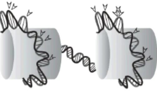
Initial experiments with murine retroviruses revealed that DNA assembled with nucleosomes constitutes a better substrate for integration as compared to naked DNA. Analysis of integration hotspots in chromatinized DNA indicates that these are sites at which DNA is probably distorted and exposed, due to the wrapping of DNA around nucleosomes (Bushman, 2005; Bushman, 1994; Muller, 1994; Pruss, 1994; Pryciak, Sil, 1992). Thus, integration preference at this spots can be explained by the fact that the outside surface of the bend is easily accessible for integration.
Although extensive analyses of the sequences flanking the integration sites have revealed some weak biases due to different primary sequence (Carteau, 1998; Pryciak, 1992; Stevens, 1996), a real consensus DNA sequence for retrovirus integration has not been identified (Bor, 1996; Fitzgerald, 1994; Goodarzi, 1997), corroborating other evidences that the structure of the integration target site, more than the primary sequence, has the major influence on site selection during infection. In this view, the influence of certain
Figure 1-8. Preferred DNA integration sites into nucleosomal DNA. The arrows indicate favorable sites for retroviral integration. These are located into the major groove on the exposed face of the DNA, as it bends around nucleosomes (schematically represented by cylinders). (Cereseto and Giacca, 2004).