CXCR4
pos
circulating progenitor cells coexpressing
monocytic and endothelial markers correlating with
fibrotic clinical features are present in the peripheral
blood of patients affected by systemic sclerosis
Diana Campioni,
1Andrea Lo Monaco,
2Francesco Lanza,
1Sabrina Moretti,
1Luisa Ferrari,
1Maria Fotinidi,
2Renato La Corte,
2Antonio Cuneo,
1and Francesco Trotta
21Department of Biomedical Sciences and Advanced Therapies, Hematology Section and BMT Unit, University of Ferrara-S.Anna Hospital, Ferrara; 2Department of Clinical and Experimental Medicine, Section of Rheumatology, University of Ferrara-S.Anna Hospital, Ferrara, Italy
ABSTRACT
There is still controversy regarding the role of circulating endothelial and progenitor cells (CECs/CEPs) in the pathogenesis of systemic sclerosis (SSc). Using a sequential Boolean gating strategy based on a 4-color flow cytometric protocol, an increased number of CD31pos/CD184pos(CXCR4)/CD34pos/CD45posand CD31pos/CD117pos(c-kit-R) /CD34pos/ CD45poshematopoietic circulating progenitor cells
(HCPCs) was detected in SSc patients compared with healthy subjects. In SSc, no circulating mature and progenitor endothelial cells were observed, while an enhanced generation of erythroid progenitor cells was found to be correlated with the presence of CD117+ HCPCs. The presence of freshly detected CXCR4posHCPC was correlated either to the in vitro cultured spindle-shaped endothelial like cells (SELC) with an endo/myelomonocytic profile or to SDF-1 and VEGF serum level. These data are related to more fibrotic clinical features of the disease, thus supporting a possible role of these cells in fibrosis.
Key words: circulating hematopoietic progenitor cells, endothelial-like cells, monocytes, CXCR4, systemic sclerosis.
Citation: Campioni D, Lo Monaco A, Lanza F, Moretti S, Ferrari L, Fotinidi M, La Corte R, Cuneo A, and Trotta F. CXCR4poscirculating progen-itor cells coexpressing monocytic and endothelial markers correlating with fibrotic clinical features are present in the peripheral blood of patients affected by systemic sclerosis. Haematologica 2008 ; 93:1233-1237. doi: 10.3324/haematol.12526
©2008 Ferrata Storti Foundation. This is an open access paper.
Introduction
Recent studies demonstrated the presence of circulating EPCs and CEPs that have been shown to contribute to tissue regeneration and therapeutic vasculogenesis.1-2 Circulating
endothelial cells (CECs) are another cell population present in a wide variety of human inflammatory and pathological con-ditions (cancer, infectious and cardiovascular disease) suggest-ing a role for these cells as a biomarker of vascular disease.3-5
The recruitment of circulating progenitor cells for vascular repair seems to be correlated to the expression of specific
hom-ing receptors, such as CXCR4 (CD184), and to the
concentra-tion of the stromal derived factor-1 (SDF-1)/CXCL12 (CXCR-4/SDF-1 axis) as their specific ligand.6Pro-angiogenetic factors
(vascular endothelial growth factor-VEGF, erythropoietin, etc.)
may also be involved in the mobilization process.7Systemic
sclerosis (SSc) is an autoimmune disease of unknown etiology characterized by excessive fibrosis and microvascular abnor-malities.8The presence of CECs and CEPs in this disease
con-tinues to be controversial. In fact, an enhanced generation of CEPs has been demonstrated in SSc9and high levels of CECs
were found to be linked to the severity of pulmonary hyper-tension in its early stages.10Conversely, it has been suggested
that a defective vasculogenesis related to the absence of CEPs/EPCs can explain vascular disease in SSc and in rheuma-toid arthritis.11,12
Due to the fact that no standardized methods are used to define and detect these cells, the results are sometimes con-flicting, and a functional and phenotypic overlap between hematopoietic and endothelial progenitor cells has often been
Funding: this work was supported by grants from PRIN 2007, AIRC regional funds, AIL-Ferrara, and Cassa di Risparmio di Ferrara foundation. Manuscript received November 19, 2007. Revised version arrived on January 23, 2008. Manuscript accepted February 22, 2008.
Correspondence: Francesco Lanza, Section of Hematology, BMT Unit, University of Ferrara-S.Anna Hospital, Corso Giovecca n. 203, 44100 Ferrara, Italy. E-mail: [email protected]
a source of misunderstanding.13,14Since the
methodolog-ical approach must be able to adequately redefine the nature of circulating progenitor cells, in this study we proposed a sequential gating multiparametric flow-cyto-metric analysis and progenitor cell replating assays with the aim of better characterizing and enumerating the cir-culating hematopoietic (HCPC) and/or endothelial pro-genitor cells in SSc. Serum levels of angiogenic cytokines, in particular the SDF-1, were correlated to the presence of either freshly detected or cultured circulat-ing progenitor cells together with the clinical features in order to evaluate their possible role in SSc pathogenesis.
Design and Methods
Patients
Forty patients with SSc fulfilling the criteria for classi-fication proposed by Le Roy et al.15 and according to
Medsger and Steen8were enrolled in this study (Table
1). Human peripheral blood (PB) samples were also obtained from 10 healthy subjects (NS) as controls (mean age 43±7, range 27-49; 3 males/7 females) after obtaining informed consent. All patients were evaluated for modified Rodnan skin score (mRSS) and lung high resolution computed tomography (HRCT) scored as reported by Warrick et al.16This study was carried out
with the approval of the local ethics committee.
Flow cytometric analysis of circulating progenitor
cell subsets from PB
A 4-color cytometric analysis of whole fresh peripher-al blood (PB) samples was performed on a FACSCperipher-alibur equiped with the four-color option (Becton Dickinson, CA, USA) as previously described.17 In particular, the
presence of the supposed endothelial and progenitor cells was evaluated in the gate of both CD31+CD45–and
CD34+CD45–cells and on the basis of their further
pos-itivity for other endothelial markers such as CD133, CD146, CD105. Platelets or non-specifically stained events, present in the gate of CD31+CD45–and
identi-fied as CD61+CD45– cells, were excluded from the
analysis. Negative controls with isotype matched non-relevant monoclonal antibodies (mouse IgG1, IgG2a, IgM) were tested in all experiments. The presence of hematopoietic circulating progenitor cells, was evaluat-ed in the gate of both CD31+CD45+and CD34+CD45+
cells and on the basis of their further positivity for CD184, CD117, CD33, CD4,CD11c antigens.
Cell culture and adherent clonogenic assay
The PB samples from SSc patients were collected for circulating progenitor cell isolation and the cell culture was performed in four different liquid media.18 The
detection of possible endothelial colonies, were moni-tored as previously described.4,19,20 The same PBMNC
were also seeded into semisolid medium (Methocult H4434, Stem Cell Technologies Inc., Vancouver, Canada) specific for the hematopoietic long-term cul-ture colony forming unit assay and the clonogenic out-put of hematopoietic progenitors was evaluated by scoring in vitro colonies at day 14.
Immunohistochemical analysis
Adherent cell colonies from SSc patients were stud-ied in situ performing: tartrate-resistant acid phos-phatase (TRAP), non specific a-naphthyl acetate esterase (NSE) and inhibited a-naphthyl acetate esterase (all from Sigma Aldrich), as recommended by the International Committee for Standardization in Haematology (ICSH) manufacturer’s guidelines.21
Cytokines assay
The presence and the serum levels of angiogenic cytokines (VEGF, HGF, bFGF, PDGF-BB) were analyzed using a Searchlight human angiogenesis array 2-multi-plex assay (Tema Ricerca, Bologna Italy). Stromal derived factor -1 (SDF-1) serum levels were also meas-ured using a single ELISA kit (R&D Systems Inc). Data were calculated using a standard curve generated with specific standards, according to manufacturer’s recom-mendations.
Statistical analysis
The number of freshly detected HCPCs and the num-ber of in vitro adherent colonies was compared with the different SSc subsets and clinical features using non-parametric statistics (Wilcoxon test). Data were com-puted with the SystatTM for WindowTM. Correlation
between the different hematopoietic and non-hematopoietic colonies with clinical and laboratory findings was evaluated using Pearson’s test.
Results and Discussion
There is still controversy regarding the role of circulat-Table 1. Summary of the clinical characteristic of SSc patients
(pts). Patients were untreated at the time of the study.
Age Disease Autoantibodies bILD cmRSS duration (Anti-topoisomerasi (0-51) (months) I I/ ACA/ nucleolar/othersa) dSSC 48.3 yrs 38.6 6/0/3/3 6 18 (12 pts) (range (range 23-60) 5-60) lSSC 63.8 yrs 98 1/20/3/4 5 7 (28 pts) (range (range 37-74) 8-125) Whole 56 yrs 68.3 7/20/6/7 11 12.5 group (40 pts)
Some patients were treated with low-doses of steroid until three days before the study. SdSSC: patients affected by a diffuse form of SSc; lSSc: patients affected by a limited form of SSc. aothers= anti-nuclear antibodies (ANA), with different pattern
and anti-extractable nuclear antigen antibodies (ENA); ACA= anti-centromere autoantibodies; b
ILD= interstitial lung disease, c
mRSS= modified Rodnan total skin score (Clements P et al. J Rheumatol 1995; 22:1281-85). We considered a Warrick total score of ≥7 as the minimum level for HRCT (lung high resolution computed tomography) abnormalities to be indicative of significant interstitial lung disease. lSSc resulted older than dSSc and healthy subjects (43±7).
ing endothelial and progenitor cells (CECs/CEPs) in the pathogenesis of the systemic sclerosis (SSc). The results are sometimes conflicting due to a phenotypic overlap between hematopoietic and endothelial progenitor cells13,14 and to the lack of a standardized analytical
methodological approach.
In this study, whole fresh PB samples were analyzed by a sequential Boolean gating strategy based on a 4-color cytometric protocol as previously described.17The
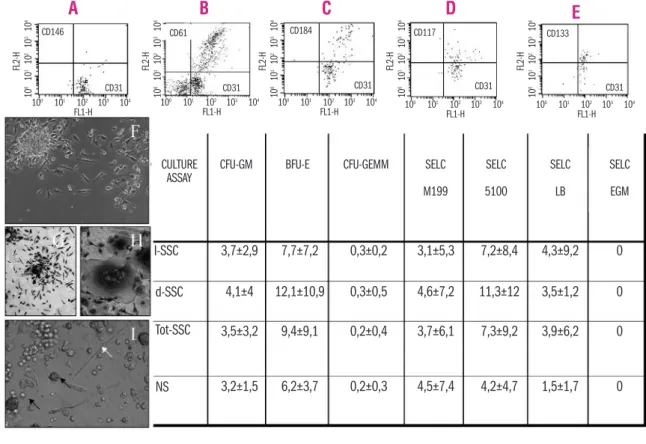
immunophenotypic results showed the lack of in partic-ular CD146+ and CD133+endothelial, respectively,
mature and progenitor CD31+CD45– endothelial cells
either in patients or in normal controls (Figure 1 A). However, in agreement with Strijbos et al.22a small
pop-ulation of CD31+CD45–circulating cells expressing some
endothelial associated markers (SSc patients, whole group: 0.5%±0.4 SD; 0.6%±0.3 SD in l-SSc vs. 0.4%±0.4 SD in d-SSc) was observed and excluded from the analysis on the basis of their positivity for platelet asso-ciated CD61 marker, high SSC scatter and negativity for CD34 and CD133 thus indicating their non-endothelial derivation (Figure 1 B).
To further investigate the presence of endothelial cells, the immunophenotypic results were combined to different progenitor culture assays 4,19,20that we
routine-ly use for the growth of CECs and CEPs in patients with hematologic disorders.19 In this study, progenitor and
mature endothelial cells were not detected in any of the samples examined. Furthermore, we did not observe in
vitro differentiation of circulating progenitor cells
towards the endothelial lineage in any patients even in the presence of different angiogenic cytokines for extended long-term cultures (Figure 1, Table of culture assay).
These data, supporting the notion that neither mature or endothelial progenitor cells are detectable in the blood of SSc, are in agreement with the hypothesis of a possible defective vasculogenesis, as previously suggest-ed by other authors.11 Nevertheless, at day 5-7 small
adherent clusters of (10-100 cells) elongated and sprout-ing cells with a characteristic spindle-shaped endothe-lial-like (SELC) morphology appeared in liquid culture for long-term hematopoietic cells (Figure 1 F). SELC grow in a granulo-monocytic manner. The cytochemical
Figure 1. Whole fresh PB samples were analyzed by a sequential gating strategy based on a 4-color cytometric protocol. (A) Circulating progenitor cells are gated on viable CD34+CD45– and CD31+CD45–cell subsets; no supposed endothelial cells were observed in the
gate of CD34+CD45–. (B) Platelets or non-specifically stained events, present in the gate of CD31+CD45–and identified as CD61+CD45–
cells, were excluded from the analysis. (C-D-E) The presence of the hematopoietic circulating progenitor cells was assessed in parallel and analyzed in the gate of CD34+CD45+for their positivity also for other stem cell hematopoietic and endothelial/monocytic markers
(CD184, CD117, CD133). (F) An example of SELC colony, cultured in liquid long-term medium is shown. SELC colonies appeared as spin-dle/elongated shaped cells, growing in a granulo-monocytic manner, positive for specific and non-specific α-naphthyl acetate esterase (NSE) at day 7 (G). With increasing culture age, SELC tend to fuse by giving rise to a multinucleated giant cells that resulted positive for the specific tartrate-resistance reactivity (TRAP) reaction (H), as expected for osteoclasts. (I) The presence of elongated endothelial-like cells (SELC) are also visible in semisolid medium during short-term clonogenic assay for hematopoietic progenitors (white arrow). Small BFU-E colonies are also visible (black arrows). In the table, the clonogenic output of PB mononuclear cells either in liquid (M199, M5100, LB, EBM) or in semisolid medium (for the growth of colony forming units granulocytes, monocytes and erythroblasts: CFU-GM,CFU-GEMM,BFU-E) is reported and expressed as average ± standard deviation of the number of colonies scored.
A
B
C
D
E
CULTURE ASSAY 100 101 102 103 104 CD146 CD31 CD31 CD31 CD31 CD31 CD133 CD117 CD184 CD61 FL1-H FL 2 -H 100 101 102 103 104 FL1-H 10 0 101 102 103 104 1 0 0 1 0 1 1 0 2 1 0 3 1 0 4 FL 2 -H 1 0 0 1 0 1 1 0 2 1 0 3 1 0 4 FL 2 -H 1 0 0 1 0 1 1 0 2 1 0 3 1 0 4 FL 2 -H 1 0 0 1 0 1 1 0 2 1 0 3 1 0 4 FL 2 -H 1 0 0 1 0 1 1 0 2 1 0 3 1 0 4 FL1-H 10 0 101 102 103 104 FL1-H 100 101 102 103 104 FL1-H I-SSC 3,7±2,9 4,1±4 3,5±3,2 3,2±1,5 7,7±7,2 12,1±10,9 9,4±9,1 6,2±3,7 0,3±0,2 0,3±0,5 0,2±0,4 0,2±0,3 3,1±5,3 4,6±7,2 3,7±6,1 4,5±7,4 7,2±8,4 11,3±12 7,3±9,2 4,2±4,7 4,3±9,2 3,5±1,2 3,9±6,2 1,5±1,7 0 0 0 0 d-SSC Tot-SSC NSCFU-GM BFU-E CFU-GEMM SELC
M199 SELC 5100 SELC LB SELC EGM
reaction for α-naphthyl acetate esterase (NSE) resulted positive on SELC (Figure 1G), but its inhibition with NaF abrogated this positivity, as normally expected for monocytes, thus confirming the monocytic nature of these cells.13,14 After 30 days of culture, SELCs tended to
fuse, giving rise to multinucleated giant cells that result-ed positive for the tartrate-resistant acid phosphatase (TRAP) reaction (Figure 1H), as expected for osteoclasts. Although cultured in different liquid media, SELC did not display either in vitro adherent expansion capacity or endothelial differentiation potential.
Short-term hematopoietic clonogenic assays revealed a statistically significant increase of circulating erythroid hematopoietic clonogenic potential (Figure 1 I), assessed as a number of cultured (burst forming unit erythrob-lasts) BFU-E/105 PBMNC in the whole SSc patients
group (Figure 1 I) (frequency: 35% of limited form, and 46% of the diffuse form) compared with NS (all p val-ues <0.005).
On the other hand, an increased number of circulating hematopoietic progenitor cells (in particular CXCR4 and c-kit-CD117-CD45 positive subsets) co-expressing endothelial and monocytic markers were observed in PB of SSc patients. In particular, a significantly increased level of the different cell subsets gated on CD34+CD45+
was seen in the PB of SSc compared with that of NS, CD184+ (CXCR4+ HCPCs: 0.047±0.009% SD vs.
0.019±0.002% SD in NS, p<0.002); CD117+
(c-kit-HCPCs, also called stem cell factor-R: 0.027±0.008% SD
vs. 0.015±0.0014% SD in NS, p<0.005), and CD133
(0.018±0.010% SD vs. 0.012±0.0014% SD in NS,
p>0.003) (Figure 1 C, D, E). Comparable values were
found in dSSc and lSSc clinical forms.
Interestingly, in our study, the CXCR4+
hematopoiet-ic progenitor cells subset, implhematopoiet-icated in the stem cell homing and mobilization process, was found to be cor-related to the presence of cultured SELC, spindle-shaped endothelial-like cell (Pearson test: r=0.79, p<0.005) that resulted positive for endothelial, and myelomonocytic surface markers and that showed a selective optimal outgrowth under hematopoietic culture conditions.
As far as the presence of c-kit -CD117- HCPCs cell subsets is concerned, in SSc patients and, especially in the dSSc subgroups, we further showed that CD117-HCPCs cell subset positively correlated with the num-ber of PB-BFU-E (p<0.005) therefore suggesting a prefer-ential in vitro erythroid commitment of this circulating hematopoietic cell subset. These findings are in agree-ment with evidence that CD31 and CD117 antigens could also be expressed on hematopoietic progenitor cells committed in particular to the erythroid pathway.23
High serum levels of angiogenic cytokines (VEGF, PDGF, EGF, IGF) and an increased concentration of SDF-1 chemokine, particularly evident in the dSSc subgroup, were observed (Figure 2), dSSc form differed significant-ly in the levels of SDF-1 (p<0.003) and to a lesser extent of VEGF (p<0.005) and HGF cytokines (p<0.003) as compared with normal subjects. This, together with an assumed defective vasculogenesis as suggested for SSc, led us to speculate that elevated levels of angiogenic cytokines, could be responsible for promoting the circu-lating hematopoietic cells erythroid commitment rather
than the endothelial one, since in dSSc of the disease the number of BFU-E in PB positively correlated with the levels of SDF-1 (p=0.002) and of circulating CD117-CPCs cell subset (p<0.005). These results are in agree-ment with the findings in literature supporting the view that these two populations share the same progenitor known as hemoangioblast.23However, this finding may
also be correlated to a subclinical hypoxiemic status in SSc patients due to pulmonary disease. But no correla-tion was found between hemoglobin levels and BFU-E count in SSc patients, or between SSc and controls.
Clinical data showed that the number of freshly cir-culating CXCR4+ HCPCs positively correlated with
mRSS (p=0.005), pulmonary involvement (p<0.005), and the severity score used to evaluate the interstitial lung disease (p<0.002). A positive correlation among CXCR4+HCPCs and two factors, implicated in the
pro-genitor cell recruitment such as SDF-1 (p=0.005) and VEGF (p=0.003) were still observed, especially in the diffuse form of SSc.
All these data seem to suggest a possible role for circu-lating hematopoietic progenitor cells in the fibrotic process of SSc. In particular, high SDF-1/VEGF serum levels could play a role in facilitating the organ homing of CXCR4pos-HCPCs and their perivascular positioning
and retention. During the course of the disease, if a mechanism for vascular repair is defective, recruited hematopoietic CXCR-4poscells that are exposed to
angio-genic stimulatory factors could enhance the proliferation of the in situ resident endothelial cells or could respond to the microenvironmental conditions, as alternatively polarized monocytes or fibrocytes, that we identify as in
vitro SELC, capable of strong stimulation of the fibrotic
process. Our data therefore agree with other findings observed in a murine model of bleomycin-induced pul-monary fibrosis24 where spindle-shaped fibrocyte-like,
resembling our SELC, has been found to play a role in the process of lung fibrosis. In conclusion, the presence of SELC could represent a useful biomarker in fibrotic disease as well as in other pathological conditions. Figure 2. The serum levels of different angiogenic cytokines such as VEGF, PDGF, HGF, bFGF and the chemokine SDF-1, evaluated on the serum of SSc patients is reported. VEGF and HGF displayed the highest serum levels either in normal samples or in SSc patients. SSc patients, in particular d-SSc differed significantly in the levels of SDF-1, and to a lesser extent in the levels of VEGF and HGF cytokines compared with normal subjects.
VEGF pg/mL bFGF pg/mL HGF pg/mL PDGF pg/mL SDF-1pg/mL NS L-SSc D-SSC SSc tot 0 200 400 600 800 1000 1200 1400 1600
Authorship and Disclosures
DC and ALM contributed equally to the work; DC performed cell cultures studies, immunohistochemistry and part of flow cytometry analysis; she was also
responsible for the supervision of laboratory investiga-tions; FL wrote the paper, and was responsible for the design of the study and data interpretation together with AC and FT. SM and LF performed flow cytometry analysis; MF, RLC and FT supervised data collection and clinical management of patients with multiple sclerosis. The authors reported no potential conflicts of interest.
References
1. Asahara T, Masuda H, Takahashi T, Kalka C, Pastore C, Silver M, et al. Bone marrow origin of endothelial progenitor cells responsible for post-natal vasculogenesis in physiological and pathological neovascularization. Circ Res 1999;85:221-8.
2. Rafii S, Lyden D. Therapeutic stem and progenitor cell transplantation for organ vascularization and regen-eration. Nat Med 2003;9:702-12. 3. Dignat-George F, Sampol J, Lip G,
Blann AD. Circulating endothelial cells: realities and promises in vascu-lar disorders. Pathophysiol Haemost Thromb 2004;33:495-9.
4. Hill JM, Zalos G, Halcox JPJ, Schenke WH, Waclawiw MA, Quyyumi AA, et al. Circulating progenitor cells, vascular function and cardiovascular risk. N Engl Med 2003;348:593-600. 5. Beerepoot LV, Mehra N, Vermaat JSP,
Zonnenberg BA, Gebbink MFGB, Voest EE. Increased levels of viable circulating endothelial cells are an indicator of progressive disease in cancer patients. Ann Oncol 2004;15: 139-45.
6. Dabusti M, Lanza F, Campioni D, Castagnari B, Tieghi A, Moretti S, et al. CXCR-4 expression on bone mar-row CD34+cells prior to mobilization
can predict mobilization adequacy in patients with hematologic malignan-cies. J Hematother Stem Cell Res 2003;12:425-34.
7. Ruiz de Almodovar C, Luttun A, Carmeliet P. An SDF-1 trap for myeloid cells stimulates angiogene-sis. Cell 2006;124:18-21.
8. Medsger TA Jr, Steen VD. Classification, prognosis. In: Clements PJ, Furst DE, editors. Systemic sclerosis. 2nd ed. Philadelphia: Lippincott Williams & Wilkins;1996. p. 51-79.
9. Allanore Y, Batteux F, Avouac J, Assous N, Weill B, Kahan A. Levels of circulating progenitor cells in sys-temic sclerosis. Clin Exp Rheumatol 2007;25:60-6.
10. Del Papa N, Colombo G, Fracchiolla N, Mazzeo Moronetti L, Ingegnoli F, Maglione W, et al. Circulating endothelial cells as a marker of ongo-ing vascular disease in Systemic Sclerosis. Arth Rheum 2004;50: 1296-304.
11. Kuwana M, Okazaki Y, Yashuoka H, Kawakami Y, Ikeda Y. Defective vas-culogenesis in Systemic Sclerosis. Lancet 2004;364:603-10.
12. Grisar J, Aletaha D, Steiner CW, Kapral T, Steiner S, Seidinger D, et al. Depletion of endothelial progenitor cells in the peripheral blood of patients with Rheumatoid Arthritis. Circulation 2005;11:204-11. 13. Schmeisser A, Strasser RH.
Phenotypic overlap between hematopoietic cells with suggested angioblastic potential and vascular endothelial cells. J Hematother Stem Cell Res 2002;11:69-79.
14. Zhao Y, Glesne D, Huberman E. A human peripheral blood monocyte-derived subset acts as pluripotent stem cells. PNAS 2003;100:2426-31. 15. Le Roy EC, Black C, Fleischmajer R,
Jablonska S, Kreig T, Medsgeeer TA. Scleroderma (systemic sclerosis): classification, subsets and pathogen-esis. J Rheumatol 1988;15:202-5. 16. Warrick JH, Bhalla M, Schabel SI,
Silver RM. High resolution comput-ed tomography in early scleroderma lung disease. J Rheumatol 1991;18: 1520-8.
17. Campioni D, Moretti S, Ferrari L, Punturieri M, Castoldi GL, Lanza F. Immunophenotypic heterogeneity of bone marrow derived mesenchy-mal stromesenchy-mal cells from patients with hematologic disorders: correlation with the bone marrow
microenvi-ronment. Haematologica 2006;91: 364-8.
18. Campioni D, Lanza F, Moretti S, Ferrari L, Cuneo A. Loss of Thy-1 (CD90) antigen expression on mes-enchymal stromal cells from haema-tologic malignancies is induced by in vitro angiogenic stimuli and is associ-ated with peculiar functional and phe-notypic characteristics. Cytotherapy 2007;21:378-81.
19. Dominici M, Campioni D, Lanza F, Luppi M, Barozzi P, Pauli S, et al. Angiogenesis in multiple myeloma: correlation between in vitro endothelial colonies growth (CFU-En) and clinical-biological features. Leukemia 2001;15:171-6.
20. Yoder MC, Mead LE, Prater D, Krier TR, Mroueh KN, Fang Li, et al. Redefining endothelial progenitor cell via clonal analysis and hematopoietic stem/progenitor cell principals. Blood 2007;109:1801-9. 21. Lanza F, Latorraca A, Moretti S,
Tomasi P. Colorazioni e tecniche di indagine morfologica e immunolo-gia. In: Piccin, Padova Editor. Trattato Italiano di Medicina di Laboratorio; Vol. V 1999.
22. Strijbos MH, Kraan J, den Bakker MA, Lambrecht BN, Sleijfer S, Gratama JW. Cells meeting our immunophenotypic criteria of endothelial cells are large platelets. Cytometry B Clin 2006;70B:56-62. 23. Bauman CI, Bailey AS, Li W,
Ferkowicz MJ, Yoder MC, Fleming WH. PECAM-1 is expressed on hematopoietic stem cells throughout ontogeny and identify a population of erythroid progenitors. Blood 2004;104:1010-16.
24. Phillips RJ, Burdick MD, Hong K, Lutz MA, Murray LA, Jing Xue, et al. Circulating fibrocytes traffic to the lungs in response to CXCL12 and mediate fibrosis. J Clin Invest 2004; 114:438-46.