PhD Course
Clinical And Experimental Immunorheumatology
And Oncology, Bioethics And Tumor Epidemiology
XXVII Cicle
PhD Final Report
M
AGNETIC
R
ESONANCE
E
NTEROGRAPHY
(MRE)
C
HANGES
AFTER
A
NTIBODY TO
T
UMOR
N
ECROSIS
F
ACTOR
(
ANTI
-TNF)
A
LPHA
T
HERAPY IN
C
ROHN
’
S
D
ISEASE
Tutor PhD Student
Prof. Luca Macarini Luca Pio Stoppino
Supervisor
C
ONTENTSAcknowledgements
.5Abbreviations
.6Summary
.91. Introduction
.111.1 Epidemiology and outcome of Crohn’s disease .11 1.2 Aetiology and pathogenesis of Crohn’s disease .12
1.3 Genetics .12
1.4 Environmental factors .13
1.5 Role of microbiota .13
1.6 Role of immune response .14
2. Disease classification by phenotype
.153. Diagnosis
.16 3.1 Clinical presentation .17 3.2 Laboratory tests .18 3.3 Endoscopy .19 3.4 Histology .22 3.5 Imaging Techniques .234. Treatment
.37 4.1 Medical therapy .38 4.2 Surgery .41 4.3 Nutritional therapy .425. Assessment of disease activity
.425.1 Clinical activity .43
5.2 Endoscopic activity and mucosal healing .47 5.3 Endoscopic scoring of inflammatory activity .49
5.4 Histological activity .54
5.6 Intestinal permeability tests .55
5.7 Faecal tests .56
5.8 Combined activity scores .59
5.9 Tools for assessing efficacy of anti-TNF therapy in CD .60
Aims of the Study
.62Material and Method
.63Results
.68Discussion
.75Acknowledgements
This research was a close cooperation between the Department of Surgical Sciences, section of Diagnostic Imaging, University of Foggia, and the Department of Clinical Medicine, research group of Gastroenterology, University Hospital of Foggia. This project was carried out between 2012 and 2015. I wish to thank the patients who agreed to participate in the study, without their contribution this research project would not have to be possible.
Thanks to Luca Macarini, for being an excellent mentor. I am forever grateful to you for believing in me and inviting me into your group. Your scientific mind, optimism and impressive experience have been essential in this process.
Thanks to Nicola Della Valle, my friend and co-advisor, for introducing me into the exciting filed of IBD. Thank you for your excellent guidance and discussions throughout the entire project. Your presence in this research group is essential and I hope to work closely with you in the future.
Thanks to co-author and colleague Stefania Rizzi for being meticulous about the MRE performance, systematic in your recordings and for always being willing to help. I really enjoyed the hours we spent together.
Finally, I want to thanks my parents and my family for supporting me during these years. Thanks to Stefania and Maria Sofia for reminding me of what is important in life.
Abbreviations
anti-TNF anti-tumour necrosis factor-α antibodies
ADA adalimumab
ASCA anti-Saccharomyces cerevisiae antibodies
ATG16L1 autophagy-related protein 16-1 gene
CARD caspase-activating recruitment domain
CBir1 anti-flagellin antibody
CD Crohn’s disease
CDAI Crohn’s disease activity index
CDEAS Crohn’s disease endomicrosopic activity score
CDEIS Crohn’s disease endoscopic index of severity
CLE confocal laser endomicroscopy
51Cr-EDTA chromium-ethylene diamine tetra-acetic acid
CRP C-reactive protein
CT computed tomography
CTE computed tomography enterography
CTEC computed tomography enteroclysis
DBE double-balloon enteroscopy
ESR erythrocyte sedimentation rate
GI gastrointestinal
HBI Harvey-Bradshaw index
Hct haematocrit
hsCRP high sensitivity C-reactive protein
kDa kilodalton
IBD inflammatory bowel disease
IBD-U inflammatory bowel disease – unclassified
IBS irritable bowel syndrome
IFN interferon
IFX infliximab
IL interleukin
MAP Mycobacterium avium paratuberculosis
MRE magnetic resonance enterography
MRI magnetic resonance imaging
NBI narrow-band imaging
NOD nucleotide oligomerisation domain
ompC outer-membrane porin C
pANCA anti-neutrophil cytoplasmic antibody with perinuclear staining pattern
PDAI perianal Crohn’s disease activity index
PEG polyethylene glycol
PMN-e polymorphonuclear neutrophil elastase
ROC curve receiver operator characteristic curve
SBCE small bowel capsule endoscopy
SBE small bowel enteroclysis
SBFT small bowel follow-through
SES-CD simple endoscopic score for Crohn’s disease
99Tc-DPTA diethylene triaminepentaasectic acid
Th T-helper
TNFα tumour necrosis factor-α
UC ulcerative colitis
Summary
Crohn’s disease (CD) is a disabling transmural and segmental chronic inflammatory bowel disease (IBD) with a relapsing and remitting course. Its inflammatory lesions can affect the entire gastrointestinal (GI) tract, in contrast to the other major type of IBD, ulcerative colitis (UC), which is limited solely to the colon [1]. The aetiology and pathogenesis of CD is incompletely known, but the most widely accepted hypothesis is that it arises from interactions between immunoregulatory, genetic, and environmental factors [2]. Exacerbations in CD are characterised by symptoms such as diarrhoea, abdominal pain, and rectal bleeding.
Assessment of CD activity has traditionally been based on symptoms and clinical signs, although symptoms sometimes fail to correlate with bowel inflammatory activity. Despite great advances in development of medical therapy during the last two decades, CD is still considered incurable, and in many patients leads to multiple complications [3]. Although surgery as a treatment option is limited to complications such as strictures and fistulae, despite optimized medical therapy, surgery will ultimately be necessary in up to 70% of cases [4].
During the era of therapy with anti-tumour necrosis factor-α antibodies (anti-TNF), complete disappearance of mucosal ulcerations has been associated with favourable outcome, and after initiation with anti-TNF, complete mucosal healing has been the only factor predicting long-term steroid-free remission [5, 6]. Accordingly, for assessing CD activity, for tailoring therapy, and for measuring treatment response, objective determination of inflammatory activity should be essential. In anti-TNF-treated CD, the cost-effect dimension and the fact that a significant proportion of patients may fail to respond to therapy make identifying predictors of response to anti-TNF also important. These predictors, by allowing a better selection of patients, would thereby reduce the associated health care costs. The gold standard for
assessment of luminal inflammation in CD is endoscopy with biopsies, but the role of Magnetic Resonance Imaging (MRI) as a disease-activity monitoring and prognostic tool in anti-TNF-treated CD is insufficiently established. Additionally, because endoscopic procedures are time-consuming, expensive, and unpleasant for patients, surrogate markers of mucosal inflammation, such as haemoglobin, C-reactive protein (CRP), or erythrocyte sedimentation rate (ESR) are currently under investigation.
This thesis aims to evaluate the role of Magnetic Resonance Enterography (MRE) in monitoring maintenance TNF therapy and predicting long-term response to anti-TNF, to analyse the role of surrogate markers and clinical indices in comparison to endoscopic disease activity in active luminal CD, and further to develop methods for predicting long-term efficacy of anti-TNF treatment. Additionally, it aims to assess the accuracy of surrogate and clinical indices, alone or in combination, in identifying endoscopically determined remission.
1. Introduction
Crohn’s disease, also known as regional enteritis, terminal ileitis, granulomatous ileitis, hyperplastic ileitis, and chronic ulcerative ileitis, is named after the American gastroenterologist Burrill B. Crohn, who, together with his colleagues Leon Ginzburg and Gordon D. Oppenheimer, described 14 patient cases with regional enteritis over 80 years ago [7]. A transmural inflammatory disease of the GI mucosa capable of affecting any part of the GI tract, CD is characterized by a chronic course with phases of remission interrupted by unpredictable worsening episodes or relapses. Some patients may have chronically active disease, meaning continuously active inflammation [1].
1.1 Epidemiology and outcome of Crohn’s disease
CD shows a small female preponderance, gender ratios depending, however, on age and geographic region [8]. Typically, CD manifests in adolescents, but approximately 20% develop symptoms in childhood [9, 10]. The highest incidence and prevalence rates occur in developed countries with annual incidences of up to 16.3/100,000 and prevalence of 213/100,000 [2, 11-13]. Annually, Europe has an estimated 23,000 to 41,000 new CD cases, with the incidence still rising [11]. Distinct north-south and west-east gradients exist within Europe, with the highest rates in northern and western countries, but the incidences in southern and eastern Europe are increasing faster [14, 15]. Age below 40 years, perianal involvement, and need for corticosteroid therapy at the time of diagnosis are factors predicting a more disabling course [16]. The life expectancy of patients with CD is slightly lower than average [17, 18]. Despite improved medical knowledge and available therapy, no significant decrease has occurred in mortality for CD patients over the last several decades [3].
1.2 Aetiology and pathogenesis of Crohn’s disease
Despite marked improvement in the understanding of immunological mechanisms during recent years, the exact aetiological factors involved in the pathogenesis of CD remain elusive. The prevailing hypothesis is that a disturbed interaction of the host immune system with its commensal microbiota and other luminal agents leads to damaged bowel mucosa [2].
1.3 Genetics
The strongest risk factor for CD is having a relative with that same disease. First-degree relatives of patients with CD have a 12- to 15-fold greater risk for developing the disease than do people of comparable age in the general population [19]. Patients with CD have a first-degree relative with IBD in up to 22% of cases [20]. The inherited predisposition is also demonstrated by the higher prevalence of CD among Jewish people than among any other ethnic group, and by a pooled concordance of 36% in monozygotic twins [21]. Several genes have been related to CD. Those genes are related to innate pattern-recognition receptors, to epithelial barrier homeostasis and maintenance of epithelial barrier integrity, to autophagy, and to lymphocyte differentiation. Thus far, the strongest and most often replicated associations with CD have been done with CARD15/NOD2, IL23R, and ATG16L1 genes. The gene encoding caspase-activating recruitment domain 15 (CARD15) – also known as the nucleotide oligomerisation domain 2 (NOD2) – plays a key role in innate host defence, and its mutations associate strongly with CD affecting the ileum and particularly with stricturing disease [20]. CARD15 seems to be not only a susceptibility gene, but also a disease-modifier gene for CD. Furthermore, studies have revealed that CD susceptibility is associated with several polymorphisms of the IL-23 receptor (IL-23R) gene locus and single nucleotide polymorphisms in the
regions of the autophagy gene ATG16L1 [22, 23]. Recently, a genomewide association meta-analysis defined more than 70 distinct CD susceptibility loci [24].
1.4 Environmental factors
Diet, microbiota, use of non steroidal anti-inflammatory drugs, and high hygiene level have all attracted study interest as triggers for CD. For example, diets high in sucrose, refined carbohydrates, and omega (ω)-6 polyunsaturated fatty acids, and diets low in fruits and vegetables seem associated with increased risk for CD [25]. Nevertheless, the associations between cigarette smoking and CD are by far the best studied: current smoking elevates the risk for developing the disease, it adversely affects the course of the disease, raises exacerbation rates, promotes complications and risk for surgery; and smoking cessation may lead to lessening of disease severity [26-28].
1.5 Role of microbiota
Metagenomic research suggests that up to four major bacterial phyla (Bacteroidetes, Firmicutes, Actinobacteria, and Proteobacteria), consisting of thousands of mostly anaerobic species, colonise the human gut. Variation in bacterial-species diversity in the gut depends upon temporal, individual, dietary, and drug-induced factors [29-32]. However, healthy intestinal microbiota variation is generally stratified and not continuous [33]. In patients with CD, studies have showed clustering and reduced diversity, especially within the Firmicutes and Bacteroides phyla; reduction in the Firmicute Faecalibacterium prausnitzii has been associated with increased risk for postoperative recurrence of ileal CD [34-37].
Higher levels of Mycobacterium avium paratuberculosis (MAP) may occur in the tissues and blood of CD patients than in controls, but despite considerable research,
the role of MAP in CD pathogenesis remains inconclusive [38, 39]. MAP in CD currently attracts, however, mostly academic interest because there exists no clinically useful test to identify its presence nor any evidence to support the use of antimicrobials to eradicate it [40, 41].
1.6 Role of immune response
In CD, the chronic autoimmune intestinal inflammatory process results from pathological interaction of the immune system with commensal enteric bacteria [42]. The mucosal host defence deteriorates due to abnormalities in the innate immune response and adaptive immune system. The innate immune system provides the nonspecific defence against pathogens by means of macrophages, dendritic cells, natural killer cells, neutrophils, and the complement system. Responses of these components are inborn and not tailored to any particular immunological challenge. As a consequence of mucus biofilm insufficiency and decreased excretion of antimicrobial agents in epithelial cells, the normally tight seals between cells become leaky, resulting in increased permeability and an access of luminal antigens into the lamina propria [43, 44]. Dendritic cells express a wide range of pattern recognition receptors and interpret microbial patterns to direct other immune cells towards immunity or tolerance [45]. When dendritic cells lose their ability to induce regulatory T cells, this leads to loss of tolerance of microbial antigens or to the induction of cross-reactive autoimmune responses [46, 47].
The adaptive immune system is slower than the innate immune system, and the secondary response is more specifically tailored through function of T- and B-lymphocytes.
In CD, this system is thought to mediate and maintain, but probably not initiate, intestinal inflammation [2]. The patchy transmural inflammation characteristic of CD is associated with activation of types 1 and 17 T-helper (Th) cells in response to
production of interleukins (IL) and transforming growth factor β by antigen-presenting cells and macrophages. Th1 and Th17 cells, in turn, cause increased secretion of the pro-inflammatory cytokines IL-2, IL-17, interferon (IFN)-γ, and tumour necrosis factor-α (TNFα). These cytokines feed into a self-sufficient cycle whereby they stimulate antigen-presenting cells, macrophages, fibroblasts, and endothelial cells to produce TNFα, IL-1, IL-6, IL-8, IL-12, and IL-18 [24, 48, 49].
2. Disease classification by phenotype
Typical presentations of CD include discontinuous involvement of various portions of the GI tract and development of disease complications such as strictures, fistulae, or abscesses. At diagnosis, about half the patients present with purely terminal ileitis, in approximately one-quarter both the terminal ileum and colon are affected, and in about one-quarter only the colon is involved [1]. In less than one tenth of all patients, CD may affect the ileum out of reach of ileocolonoscopy or involve the more proximal small bowel or the upper GI tract. Additionally, at the time of diagnosis, 15% of patients have penetrating lesions, meaning fistulae or abscesses [50]. Disease classification allows clinicians to differentiate among the features and behaviours of CD. The 2005 Montreal revision of the Vienna classification is regarded as the international standard of CD phenotype subtyping [51, 52] (Table 1).
A well-established fact is that after diagnosis of adult patients, location subtyping remains stable, whereas behavior subtyping changes continuously, with an increasing proportion of patients progressing from inflammatory disease to stricturing or penetrating disease [53, 54]. According to one follow-up study of new CD cases, however, changes in disease location were apparent at 5 years in 13.5% and in disease behavior in 17.5% of patients. In ileal CD patients, structuring complications were evident in 64%, but in only 6% of patients with colonic CD [55].
3. Diagnosis
As there exists no single method to diagnose CD, Lennard-Jones and Shivananda (1997) with the European IBD study group have defined macroscopic and microscopic criteria for establishing diagnosis. Macroscopic diagnostic tools include physical, endoscopic, and radiological examination, and examination of a surgical specimen. Microscopic features can only in part be analysed by mucosal biopsy, but can be completely analysed in a surgical specimen. Diagnosis is based on the finding of noncontinuous and often granulomatous intestinal inflammation. Current opinion is that diagnosis is, in practice, established by a loosely defined combination of clinical presentation, endoscopic features, radiological findings, histological appearance, surgical findings and, more recently, serological abnormalities [50]. In clinical practice, CD and UC can typically be differentiated by their clinical characteristics. The main differences between CD and UC are location and nature of the inflammatory changes (Table 2).
The term – inflammatory bowel disease – unclassified (IBD-U) is appropriate in situations where a definitive distinction between CD, UC, or other causes of colitis is impossible despite appropriate diagnostic assessment [51]. Indeterminate colitis is a pathological-anatomical diagnosis reserved for pathologists to describe a colectomy specimen with overlapping features of CD and UC [51, 56].
3.1 Clinical presentation
Symptoms in CD are heterogeneous and depend on disease location and behavior. Chronic diarrhea is the most common symptom, affecting up to 85% of patients [57]. Abdominal pain occurs in approximately 70% and weight loss in 60% of patients before diagnosis, and in CD patients with colonic disease, bloody or mucous stools or both occur in up to 50% [58]. Fever, rectal pain, and fatigue may also be present. More acute presentations may occur, and acute terminal ileal CD may even be
mistaken for acute appendicitis. CD can also cause unexplained anemia, chronic non-specific symptoms resembling irritable bowel syndrome (IBS), and, in children, growth failure [59]. On the other hand, as IBS is up to three times as prevalent in IBD as in the non-IBD population, symptoms compatible with IBS can sometimes dominate the clinical picture despite IBD remission [60]. Up to 30% of patients also present with extraintestinal manifestations such as peripheral arthropathy, axial arthritis, ocular (uveitis, episcleritis), cutaneous (erythema nodosum, pyoderma gangraenosum), or hepatobiliary disease (primary sclerosing cholangitis). Extraintestinal manifestations are most common when CD affects the colon. At diagnosis, 10% of patients have perianal fistulae [50, 61]. In physical examination, a CD patient may be underweight and even malnourished. Ileal CD can present with pain in the right lower abdomen. Palpation may indicate an abdominal mass or may cause pain. Discovery of perianal fistulae or fissures may follow anal inspection or rectal palpation. Small aphthous ulcers may be evident in the oral cavity [50].
3.2 Laboratory tests
In the full blood count of CD patients, anemia and thrombocytosis are the most common changes. ESR and CRP may be elevated, and albumin levels low. Stool tests for investigation of pathogenic bacteria, especially Clostridium difficile, and parasites are necessary to differentiate between IBD and infectious colitis. Stool tests can additionally reveal elevated levels of fecal inflammatory markers [62]. Anti-Saccharomyces cerevisiae antibodies (ASCA) directed against Candida albicans, and perinuclear anti-neutrophil cytoplasmic antibodies (pANCA) may, particularly in difficult cases, be useful in improving diagnostic precision and differentiation between UC and CD [63]. In a meta-analysis of studies involving detection of CD in 4,019 patients, positive detection of ASCA in combination with lack of pANCA resulted in 55% sensitivity and 93% specificity [64]. Population studies have revealed a particular strength of ASCA in its predicting a complicated, severe course
of the disease [65]. Positive detection of ASCA is, however, a nonspecific finding occurring in up to 60% of patients with coeliac disease, suggesting an immune response to commensal microbes inducing mucosal damage [66]. Other serum antimicrobial antibodies of IBD patients include the CD-related protein from Pseudomonas fluorescens (anti-I2), a flagellin-like antigen (anti-Cbir1), and Escherichia coli outer membrane porin C (anti-OmpC) I2 antibodies [67, 68]. These antibodies are detectable in about 50% of CD patients but in only 10% with UC. Anti-CBir1 expression is associated independently with small bowel, with penetrating, and with stricturing disease [69]. Patients concurrently positive for ASCA, for anti-ompC, and for anti-I2 are eight times as likely as are seronegative patients to require small bowel surgery [67]. These antibody responses may play a role in subtyping CD patients, in prediction of disease course, or in differentiation of IBD-U. Serological testing currently available may serve as a complement to diagnosis in clinical practice, but because of their inaccuracy, even the best available tests are of little use in routine clinical diagnosis [64]. Despite huge advances in the field of CD genetics, currently no laboratory genetic test exists that can be recommended routinely for diagnosis [50].
3.3 Endoscopy
The gold standard as a first-line diagnostic procedure for suspected CD is full ileocolonoscopy providing multiple biopsy specimens. With practice, the ileum can be reached in at least 85% of colonoscopies, which enhances diagnosis of CD in patients presenting with symptoms of IBD [70]. The central endoscopic features of CD are discontinuous involvement, anal lesions, and cobblestoning. Anatomical criteria of severe disease are deep ulcerations eroding the muscle layer, or mucosal detachments or ulcerations limited to the submucosa but extending to more than one-third of a specific colonic segment (right, transverse, or left colon) [71]. In severe, active disease, however, full ileocolonoscopy leads to increased risk for bowel
perforation, and diagnostic errors are more frequent. In such circumstances, initial flexible sigmoidoscopy is safer, and full ileocolonoscopy should be delayed until the patient’s condition improves [50]. A further diagnostic limitation of endoscopy is its inability to detect disease activity beyond the mucosa. Irrespective of the findings in ileocolonoscopy, further investigation is recommended to determine the location and extent of the disease in the small bowel and upper GI tract [50]. CD affecting the upper GI tract is almost always accompanied by small- or large-bowel involvement. Gastric biopsies may prove useful in a patient with IBD-U, as CD may include focal active gastritis in the absence of ulceration [72]. Because prevalence rates for CD in the upper GI tract can be high (17–75%), especially in symptomatic patients (dysphagia, chest pain, heartburn, dyspepsia, epigastric pain), some experts have suggested that all newly diagnosed patients with CD should have at least one upper endoscopy [5].
Recently, wireless small-bowel capsule endoscopy (SBCE), device-assisted enteroscopy, and new imaging modalities have offered novel possibilities for detecting inflammatory lesions in the small bowel where traditional endoscopic and radiologic approaches have limitations. SBCE, a technique using a wireless miniature encapsulated video camera designed to examine the entire small bowel and directly visualize small bowel lesions, is useful in suspicion of small bowel CD and in assessment of its extent and severity [73]. In patients with IBD-U, SBCE may help distinguish between UC and CD. SBCE is superior to small bowel follow-through (SBFT), barium enteroclysis (SBE), and conventional computed tomography (CT) in establishing the diagnosis and estimating disease extent and is widely considered a first-line examination after negative ileocolonoscopy and upper endoscopy [74-76]. SBCE is, however, limited by cost and its inability to provide either tissue samples or therapy. Furthermore, because SBCE produces picture data at constant speed irrespective of the capsule’s pace through the small bowel, localization of lesions is tricky. Absolute contraindications for SBCE are suspected or documented intestinal
obstruction or strictures. In suspected or verified CD, potential risk for capsule retention should therefore always be considered [5].
Double–balloon enteroscopy (DBE) is a device–assisted enteroscopy technique for reaching lesions throughout the entire small bowel [77]. The scope may be inserted either orally or anally. As the availability of DBE is limited, it should be reserved for situations in which biopsy samples are vital for diagnosis or in which dilatation of strictures is required. DBE plays an additional role in retrieval of retained capsules, which may avoid surgery. Newer modalities of device–assisted enteroscopy are single-balloon enteroscopy and spiral enteroscopy [78].
Chromoendoscopy uses various techniques during endoscopy to enhance mucosal detail and submucosal vascular pattern. It can be divided into dye-based and dye-less imaging. Although dye-based chromoendoscopy yields additional diagnostic value with a three- to four-fold higher detection rate of intraepithelial neoplasia, it is time-consuming and costly [79, 80]. Dye-less chromoendoscopy, also called virtual chromoendoscopy, has therefore been developed. Virtual chromoendoscopy such as narrow-band imaging (NBI) and Fujinon intelligent color enhancement uses light of blue and green wavelengths to enhance detail of the mucosal surface and its capillary patterns [81]. Confocal laser endomicroscopy (CLE) is a recently introduced endoscopic tool making it possible to carry out microscopic examination with 1000-fold magnification of the mucosal layer while endoscopy is ongoing. Different types of tissue are recognizable, and diseases can be diagnosed immediately, facilitating early identification of intraepithelial neoplasia. Analysis of in vivo microarchitecture may be helpful in targeting biopsies to relevant areas [79]. In current diagnostic work-up, however, the role of chromoendoscopy and of CLE is insignificant.
3.4 Histology
Analysis of a full ileocolonoscopy biopsy series obtained from all segments of the colon (right colon, transverse colon, left colon and sigmoid, and rectum) and the ileum produces the most reliable diagnosis of CD [50]. Samples preferably come both from areas involved in the disease and from uninvolved areas. Histological examination is routine for IBD diagnosis and is helpful in histological distinction between UC and CD. In UC, inflammation is limited to the colon and is superficial, whereas in CD it is generally transmural, multifocal, and may contain granulomas. Focal (discontinuous or segmental) chronic and patchy inflammation, focal crypt irregularity, and granulomas unrelated to crypt injury are the most accepted microscopic features, which allow CD diagnosis. The presence of granulomas is also the central histologic criterion among the Lennard-Jones criteria [58]. The transmural character of CD inflammation can be identified only when surgical samples are available. Other microscopic features detectable in surgical specimens of CD patients are aggregated inflammatory pattern, transmural lymphoid hyperplasia, submucosal thickening, fissures, sarcoid granulomas, abnormalities of the enteric nervous system, and relatively normal epithelial mucin preservation [50]. Distinguishing between CD and intestinal tuberculosis is a diagnostic challenge, as they present analogous histological features, in addition to overlapping clinical, radiological, and endoscopic features [82]. As anti-TNF therapy is associated with a higher incidence of tuberculosis with extraintestinal and disseminated infection, the recommendation is that patients with a suspicion of infection are thoroughly investigated before start of that therapy [83].
3.5 Imaging Techniques
Barium
Historically, small bowel follow through (SBFT) studies have been the standard approach to assess active disease. However, only 70–80% of patients have small bowel involvement [84]. The colon is affected in approximately 50% of patients’ disease and is naturally undetected on SBFT. Although well-supervised SBFT studies allow excellent visualisation of the bowel mucosa, small bowel enteroclysis (SBE) offers a more sensitive and accurate assessment of mucosal abnormality and strictures. When performed using Hertinger’s technique [85], with a nasojejunal (NJ) tube placed under fluoroscopic guidance, insufflating the small bowel with Barium and air, a double contrast distended view of the bowel is produced. Double contrast SBE produced in this way is suggested to have higher detection rates of early mucosal changes compared with standard SBFT [86-89].
Radiological features of CD on fluoroscopic studies are well described and include irregular thickening and distortion of the valvulae conniventes, mesenteric and mural thickening causing bowel loop separation, and loop adhesions resulting in a mass effect [90]. A cobblestone appearance arises due to the longitudinal distribution of ulcers with surrounding islands of edematous mucosa. Strictures are separated by lengths of normal bowel forming skip lesions (Fig. 1).
Rigid stenotic sections form due to fibrosis, which demonstrate impaired peristalsis. Although conventional enteroclysis can accurately detect the location and extent of CD [91], no information is obtained regarding extra-luminal disease and is of course limited by failure to visualize the colon. SBFT is also limited in assessing stricturing disease when mechanical obstruction of the contrast medium results in small bowel distal to the stricture not being accurately visualized with the potential to miss skip lesions. The use of SBFT is naturally decreasing as CTE and MRI becomes more widespread. However, well-performed SBFT or enteroclysis remains a sensitive tool
for the diagnosis of early CD particularly aphthous ulcers [92]. SBFT is also useful to demonstrate reduction or loss of bowel motility as a marker of disease activity.
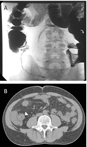
Figure 1. ‘String of Kantor’ identified on a Barium follow through demonstrating terminal ileum
stricture with separation of adjacent bowel loops (A). Corresponding axial CT (B) enteroclysis displays the thickened terminal ileum with fatty replacement of bowel wall (arrowhead), typical of chronic disease including a ‘target sign’.
Ultrasound
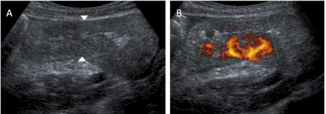
The use of trans-abdominal ultrasound (US) for assessment of patients with CD is gaining popularity. The lack of ionizing radiation is particularly attractive when examining young patients with frequent follow up. US is widely available and relatively inexpensive but experience in the use of US for bowel disease is limited. Optimal evaluation with US requires patients to fast for at least 4 h. A curvilinear 3.5 MHz or 5 MHz trans-abdominal transducer is recommended for a generalized survey of the abdomen. Regions of interest are interrogated with a 5 MHz curved-array probe. High frequency transducers including 7.5 MHz and 10 MHz are used to assess sinus tracts or fistulae [93]. US enables direct visualization of the bowel wall (Fig. 2) to assess enteric and peri-enteric changes.
Mesenteric masses including nodes, phlegmon, abscesses or matted bowel loops and the phenomenon of creeping fat can be accurately demonstrated by skilled operators. Color Doppler is used to aid detection of bowel wall hyperemia (Fig. 3) and assist in the distinction between chronic versus active disease. Doppler assessment may also have a role in demonstrating response to treatment [94]. US sensitivity may approach 87% for the detection of abnormal bowel [95] demonstrating features similar to those identified on cross-sectional imaging including mural thickening, strictures, fat creeping, and hyperemia. Normal bowel wall consists of five concentric alternating hyper and hypoechoic layers. Normal bowel wall thickness is 2–5 mm, measured in transverse section from the central hyperechoic line to to the outer margin of the edematous wall (Fig. 2) [96].
Figure 2. US study demonstrating thickened bowel wall with luminal narrowing, and fatty
replacement of the bowel wall seen in chronic disease. Normal bowel wall thickness is up to 5 mm and typically >2 cm in Crohn’s disease.
Figure 3. US study identifying thickened ileo-caecum of acute Crohn’s with small bowel obstruction.
Mural thickening greater than 5 mm is a typical finding in CD with areas of active disease often greater than 2 cm thick (Figs. 2 and 3) [30], which can be seen as the ‘target sign’ or ‘pseudokidney’ appearance. With chronic disease, there may be loss of the bowel wall signature of alternating layers due to in fatty infiltration, edema or fibrosis. The bowel appears diffusely hypoechoic with a central hyperechoic line representing the stenotic lumen. These segments can be angulated, aperistaltic, rigid and incompressible and lack the normal haustra [97].
Chronic disease can also be identified by sacculations forming on the anti-mesenteric border on US. Strictures are seen as fixed areas of mucosal thickening with proximal dilatation. The phenomenon of creeping fat implies transmural inflammation, seen as finger like hyperechoic projections into the mesenteric fat that ‘creep’ over the bowel towards the anti-mesenteric border, separating adjacent bowel loops. This is typically seen within the cephalic margin of the terminal ileum and medial aspect of the caecum [97]. Skilled operators may be able to identify mucosal abnormalities including deep fissures and intramural ulcers as linear echogenic lines traversing thickened bowel wall. Concern for fistula formation is raised when these ulcers are identified extending across closely apposed bowel loops. Complications of CD demonstrated by US include bowel obstruction, abscess formation, fistulae and appendicitis. Abscesses are hypoechoic fluid collections with thickened hyperechoic walls, which may contain echogenic air or debris. Phlegmon is differentiated from abscess due to irregular, ill-defined hyperechoic borders with a central hypoechoic region. US is better than CT in distinguishing between fluid or solid abscess content. The real-time imaging by US facilities percutaneous drainage of superficial collections.
Fistulae formation and identification is particularly useful with regards to further characterization of small fluid tracts not easily detected on CT. Hypoechoic sinus tracts are visualized with dynamic palpation of bowel loops during US examination with the resultant movement of gas bubbles passing through them [97]. Furthermore, US can be used to identify potential fistulae formation by localizing air within the
urinary bladder, vaginal vault, urachal remnant or in the subcutaneous tissues [97]. US examination is limited by intervening bowel gas but disease affected segments are often relatively gas free. Increased body mass index can influence depth of penetration of US and subsequent visualization of bowel loops. Typically patients with CD have a low BMI and therefore US penetration is not an issue but abdominal pain may be a limiting factor. The wide range of published sensitivity and specificities reflects inter-observer variability and operator skill both of which have led to limited use of US in the evaluation of patients with IBD.
Contrast enhanced US (CEUS) is an emerging technique providing real-time demonstration of bowel wall perfusion utilizing intravenous microbubble contrast agents. In addition to enhanced visualization injection of micro-bubbles (2-6 μM in diameter) provides real-time data for disease quantification [98]. Quantitative parameters, including time-intensity and brightness–time curves, are calculated using integrated software [99]. Motion artifact limiting the diagnostic accuracy of conventional grey scale US has no effect on CEUS thereby improving reproducibility [99].
Computed Tomography
Computed tomography enterography (CTE) is a non-invasive imaging technique which utilizes neutral oral contrast agents and intravenous contrast medium to identify small bowel inflammation and visualize extra-enteric structures directly (Fig. 4) [100-107]. CTE is indicated for the diagnosis, complications, disease monitoring and recurrence of CD. A large volume of intraluminal contrast agent is either administered orally (enterography, CTE) or via an NJ tube (enteroclysis, CTEC). Neutral intraluminal contrast agents (such as water) are required to enable adequate visualization of enhancing mucosal lesions which would otherwise be masked by positive contrast agents such as Barium. The gastrointestinal tract should preferably be clean and empty with a distended lumen for adequate interrogation
[108-110]. Collapsed bowel can mimic pathology such as wall thickening, abscess, or enlarged lymph nodes [103, 110]. An example of protocol may be 1 l of Klean Prep ™ (polyethylene glycol solution, Norgine pharmaceuticals) followed by 1 l of water, one hour prior to scanning for bowel preparation. Anti-peristaltic agents such as hyoscine butylbromide are preferably administered intravenously at the time of scanning to reduce motion artifact.
CTE is performed during intravenous contrast enhancement utilising 100–150 ml of iodinated contrast medium intravenously at 3-4 ml/sec in the enteric or portal venous phase (40-70 sec delay). Patients are better imaged in the prone position, if tolerated, to separate individual bowel loops. Thin axial sections allow near isotropic voxels for 3D reconstruction to aid visualisation of diseased segments of bowel.
The sensitivity of CTE is quoted at >95% for the detection of CD [101, 104], but due to the lower spatial resolution compared to conventional enterography there are lower rates of early disease detection [111]. Multi-detector CTE has been shown to be highly accurate for imaging mural and extra-luminal disease, but conventional enterography remains superior for luminal abnormalities and ulceration [112, 113]. CTE is a validated imaging technique when compared to clinical, pathological, endoscopic and other established imaging techniques [107 114-120]. Recent studies have shown that the use of CTE has altered therapeutic management in 50-61% of Crohn’s patients [121]. The differences in reported specificities and sensitivities for CTE are likely due to the lack of standard protocols [122]. At the more advanced stages of the disease, CT can identify involved bowel segments and lesion activity to plan surgical treatment and monitor the effects of medical management. CT is also reliable for the detection of extra-luminal extension of CD such as fistulae, abscesses and lymphadenopathy [115].
Figure 4. Axial CT enteroclysis demonstrating the ‘target sign’; three concentric layers of bowel wall
with inner and outer rings of high density due to hyperaemia of the mucosa, muscularis propria and/or serosa. Low density ring due to submucosal oedema.
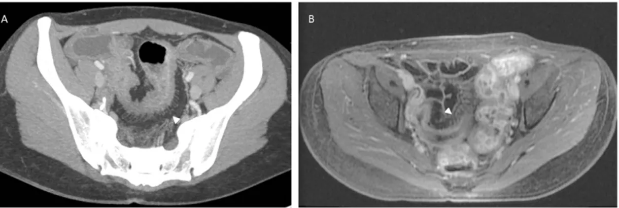
Evidence has suggested that CT enteroclysis is superior to CT enterography by providing a more uniform distension of the bowel lumen and thereby allowing accurate assessment of wall thickness [123]. Comparative studies have shown oral enterography to be both reliable and better tolerated by patients [124-126]. Following surgical resection, CTE is good for imaging ileocolic anastamoses to assess for disease recurrence and progression (Fig. 5) [127].
Figure 5. Axial CT enteroclysis (imaged prone). Disease recurrence with thickening of the
neo-terminal ileum at the anastamotic site. ‘Target sign’ (arrowhead).
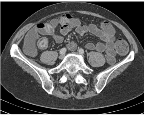
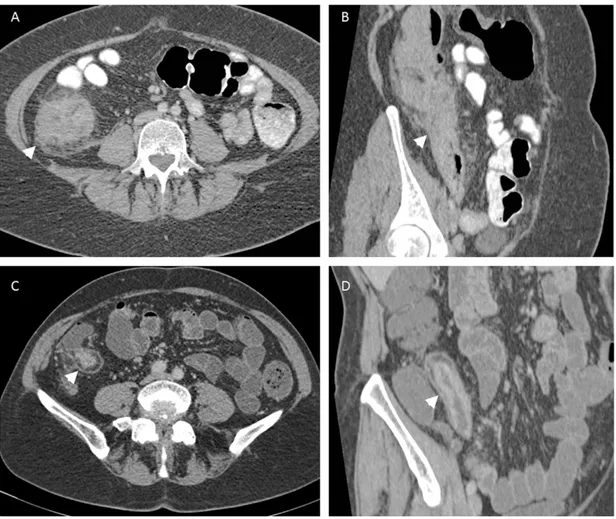
The presence of a pathological bowel segment on CTE can be detected by measuring bowel wall thickness and assessing the mucosal enhancement. With luminal distension normal small bowel thickness should measure 1–2 mm and colon 3 mm [108, 109, 128]. If the bowel wall measures more than 4mm thick then it is abnormal [129]. 1-2 cm bowel wall thickening is the most consistent feature of CD on cross-sectional imaging (Fig. 4) [111]. Differential enhancement of the bowel wall layers with a dense mucosa, oedematous submucosa and enhancing serosa gives pathological segments a stratified radiological appearance known as the ‘target sign’ (Figs. 4, 5, 6) [130]. Engorgement of the vasa recta within the small bowel mesentery suggesting mesenteric hypervascularisation is consistent with local active inflammation and has the appearance of a fine toothed comb (‘comb sign’) (Fig. 7) [106]. Luminal narrowing associated with a fibrotic stricture with pre-stenotic dilatation of small bowel is easily identifiable on CT (Fig. 6).
Figure 6. Acute Crohn’s disease seen on axial (A) and sagittal (B) views with hyper-enhancing,
thickened terminal ileum with adjacent mesenteric fatty stranding. Luminal narrowing is best appreciated on the coronal view (arrowheads). Chronic disease (C, axial and D, sagittal) and is identified by bowel wall fatty replacement and no acute adjacent inflammatory change. Both axial views (A and C) demonstrate the ‘target sign’ (arrowheads).
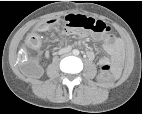
Fibrofatty proliferation within the mesentery has also been described as a feature of IBD with a subtle increase in the density of fat separating the bowel loops. Complications including local perforation and abscess formation are well delineated on CT. Abscesses within the mesentery (Fig. 8), abdominal wall, retroperitoneum and peri-anal regions are readily identifiable on CT.
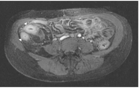
Figure 7. ‘Comb sign’ (arrowheads) as demonstrated on CT (A) and MRI (A). Axial enhanced
maximum intensity projection CT (A) and axial T2 fat sat MRI (B). Stenosis of a long segment of small bowel with bowel wall thickening and engorged mesenteric vessels.
Figure 8. Complications of Crohn’s disease. Axial enhanced CT demonstrating rim enhancing
mesenteric abscess (arrow).
CD patients are often young and may have a chronic relapsing course. The need for repeated imaging may lead to a cumulative radiation dose. The use of ionising radiation therefore warrants discussion, especially as there is increasing awareness regarding radiation dose from both published articles and the media. Medical
radiation exposure is growing exponentially, and is of particular concern in the young population with CD who are both more sensitive to ionising radiation and have a life-long chronic disease [131]. Studies of atomic bomb survivors who have a large immediate dose and radiation workers in the nuclear industry who have a slow cumulative dose have shown an increased risk of cancer and cancer related death which is directly related to the cumulative exposure [132]. This risk decreased with increasing age and therefore it is most important to limit dose in a young population group. The biological effects of low-dose radiation exposure from CT are not well established but the cumulative dose in patients with repeated studies is of increasing concern [133, 134]. There are newer low-dose CT techniques with low kVp and automatic tube current modulation with better noise reduction algorithms [135-137]. Centres have now been able to reduce the dose of an abdomen and pelvis CT to below 1 mSV [138]. A lower tube voltage (kVp) decreases the dose and also increases the visualisation of bowel wall enhancement as iodine has a greater attenuation at lower beam energies, allowing better detection of mural inflammation and engorged mesenteric veins [137].
Radiation exposure is less of a concern in elderly patients where the risk is lower and in acutely unwell patients where the benefit is high. Despite valid concern in regard to radiation CTE remains too clinically useful and cost effective [40] to avoid use based on radiation concerns. CTE is fast, reliable and easier for both patient and radiologist than MRE. Numerous studies comparing the sensitivity of CTE versus MRE in imaging CD have shown them to be equivocal [139-141]. In one study comparing the sensitivity of colonoscopy, including terminal ileum evaluation and biopsy, with a combination of clinical, serological and radiological findings, over half of those patients with negative histology were subsequently found to have active disease [142].
Magnetic Resonance Imaging
MRI is an equally capable alternative to CT without employing ionising radiation. Access to MRI may be limited and reporting expertise not as widespread. MRI, including MR enterography (MRE) and enteroclysis (MREC), is capable of demonstrating a wide range of pathological features of CD. As an additional advantage, the lack of ionising radiation makes MRI invaluable for follow up and disease monitoring. MRE and in particular MREC ensures luminal distension maximising mucosal visualisation and demonstrating subtle strictures and proximal hold up. T2 weighted imaging (WI) fast spin echo imaging following distension with a luminal agent (usually polyethylene glycol) clearly delineates mucosal inflammation which can be confirmed by Gadolinium enhancement during T1 WI. Transmural disease, focal perforation and mesenteric abscess formation are also clearly demonstrated by intravenous Gadolinium enhancement [143]. Intense mucosal enhancement post-intravenous Gadolinium is typical of active disease. Similar to the ‘comb sign’ seen on CT, distended, enhancing mesenteric vessels adjacent to a segment of inflamed bowel is well documented (Fig. 7).
Mirroring the changes demonstrated by contrast enhanced CT the ‘target sign’ with a central enhancing high SI following intravenous (IV) Gadolinium administration, identifies acute inflammatory change (Fig. 9).
Mucosal hyperenhancement has occasionally been found to be the only feature of recurrent disease in the absence of typical imaging findings. MRI can be used to monitor disease activity in addition to aid diagnosis. The peak SI post Gadolinium has been shown to correlate with the degree of disease active [144-146]. The ‘halo sign’ featured in chronic disease due to fibrofatty proliferation comprises a low SI halo with thickening of the extramural fat wrapping, separating bowel loops resulting on chronic stricture formation secondary to fibrosis [147]. MRI aids the management of strictures by distinguishing between fat hypertrophy seen in chronic strictures and the bowel wall spasm with hyperenhancement seen in acute inflammatory change.
CD complications, including transmural ulceration progressing to fistulae formation (Fig. 10) can be clearly demonstrated by MR with sensitivity between 83 and 84% and specificity of 100% [148].
Figure 9. Axial MRI T1 post IV Gadolinium demonstrating the ‘target sign’ (arrowheads) as seen on
MRI.
Figure 10. Axial (A) and coronal (B) T2 fat saturated MRI perineum. High SI (arrows and arrowhead)
Perineal fistulae can be clearly delineated with multi-planar MR as linear areas of intermediate SI originating from the bowel wall and best seen on small field of view T2 weighted, fat saturated, axial and coronal imaging. IV Gadolinium enhanced imaging may be useful to determine active inflammatory fistulae from chronic disease, with avid enhancement identifying active fistulae [149]. Stricture formation with no associated mural thickening of mesenteric inflammatory change is typical of CD [150]. MR fluoroscopy is able to demonstrate fixation of involved bowel segments, typical of chronic disease, as well as low SI T1 and T2 strictures with minimal heterogeneous enhancement post IV Gadolinium. Subsequent asymmetric shortening due to fibrosis and ulceration results in pseudo-sacculation of the anti-mesenteric border, best seen on coronal views. Further radiological aids to clinical management include the development of a robust, objective MR scoring method of disease activity when correlated with endoscopy findings. These studies however are compromised by limitations including variability in MR technique and moderate inter-observer agreement [151]. Image analysis software may overcome these failures, providing a reproducible method of quantifying and scoring disease activity and treatment response. Novel sequences and techniques including MR perfusion are currently being investigated, to add further value to MRI in aiding diagnosis, management and assessment to new treatment regimes.
4. Treatment
Treatment of active CD requires recognition of disease activity, localization (ileal, ileocolonic, colonic, or other), and behaviour (inflammatory, stricturing, or fistulating). Even in cases with mild disease, leaving patients without treatment is
seldom an option. As smoking cessation is associated with a 65% reduction in risk for relapse, stopping smoking should be encouraged [21].
4.1 Medical therapy
Before the era of corticosteroids, IBD was a fatal disease for a great proportion of patients; no other medication has had such a great impact on outcome [152, 153]. Despite the development of more potent drugs against CD during recent decades, the disease is still considered incurable and in many patients it leads to surgery and disability. Medical therapy for CD can be divided into therapy aimed at induction and at maintenance of remission (Table 3).
Anti-TNFα antibodies
In patients with CD, the proinflammatory cytokine TNFα plays a role in the inflammatory cascade. Anti-TNFs neutralize by several mechanisms this cytokine and thus interrupt the inflammatory cascade. As anti-TNFs are created by biological processes, they are also called biological drugs or more simply biologicals.
Infliximab is an intravenously administered murine-derived chimeric monoclonal TNFα inhibitor antibody of the immunoglobulin G1 subset; it is the most extensively investigated biological drug available for the treatment of IBD. Approximately two thirds of patients achieve significant clinical improvement, and nearly half maintain clinical remission after one year of maintenance therapy [154]. Additionally, infliximab induces – as early as four weeks after initiation of therapy – mucosal healing based on endoscopic evaluation. This has been shown to reduce risk for recurrence, in addition to reducing hospitalization and surgery [155-157].
Infliximab is also effective as induction and maintenance therapy for fistulizing CD [57] and may serve as monotherapy or be useful in combination with other immunomodulating agents, usually given as induction doses at weeks 0, 2, and 6, followed by maintenance infusions every 8 weeks [158].
Adalimumab is a subcutaneously administered immunoglobulin G1 isotype monoclonal human antibody against TNFα. Evidence indicates that adalimumab is effective as a weekly or biweekly dosage for both induction and maintenance of remission [159, 160]. It also shows efficacy in treatment of fistulizing CD. The EXTEND trial demonstrated complete mucosal healing after 52 weeks in 24% of patients on adalimumab compared to 0% on placebo [161]. Patients who have developed antibodies against infliximab may still benefit from adalimumab [162]. No randomised controlled trials that systemically compare in CD the efficacy of
adalimumab and infliximab exist, although based on studies with more or less similar study designs and populations, their efficacy has been considered comparable. A recent retrospective study of 200 matched anti-TNF naïve CD patients reported no significant difference in steroid-free response rates or in adverse effects after one and two years [163]. Because of its retrospective design, that study lacked data on clinical or endoscopic activity.
Certolizumab pegol is a pegylated, subcutaneously administered humanized TNFα- binding Fab fragment (Schreiber et al. 2005). Multiple studies have shown its efficacy as similar to that of the other anti-TNF agents, its effects being more pronounced if the patient is anti-TNF naïve. In one randomized, double-blind, placebo-controlled trial of adults with moderate-to-severe CD, those who responded to induction therapy with certolizumab were more likely to maintain their response and sustain remission at 26 weeks with continuous certolizumab than were those switched to placebo [10]. The need is strong for further biological drugs, because when one antibody loses effectiveness, switching to another becomes necessary. Under study are several biological therapies targeted at mechanisms other than blockade of TNFα, including modulation of other cytokines, blockade of T cells, and blockade of inflammatory cell migration and adhesion [164].
Natalizumab, a blocker of α4-integrin, has shown promising results in CD treatment, but severe adverse effects such as reactivation of a human polyomavirus leading to progressive multifocal leukoencephalopathy limit its use [165]. Vedolizumab is a fully humanized α4 7 integrin antibody that has passed a number of phase-III clinical trials. In clinically moderate to severe CD it has been more efficient than placebo in inducing and maintaining clinical remission. As vedolizumab modulates gut but not brain lymphocyte migration, it is at least theoretically less likely than natalizumab to cause progressive multifocal leukoencephalopathy [166]. Apilimod is an inhibitor of the transcription of IL-12 and IL-23, whereas ustekinumab and briakinumab both target the p40 subunit common to IL-12 and IL-23.
As trials investigating these drugs are still in phases I and II, their long-term effects are unclear. However, results indicate that ustekinumab might be useful in patients who have failed to respond to anti-TNF therapy [167]. Golimumab is a TNFα-blocking monoclonal antibody newly approved for treatment of UC. Its advantages compared with adalimumab and infliximab are its once-monthly dosing by either intravenous or subcutaneous administration [168, 169]. It has not yet advanced into CD trials.
4.2 Surgery
Although surgery in CD should be limited to complications of the disease such as strictures and fistulae, ultimately, a large majority, up to 70%, despite optimized medical therapy, will need surgery. Furthermore, up to 40% will need secondary surgery because of disease recurrence [4]. Despite its revolutionizing effect on CD treatment, evidence is still limited as to anti-TNF impact on need for surgery. Population surveys during the last two decades have shown inconsistent results, with both declining trends in and no changes in need for surgery [170, 171]. Subgroup analyses of anti-TNF-responding patients seem to suggest a reduction in the need for surgery at a median follow-up of up to three years [172]. The short follow-up and exclusion of patients with imminent surgical need could, however, cause bias. CD patients in northern Europe seem more likely to undergo surgery than in southern Europe, suggesting a north-south disease-severity gradient [18]. To preserve bowel function and minimize risk for intestinal failure, a more conservative surgical approach has been adopted during recent decades. In cases with medical intractability, internal fistulae, abscesses, symptomatic bowel obstruction, severe bleeding, toxic dilatation, or acute perforation, however, surgical resection inevitably becomes necessary [173]. Patients with perianal or rectovaginal fistulae often need a combination of surgery and medical treatment. In the surgical management of small bowel CD, strictureplasty plays a central role. Often considered for strictureplasty are
isolated strictures under 10 cm in length. A majority of patients achieve symptomatic relief, with secondary surgery rates of between 34 and 44% during a seven-year follow-up [174].
4.3 Nutritional therapy
Unlike the management of CD in paediatric and adolescent patients, no placebo controlled trials involve nutritional therapy for active CD in adult patients. In one Cochrane systematic review, however, elemental or polymeric diets were less effective than corticosteroids in inducing remission [175]. Enteral therapy is regarded as appropriate only for adjunctive treatment to support nutrition, not for primary therapy in active CD [176]. Omega (ω)-3 fatty acids may have anti-inflammatory effects by reducing production of leukotriene B4. Due to the heterogeneous study data, however, the efficacy of ω-3 fatty acids in maintaining remission remains controversial [177]. Data in ten systematically reviewed studies suggest that enteral nutrition as a complement to an ordinary diet may be useful for maintaining remission in patients with CD, although the evidence level is low [178].
5. Assessment of disease activity
In clinical practice, disease activity assessment relies on clinical history and a combination of clinical, laboratory, endoscopic, and radiological findings. Need for standardisation and quantification of disease severity in clinical trials has led to development of several disease activity indices based on findings or symptoms or their combinations. Table 4 presents a summary of those assessment methods of clinical activity most commonly used in both clinical practice and trials.
5.1 Clinical activity
In clinical trials, the score most commonly used is the Crohn’s disease activity index (CDAI), which comprises one serological and seven clinical variables (Table 5), with scores ranging between 0 and approximately 650 [179]. A CDAI <150 has defined clinically inactive disease and >450 severe disease. Some investigators have arbitrarily further labeled CDAI scores of 150 to 219 as mildly, and 220 to 450 as moderately active disease. One definition for clinical response is a reduction of ≥100 points in the CDAI, although some clinical trials have defined response as a reduction of ≥70 points [50]. The CDAI score is infrequent in everyday clinical work because of its complex and time-consuming calculation and the need for a seven-day diary of symptoms. Although it has seemed to perform quite well recently in a
postoperative setting, it is unsuitable for use as a primary outcome measure in patients with a history of extensive surgery [180]. Further, it is unreliable in patients with a mainly fistulating and stricturing disease. An additional feature of the CDAI score is the considerable weight given for scores on ― general well-being and ― intensity of abdominal pain, which are completely subjective [181]. Correlation of the CDAI with ileocolonoscopy findings is weak, and the CDAI underestimates endoscopically determined inflammatory activity [182-184]. For scoring of clinical disease activity of children and adolescents, we have a paediatric Crohn’s disease activity index (PCDAI) [185].
Unlike the CDAI, the Harvey-Bradshaw index (HBI) notes only symptoms and signs over the preceding 24 hours (Table 6). It is based on five clinical variables: general wellbeing, graded from 0 to 4 points, abdominal pain and palpable abdominal mass, each graded from 0 to 3 points, number of liquid stools per day, and
complications/extraintestinal features, each graded as one point [186]. It has been suggested that HBI scores ≤4 or <4 indicate clinical remission [187, 188].
Both the Oxford Index, based on one laboratory variable and nine clinical variables, and the Cape Town index, originally based on one laboratory variable and nine clinical variables, correlate with the CDAI [181, 189, 190]. Because the major contribution of subjective variables to the CDAI has attracted criticism, researchers have attempted to develop disease activity indices on objective grounds only. One instrument that eliminates subjective criteria is the van Hees or Dutch index; it is made up of two laboratory variables, of which serum albumin contributes most, and seven clinical features from patient history or physical examination [191]. Although the correlation between the van Hees index and the CDAI is poor, both seem to be predictive of CD exacerbations [189]. The CDAI describes poorly the activity of perianal and fistulizing CD; the perianal disease activity index (PDAI) currently represents the gold standard for evaluating perianal disease severity [181, 192]. Recently, the short CDAI was developed and validated [193]. Of the eight variables in the CDAI, the short version includes only three that are clinical self-reported symptom variables. Though it shows a strong correlation with the CDAI, its self-reporting of subjective symptoms and well-being, means that it has disadvantages similar to those of the original index (Table 7).
5.2 Endoscopic activity and mucosal healing
In patients with long-standing chronic ileocolonic CD, correct diagnosis requires clearcut indications for endoscopy, assessment of disease activity and extension, dilation of strictures, and surveillance [5]. Follow-up endoscopies are required when disease activity or disease location is uncertain. In CD, blood tests and symptoms do not necessarily correlate with endoscopic disease activity; intestinal inflammation can occur in patients free from symptoms [182]. Ileocolonoscopy has, however, several drawbacks: it is time-consuming and expensive, it requires bowel preparation, and most patients consider it unpleasant.
Since the 1960s, clinical studies on UC have suggested a more favourable outcome after a corticosteroid course in UC patients achieving clinical and endoscopic remission than in those achieving only clinical remission. Up until the late 1990s, studies reported no such correlation in CD patients [194]. The introduction of anti-TNF therapy completely changed investigators’ and clinicians’ attitudes towards mucosal healing; healing of the mucosa for the first time became possible. Since then, constantly growing interest in mucosal healing has revealed its clinical importance as a predictive marker of favourable outcome. Now, assessment of mucosal healing during therapy has become essential for clinical practice and for evaluation of response in clinical trials [5, 6].
Anti-TNF therapy and mucosal healing
Schnitzler and coworkers [157] analysed retrospectively for a median of almost five years 214 CD patients who had undergone endoscopy before start of infliximab therapy. Scheduled infliximab therapy led to mucosal healing that was associated with the best long-term outcome; nearly 80% showed sustained clinical benefit from infliximab until the end of the study. Those achieving mucosal healing underwent
significantly less surgery and needed less hospital treatment than did those with active disease seen in the follow-up endoscopy done approximately seven months after the start of therapy. In IBD, studies and reviews suggest mucosal healing as a therapeutic goal, with absence of mucosal ulcerations serving in most anti-TNF trials as the definition of mucosal healing [161, 195].
The importance of minor changes such as an aphthous ulcer in otherwise healed mucosa remains unclear, however. Most studies on predictive factors in anti-TNF-treated CD have focused on clinical outcome. We know relatively little about factors predicting mucosal healing during anti- TNF therapy. In 201 Hungarian CD patients treated with adalimumab, Kiss and coworkers [196] found that low CRP (<10 mg/l) at week 12, clinical remission at week 24, and non-smoking were all associated with endoscopic improvement or healing at one year. They defined clinical remission as CDAI<150, and mucosal healing as the absence of any mucosal lesions or signs of active inflammation. Hébuterne and coworkers [197] have described the similarity of endoscopic findings in early endoscopic evaluation and in endoscopy one year after start of anti-TNF. Their open-label MUSIC trial involved 89 CD patients treated with certolizumab pegol for 54 weeks. They defined endoscopic response as a decrease in CDEIS score of more than 5, defined maintenance of endoscopic effect as unaltered CDEIS between weeks 10 and 54, and defined complete endoscopic remission as CDEIS<3. In a subpopulation of 52 patients with 54-week endoscopic data, of the 37 who showed an endoscopic response at 10 weeks, 28 (76%) maintained their response at week 54. Of 7 patients in complete endoscopic remission at week 10, 5 (71%) maintained remission at week 54. Although the EXTEND trial focused mainly on comparing endoscopic outcome after adalimumab or placebo following a two-week adalimumab induction, its results also suggested that mucosal healing may be more difficult to achieve for patients with more severe ulcerations at baseline [161]. Optimal timing for determination of mucosal healing remains unsettled [195]. Recently, the concept of deep remission, defined as a combination of clinical and endoscopic remission, has become recognized as a potential predictive CD marker
[161, 198, 199]. During adalimumab therapy, deep remission has been associated with lower health-care costs and a favourable long-term outcome in terms of hospitalizations and quality of life [200]. The definition of deep remission is, however, still evolving.
5.3 Endoscopic scoring of inflammatory activity
The need for reproducibility and standardisation in the management and follow-up of IBD has led to development of several endoscopic grading scores. Endoscopic scores originally classifying disease activity have also been proposed as means to define mucosal healing [5].
CDEIS
The Crohn’s disease endoscopic index of severity (CDEIS) was developed at the end of the 1980s by the French Groupe d’Etude des Affections Inflammatoires Digestivesis (GETAID) [201]. The CDEIS is validated in terms of reproducibility and global endoscopic evaluation of lesion severity and has become the gold standard for assessment of endoscopic activity in CD [181].
Calculation of the CDEIS requires considering the colon and the terminal ileum as comprising five segments: (1) rectum, (2) left colon and sigmoid, (3) transverse colon, (4) right colon, and (5) ileum (Table 8).
From each segment, the presence of nine mucosal lesion types would be recorded: (1) pseudopolyp, (2) healed ulceration, (3) erythema (plaques, bands, or diffuse), (4) swollen mucosa, (5) aphthous ulceration, (6) superficial or shallow ulceration, (7) deep ulceration, (8) non-ulcerated stenosis, and (9) ulcerated stenosis. The percentage of segmental surfaces involving the disease and ulcerations are posited on a 10-cm analogue scale between 0 and 10 (no lesion=0, lesions or ulcerations involving 100% of the segment=10). For the terminal ileum and for those colonic segments only partly explored, the 10-cm scale represents the area actually seen. The CDEIS can range between 0 and 44, with higher scores depicting more severe endoscopic activity. Although the CDEIS has served for endoscopic scoring in
several studies, threshold values for remission or for mild, moderate, or severe disease, or for significant response are still lacking. After revisiting the endoscopy findings for the validation of the CDEIS, the GETAID study group suggested a cut-off value of 3 or 3.5 for complete mucosal healing, defined as no lesions or scars, and a rougher cut-off for endoscopic remission set at CDEIS levels of between 6 and 7, defined as no lesions or scars but accepting minor lesions, and for endoscopic response a decrease in the CDEIS of more than 5 [202, 203]. The CDEIS correlates poorly with clinical activity [182].
SES-CD
The time-consuming and complex structure of the CDEIS has prevented it from becoming a tool in everyday clinical practice. To simplify endoscopic assessment of inflammatory activity in CD, the simple endoscopic score for Crohn’s disease (SES-CD) was developed and validated nearly ten years ago. Its construction and validation is based on correlations with the CDEIS and, to a lesser extent, the CDAI [204]. The SES-CD shows a strong correlation with the CDEIS and is easier and quicker to calculate. It is based on four variables scored in the same five ileocolonic segments as in the CDEIS (Table 9). The ileum is scored for the full extent to which it is examined, but the ileal score specifically excludes the ileocaecal valve or any ileocolonic anastomosis, which are both included in the neighbouring distal segment. Additionally to the ileocaecal valve, the right colon includes the caecum and the ascending colon up to the hepatic flexure. The transverse colon is defined as the segment between the hepatic and splenic flexures. The left colon includes the descending colon and sigmoid colon. The rectum is defined as that portion distal to the rectosigmoid junction. The SES-CD can range from 0 to 60, with higher scores for increased inflammatory activity. No consensus on cut-offs for remission or different stages of inflammatory activity exists. Suggested definitions on endoscopic remission for the SES-CD have been a score of 0–2 and 0–3 [183, 205]. Moskovitz